Selective Amygdalohippocampectomy:
Indications and Follow-up
Heinz Gregor Wieser
ABSTRACT: Selective amygdalohippocampectomy (AHE) offers a real chance of cure only in patients with well-defined, precisely localized "epileptogenic area", i.e. seizure focus. Therefore, a priori only a small proportion of all patients with epilepsy can meet the criteria for selective surgical interventions. From the evidence in patients meeting the criteria for AHE, we conclude that this technique is to be preferred to the "standard" anterior temporal lobectomy and represents a more selective but still effective surgical treatment of epilepsy.
RESUME: L'amygdalo-hippocampectomie selective: Indications et suivi. L'amygdalo-hippocampectomie selec-tive offre de replies chances de guerison de l'epilepsie seulement chez les patients avec une "zone epileptique" bien deTinie, bien localised et de dimension restreinte, c'est-a-dire un foyer epileptique. Done, a priori, seulement une faible proportion des patients epileptiques pourront satisfaire auz criteres les rendant eiigibles a une chirurgie selective. En se basant sur les r6sultats des series que nous avons presentees, nous sommes en mesure d'affirmer qu'une amydaloj-hippocampectomie selective, chez des patients rencontrant les criteres d'eiigibilite, est preferable a une lobectomie temporale anterieure. En conclusion, nous esperons que l'amygdalo-hippocampectomie selective, telle que decrite plus haut, constituera un pas vers une approche chirurgicale plus cibiee, et tout aussi efficace, de l'epilepsie.
Can. J. Neurol. Sci. 1991; 18: 617-627
Since the pioneering work of the Montreal school1-6 the sur-gical treatment of epilepsy has continued to achieve worldwide acceptance as an alternative treatment for selected patients with medically refractory seizures. Patients with temporal lobe (TL) seizures still constitute the most important group in the surgical series. The advent of modern diagnostic methods has aided the recognition of certain subtypes of complex partial seizures.7-8 Improved diagnosis and better understanding of the importance of the mesial temporal region in the pathogenesis of TL seizures has resulted in more selective operations. New diagnostic meth-ods, such as the foramen ovale electrode9 and other special extracranial electrodes technologies have led to better presurgi-cal assessment.10
The first selective temporal lobe resections for treatment of mesiobasal limbic epilepsy7' "-1 3 were performed at our University Hospital by Yasargil in 1975. Operative anatomy, surgical techniques, criteria for operation, investigative techniques and results of the "selective amygdalohippocampec-tomy" (AHE) have been previously reported.n.14-18
We have recently completed three studies on our patients who underwent AHE: 1) a detailed volumetric examination of surgical resection on pre- and postoperative MR imaging (N = 30),19 2) a year by year post-surgical follow-up (N = 20),20 and 3) a detailed analysis of 320 seizures recorded in 77 patients21 with foramen ovale electrodes. The aim of this paper is to corre-late the above three studies and to summarize indications and results of AHE.
Indications for Selective Amygdalohippocampectomy The microsurgical AHE procedure is the result of both a pre-cise identification of the seizure-initiating structures (and prefer-ential pathways of seizure spread) and the refinements of neuro-surgical techniques.22-23 Our analysis of seizures recorded by stereo-electroencephalography (SEEG)24 indicated that the "classical" anterior TL resection may be a needless and aggres-sive operation for patients with an unequivocally demonstrated seizure focus in the mesiobasal limbic area. At our institution, the majority of patients, evaluated for medically refractory com-plex partial seizures (cps), showed a strictly focal unilateral mesiobasal seizure focus involving the hippocampal formation and the amygdala. In our series, approximately 65% of the patients showed high-frequency low-amplitude discharges simultaneously in the hippocampus and the amygdala at the onset of each seizure (Figure 1). Seizure discharges recorded only in the hippocampal formation and initially not affecting the ipsilateral amygdala constituted another 25% (Figure 2). About 7% of mesiobasal limbic seizures started in the amygdala and propagated to the hippocampus within 3 to 5 sec. Only 3% of the patients showed tonic discharges exclusively confined to the amygdala without hippocampal spreading at the onset of seizure.
Specific requirements for "causal" AHE are the following: 1) the seizure focus must be in the mesiobasal limbic structures ipsilateral to the side chosen for resection, and 2) the contralat-eral mesiobasal TL must be functional.
From the Department of Neurology, University Hospital, Zurich, Switzerland
ECG J U J L _ J L _ J L _ J L J U J L _ L _ . I _ J L J L .
EDAR
n_n_n_JLjy
f~u—uJ L J U - U J - J U U U J U J U J U U L
U U L J U L J U J / U
>i ^ - • ^ • • A - ' A J ) . i - * i •<•« . H L " A H •*•• •>•• I — I . .www
1.8 - 9 - A — "*>V /""W-"^/»*'-V'\/~r;ji!!iiTiy\nn^fv/
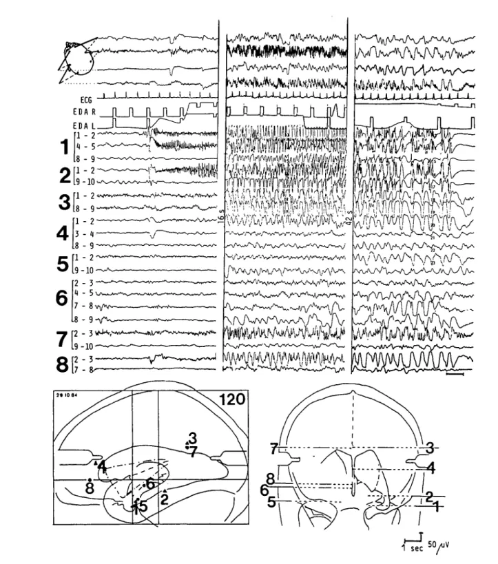
50 AJV 1 sec ^ YFigure I — Combined scalp- and depth EEG recordings at onset, peak, and end of a spontaneously occurring complex partial seizure. The initial epileptic discharge is simultaneously recorded in the left amygdala (111-2 and 114-5) and left hippocampus (211-2). Note the increased electroder-mal activity (EDA L) recorded from a left hand finger at the onset of seizure, the tachycardia (ECG) at the peak of seizure, and the postictal depres-sion in the depth-EEG left temporal derivations.
The seizure was characterized by an epigastric aura, staring, progressive impairment of consciousness, unresponsiveness and oral-alimentary automatisms (second section).
The position of the 10-polar hollow-core depth electrodes (bold numbers) is indicated in the brain maps. The small numbers refer to the contacts of each depth electrode. The length of the contacts and the intercontact spacing is 2 mm with the exception of intercontact distance 5-6. Contact 1 is the deepest and contact 10 is the most superficial in each electrode. The external diameter of the stainless steel electrodes is 0.9 mm.
ECG J - J U L - L J U . .L.i. JL1.. J U L J U U U U J L - L J J U U L J L J a J L X J U L ^ 1 ' "•' } ^ — ^ A \.—^—^ — v ^-—WN^-W—^. /-w——• . -> ^ — ^ - ^ _ ^ . - _ ^ - ^ . . 9-10
, _
2^O-^^^^^f^
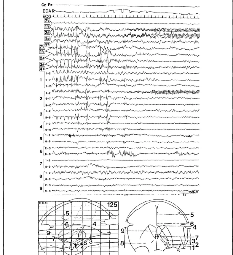
1-2 VA V ^ / W U A N V ^ V ^ A/V/W - \ A ^ ^ / V < > V ^ ^ 8-9Figure 2 — Example of depth EEG recording at the onset of a seizure, characterized by an aura of detachment. Note the special montage, which con-sists of uninterrupted bipolar long distance derivations in the parasagittal plane (... in the brain map), connecting the deep (channels 4-7) and the superficial contacts (channels 8-11) of electrodes 7,1,2,3,4 and of interrupted short-distance derivations along electrodes 1-9 (channels 12-31). Note that the pre-ictal epileptiform activity consists of fairly regular 2/sec spike-slow-wave complexes with a clear predominance in the anterior lateral temporal neocortex (left), whereas the low-voltage high-frequency ictal discharge (right) is clearly localized to the anterior hippocampus (electrode 2, contact 2). In addition to the depth EEG, one scalp derivation (Cz-Pz), the electrocardiogram (ECG) and the electrodermal activity recorded from the right fingers (EDA R) are shown. Calibration refers to surface and depth EEG only. (From Wieser, 1987b).
In reality, the first requirement is somewhat relative. Since 1979, we considered selective AHE to be indicated also in patients with either a seizure focus greater than the eventually resected amygdala and hippocampus or with a seizure focus out-side the amygdalohippocampal complex, but with the mesiobasal limbic structures acting as a "secondary pacemaker". These two events constitute the rationale for the so-called "pal-liative" AHE (as opposed to "causal" AHE with a seizure focus strictly localized within the resected structures).
To better assess the preoperative risks of selective AHE on higher cognitive functions, such as learning and memory, we developed three selective TL-Amytal procedures.2526 During preoperative neuropsychological evaluation we are now using one of the following techniques for the Amytal test: 1) temporal balloon-occlusion distal to the anterior choroidal artery (a.ch.a.) with subsequent Amytal injection into the a.ch.a., the communi-cans posterior and the ophthalmic artery; 2) selective catheteri-zation of the a.ch.a.; and c) the selective charactericatheteri-zation of the P2 segment of the posterior cerebral artery. These modifications of the "Wada-Test"6-27"28 have been proven to be better predic-tors of neuropsychological outcome for selective AHE patients.29 Further studies investigating individual variations of the vascular territories of injected vessels are necessary.
Rationale for Amygdalohippocampectomy
The aim of our standard AHE technique is the complete removal of the amygdala, the anterior hippocampus, and part of the parahippocampal gyrus (Figure 3). It is important to note that the resection of the subiculum which can act as a "hyper-excitable fringe" and therefore as an amplifier7'30 constitutes an important aspect of this operation. Furthermore, the surgical trans-Sylvian approach to the amygdala (the first target of the neurosurgeon) is through the medial aspect of the first temporal gyrus. This initial operative step interrupts the anterior commis-sure and at the same time the uncinate fasciculus. Thus, the resection of the epileptic focus is combined with the interruption of the main propagation pathways operative in most mesiobasal limbic seizures31 (see also Figure 4).
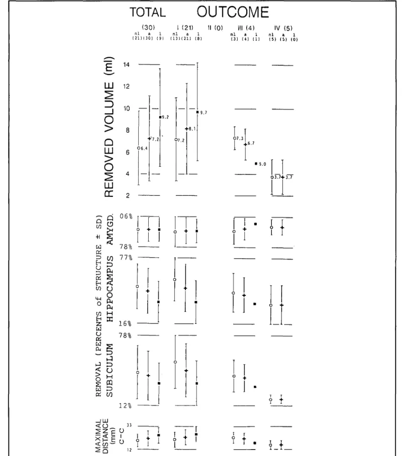
Recently, we completed a study in which we measured the total volume of surgically resected brain in AHE and the per-centage of the following structures: amygdala, hippocampus, and parahippocampal gyrus. The percentage of resection of pes hippocampi, dentage gyrus, uncus, and subiculum (as defined by Duvernoy)32 was also measured. The study was performed in 30 patients who had special pre- and postoperative MR imaging and a postoperative clinical follow-up of at least one year. Postoperative seizure control was correlated with the total resected volume and with the proportions of resected volume of each of the above mentioned limbic subcompartments (as judged independently by two investigators). The following results were obtained.19
The mean volume of the removed tissue was 7.2 ml (range 2.1 to 17.7). The percentage scores of the limbic subcompart-ments were: amygdala 92%, hippocampus 46%, parahippocam-pal gyrus 32%, pes hippocampi 92%, dentate gyrus 45%, uncus 92%, and subiculum 40%. Although a small resection did not exclude a good outcome, there was a trend showing that a better outcome was associated with a larger resection. Of note is that the incomplete resection of the parahippocampal gyrus, and in particular of the subiculum, resulted in a less satisfactory
out-come (Figure 5). These data show that the rationale for AHE aims at both, elimination of the seizure generating structures and interruption of pathways important for the maintenance and spread of seizure discharges.
Results of Amygdalohippocampectomy
Two hundred sixty-six patients with medically refractory mesiobasal TL epilepsy underwent selective TL surgery in the University Hospital of Zurich since 1975. Excluding patients who underwent selective TL resections outside the amygdala and hippocampus, 215 patients underwent selective AHE. These 215 patients have been classified according to the neuropatho-logical findings of the resected specimen. All specimens were examined by routine and immunohistochemical staining proce-dures including antibodies to glial fibrillary acidic protein (GFAP) and synaptophysin. Brain tumors were found in 110 patients (48%). The tumors included fibrillary astrocytoma (22%), mixed oligo-astrocytoma (14%), ganglioglioma (13%), pilocytic astrocytoma (13%), oligodendroglioma (11%), glioblastoma (10%), anaplastic astrocytoma (8%), and others (9%). Vascular malformations were diagnosed in 12%, and neu-ronal, glial and neuro-glial hamartomas in 7% of the total num-ber of cases. In 37 patients (17%) reactive gliosis in the hip-pocampus was present and in 2 patients (1%) findings consistent with sequelae of an encephalitis were found. In 34 patients (16%) no structural alterations were detected.
The male to female ratio was 120 vs 95. In 114 patients the operation was on the right and in 101 patients on the left side. Mean age at the onset of the seizure disorder was 18.8 years (SD 15.3), and the mean age at operation was 29.7 years (SD 13.7). The calculated mean duration of the seizure disorder prior to operation was 11.0 (SD 10.2) years. Patients with benign tumors were operated at younger age (mean 29.2; SD 29.6 years) than those with malignant tumors (mean 37.3; SD 16.7 years).
Patients with no microscopical pathology were operated ear-lier (mean 28.0; SD 8.8 years) than those with slight gliosis (mean 30.1; SD 10.6 years). Patients with moderate gliosis (mean 32.2; SD 9.6 years) were operated earlier than those with severe gliosis (mean 34.1; SD 7.3 years).
The shortest duration of the seizure disorder prior to the operation was found in patients with malignant tumors (mean 3.7; SD 5.8 years), followed by patients with semibenign tumors (mean 6.0; SD 7.2 years) and patients with benign tumors (mean 9.0; SD 7.1 years).
The mean duration of epilepsy prior to surgery was 18.5 (SD 8.1) years in patients with slight gliosis, 21.9 years (SD 9.1) in cases with moderate gliosis, and 28.8 years (SD 4.5) in patients with severe gliosis.
The epileptological outcome was classified according to Engel's recommendations33 into the following categories: I, seizure free; II, rare seizures (not more than 1-2 per year); III, worthwhile improvement (> 90% reduction); and IV, no worth-while improvement. A postoperative follow-up of at least 12 months was required prior to classification in one of the above categories. One hundred seventy-seven patients out of the 215 who underwent AHE met the above criterion for classification. The most recent epileptological outcome together with the fol-low-up period is reported in Table 1. The year-by-year outcome classification shows that the percentage of patients in each
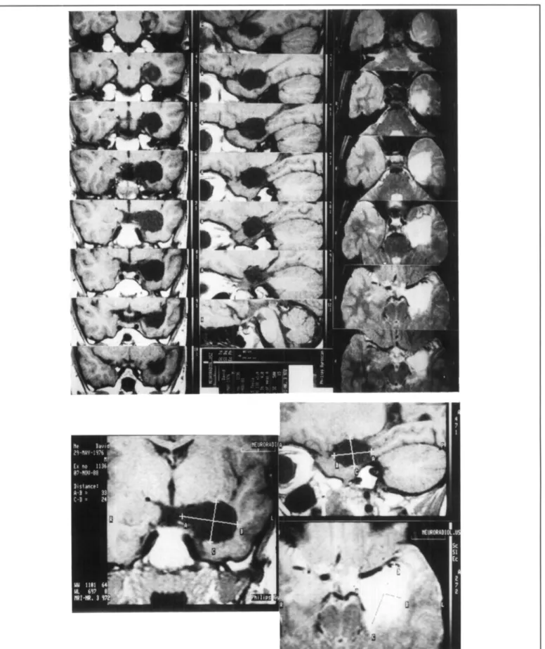
out-Figure 3 — Serial postoperative MR-images in all three planes showing the extent of a left selective amygdalohippocampectomy. Coronal and sagittal images are Ti-weighted, transversal images are T2- weighted. 1J Tesla system (Philips). Repetition time 500 msec, echo time 15 msec, (T2: dou-ble echo 501100, TR 2000); 256 x 256 matrix, field of view 200 mm; slice thickness 4 mm.
213 MS L . AMYGDALA L. H1PP0CAMP L . G C I N G p / m L. FORB MED
2S|>£
B
"•A^^W-^;1
2
3
4
5
6
7
8
9
10
^ \ / V S y - V v ^ ^ ^ ^ ; ' ! , l'!|l'\ - 9 * , - 9 "*•"—-—'-*«*—- •—•*~>«>*'**"J'yi|(l|^^ - 1 0 YV^ftvy<)H^vv^r«^ - 8 - 9 r v - v - w ^ * * ^ ^ ^ ^ - 9 ^ - ^ — k ^ ^ ^ - ^ ^ i ^ ^ ^ ^ - 10^'Vv*VvyVvnrwy>-*'>A^ft" w ^
3 ^ " • / \ * ' A ^ *, , ^ > ' \ / . ' k ' ^ ^ A-V'^'I , \ . v 10 S 100 /UV SURFACE ! Z ^ i 50 /jV DEPTH _ 11STIM-.L HIPPOC
9 8"v/il
/Vix
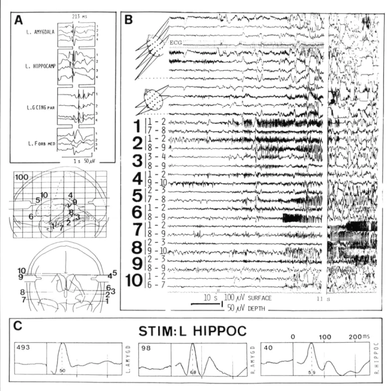
0 100 1 40 200msFigure 4 — Interictal spike (A) and ictal discharge propagation (B) to ipsilateral frontal regions, most probably mediated through the uncinate fasciculus. Ipsilateral (C left) and contralateral (C middle) amygdalar and contralateral hippocampal response (C right) following repetitive left
hippocampal single pulse stimulation (I0V, 0.7 ms, 1 Hz; averaged response, N=102) are also shown.
Parts A and B are from the same patient, whose brain map with the electrode position is shown. In A, four spontaneously occurring spikes are depicted (numbered by small numbers 1-4, on the right margin) and arranged vertically in order to better visualize the stereotype propagation. The primary epileptogenic area has been localized in the left amygdala and hippocampus, based on the spontaneous seizures (see B). These find-ings were confirmed by complete seizure control following selective left amygdalohippocampectomy. Note that both the interictal spikes as well as the ictal discharge propagate to the left frontal regions (6/8-9). The four right sections of EEG-recording represents the "tertiary" seizure propa-gation to the contralateral hemisphere.
Tl
n (2 LU 123 10
_ J 1UO
> 8Q
LU
6>
o
^
4 LU CC 2 — /-< Ofi% +i 2 CJ 7R9-g CO 77% a 2 EH <! CO U° s
CO H EH K 2 16% W £ 78% w «-, P-I e ~ i-q> °
K CO 12% _ I L U <rT-|-1 33 MAXIM / DISTAN C (mm )c-c
3TAL OUTCOME
(30) 1 (21) II (0) III (4) IV (5) 1 a 1 n l a 1 n l a 1 n l a 1 1 ) 1 3 0 ) ( 9 1 ( 1 3 ) 1 2 1 ) ( 8 1 ( 3 ) ( 4 ) ( 1 ) ( 5 ) ( 5 ) ( 0 ) 3 6 . 4 K 7 . 2 ••1
3 + 1i
)
I 3•
I• H
19.?" '
C < I c1
>7.2 K8.11
'II
• i • • iT I
t "
' 9 . 7 I C 1 cI I
1
1
• 5.0 3 3 . 7 + 3 . 71 I • u
[ 1 ' '
' • • • < > ••
[I
If-T r O + T +
Figure 5 — Removed volume in ml (top), percentage of removal per indicated limbic suhcompartment (middle), and maximal length of the resection in the cranio-caudal axis (bottom), in 30 patients who underwent selective amygdalohippocampectomy. Data are displayed for the total number of patients and according to the epileptological outcome (l-IV, for definition see text: further abbreviations: nl, non-lesional: a, all; I, lesional). Number in brackets refers to the underlying patients. The numerical values of the means are given with the figure.
Table 1: Most recent and year-by-year epileptological outcome of patients treated with selective amygdalohippocampectomy. The bottom part of the table shows the outcome of 58 non-lesional patients preoperatively classined into "causal" and "palliative" operations (see text), x, mean in months; SD, standard deviation in months.
Follow-up Groups Total
Outcome Categories
I II III IV I II III
(Number of Patients) (Percents) IV
Most Recent Follow-Up (x 47; SD 30.8) Year-by-Year Outcome 1 year 2 years 3 years 4 years 5 years 6-8 years 9-13 years Causal AHE (x 50.4; SD 35.3) Palliative AHE (x 55.7; SD 31.0) 177 30 28 105 22 13 20 39 16 59 73 12 20 31 22 177 138 106 91 73 34 10 115 82 63 57 43 20 7 9 9 4 4 5 1 1 20 17 13 12 9 4 1 33 30 26 18 16 9 1 65 59 59 63 59 59 70 5 7 4 4 7 3 10 11 12 12 13 12 12 10 19 22 25 20 22 26 10 57
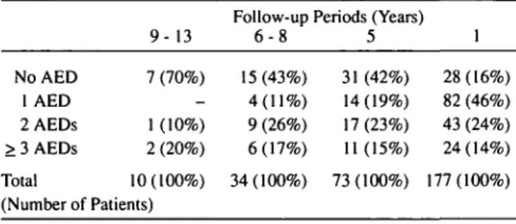
Table 2: Antiepileptic drug (AED) treatment at different postoper-ative follow-up periods.
NoAED 1 AED 2AEDs > 3 AEDs Total 9- 13 7 (70%) -1 (-10%) 2 (20%) 10(100%) (Number of Patients) Follow-up 6 - 8 15 (43%) 4(11%) 9 (26%) 6(17%) 34(100%) Periods (Years 5 31 (42%) 14(19%) 17 (23%) 11(15%) 73(100%) 1 28(16%) 82 (46%) 43 (24%) 24(14%) 177(100%)
Table 3: Antiepileptic drug (AED) treatment versus postoperative seizure outcome classification (I - IV) 1 year after selective amyg-dalohippocampectomy. Outcome Categories I II III IV Total (N 177) NoAED N(%) 28(100%) -28(100%) AED Treatment year after selective AHE
1 AED 2 AEDs N (%) N (%) 57 (70%) 6 (7%) 8(10%) 11(13%) 82(100%) 22(51%) 2 (5%) 7(16%) 12(28%) 43 (100%) > 3 AEDs N(%) 8 (33%) 1 (4%) 5 (21%) 10 (42%) 24(100%)
come category remains fairly stable throughout the years. A pre-operative classification into "causal" and "palliative" AHE was done in patients without tumors, vascular malformations and hamartomas. The bottom part of Table 1 shows that "causal" AHE yields to better epileptological outcome.
In order to describe the success rate of the selective AHE it is necessary to consider the pre-/postoperative modifications in the
antiepileptic drug (AED) regimen. Whereas preoperatively all our patients had high doses of AEDs in polytherapy, postopera-tively 49 out of 177 patients (28%) had a complete discontinua-tion of AEDs. AEDs are not required by 70% of the patients in the longest follow up group (9-13 years, N = 10), 43% of the patients with a postoperative follow up of 6-8 years (N = 34), and 42% of patients with a follow up of 5 years (N = 73, see Table 2).
The mean postoperative period before discontinuation of the AEDs was 20.1 months (ranging from 6 to 60, see also Table 3). Forty-six patients (94%) remained seizure free following AED discontinuation. Two patients had recurrence of seizures: one patient had persistent seizures despite immediate de-novo AED treatment (AEDs discontinued 6 months postoperatively, follow up with de-novo AED treatment 6 months), the other patient became seizure free after readministration of AEDs (discontin-ued 6 months postoperatively, follow up with de-novo AED treatment for 60 months). One patient experienced rare auras after AED discontinuation and elected not to be treated with AEDs (AED discontinuation 42 months postoperatively; follow up period without AEDs 24 months).
Phenytoin was given in 56% of the 82 patients as monothera-py, phenobarbital or primidone in 22% and carbamazepine in 21%. When two AEDs were given (N = 43) 79% of the patients received phenytoin, 44% phenobarbital or primidone, 37% car-bamazepine, 26% valproate, and 14% diazepame or other AEDs. Neuropsychological Outcome Following
Amygdalohippocampectomy
Several studies 11.34-36 f,ave been conducted with patients of our AHE series comparing pre- and postoperative neuropsycho-logical performances, in particular learning and memory. Birri et
al.34 and Gonser36 reported that postoperative
neuropsychologi-cal performance was better in patients with selective AHE com-pared to patients with a classical anterior two-thirds TL resection. Furthermore, we showed that the postoperative
neuropsycholog-ical performance is related to the epileptologneuropsycholog-ical outcome." Patients seizure-free after selective AHE did much better than those with persisting seizures. Moreover, it has been shown that the performance of the non-operated hemisphere improves more than the operated one. This phenomenon was explained by the cessation of the pathological interference from the contralateral side after the operation.
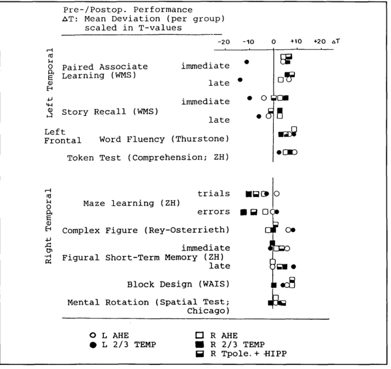
Although more detailed neuropsychological studies of the effects of AHE have to be done, the main findings of the study of Birri et al34 remain still valid (see Figure 6). The updated
results based on 70 patients showing the pre-/postoperative per-formance changes obtained with Nadig's learning and memory test are shown in Figure 7. The learning performance was exam-ined by the Nadig's test consisting of three series of 15 items each drawn or written in black ink on white cards (15 xlO cm). The items of the first series were nonsense designs ("designs"). The second consisted of drawings of common objects ("draw-ings") and the third of concrete nouns ("nouns"). The items were presented until the patient was able to reproduce at least 12 items, but for a maximum of 5 trials. The reproduction of as
Pre-/Postop. Performance
AT: Mean Deviation (per group)
scaled in T-values
-20
o Paired Associate immediate
§* Learning (WMS)
<u late
EH£j immediate
u Story Recall (WMS)
late
Left
Frontal Word Fluency (Thurstone)
Token Test (Comprehension; ZH)
^ trials
^ Maze learning (ZH)
a errors
E
E"
1Complex Figure (Rey-Osterrieth)
•§, immediate
TJ Figural Short-Term Memory (ZH)
m
late
Block Design (WAIS)
Mental Rotation (Spatial Test;
Chicago)
-10 0 +10 • •a
• o
6am
• SB
• B DC
«£
• O D
o
a f
cm
not •
i
&
O L AHE D R AHE
• L 2/3 TEMP • R 2/3 TEMP
0 R Tpole, + -HIPP
+20 A TFigure 6 — Comparison of pre-/postoperative performance in patients with different types of temporal lobe resections for treatment of medically refractory complex partial seizures: Selective amygdalohippocampectomy (AHE; left N = 3, right N = 3), removal of right temporal polar area, amygdala and hippocampal formation (R Tpole + HIPP; N = 3), and "classical" two-thirds temporal lobe resection (2/3 TEMP; left N = 2; right N = 3). (Modifiedfrom Birri et al., 1982).
many items as possible of each series (design, drawing, nouns) after a distraction interval of 30 minutes made up the memory performance. In order to avoid memory savings from pre- to postoperative testing, two similar but not identical parallel ver-sions (A/B) of this test were used. Half of the patients were test-ed preoperatively with version A and postoperatively with Version B and vice versa.
We have recently examined the role of TL lesions in memo-rizing auditory nonverbal material (pure tones). Patients treated with selective AHE were included in this study. Controls and patients with mesiobasal TL or frontal lesions had to identify target tones in series consisted of target and distractor tones ran-domly presented. Patients with right TL lesions showed markedly reduced task performances when more than three different target tones had to be identified. The decrement in performance of these patients was attributed to poor structuring of the stimuli to reduce memory load and to lack of selection for adequate response. This study37 confirms the results presented by the
Montreal group analyzing data from patients with varying extents of unilateral hippocampal excisions38. We support that the hippocampus plays a role in some, but not all, memory tasks depending on the kind of stimuli and complexity of the task. Even small differences in the experimental protocol may be suf-ficient to lower the performance on a task that normally is not under the influence of the hippocampal function.
CONCLUSION
Selective AHE offers a high probability of cure only in patients with a well-defined, precisely localized "epileptogenic area", i.e., seizure focus. Only a small portion of all patients with epilepsy can meet the criteria for selective surgical inter-ventions. However, we conclude that if the criteria are met, selective AHE is preferable to the "standard" anterior temporal lobectomy and represents an approach towards a more selective but still effective surgical treatment.
12© 1 0
-t
> 8 -O y UJ 6 "4-
2-o ^ 1 11
oc
-• i
y \
r
0 ! 1 1 P R E POST P R E -N 0 U -N S — 1 1 — D E S D t f j T C n flllF l u u n i J U L m i l .i
C
. >T
1
o •• POST P R E -I G N S H I N i i i ic
Pos
D U N S LEFT S > >1
i
T- PRE-— I rPRE-—D E S - i AlIP i L HI1L25-
20-9
15
"
•10-
5- POST-I G N S H H •t
- o
CDA R
N
I
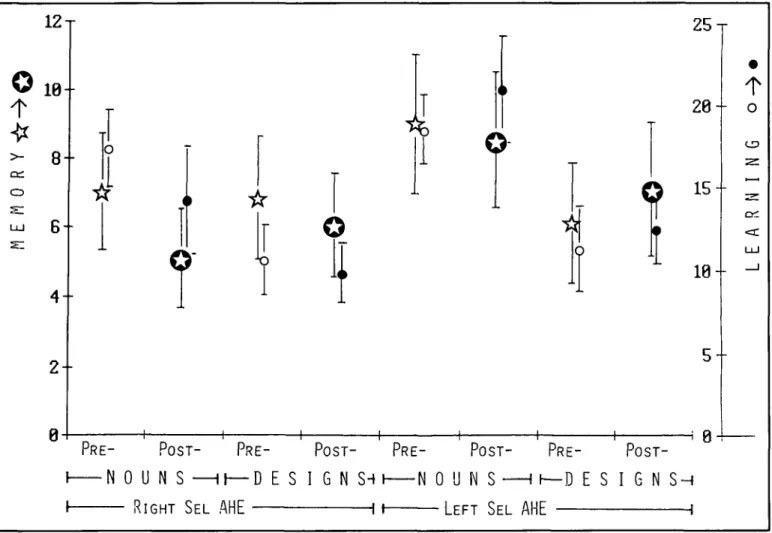
1Figure 7 — Pre- and postoperative performance changes obtained with Nadig's learning and memory test battery in 35 patients with right and 35 patients with left selective amygdalohippocampectomy. Stars refer to memory, circles to learning. Empty symbols indicate preoperation results, full symbols are postoperative. Symbols represent means and standard deviation. The learning performance is based on the number of trials
nec-essary to achieve the correct reproduction of 12115 items (see text). The performance values of the learning scale (0-25) have the following mean-ings: 4=2 trials, 8 = 3 trials, 12 =4 trials, 16 = 5 trials.
If at the fifth trial the patient is still unable to reproduce 12/15 items, the sum of the correctly reproduced items are taken into account. The memo-ry scale (0-12, left) indicates the number of items that could OM be reproduced after a distraction interval of 30 minutes, i.e. a memomemo-ry perfor-mance of 5 indicates that 10 out of 15 items were correctly reproduced, whereas a score of 8 means that only 7 items were correctly reproduced.
I want to thank Dr. Isabelle Germano for editorial suggestions.
R E F E R E N C E S
1. Jasper HH. Localized analyses of the function of the human brain by the electroencephalogram. Arch Neurol & Psychiat 1936; 36:
1131-1134.
2. Penfield W, Jasper H. Epilepsy and the Functional Anatomy of the Human Brain. Little, Brown, Boston; 1954.
3. Feindel W. Temporal lobe seizures. In: Magnus O, Lorentz de Haas AM, eds. The Epilepsies (Handbook of Clinical Neurology, Vol.
15). North-Holland Publ Company, Amsterdam, 1974; 87-106. 4. Rasmussen T. Surgical treatment of patients with complex partial
seizures. In: Penry JK, Daly DD, eds. Advances in Neurology. Raven Press, New York, 1975; Vol. 11, 415-449.
5. Gloor P. Electroencephalography and the role of intracerebral depth electrode recordings in the selection of patients for surgical treat-ment of epilepsy. In: Porter RJ, Mattson R, Ward AA, Dam M, eds. Advances in Epileptology: XVth Epilepsy International Symposium. Raven Press, New York; 1984: 433-437.
6. Milner B, Branch C, Rasmussen T. Study of Short-term memory after intracarotid injection of Sodium Amytal. Trans Am Neurol Assoc 1962; 87: 224-226.
7. Wieser HG. Electroclinical features of the psychomotor seizure. Gustav Fischer-Butterworths, Stuttgart-London; 1983.
8. Williamson PD, Wieser HG, Delgado-Escueta AV. Clinical charac-teristics of partial seizures. In: Engel J Jr, ed. Surgical Treatment of the Epilepsies. Raven Press, New York; 1987; 101-120. 9. Wieser HG, Moser S. Improved multipolar foramen ovale electrode
monitoring. J Epilepsy 1988; 1: 13-22.
10. Quesney LF, Gloor P. Special extracranial electrodes. In: Wieser HG, Elger CE, eds. Presurgical Evaluation of Epileptics. Springer, Berlin, Heidelberg, New York; 1987: 162-176. 11. Wieser HG. Selective amygdalohippocampectomy: Indications,
investigative technique and results. In: Symon L et al., eds. Advances and Technical Standards in Neurosurgery. Springer, Vienna, New York; 1986a; Vol. 13: 39-133.
12. Wieser HG. Psychomotor seizures of hippocampal-amygdalar ori-gin. In: Pedley TA, Meldrum BS, eds. Recent Advances in Epilepsy 3. Churchill Livingstone, Edinburgh, London, Melbourne, New York; 1986b; 57-79.
13. Wieser HG. The phenomenology of limbic seizures. In: Wieser HG, Speckman EJ, Engel J Jr, eds. The Epileptic Focus. John Libbey & Co, London, Paris; 1987a; 113-136.
14. Wieser HG, Yasargil MG. Selective amygdalohippocampectomy as a surgical treatment of mesiobasal limbic epilepsy. Surg Neurol
1982; 17: 445-457.
15. Yasargil MG, Teddy PJ, Roth P. Selective amygdalohippocampec-tomy: operative anatomy and surgical technique. In: Symon L et al., eds. Advances and Technical Standard in Neurosurgery, Vol.
12. Springer, Vienna, New York; 1985; 93-123.
16. Wieser HG. Selective amygdalo-hippocampectomy for temporal lobe epilepsy. Epilepsia 29, Suppl 2, 1988b; 100-113.
17. Yasargil MG, Wieser HG. Selective amygdalophippocampectomy at the University Hospital, Zurich. In: Engel Jr J, ed. Surgical Treatment of the Epilepsies. Raven Press, New York; 1987a; 653-658.
18. Yasargil MG, Wieser HG. Selective microsurgical resections. In: Wieser HG, Elger CE, eds. Presurgical Evaluation of Epileptics. Springer, Berlin, Heidelberg, New York; 1987b; 352-360. 19. Siegel AM, Wieser HG, Wichman W, et al. Relationship between
MR-imaged total amount of tissue removed, resection scores of
come following selective amygdalohippocampectomy. Epilepsy Res 1990; 6: 55-65.
20. Siegel AM, Wieser HG. A detailed follow-up study on the Zurich amygdalhippocampectomy series. J Epilepsy (submitted); 1991. 21. Wieser HG, Siegel AM. Analysis of foramen ovale electrode
recorded seizures and correlation with outcome following amyg-dalohippocampectomy. Epilepsia (submitted); 1990.
22. Yasargil MG. Microneurosurgery, Vol. 1. Thieme, Stuttgart; 1984. 23. Renella RR. Microsurgery of the Temporo Medial Region.
Springer-Verlag, Wien, New York; 1989.
24. Wieser HG. Stereo-electroencephalography. In: Wieser HG, Elger CE, eds. Presurgical Evaluation of Epileptics. Springer, Berlin, Heidelberg; 1987b; 192-204.
25. Wieser HG, Valavanis A, Roos A, Isler P, Renella RR. "Selective" and "superselective" temporal lobe Amytal tests: 1. Neuroradiological, neuroanatomical, and electrical data. In: Manelis J, Bental E, Loeber JN, Dreifuss F, eds. Advances in Epileptology, Vol. 17. Raven Press, New York; 1989a; 20-27. 26. Wieser HG, Landis T, Regard M, Schiess R. "Selective" and
"superselective" temporal lobe Amytal tests: 2. Neuropsycho-logical test procedure and results. In: Manelis J, Bental E, Loeber JN, Dreifuss F. eds. Advance in Epileptology, Vol. 17. Raven Press, New York; 1989b; 28-33.
27. Wada J. A new method for the determination of the side of cerebral speech dominance. A preliminary report on the intracarotid injection of Sodium Amytal in man. Igaku to Seibutssugaku (Medicine and Biology), 1949; 14: 221-222.
28. Wada J, Rasmussen T. Intracarotid injection of Sodium Amytal for the lateralization of cerebral speech dominance. Observations on 123 patients. J Neurosurg 1960; 17: 266-282.
29. Wieser HG. Selective Amytal memory tests: correlation with post-operative results. Epilepsia 1989; 30: (5), 724.
30. Babb TL, Brown WJ. Neuronal, dentritic, and vascular profiles of human temporal lobe epilepsy correlated with cellular physiolo-gy in vivo. In: Delgado-Escueta AV, Ward AA Jr, Woodbury DM, Porter RJ, eds. Basic Mechanisms of the Epilepsies. Advances in Neurology, Vol 44. Raven Press, New York; 1986; 949-966.
31. Wieser HG. Human limbic seizures: EEG studies, origin, and pat-terns of spread. In: Meldrum BS, Ferrendelli JA, Wieser HG, eds. Anatomy of Epileptogenesis. John Libbey & Co, London, Paris; 1988a; 127-138.
32. Duvernoy HM. The Human Hippocampus. Bergman Verlag, Munich; 1988.
33. Engel J Jr. Outcome with respect to epileptic seizures. In: Engel J Jr, ed. Surgical Treatment of the Epilepsies. Raven Press, New York; 1987; 553-571.
34. Birri R, Perre E, Wieser HG. Der Einfluss verschiedener Temporallappenoperationen auf das Gedachtnis bei Epileptikern. Nervenarzt 1982; 53: 144-149.
35. Nadig T, Wieser HG, Perret E. Learning and memory performance before and after unilateral selective amygdalohippocampectomy.
In: Will BE, Schmitt P, Dalrymple-Alford JC, eds. Brain
Plasticity, Learning and Memory. Plenum Publ Co, New York; 1985: 397-403.
36. Gosner A, Perret E, Wieser HG. 1st der Hippokampus fur Lernund Gedachtnisprozess notwendig? Nervenarzt; 1986; 57: 269-275. 37. Wittlieb-Verpoort E, Wieser HG. Tone discrimination in patients
with temporal lobe lesion (submitted to Neuropsychologia); 1991. 38. Jones-Gotman M. Comment: psychological evaluation. In: Engel J Jr, ed. Surgical Treatment of the Epilepsies. Raven Press, New York; 1987; 197-201.