Patricia Lariguet, Christophe Dunand
Department of Plant Biology, University of Geneva, Quai Ernest Ansermet 30, CH-1211 Geneva 4, Switzerland Received: 5 October 2004 / Accepted: 4 June 2005 [Reviewing Editor: Dr. Ru¨diger Cerff]
Abstract. Plants possess photoreceptors to perceive light which controls most aspects of their lives. Three photoreceptor families are well characterized: cryp-tochromes (crys), phototropins (phots), and phyto-chromes (phys). Two putative families have been identified more recently: Zeitlupes (ZTLs) and UV-B photoreceptors (ULI). Using Arabidopsis thaliana and Oryza sativa photoreceptor sequences as refer-ences, we have searched for photoreceptor encoding genes in the major phyla of plant kingdom. For each photoreceptor family, using a phylogenetic tree based on the alignment of conserved amino acid sequences, we have tried to trace back the evolution and the emergence of the diverse photoreceptor ancestral se-quences.The green alga Chlamydomonas contains one cry and one phot sequence, probably close to the corresponding ancestral sequences, and no phy-re-lated sequence. The putative UV-B photoreceptors seem to be restricted to the Brassicacae. Except for mosses and ferns, which contain divergent photore-ceptor numbers, the composition of the diverse pho-toreceptor families is conserved between species. A high conservation of the residues within domains is observed in each photoreceptor family. The complete phylogenic analysis of the photoreceptor families in plants has confirmed the existence of crucial evolu-tionary nodes between the major phyla. For each photoreceptor class, a major duplication occurred before the separation between Mono- and Eudicoty-ledons. This allowed postulating on the putative ancestral function of the photoreceptors.
Key words: Cryptochromes — Phototropins — Phylogeny — Phytochromes — UV-B photoreceptors — Zeitlupes
Introduction
Because plants are sessile in nature, their survival and reproduction depend on their ability to adapt their development in response to environmental changes such as light, water availability, temperature, and pathogens. In particular, land plants need to deal with various light environments depending on the degree of shading by soil, foliage, or clouds, time of day, and time of year. Plants are able to perceive mainly the UV, blue (B), green, red (R), and far-red (FR) wavebands through different photoreceptor families (Fig. 1): cryptochromes (crys), phototropins (phots), phytochromes (phys), zeitlupes (ZLPs), and a putative UV-B photoreceptor (Gyula et al. 2003; Somers et al. 2000; Suesslin and Frohnmeyer 2003).
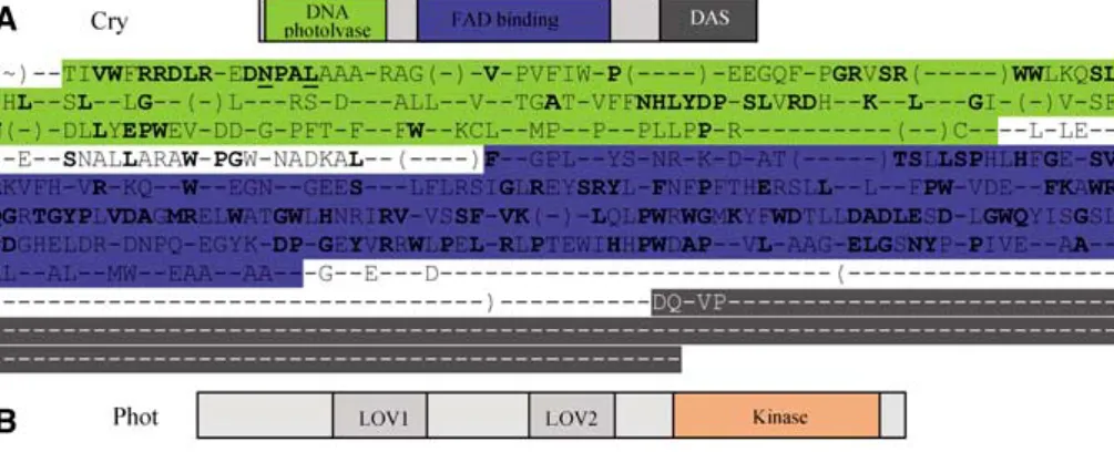
Crys flavoproteins are receptors perceiving B and UV-A radiations (320–520 nm). They consist of three members, cry1, cry2(Lin 2000), and CryDASH (cry3 in Arabidopsis) (Brudler et al. 2003). Crys flavopro-teins are responsible for photomorphogenesis, flow-ering, and clock resetting in plants (Lin and Shalitin 2003). They have sequence homologies with light-dependent DNA repair photolyases without DNA photolyase activity (Sancar 2003). Photolyases and crys are characterized by a flavin adenine dinucleo-tide (FAD) chromophore and contain either deaza-flavin or pterin as light-harvesting chromophore
Correspondence to: Christophe Dunand; email: christophe.
(Sancar 1994) (Fig. 2A). In addition, plant crys flavoproteins contain an extra C-Term extension, which can interact with COP1, a E3 ubiquitin ligase (Wang et al. 2001; Yang et al. 2001). This extension is not present in the CryDASH family. Crys homo-logues are apparently present in several phyla including animals, yeasts, plants, and bacteria (Daiyasu et al. 2004; Sancar 2003). In animals they function as circadian clock components, possibly within the clock oscillator in Vertebrates and in the clock input in Drosophila (Panda et al. 2002). Phy-logenetic analysis of the crys/photolyase family re-vealed five evolution branches (Lin 2000b; Brudler et al 2003). However, those studies included only a few plants, leading to limited knowledge concerning the evolution of crys in plants.
The phots family is composed in Arabidopsis of two members, namely, phot1 and phot2(Briggs and Christie 2002), and perceives UV-A, UV-B, and green lights (320–550 nm). Their main functions are pho-totropism, chloroplast relocation, and stomata opening (Briggs and Christie 2002; Kinoshita et al. 2001). Phots are flavoproteins containing two LOV domains at the N-terminal and a serine/threonine kinase domain at the C-terminal part (Fig. 2B) (Hu-ala et al. 1997; Sakai et al. 2001). They represent a class of receptor kinases that appear to be exclusive to plants, although no phylogenetic analysis is available to our knowledge (Briggs and Christie 2002).
Arabidopsis has five distinct phys, phyA- to -E, which are broad-range sensing photoreceptors with maximum absorption for R and FR lights (600– 750 nm). PhyA and phyB can also act as B light photoreceptors (Lagarias et al. 1997; Lariguet and Fankhauser 2004b; Moller et al. 2002). They have different but overlapping functions in photomor-phogenesis, shade avoidance, clock resetting, and gravitropism inhibition (Lariguet and Fankhauser 2004a; Moller et al. 2002). The phytochrome
apoproteins contain a chromophore binding site in the N-terminal part and two histidine kinase-related domains (HKRD1 and -2). The HKRD1 domain contains two PAS domains with a putative function for protein–protein interaction (Fig. 2C). Several phylogenetic analyses were performed concerning phys (Alba et al. 2000a; Lamparter 2004; Mathews et al. 2003; Mathews and Sharrock; Montgomery and Lagarias 2002). They have shown that phy-like genes are present not only in all green plants but also in certain bacteria and fungi (Lamparter 2004; Mathews and Sharrock 1997). The plant phy family results from several gene duplications, with the first one preceding the origin of seed plants (Mathews and Sharrock 1997). An exhaustive phylogenetic analysis was performed in Gymnosperms (Schmidt and Schneider-Poetsch 2002). But the use of very small phys Gymnosperm sequences did not allow a global analysis with the phy sequences of other phyla. No global phylogenetic analyses including Gymnosperm, Angiosperm, and older phy sequences such as Bryo-phytes and PteridoBryo-phytes have been reported. This global approach would allow visualizing the hot spot of the phy sequences evolution.
The ZTL family consists of three members in Arabidopsis—ZTL; flavin-binding, kelch repeat,
F-box1 (FKF1); and LOV kelch protein 2
(LKP2)—and is predominantly involved in the con-trol of the circadian period in plants (Imaizumi et al. 2003; Mas et al. 2003; Schultz et al. 2001; Somers et al. 2000). They possess a PAS-like LOV domain, an F-box domain, and six kelch repeats (Fig. 2D) (Somers et al. 2000). The PAS region is highly similar to the LOV domain of phot1, the F-box is involved in substrate recognition for degradation, and the kelch repeats are implicated in protein–protein interactions. Therefore the ZTL proteins could be involved in the degradation of circadian clock components (Han et al. 2004). The similarity of the ZTL PAS domain to the phots LOV domain and the strong light
depen-Fig. 1. The plant photoreceptor
absorption zones. The wavelength ranges perceived by the different known and putative Arabidopsis photoreceptors have been are visualized. Note that 550–660 nm (yellow) does not seem to be sensed by any known photoreceptor, while 450–520 nm is absorbed by crys, phots, phys, and ZTL. Modified from Sullivan and Deng (2003).
dence of their functions suggest that ZTL may be a novel class of blue light receptors (Imaizumi et al. 2003; Somers et al. 2000). ZTL sequences were found in Arabidopsis thaliana, Adiantum capillus-veneris, and Neurospora crassa, but with very low amino acid identity (Somers et al. 2000).
The UV-B region (280–320 nm) is not entirely absorbed by the ozone layer. In plants, a portion of this radiation not only triggers multiple cellular
damages, but also could act as an informational signal. The above-mentioned photoreceptors are apparently not the UV-B sensors (Suesslin and Frohnmeyer 2003). Recently, ULI3 has been iden-tified as a component of the UV-B signaling path-way. ULI3 encodes for an unknown protein containing a putative heme and diacylglycerol binding sites (Suesslin and Frohnmeyer 2003). To our knowledge, no data are available in the
litera-Fig. 2. Crys, phots, phys, and
ZTLs. Primary structure, domains, and consensus sequences. (A) Cryptochromes; (B) phototropins; (C) phytochromes; (D) zeitlupes. Each domain is represented on the schematic structure and on the consensus sequences. The 60% consensus sequences were obtained from the complete sequences. The 100% consensus is represented in boldface. The residues underlined are also conserved in the non–green plant organisms. The sequences in brackets correspond to regions that are variable or not present in all the genes.
ture concerning the repartition of those putative UV-B sensors in the tree of life.
Numerous publications report independent studies of the different plant photoreceptors or the situation in a single specie. Until now, the presence of the different plant photoreceptors in the various species and their evolution have not been described in an exhaustive manner. The availability of an increasing number of plant sequences gave us the opportunity to search for photoreceptor encoding sequences in the major phyla of the plant kingdom. The elucidation of the individual photoreceptor function is often diffi-cult due to their redundant and overlapping action. For each family of photoreceptors, using a phyloge-netic tree based on the alignment of conserved amino acid sequences, we have traced back the evolution and the emergence of the diverse photoreceptors from an ancestral sequence. We have also tried to correlate the presence or the absence of particular photore-ceptors with specific roles in the plant.
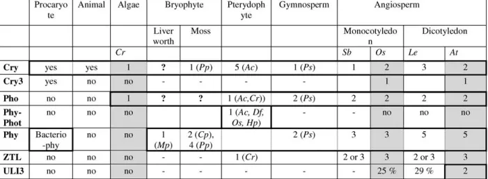
Our global phylogenetic analysis revealed that crys are highly divergent in ferns, phots are specific to green plants, and Chlamydomonas possesses only one cry and one phot, probably close to the origin of crys and phots plant sequences but no phys. ZLPs were found in Pteridophytes and Angiosperms. The puta-tive UV-B photoreceptor ULI3 is actually restricted to Brassicacea. The composition of the nonmoss and nonfern photoreceptor families is conserved between species. The existence of a crucial evolution node suggested by partial crys analysis was confirmed: the cry1 and the cry2branches were separated before the divergence between the Mono- and the Dicotyledons. The same appeared to be true for phots, where the phot1 and phot2branches separated before the divergence between the two clades. Finally, the time of occurrence of the evolutionally nodes of phys was specified.
Materials and Methods
Retrieval of the Photoreceptor Sequences
Arabidopsis photoreceptor protein sequences from the different
photoreceptor families have been used as a starting point for the data mining. Each of the different amino acid sequences of
Ara-bidopsiswas submitted against the whole genomic rice database
with a tblastn search on the Rice Genome Project (RGP) Web site (http://rgp.dna.affrc.go.jp/).
In order to find photoreceptor sequences in other organisms, protein sequences from rice or Arabidopsis were used as input se-quences in TBLASTN searches in different databases. Photore-ceptors were sought at the NCBI Web site (www.ncbi.nih.gov/ BLAST) and in several specialized databases such as the PEP EST
database (www.moss.leeds.ac.uk), the PlantGDB database
(www.plantgdb.org/), the DOE Joint Genome Institute (JGI) Web site (genome.jgi-psf.org/), MaizeGDB (www.maizegdb.org/), the Solanaceae genomic network (soldb.cit.cornell.edu), and the Plant Genome network (http://pgn.cornell.edu/). Nonannotated
se-quences were analyzed for the presence of the gene with different
programs such as FGenesh
(http://www.softberry.com/ber-ry.phtml) and GenScan (http://genes.mit.edu/GENSCAN.html). The corresponding coding sequence (CDS) was translated with the ‘‘translate’’ tool on Expasy (http://us.expasy.org/tools/dna.html) and controlled for specific motifs.
The photoreceptor sequences of the following organisms have been used for the comprehensive phylogenetic analysis. Adiantum capillus-veneris, Ac; Arabidopsis thaliana, At; Agrobacterium
tum-efaciens,At; Avena Sativa, As; Ceratopteris richardii, Cr; Ceratodon
purpureus, Cp; Chlamydomonas reinhardtii, Cr; Dryopteris filix-mas, Df; Gloeobacter violaceus, Gv; Hypolepis punctata, Hp; Ip-omoea nil, In; Lycopersicon esculantum, Le; Marchantia
polymor-pha, Mp; Mesembryanthemum crystallinum, Mc; Mougeotia
scalaris, Ms; Neurospora crassa, Nc; Nicotiana plumbaginifolia, Np; Nicotiana tabaccum, Nt; Anabaena (Nostoc), Nos; Onoclea sensi-bilis, Os; Oryza sativa, Os; Physcomitrella patens, Pp; Pinus
syl-vestris,Ps; Pisum sativum, Ps; Populus balsamifera, Pb; Rhyzobium
leguminosarum, Rl; Sorghum bicolor, Sb; Solanum tuberosum, St; Spinacia oleracea, So; Stellaria longipes, Sl; Tricicum aestivum, Ta; Vicia faba, Vf; Xanthomonas axonopodis, Xa; and Xenopus laevis,
Xl.All corresponding accession numbers are summarized in Fig. 3
and Supplemental Table.
Comprehensive Phylogenetic Analysis of Photoreceptor Sequences Identified in Plants
Among the high number of available photoreceptor sequences in the databases, only two sequences for each photoreceptor in Mono-and Eudicotyledons phyla were used to build the different trees. Complete sequences were used in most cases when the available sequences allowed it. The very short sequence of PsPhyO was aligned only with the Arabidopsis phys and the two other Pinus sequences. In the global phys tree, the branch length of PsPhyO is approximate, but not the position. Photoreceptor protein se-quences present in various plants were aligned using Clustal W (Thompson et al. 1994). The alignment was further inspected and visually adjusted and realigned with Clustal X. Due to the large number of sequences analyzed; only a distance analysis was per-formed. The distance tree was constructed with the NEIGHBOR option of the PHYLIP 3.6a3 package (Felsenstein 1993) under the JTT substitution frequency matrix, and 1000 bootstrap replicates were carried out. The maximum likelihood tree was inferred with the PHYML algorithm, under the JTT substitution frequency matrix, by using the BIONJ starting tree (Guindon and Gascuel 2003). Njplot software was used to visualize phylogenetic trees, and BioEdit software to obtain the different consensus sequences. The consensus sequences represented in Fig. 2correspond to the com-plete sequences of plants; 100% and 60% conserved residues are represented.
Results Cryptochromes
Crys have homologous isoforms in all kingdoms (Cashmore et al. 1999). The crys/photolyase protein family is divided into five isolated and independent phylogenetic branches: the classes I and II CPD photolyases, the CryDASH, the plant crys, and, fi-nally, the (6-4) photolyases and animals crys together (Brudler et al. 2003). Very few data were available in plants. The evolutional situation in plants has been investigated by looking at sequences of a large
number of representative plant organisms. Except for the class I CPD photolyases, which do not exist in plants, the four other divisions were confirmed (Fig. 4). The class II CPD photolyases branch forms an independent group with any duplication suggest-ing that it is closer to an ancestral sequence. This ancestral branch led to the emergence of three inde-pendent phylogenetic groups: CryDASH, (6-4) pho-tolyases, and plant cryptochromes (step 1 and step 1bis; Fig. 4). Interestingly the green alga Chlamydo-monas reinhardtiicontained only a single cry sequence (CrCPH; shaded gray in Fig. 4). The Chlamydomonas cry sequence has been situated between the bacterio-cry sequence from Agrobacterium tumefaciens (At-Cry) and the other plant cry sequences. Therefore the origin of the plant cryptochromes could be related to the unicellular green alga cry sequence (Fig. 4). The duplication of this single ancestral sequence led to cry1 and cry2sequences, found, respectively, in two independent branches (step 2; Fig. 4). Mono- and Eudicotyledon cry sequences were present in both cry1 and cry2branches. This indicated that the duplication event that resulted in the separation of these cry1/cry2branches occurred after the origin of the seed plants but before the Mono- and Eudicoty-ledon separation (step 3; Fig. 4). This idea was con-firmed by the existence of two independent cry sequences in Amborella tricopoda, a basal Angio-sperm. Unfortunately these sequences were too short to be incorporated in the phylogentic analysis. Sur-prisingly, five cry isoforms resulting from four duplication events were present in the fern Adiantum (AcCry1 to AcCry5) and constituted an independent group in the cry1 branch. Apart from the special cases of Adiantum and of Lycopersicon, which con-tain two cry1 sequences (LeCryc and -b), two copies
of cry sequences were detected in all examined spe-cies. The separation between the two clades (Mono-and Eudicotyledons) is well respected.
The existence of homologies in Prokaryotes for cry1/2and CryDASH, respectively, supported the idea that cry1/2and CryDASH resulted from one duplication event in an ancestral organism and evolved separately after the divergence of Prokaryotes and Eukaryotes (Brudler et al. 2003) (Fig. 4). How-ever, the possibility of convergent evolution within the cry family remains. This idea was also supported by the molecular structure: the DNA photolyase and FAD binding domains were highly conserved through the various species and between the different cry se-quences, whereas the C-Term DAS domain, which was absent in CryDASH sequences, was poorly con-served within the cry1/2branch (Fig. 2A). Therefore, the conserved region (DNA photolyase and FAD binding domains) of the crys probably corresponds to the ancestral sequence. The gene structure also dif-fered between CryDASH and cry1/2, which con-tained, respectively, 11 and 3 introns in Arabidopsis. These divergences could be directly the result of the evolution of cry1/2. This hypothesis was supported by the absence of CryDASH sequence in Chlamydo-monasand the low level of identities between the plant and the animal sequences (27% between Arabidopsis and human). Despite the significance of the acronym DASH (Drosophila, Arabidopsis, Synechocystis, Hu-man) (Brudler et al. 2003), cryDASH sequences were not found in insects or in Mammals.
Phototropins
Contrary to crys that exist in various phyla, phots sequences were found only in green organisms
Fig. 3. Summary of the photoreceptors composition in the
dif-ferent phyla. Gray boxes correspond to completed genome pro-jects. Question marks represent the absence of photoreceptor sequence, probably due to the low number of EST. Minus marks correspond to the absence of photoreceptor sequence. Plus marks
are used for photoreceptor sequences that are present in various numbers according to the species. The cells surrounded by bold lines correspond to a group of organisms in which the emergence and the evolution of a photoreceptor family are well defined. For the initials of the species refer to Materials and Methods.
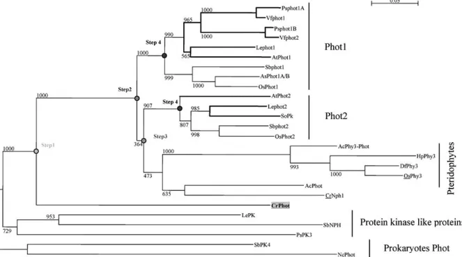
(Fig. 5). To our knowledge, no phot phylogenetic analysis has been reported in plants. Our analysis showed that Chlamydomonas contained only one phot sequence, called CrPhot1. CrPhot1 was anterior to the plant phot ancestors and independent of the protein kinase-like proteins (used to root the phot branch). Thus CrPhot1 could be related to the phot ancestral sequence from which the plant phots evolved (step 1; Fig. 5). No phot-encoding sequence has been found in liverworts and mosses, probably due to the low level of expression of phots (for example, four ESTs in Arabidopsis). One major duplication event seemed to have occurred since the origin of the seed plants, which generated the Phot1 and Phot2branches (step 2; Fig. 5). Phy–Phot chimeric sequences were found in Pteridophytes (Kawai et al. 2003). They formed an independent branch from the seed plant phots and have evolved from one single phot sequence after the appearance of the seed plants (step 3; Fig. 5). Both phot1 and phot2branches contained Mono- and Eudicotyledon phot sequences. This indicated that the Mono- and Eudicotyledon separation occurred after the duplication event that led to the separation of the phot1/phot2branches (step 4; Fig. 5). A more recent duplication led to the existence of two Phot1 se-quences in the Eudicotyledonous Pisum sativum and Vicia faba.
At the molecular level, the sequences of the two LOV domains and kinase domain were highly
con-served between the isoforms and the species (Fig. 2B). Supporting the importance of these do-mains, the interdomain sequences were poorly con-served.
Phytochromes
Phys are present in plants, prokaryotes, and fungi (Montgomery and Lagarias 2002). The ancestral phytochrome-like protein could have originated both the bacteriophytochromes and the phys of photo-synthetic organisms (step 1; Fig. 6). Surprisingly, no phy-like protein sequence has been found in Chla-mydomonas. From the emergence of the terrestrial plants to the higher plants, the phy family has fol-lowed several evolutions. As for crys, Bryophytes and Pteridophytes phys sequences constituted an inde-pendent branch versus phys of higher plants (step 2; Fig. 6). Phylogenetic analyses of Gymno- and An-giosperms phys protein sequences revealed four ma-jor duplication events. Analysis of complete or significant sequences indicated that Pinus contained three phys as previously shown in a study dedicated to the Gymnosperm phys and using short sequences (Schmidt and Schneider-Poetsch 2002). The global phylogenetic analysis in the major plant phyla al-lowed positioning of the first two duplications. PsphyN and PsphyO from Pinus were related to phyA and phyC, respectively, and PsPhyP to the
Fig. 4. Phylogenetic relationships of the cryptochromes from
plants based on protein sequence alignments. Several bacterio-cryptochrome protein sequences were used to root the tree. The bold lines represent cry sequences from Eudicotyledons. The main
nodes corresponding to putative duplications are symbolized with a gray circle and a step number. All branches are drawn to scale and the scale bar represents 0.05 substitution per nucleotide.
phyB/D/E branch. Therefore, the first two duplica-tions arose before the separation between Gymno-sperms and AngioGymno-sperms. The first one generated the phyA/C and phyB/D/E lines (step 3; Fig. 6) and the second one led to the separation between phyA to phyC (step 4; Fig. 6).
In Angiosperms, two independent events separated phyB/D from phyE (step 5; Fig. 6) and phyB from phyD (step 6; Fig. 6). Only one sequence of the phyB/ D/E subfamily was found in the Monocotyledons. This suggested that the two latest duplication events within the phyB/D/E branch likely occurred after the separation of the two clades and only in the eudi-cotyledonous plants. A special evolution could be observed for the last duplication between PhyB and phyD. PhyB- and phyD-related sequences were closer between paralogues than between orthologues (sup-plemental data) (Pratt 1995). Two hypotheses could explain this phenomenon. First, the last duplication could have occurred independently in the various eudicotyledonous species. Second, and more proba-bly, the phyB/D separation occurred before the emergence of the major Eudicotyledon families and an evolutional convergence within each species led to the actual phylogenetic situation.
The level of phys sequence conservation, even within domains, is much lower than that of the phots and crys photoreceptors (Fig. 2C). This low conser-vation rate could be due to the higher number of duplications (steps 1 to 6; Fig. 6).
Zeitlupe
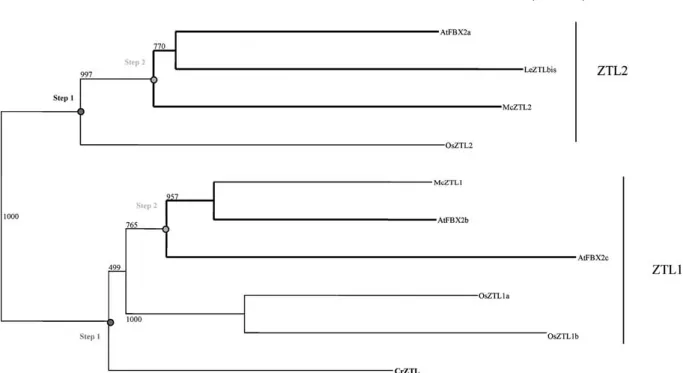
Concerning plants, previous studies revealed the presence of ZTLs only in Adiantum capillus-veneris and in Arabidopsis thaliana (Somers et al. 2000). The analysis we conducted showed that ZTL sequences were actually present in Pteridophytes and Angio-sperms (Fig. 7). No ZTL homologue was found in prokaryotic organisms, in Chlamydomonas, in Bryo-phytes, in Gymnosperms, or in animals. The small size of the EST databases and the low expression level of the ZTL gene (10 ESTs in Arabidopsis) might ex-plain the absence of ZTL homologues in Gymno-sperm and Bryophyte species. Interestingly, the ZTL sequence contained homologies with the two phot LOV domains. Three homologues have been found in the Mono- and Eudicotyledons, resulting from two duplication events (Fig. 7). Incomplete sequences from Sorghum bicolor and Lycopersicum were not represented but confirmed the previous idea. The first duplication occurred before the separation of Mono-and Eudicotyledons Mono-and led to the separation of the ZTL1 and ZTL2branches (step 1; Fig. 7). The sec-ond appeared more recently, after the separation between Mono- and Eudicotyledons (step 2; Fig. 7). The ZTL sequence from the fern Ceratopteris, named CrZTL, appeared in the AtFBX2b/c branch with a low bootstrap support. The weakness of its position and the fact that only one sequence of a Pteridophyte has been found suggested that CrZTL could be close
Fig. 5. Phylogenetic relationships of the phototropins from plants
based on protein sequence alignments. Phot sequences from prokaryotes were used to root the tree. The bold lines represent the phot sequences from Eudicotyledons. The main nodes
corre-sponding to putative duplications are symbolized by a gray circle and a step number. All branches are drawn to scale and the scale bar represents 0.05 substitution per nucleotide.
to the ancestral sequence. The observation that ZTLs appear to be present only in Pteridophytes and An-giosperms suggests that ZTLs arose later during plant
evolution and that they have arisen twice (in Pteri-dophytes and then in Angiosperms) or that they have been lost in Gymnosperms.
Fig. 6. Phylogenetic relationships of the phytochromes from
plants based on protein sequence alignments. Several bacterio-phytochrome protein sequences were used to root the tree. The gray and the bold lines represent the phy sequences from Gym-nosperms and Eudicotyledons, respectively. The approximate
length of the PsPhyO branch is represented by a dashed line. The main nodes corresponding to putative duplications are symbolized by a gray circle and a step number. All branches are drawn to scale and the scale bar represents 0.05 substitution per nucleotide.
Fig. 7. Phylogenetic relationships of the zeitlupes from plants based on protein sequence alignments. The bold lines represent the ZTL
sequences from Eudicotyledons. The main nodes corresponding to putative duplications are symbolized by a gray circle and a step number. All branches are drawn to scale and the scale bar represents 0.02substitution per nucleotide.
Brassicaceae-specific. Maximum-Likelihood Tree
Analyses with maximum-likelihood (ML) trees (data not shown) have been performed for each plant photoreceptor family and supported that the group-ing and evolution observed with the distance trees are not the result of long branch attraction.
Discussion
Monocotyledons (rice) and Eudicotyledons (Arabid-opsis) diverged from a common ancestor, some 150 MY ago (Wikstrom et al. 2001). It can be assumed that, at their origin, the first Monocotyledon and Eudicotyledon had a similar number of photorecep-tor genes. A differential evolution leading to todayÕs Arabidopsisand rice yields to a variation in the gene numbers and sequences. The evolution rate varies between the phyla but also between the photorecep-tor families and probably between the different do-mains. Indeed the N-Term domain (putative output domain) of the phy is evolving quickly (Alba et al. 2000b; Matsushita et al. 2003).
Stability in Photoreceptor Copy Number
Compared to the high duplication rate of the perox-idase genes (Passardi et al. 2004), the photoreceptor families showed a slow evolution in copy number since the emergence of the green plants. The large increase in peroxidase copy number could result from conservation of duplicated peroxidase genes through evolution. The elevated peroxidase copies could be related to the large range of physiological processes in which the proteins are implicated (Hiraga et al. 2001). On the contrary, the size conservation of the photo-receptor families could be associated with the major functions of the photoreceptors in light perception.
The photoreceptor families have followed a different evolution despite the conservation of gene number. Specific appearance and evolution of certain photo-receptor families could be related to particular phyla or to a modification of the biotope (Fig. 3). For example, in the phy family the separation between phyB/D/E and phyA/C occurred before the Mono-cot/Eudicotyledon divergence, whereas the separa-tion between phyB/D and phyE and between phyB from phyD occurred after the Monocot/Eudicotyle-don separation.
Within the different photoreceptor families, the homologies were higher between orthologues than between paralogues. This rule was not respected in the case of phyB and phyD. Indeed phyB- and phyD-related sequences were closer between paralogues within each Eudicotyledon group. This situation could be the result of a convergent evolution that led to the actual phylogenetic image of the Eudicotyle-don family evolution (Wikstrom et al. 2001). The result of exhaustive phyB/D/E phylogenetic analysis (supplementary data) showed, as expected, that Caryophyllales were anterior to Rosids and Asterids. In opposition to the usual phylogenetic analysis (Wikstrom et al. 2001), in our analysis, the Asterids looked posterior to the Rosids. This observation was strongly supported by high bootstraps. The discrep-ancies concerning the relative position of the major Eudicotyledon groups in global evolution could be addressed with a systematic research of the phyB-and phyD-related sequences phyB-and their phylogenetic analysis.
Photoreceptor Evolution and Functions
Many data concerning photoreceptor functions have been accumulated for Arabidopsis. They can be used as a reference to understand the function of the photoreceptors in relation to their evolution.
Phys and crys encoding sequences were found in many organisms with variations of the sequence and
an increase in copy numbers. Regarding their major role in circadian clock entrainment (Chen et al. 2004), these two protein families are probably important for a normal growth in these organisms. In Arabidopsis, phys and crys are also implicated in other light-reg-ulated processes such as flowering, de-etiolation, and gravitropism inhibition (Briggs et al. 2001; Lariguet and Fankhauser 2004a; Mockler et al. 2003). These specific functions could be achieved thanks to par-ticular phys or crys isoforms or to specific interac-tions of these photoreceptors with privileged partners. For example, the phy binding proteins Phytochrome Kinase Substrate (PKS) are only found in superior Angiosperms, and not in basal Angio-sperms such as Amborellacea and Gnetacea (sup-plementary data) or in ‘‘lower’’ organisms. Other partners, such as COP1, HY5, and DET1, are present in all plant organisms with high homology and in animals with only a lower level (30 to 40%) (data not shown).
PhyA, -B, and -E have a role in Arabidopsis seed germination (Sullivan and Deng 2003). PhyA and -B have existed since the Gymnosperms, while phyE is detected only in the Eudicotyledons. Therefore, it can be speculated that phyE could have a specific func-tion for the germinafunc-tion of Eudicotyledons. Shade avoidance is a mechanism used by the majority of Gymno- and Angiosperms to compete with plant neighbors (Gilbert et al. 2001). In addition to their own individual functions, phyB, -D, and -E, which are issued from a common ancestor, act redundantly to mediate Arabidopsis shade avoidance (Morelli and Ruberti 2002). The Pinus phy, a member of this branch, could concentrate all the functions in ger-mination of phy B, -D, and -E in a single molecule.
In Arabidopsis, phots are involved in photosyn-thesis optimization by controlling chloroplast move-ments, phototropism, and stomata opening (Briggs and Christie 2002; Celaya and Liscum 2004; Kinosh-ita et al. 2001). In accordance with the phot functions, only the chloroplast-containing organisms contain phot encoding sequences. Chlamydomonas contains a unique chloroplast that probably does not undergo relocation. In this case, optimization of light capture is accomplished by the movement of the organisms themselves (phototaxis) (Schaller et al. 1997). This unicellular algae possesses only one ancestral photo-tropin known to be required for multiple aspects of sexual development (Huang and Beck 2003) and able to function in Arabidopsis (Onodera et al. 2005). Chlamydomonaslacks phy. It is possible that its single phot is responsible for Chlamydomonas movement in response to blue light intensity. It was not possible to establish the presence of phot sequences in Bryo-phytes but phots exist in PterydoBryo-phytes. Since pho-totropism can be mediated by both phots and phys (in higher plants and mosses, respectively), determining if
phot sequences are actually absent from Bryophytes is an important issue.
Hybrid Phy–Phot in Ferns
Particular situations have been observed that di-verged from the usual photoreceptor sequences and numbers. Adiantum was of particular interest because it contained at least five crys, two phys, one phot, and one phy–phot hybrid. The lack of sequences (genomic or EST) does not allow us to determine if all the other phy–phot hybrid-containing ferns also possess a large number of other photoreceptors. Adiantum possesses highly divergent crys. Assuming that these Adiantum cry sequences are not splice variant, this observation implies that crys can undergo rapid gene duplication/ diversification in some plant species including Adi-antum. Phy–phot encoding sequence has been found only in a particular group of ferns. This hybrid se-quence probably appeared during a genomic rear-rangement which occurs rapidly after the emergence of the Pteridophytes. The acquisition of this so-called phy3 sequence could be a critical step for the prolif-eration of ferns in a low-light environment (Kawai et al. 2003).
Acknowledgments. We thank Filippo Passardi for his critical
reading and his constructive suggestions. We thank the University of Geneva and the Swiss National Science Foundation for research support: Grant 31-068003.02to C.D. and the Marie Heim-Vo¨gtlin program to P.L. and the Societe Acade´mique de Gene`ve.
References
Alba R, Cordonnier–Pratt MM, Pratt LH (2000a) Fruit-local-ized phytochromes regulate lycopene accumulation indepen-dently of ethylene production in tomato. Plant Physiol 123:363–370
Alba R, Kelmenson PM, Cordonnier–Pratt MM, Pratt LH (2000b) The phytochrome gene family in tomato and the rapid differ-ential evolution of this family in angiosperms. Mol Biol Evol 17:362–373
Briggs WR, Beck CF, Cashmore AR, Christie JM, Hughes J, Ja-rillo JA, Kagawa T, Kanegae H, Liscum E, Nagatani A, Okada K, Salomon M, Rudiger W, Sakai T, Takano M, Wada M, Watson JC (2001) The phototropin family of photoreceptors. Plant Cell 13:993–997
Briggs WR, Christie JM (2002) Phototropins 1 and 2: versatile plant blue-light receptors. Trends Plant Sci 7:204–210 Brudler R, Hitomi K, Daiyasu H, Toh H, Kucho K, Ishiura M,
Kanehisa M, Roberts VA, Todo T, Tainer JA, Getzoff ED (2003) Identification of a new cryptochrome class. Structure, function, and evolution. Mol Cell 11:59–67
Cashmore AR, Jarillo JA, Wu YJ, Liu D (1999) Cryptochromes: blue light receptors for plants and animals. Science 284:760–765 Celaya RB, Liscum E (2005) Phototropins and associated signal-ing: Providing the power of movement in higher plants. Pho-tochem Photobiol 81: 73–80
Chen M, Chory J, Fankhauser C (2004) Light signal transduction in higher plants. Annu Rev Genet 38:87–117
Huala E, Oeller PW, Liscum E, Han IS, Larsen E, Briggs WR (1997) Arabidopsis NPH1: a protein kinase with a putative redox-sensing domain. Science 278:2120–2123
Huang K, Beck CF (2003) Phototropin is the blue-light receptor that controls multiple steps in the sexual life cycle of the green alga Chlamydomonas reinhardtii. Proc Natl Acad Sci USA 100:6269–6274
Imaizumi T, Tran HG, Swartz TE, Briggs WR, Kay SA (2003) FKF1 is essential for photoperiodic-specific light signalling in Arabidopsis. Nature 426:302–306
Kawai H, Kanegae T, Christensen S, Kiyosue T, Sato Y, Imaizumi T, Kadota A, Wada M (2003) Responses of ferns to red light are mediated by an unconventional photoreceptor. Nature 421:287–290
Kinoshita T, Doi M, Suetsugu N, Kagawa T, Wada M, Shimazaki K (2001) Phot1 and phot2 mediate blue light regulation of stomatal opening. Nature 414:656–660
Lagarias DM, Crepeau MW, Maines MD, Lagarias JC (1997) Regulation of photomorphogenesis by expression of mamma-lian biliverdin reductase in transgenic arabidopsis plants. Plant Cell 9:675–688
Lamparter T (2004) Evolution of cyanobacterial and plant phy-tochromes. FEBS Lett 573:1–5
Lariguet P, Fankhauser C (2004a) The effect of light and gravity in hypocotyl growth orientation. In: Wada M (ed) 58th Yamada Conference. Springer, New York
Lariguet P, Fankhauser C (2004b) Hypocotyl growth orientation in blue light is determined by phytochrome A inhibition of gravitropism and phototropin promotion of phototropism. Plant J 40: 826–834
Lin C (2000) Photoreceptors and regulation of flowering time. Plant Physiol 123:39–50
Lin C, Shalitin D (2003) Cryptochrome structure and signal transduction. Annu Rev Plant Biol 54:469–496
Mas P, Kim WY, Somers DE, Kay SA (2003) Targeted degrada-tion of TOC1 by ZTL modulates circadian funcdegrada-tion in Ara-bidopsis thaliana. Nature 426:567–570
Mathews S, Sharrock RA (1997) Phytochrome gene diversity. Plant Cell Environ 20:666–671
Mathews S, Burleigh JG, Donoghue MJ (2003) Adaptive evolution in the photosensory domain of phytochrome a in early angio-sperms. Mol Biol Evol 20:1087–1097
Matsushita T, Mochizuki N, Nagatani A (2003) Dimers of the N-terminal domain of phytochrome B are functional in the nu-cleus. Nature 424:571–574
plants. Phytochemistry 65:1879–1893
Pratt LH (1995) Phytochromes—Differential properties, expression patterns and molecular evolution. Photochem Photobiol 61:10– 21
Sakai T, Kagawa T, Kasahara M, Swartz TE, Christie JM, Briggs WR, Wada M, Okada K (2001) Arabidopsis nph1 and npl1: blue light receptors that mediate both phototropism and chloroplast relocation. Proc Natl Acad Sci USA 98: 6969–6974
Sancar A (1994) Structure and function of DNA photolyase. Bio-chemistry 33:2–9
Sancar A (2003) Structure and function of DNA photolyase and cryptochrome blue-light photoreceptors. Chem Rev 103:2203– 2237
Schaller K, David R, Uhl R (1997) How Chlamydomonas keeps track of the light once it has reached the right phototactic ori-entation. Biophys J 73:1562–1572
Schmidt M, Schneider-Poetsch HA (2002) The evolution of gym-nosperms redrawn by phytochrome genes: the Gnetatae appear at the base of the gymnosperms. J Mol Evol 54:715–724 Schultz TF, Kiyosue T, Yanovsky M, Wada M, Kay SA (2001) A
role for LKP2in the circadian clock of Arabidopsis. Plant Cell 13:2659–2670
Somers DE, Schultz TF, Milnamow M, Kay SA (2000) ZEIT-LUPE encodes a novel clock–associated PAS protein from Arabidopsis. Cell 101:319–329
Suesslin C, Frohnmeyer H (2003) An Arabidopsis mutant defective in UV-B light-mediated responses. Plant J 33:591–601 Sullivan JA, Deng XW (2003) From seed to seed: the role of
photoreceptors in Arabidopsis development. Dev Biol 260:289– 297
Thompson JD, Higgins DG, Gibson TJ (1994) CLUSTAL W: improving the sensitivity of progressive multiple sequence alignment through sequence weighting, position-specific gap penalties and weight matrix choice. Nucleic Acids Res 22:4673– 4680
Wang H, Ma LG, Li JM, Zhao HY, Deng XW (2001) Direct interaction of Arabidopsis cryptochromes with COP1 in light control development. Science 294:154–158
Wikstrom N, Savolainen V, Chase MW (2001) Evolution of the angiosperms: calibrating the family tree. Proc R Soc Lond B Biol Sci 268:2211–2220
Yang HQ, Tang RH, Cashmore AR (2001) The signaling mecha-nism of Arabidopsis CRY1 involves direct interaction with COP1. Plant Cell 13:2573–2587
Suppleme ntal Table. Name of the variou s photore cepto r seq uences used in the phyloge netic analysis and their corres pondin g acc ession numb ers. Few access ion numbe rs are associa ted when sequen ce used has been co mpiled. TC numb ers corres pond to the access ions obt ained from TIGR (htt p://www .tigr.org/ tdb/tg i/plant.sh tml). SGN numb ers co rrespond to the access ions obt ained from Solanac eae Ge nomics Ne twork (http://so ldb.cit.c ornell.edu/). Cry Pho t Phy UV-B ZTL other Adiantu m capil lus-vene ris AcCr y1 AB 012626 AcPh ot AB 037188 AcPhy3 AB 0120 82 AcCr y2 AB 012627 AcPh y3 AB0120 82 AcPhy1 AB 0161 68 AcCr y3 AB 012628 AcPhy2 AB 0162 32 AcCr y4 AB 028928 AcCr y5 AB 028929 Agrob acterium tume facie ns AtC ry AE0080 50 AtBph B AAL43 154 Anaba ena/Nostoc No sPholya se AP003 590 Nosp hy-like AB 028873 Arabid opsis thalian a AtC ry1 AY12486 3 At Phot 1 AY04006 2 AtPhy A X173 41 AtULI3 At 5G5992 0 AtFKF 1 AAF32 298 AtHY 5 AB0054 56 AtC ry2 NM_1003 20 At Phot 2 AF0539 41 AtPhy B X17342 AtUL3b is At4g0 1350 AtZTL AF2544 13 AtCop 1 NM 128855 AtC ryDAS H AB0629 26 AtPhy C X173 43 AtLKP2 AB 0387 97 AtPKS 1 AY0 63721 At( 6-4)PHR photo lyase AtPhy D X76609 AtPKS 2AY0 88864 AB 003687 AtPhy E X76610 AtPKS 3 AK1180 AtP HR2AF 0533 66 AtPKS 4 BT01 5323 AtP HR1 AF 0533 65 Avena sativa AsP hot1 A/B AAC0 5083 , AAC0 5084 Cerato don purpure us CpPh y1 U56698 CpPh y2U72 993 CpPh y3 AY12314 9 Cerato pteris richardii CrNp h1 BE64 2226 C rNPH1 BE64 1733 CrZTL BQ0876 48 Chlam ydomona s rein hard tii CrC PH CRE DNAPL C rPhot CAC94 941 Dryopt eris filix DfP hy3 BA C5526 5 Gloeobac ter viola ceus Gvph oto lyase BA C8969 0 Hordeu m vulgar HvPKS 1 BJ469 192, BG368 823 Hypole pis punct ata Hp Phy3 AB0899 18 Ipomoea nil InPhyE U39787 Lycop ersicon esculentum LeC rya AF1304 23 Le PK AF1435 05 LePhy F AF1785 71 LeZTL 1 AW93 4013 LePKS 4 AI89 7288 and hirsutu m LeC ryb AF 348461 Le Phot 1 TC15594 2 LePhy A AJ0 01913 BF11 4438 LhPKS 3 AW61 6220 LeC ryC AF1304 25 Le Phot 2 TC16348 0 LePhy B1 AJ0022 81 LeZTL 2 TC16782 6 LePhy B2 AF1229 01 LeZTL 3 SGN-U22 0996 LePhy E AF1785 71 Marc antia paleacea MpPh y1 AB0229 17 Medicag o trunc atula MtPKS 1 BE124133 Mesembr yanth emu m crystallinum McZT L1 AY3 7129 0 McPK S3 BG269652 McZT L2AY3 712 9 1 Mougeot ia Scalaris MsPhy S52048 Neurosp ora crassa Nc Phot XM325644 NcBph P1 XM 32 5644 (Continued)
Su pplemental Ta ble. Con tinued. Nico tiana plum bagin ifolia NpPhy B Y146 76 Nico tiana tabac cum NtPhyB L10114 Onoc lea sensibilis OsPh y3 BAC55267 Oryz a sativa OsCry 2AB 073547 OsPh ot1 BA A847 80 OsPhyA X141 72 OsCry a+b AB0735 47 OsPh ot2BA A847 79 OsPhyB X575 63 OsCry DA SH AK 072287 OsPhyC AB0184 42 OsPH R2AK 105611 OsCP D-phot olyase AB0960 03 Os(6-4) PHR AP005 915 Ph yscomit rella pat ens PpCry a+b AB0275 28, AB0606 93 PpPhy1 AY1 2314 6 PpPhy2AY1 2 314 7 PpPhy3 AY1 2314 8 PpPhy4 AY1 2314 5 Pin us sylv estris PsC ry TC41388 PsPhyN AJ271627 PsPhyP X967 38 PsPhyO AJ2862 45 Pisu m sativu m PsPho t1A, AAM1 5725 PsPhyB AF 069305 PsPho t1B AY29534 8 PsPK3 U11553 Po pulus balsa mifera PbPhyB 1 AF3098 06 PbPhyB 2AF3098 07 R hyzobiu m legu minosarum RlBphb AJ4 16905 So lanum tuberosum StPhyB Y145 72 StPhyB 2NP00 5890, SGN-U25 6068 , SGN-U25 6952 , TC13036 6, SGN-U26 5279 So rghum bicolor SbC ry1 TC93286 Sbpho t2 TC9 8322 SbPhy A U56729 Sbpho t1 BM3 25071 SbPhy B AF 369906 SbNPH TC10839 1 SbPhy C U56731 SbPK4 AA7 38536 Sp inacia olerac ea SoPk X73298 Ste llaria longi pes S1PhyB AF5440 28 Tritic um aestiv um Ta Cry1 TC24007 3 Ta Cry2TC2 4969 9 V icia faba VfPho t1 AB 095909 VfPho t2AB 095910 X antho monas axo nopo dis XaPhy-like AE0118 87 X enopus laevis XlCryD ASH AY0 4903 3 Z ea mays
from Angiosperms based on the protein sequence alignments. All branches are drawn to scale and the scale bar represent 0.05 substitution per nucleotide.
Supplemental Figure. Phylogenetic relationships of the phytochromes B/D/E from Eudicotyledon based on the protein sequence