. . . .
. . . .
Long-term outcomes of fractional flow
reserve-guided vs. angiography-guided
percutaneous coronary intervention in
contemporary practice
Jing Li
1
, Muhamad Y. Elrashidi
2
, Andreas J. Flammer
3,4
, Ryan J. Lennon
5
,
Malcolm R. Bell
3
, David R. Holmes
3
, John F. Bresnahan
3
, Charanjit S. Rihal
3
,
Lilach O. Lerman
6
, and Amir Lerman
3
*
1
Division of Cardiology, Xuanwu Hospital Capital Medical University, Beijing 100053, China;2
Division of General Internal Medicine, Mayo Clinic, Rochester, MN 55905, USA;3 Division of Cardiovascular Diseases, Mayo Clinic, 200 First Street SW, Rochester, MN 55905, USA;4
Cardiovascular Center, Cardiology, University Hospital Zurich, Zurich, Switzerland; 5
Biomedical Statistics, Mayo Clinic, Rochester, MN 55905, USA; and6
Division of Nephrology and Hypertension, Mayo Clinic, 200 First Street SW, Rochester, MN 55905, USA Received 23 July 2012; revised 14 December 2012; accepted 4 January 2013; online publish-ahead-of-print 23 January 2013
See page 1321 for the editorial comment on this article (doi:10.1093/eurheartj/eht080)
Aims
Fractional flow reserve (FFR) is the reference standard for the assessment of the functional significance of coronary
artery stenoses, but is underutilized in daily clinical practice. We aimed to study long-term outcomes of FFR-guided
percutaneous coronary intervention (PCI) in the general clinical practice.
Methods
and results
In this retrospective study, consecutive patients (n ¼ 7358), referred for PCI at the Mayo Clinic between October
2002 and December 2009, were divided in two groups: those undergoing PCI without (PCI-only, n ¼ 6268) or
with FFR measurements (FFR-guided, n ¼ 1090). The latter group was further classified as the FFR-Perform group
(n ¼ 369) if followed by PCI, and the FFR-Defer group (n ¼ 721) if PCI was deferred. Clinical events were compared
during a median follow-up of 50.9 months. The Kaplan – Meier fraction of major adverse cardiac events at 7 years was
57.0% in the PCI-only vs. 50.0% in the FFR-guided group (P ¼ 0.016). Patients with FFR-guided interventions had a
non-significantly lower rate of death or myocardial infarction compared with those with angiography-guided
interven-tions [hazard ratio (HR): 0.85, 95% CI: 0.71 – 1.01, P ¼ 0.06]; the FFR-guided deferred-PCI strategy was independently
associated with reduced rate of myocardial infarction (HR: 0.46, 95% CI: 0.26 – 0.82, P ¼ 0.008). After excluding
patients with FFR of 0.75 – 0.80 and deferring PCI, the use of FFR was significantly associated with reduced rate of
death or myocardial infarction (HR: 0.80, 95% CI: 0.66 – 0.96, P ¼ 0.02).
Conclusion
In the contemporary practice, an FFR-guided treatment strategy is associated with a favourable long-term outcome.
The current study supports the use of the FFR for decision-making in patients undergoing cardiac catheterization.
-Keywords
Fractional flow reserve † Percutaneous coronary intervention † Outcome
Introduction
The benefits of percutaneous coronary intervention (PCI) are mainly
attributable to reduction of myocardial ischaemia.
1Therefore,
clinic-al practice guidelines currently recommend performing PCI only
when symptoms and/or myocardial ischaemia are identified.
2,3The accuracy of fractional flow reserve (FFR) for assessments of
the functional significance of a coronary stenosis has been well
established.
4Improved clinical outcomes have been demonstrated
in clinical trials when the decision to perform PCI was based on
available FFR.
5–7Indeed, the randomized ‘Fractional Flow
Reserve vs. Angiography for Multivessel Evaluation’ (FAME)
study
8has recently shown a favourable 2-year clinical outcome
of FFR-guided PCI compared with angiography-guided PCI in a
broad population of patients. Moreover, the FAME II trial
subse-quently reported that combination of an FFR-guided treatment
*Corresponding author. Tel:+1 507 255 4152, Fax: +1 507 255 7798, Email:lerman.amir@mayo.edu
strategy and the best available medical therapy improved outcomes
in patients with stable coronary disease, compared with the best
available medical therapy alone.
9These important studies increased
physicians’ awareness to the benefits of FFR-guided PCI, and in the
current guideline on coronary revascularization of the European
Society of Cardiology, FFR has been upgraded to a class IA
classifi-cation in multivessel PCI.
3Nevertheless, while evidence for the utility of FFR in different
patient subsets has been mounting,
6,10–18FFR is assessed in
,10% of PCI performed in the absence of clinical evidence of
is-chaemia.
19,20The operator’s decision on the use of FFR in clinical
practice is often based on angiographic findings, which frequently
fail to provide an accurate and reproducible measure of the
haemodynamic significance of a stenosis.
21,22Therefore, the
ability of routine use of FFR in clinical practice to confer any
benefit remains uncertain. This ambiguity was reflected in the
design of the FAME study,
8in which FFR-guidance was mandated
in patients randomized to the FFR arm.
The aim of this study was to test the hypothesis that the use of
FFR is associated with improved outcome in contemporary clinical
practice. For this purpose, we compared long-term outcomes of
FFR-guided to angiography-guided PCI.
Methods
Study population
Fractional flow reserve measurements were first introduced at the
Mayo Clinic in 1999, but patients were followed in a registry starting
October 2002. Therefore, consecutive patients referred for coronary
revascularization with or without adjunct FFR between October 2002
and December 2009 were included in this study. Exclusion criteria
included presentation with ST-segment elevated myocardial infarction
(MI) or cardiogenic shock; referral for coronary artery bypass surgery;
or patient refusal to allow the use of their records for research
pur-poses. The study followed the principles of the Declaration of Helsinki
and was approved by the Institutional Review Board of the Mayo
Clinic.
Patients were divided into two groups: (i) PCI-only and (ii)
FFR-guided PCI groups. The latter group was further divided into
sub-groups of patients who underwent PCI in all the lesions assessed by
FFR (a FFR-Perform group), and those in whom after FFR
measure-ment PCI was deferred in at least one vessel (a FFR-Defer group)
(Figure
1
). Medical records of all patients were reviewed to extract
in-formation on clinical, laboratory, and angiographic characteristics.
Coronary angiography
Diagnostic coronary angiography was performed using 4 – 7 French
Judkins catheters through femoral or radial approaches.
23To avoid
spasm and to achieve maximal epicardial vasodilatation, intracoronary
(0.1 – 0.3 mg) or sublingual (0.4 mg) nitroglycerine was administered
before angiography. All stenoses were assessed visually.
Intracoronary pressure measurements
Intracoronary pressure was measured using a 0.014-inch
pressure-monitoring
guidewire
(Pressure Wire, Radi
Medical,
Uppsala,
Sweden, or Wave Wire, Volcano, Rancho Cordova, CA, USA). The
pressure wire was introduced via a 5F, 6F, or 7F guiding catheter,
cali-brated, advanced into the coronary artery, and placed distal to the
assessed stenosis, as described previously.
4Fractional flow reserve
was assessed after the administration of incremental doses of
intracor-onary adenosine to achieve maximal hyperaemia (up to 42 mg for the
right coronary artery and up to 72 mg for the left coronary artery). In
19 (1.7%) of the 1090 patients with the use of FFR, adenosine was
given i.v. (140 mg/kg/min). Fractional flow reserve was calculated as
the ratio of the mean distal (trans-stenotic) coronary pressure
mea-sured by the pressure wire to the mean aortic pressure meamea-sured
by the guiding catheter at maximal hyperaemia.
24Generally, PCI was performed in patients with FFR ,0.75, and
de-ferred in those with FFR .0.8. For FFR values ranging between 0.75
and 0.80, the decision was left to the operator’s discretion.
Clinical follow-up
Patients that had undergone PCI were subsequently tracked via
tele-phone calls at 6 , 12 months, and annually thereafter. Hospital
records were obtained and reviewed to record follow-up events.
Patients in whom PCI had been deferred were followed up by
means of a single questionnaire and history review.
The primary endpoint during the follow-up was major adverse
cardiac events (MACE), defined as composite of death, MI, and any
repeat revascularization. The secondary endpoints were individual
components of the MACE. Death encompassed all-cause mortality.
Myocardial infarction was defined as (two out of three criteria):
pro-longed chest pain .20 min; levels of serum creatine kinase (or the
MB fraction) or troponin over two-fold higher than the upper
normal limit; and ST-T segment changes or new Q waves on serial
electrocardiogram indicative of myocardial damage.
25Statistical analysis
Continuous variables are summarized as mean + standard deviation
for most variables, or median (25th, 75th percentile) where indicated.
Discrete variables are summarized as frequency (group percentage).
Group comparisons are tested using Student’s two-sample t-test for
most continuous variables, the rank sum test for FFR comparisons,
and Pearson’s x
2test for discrete data. Kaplan – Meier estimates
were used to estimate survival curves, and the log-rank test to test
dif-ferences between groups. Cox proportional hazards multiple
regres-sion models were used to estimate association between FFR use vs.
deferral on long-term outcomes, after adjusting for other patient
char-acteristics that were significantly different between groups. All
signifi-cance tests were two-tailed with a 0.05 signifisignifi-cance level. All
analyses were conducted using SAS 9.2 (SAS Institute, Cary, NC, USA).
Results
Baseline characteristics
A total of 8942 PCI and/or FFR procedures were performed during
the indicated time period. Of these, 220 patients were excluded
due to denial of research authorization. We used only the first
qualifying procedure for each unique patient, resulting in 7358
patients identified for analysis (Figure
1
). Of these, 6268 (85.2%)
underwent PCI without FFR assessment, while in the remaining
1090 patients (14.8%) FFR was performed. Among the latter, in
369 (33.9%) PCI was ultimately performed and in 721 (66.1%)
PCI was deferred; in 115 patients (10.5%) PCI was performed in
a vessel with FFR .0.80, while in 39 (3.6%) no PCI was performed
in a vessel with FFR ,0.75. The annual use rate of FFR was
between 14 and 18% (Figure
2
). Multivessel intervention was
the FFR-Perform group, and 18 (2.5%) of the FFR-Defer group. In
135 (18.4%) patients of the FFR-Defer group, PCI was deferred
in one vessel after the measurement of FFR, but was performed
in another vessel. The clinical and angiographic characteristics of
the different groups are shown in Tables
1
and
2
. In-hospital
out-comes are shown in Table
3
.
Clinical outcome of patients with vs.
without use of fractional flow reserve
Follow-up information was available in 7050 (95.8%) patients. The
median follow-up duration was 44.9 months in the PCI-only group,
52.2 months in the FFR-Perform group, and 48.7 months in the
FFR-Defer group, which were not significantly different.
The unadjusted Kaplan – Meier estimates of MACE (50 vs. 57%,
P ¼ 0.016), mortality (21 vs. 32%, P , 0.001), MI (8 vs. 15%, P ¼
0.001), and mortality or MI (26 vs. 41%, P , 0.001) at 7 years
were lower in the FFR-guided compared with the PCI-only group;
on the other hand, the rate of repeat revascularization was
compar-able between the two groups (35 vs. 36%, P ¼ 0.97) (Figure
3
).
Long-term outcomes in the fractional
flow reserve -Perform and fractional flow
reserve -Defer groups
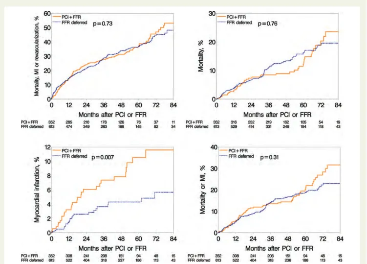
The unadjusted Kaplan – Meier fraction of MI estimated at 7 years
reached 12% in the FFR-Perform vs. 6% in the FFR-Defer groups
(P ¼ 0.007) (Figure
4
). Other outcome events showed no
signifi-cant difference between the two groups.
Cox multivariate models for follow-up
events
After adjustment for baseline characteristics in a Cox multivariable
model, patients undergoing FFR measurements tended to have
lower rates of death/MI [hazard ratio (HR): 0.85, 95% confidential
interval (CI): 0.71 – 1.01, P ¼ 0.06]; specifically, deferral of PCI
guided by FFR was significantly associated with a reduced rate of
MI (HR: 0.46, 95% CI: 0.26 – 0.82, P ¼ 0.008) (Table
4
).
After excluding patients with FFR of 0.75 – 0.80 and deferral of
PCI, the incidence of death or MI was significantly lower in the
FFR-guided group compared with the PCI-only group (HR: 0.80,
95% CI: 0.66 – 0.96, P ¼ 0.02); deferral of PCI after FFR
measure-ment was significantly associated with reduced rates of MI (HR:
Figure 2
Utility rates of fractional flow reserve between 2002
and 2009. The annual rate of fractional flow reserve use was
gen-erally between 14 and 18%. The bar represents the rate of FFR
use for that year and the bold-dashed line represents rate of
frac-tional flow reserve use quarterly. FFR, fracfrac-tional flow reserve;
PCI, percutaneous coronary intervention.
0.39, 95% CI: 0.20 – 0.73, P ¼ 0.004) and death or MI (HR: 0.64,
95% CI: 0.45 – 0.92, P ¼ 0.02) (Table
5
).
Discussion
In this registry-based study, we compared clinical outcomes of
patients undergoing FFR-guided vs. angiography-guided PCI in a
large non-selected population. Overall, the use of FFR (achieved
in 14.8% of patients) was associated with a non-significantly
lower incidence (P ¼ 0.06) of death or MI. However, after
exclud-ing patients in whom PCI was deferred in a vessel with FFR
between 0.75 and 0.80, the long-term outcome was better in
the FFR-guided group compared with the angiography-guided
group. Furthermore, the incidence of MI was significantly lower
in the FFR-Defer group compared with the FFR-Perform group.
These observations, therefore, support the use of FFR in routine
clinical practice.
Fractional flow reserve-guided
percutaneous coronary intervention in
the general clinical practice
The current registry-based study, to our knowledge, examined the
largest number of patients with FFR-guided interventions. In the
overall population, the primary endpoint (MACE) was not different
between the FFR-guided and angiography-guided groups and the
secondary
endpoint
of
death/MI
reached
only
borderline
significance levels. However, several disparities in the use of FFR
between the routine clinical practice and clinical trials may diminish
potential benefits from FFR-guided treatment.
First, in daily practice settings in many hospitals, the use of FFR is
often entirely at the operator’s discretion based on angiographic
information. Alas, the relationship between the angiographic
ap-pearance of a coronary stenosis and its functional significance is
notoriously poor.
21,22Consequently, when FFR is infrequently
used, some functionally severe lesions that by visual assessment
induce ostensibly mild to moderate vessel narrowing might have
been excluded from PCI. Conversely, some haemodynamically
in-significant lesions, which potentially have a better outcome, may be
visually interpreted as severe, and therefore undergo unnecessary
PCI. Indeed, Sant’Anna et al.
26have demonstrated that FFR
rou-tinely used for decision-making modifies treatment decisions for
32% of all stenoses and 48% of all patients, compared with
deci-sions based on angiography alone. Therefore, low usage rate of
functional assessment in general practice may lead to
underestima-tion of its potential benefits. Since FFR has been validated in almost
all clinical and anatomical subsets, its systematic use can render PCI
an even more effective and appropriate treatment than it is
currently.
27Moreover, FFR values ranging between 0.75 and 0.80 were
pre-viously considered to be in a ‘grey zone’, and required clinical
judg-ment for decision-making regarding revascularization. However,
Legalery et al.
20demonstrated that deferring PCI in lesions with
FFR under 0.80 was harmful. More recent studies adopted the
. . . .
. . . .
Table 1
Baseline characteristics of patients
Variables PCI-only (n 5 6268) FFR-guided group (n 5 1090)
All (n 5 1090) FFR-Perform (n 5 369) FFR-Defer (n 5 721) Age (year) 67.9 + 11.6* 65.7 + 11.3 65.5 + 11.3 65.8 + 11.4
Male gender, n (%) 4416 (70.4)* 683 (62.6) 255 (69.1) 428 (59.3)‡ Body mass index 30.2 + 5.9 30.4 + 5.8 30.4 + 5.7 30.4 + 5.8 Current smoking, n (%) 836 (13.3) 140 (12.8) 49 (13.2) 91 (12.6) Diabetes mellitus, n (%) 1862 (29.7) 306 (28.0) 106 (28.7) 200 (27.7) Hypertension, n (%) 4897 (78.1) 864 (79.2) 296 (80.2) 568 (78.7) Hypercholesterolaemia, n (%) 5119 (81.6)* 608 (55.7) 312 (84.5) 296 (41.0) Chest pain, n (%) 4529 (72.2)* 753 (69.0) 261 (70.7) 492 (68.2) Dyspnoea, n (%) 168 (2.6)* 89 (8.1) 5 (1.3) 84 (11.6)† ACS (1 – 7 days), n (%) 818 (13.0)* 48 (4.4) 19 (5.1) 29 (4.0) History of MI (.7 days), n (%) 1882 (31) 270 (25) 99 (27) 171 (24) Prior PCI, n (%) 1981 (31.6)* 445 (40.8) 168 (45.5) 277 (38.4)‡ CVD, n (%) 682 (10.8) 103 (9.4) 35 (9.4) 68 (9.4) PAD, n (%) 718 (11.4) 106 (9.7) 37 (10.0) 69 (9.5) Chronic obstructive pulmonary disease, n (%) 704 (11.2) 121 (11.1) 32 (8.6) 89 (12.3) Renal dysfunction, n (%) 285 (4.5) 41 (3.7) 13 (3.5) 28 (3.8) Stress test, n (%) 1008 (16.0) 112 (10.2) 67 (18.1) 45 (6.2) LVEF≤40%, n (%) 705 (11.2)* 82 (7.5) 31 (8.4) 51 (7.0)
ACS, acute coronary syndrome; CVD, cerebral vascular disease; FFR, fractional flow reserve; LVEF, left ventricular ejection fraction; MI, myocardial infarction; PCI, percutaneous coronary intervention; PAD, peripheral artery disease.
*P , 0.001 when compared with the FFR-guided group. †
P , 0.05 when compared with the FFR-Perform group. ‡
upper limit of this small transition zone as a threshold to perform
PCI in order to limit the number of ischaemic lesion left
untreat-ed.
7,11,12In the current study, in 40.0% of the 220 patients with
FFR values between 0.75 and 0.80 PCI was deferred. This might
in-fluence relative outcomes between the angiography-guided and
FFR-guided groups, probably in favour of the angiography-guided
strategy. After excluding those patients from analysis, the
FFR-guided group had a significantly lower rate of death or MI
when compared with the PCI-only group. This result is consistent
with the 2-year follow-up of the FAME study
8and indicates a FFR
value of 0.80 as an appropriate threshold to intervene.
In addition, a registy study reported that 9% of patients were
treated despite an FFR
≥0.80.
20In the FFR-guided group of the
current study, 10.5% of patients had PCI performed in a vessel
with FFR .0.80 and 39 (3.6%) did not have PCI in a vessel with
FFR ,0.75. It is not uncommon to encounter a clinical or
angio-graphic situation where the FFR result seems to disagree with
visual assessment by angiography. Although its accuracy may be
influenced by some pathological factors,
28–30when performed
correctly, false negative or false positive FFR results are relatively
rare.
31Obviously, FFR should only be measured when the
oper-ator plans to follow through on the result of this test. In fact,
the FAME study suggests that stenting of lesions with an FFR
.0.80 is detrimental.
8Thus, clinical benefits could be enhanced
when patients are treated in accordance with the FFR criteria.
The safety of deferring percutaneous
coronary intervention under guidance of
fractional flow reserve
The benefit of functional evaluation is attributed to identification of
ischaemia-causing coronary stenoses, and its contribution to
judi-cious decision-making of revascularization, which in turn may
reduce unexpected device-related diseases.
2,3Consistent with
. . . .
. . . .
Table 2
Angiography and percutaneous coronary intervention characteristics
Variables PCI-only (n 5 6268) FFR-guided group (n 5 1090)
All (n 5 1090) FFR-Perform (n 5 369) FFR-Defer (n 5 721) RCA stenosis≥70%, n (%) 3134 (50.0)* 293 (26.8) 125 (33.8) 168 (23.0)‡ LMCA stenosis≥50%, n (%) 205 (3.2)* 9 (0.8) 1 (0.3) 8 (1.1) LAD stenosis≥70%, n (%) 3474 (55.4)* 419 (38.4) 233 (63.1) 186 (26.0)† LCX stenosis≥70%, n (%) 2620 (41.7)* 250 (22.9) 104 (28.1) 146 (20.2) FFR in LMCA, n (%) — 37 (3.3) 3 (0.8) 34 (4.7)† FFR 0.75 – 0.80, n (%) 1 (0.1) 0 1 (0.1) FFR in LAD, n (%) — 713 (65.4) 244 (66.1) 469 (65.0) FFR 0.75 – 0.80, n (%) 149 (13.7) 87 (27.4) 62 (8.6)† FFR in LCX, n (%) — 192 (17.6) 51 (13.8) 141 (19.5)† FFR 0.75 – 0.80, n (%) 22 (2.0) 14 (3.8) 8 (1.1)† FFR in RCA, n (%) — 213 (19.5) 70 (18.9) 143 (19.8) FFR 0.75 – 0.80, n (%) 37 (3.4) 22 (6.0) 15 (2.1)† FFR in graft, n (%) — 23 (2.1) 4 (1.0) 19 (2.6) FFR 0.75 – 0.80, n (%) 2 (0.2) 0 2 (0.3) Median FFR value (IQR) — 0.77 (0.72, 0.82) 0.87 (0.82, 0.91)† PCI in native LM, n (%) 262 (4.1)*,‡ 7 (0.6) 6 (1.6) 1 (0.1)‡ PCI in native LAD, n (%) 2846 (45.4)*,† 299 (27.4) 260 (70.4) 39 (5.4)† PCI in native LCX, n (%) 1867 (29.7)*,† 124 (11.3) 79 (21.4) 45 (6.2)† PCI in native RCA, n (%) 2118 (33.7)*,‡ 147 (13.4) 97 (26.2) 50 (6.9)† Vein graft intervention, n (%) 475 (7.5)*,† 6 (0.5) 3 (0.8) 3 (0.4) Number of vessel treated, n (%)
1 5078 (81.0) — 296 (80.2) — 2 1087 (17.3) — 71 (19.2) —
3 99 (1.7) — 2 (0.6) —
Use of DES, n (%) 4417 (70.4)* 361 (33.1) 273 (73.9) 88 (12.2)† Use of BMS, n (%) 1632 (26.0)* 118 (10.8) 88 (23.8) 30 (4.2)† Number of stents placed 1.5 + 1.0*,‡ 0.6 + 0.9 1.4 + 0.7 0.2 + 0.7† Procedural success of stents placement, n (%) 5953 (94.9)‡ — 359 (97.3) —
BMS, bare metal stent; DES, drug-eluting stent; FFR, fractional flow reserve; LAD, left anterior descending; LCX, left circumflex; LMCA, left main coronary artery; PCI, percutaneous coronary intervention; RCA, right coronary artery.
*P , 0.001 when compared with the FFR-guided group. ‡
P , 0.05 when compared with the FFR-Perform group. †
Figure 3
Long-term adverse events in the percutaneous coronary intervention only group and fractional flow reserve-guided group.
Un-adjusted Kaplan – Meier curves during a 7-year follow-up for major adverse cardiac event (left top); death (right top); myocardial infarction
(bottom left); and for death or myocardial infarction (bottom right). FFR, fractional flow reserve; PCI, percutaneous coronary intervention;
MACE, major adverse cardiac events; MI, myocardial infarction.
. . . .
. . . .
Table 3
In-hospital events
Variables PCI-only (n56268) FFR-guided group (n 5 1090)
All (n 5 1090) Perform (n 5 369) Defer (n 5 721) In-hospital events Death, n (%) 22 (0.3) 3 (0.2) 2 (0.5) 1 (0.1) Death/Q-wave MI/stroke/CABG, n (%) 63 (0.9)§ 4 (0.3) 2 (0.5) 2 (0.2) Q-wave MI, n (%) 9 (0.1) 0 0 0 Emergency CABG, n (%) 18 (0.2) 1 (0.09) 0 1 (0.1) In-hospital CVD, n (%) 19 (0.3) 0 (0) 0 0 In-hospital any MI, n (%) 266 (4.2)* 18 (1.6) 12 (3.2) 6 (1)‡
CABG, coronary artery bypass grafting; CVD, cerebral vascular disease; FFR, fractional flow reserve; MI, myocardial infarction; PCI, percutaneous coronary intervention. §P , 0.05 when compared with the FFR-guided group.
*P , 0.001 when compared with the FFR-guided group. ‡
previous studies,
32,33our data demonstrate a favourable outcome
in the FFR-Defer group compared with the FFR-Perform group,
which strongly supports the use of FFR when evaluating whether
PCI can be safely deferred. Moreover, although some patients
underwent PCI in a vessel with FFR .0.80, the number of stents
placed was still significantly lower in the FFR-guided group.
Similar-ly, a recent analysis of the FAME study showed that the FFR-guided
PCI resulted in significant cost-saving by reducing stent use,
rehos-pitalizations, and MACE.
34Therefore, the FFR-guided treatment
could have been more economical in daily practice if
decision-making for PCI relies more strictly on FFR value.
Incidentally, the use of FFR between 2003 and 2009 showed a
transient decline in the mid-years, followed by a sudden increase
in 2009. This may reflect a change in clinical practice after
publica-tion of the landmark FAME study in January 2009.
7Limitations
This single-centre, observational study has limitations inherent to
non-randomized trials. It was performed in a non-selected
population of clinical practice with unequal baseline characteristics,
and involved multiple operators. Multiple regression analysis may
mitigate bias after adjustment of confounding factors, but
unmeas-ured indicators leave room for residual bias.
In the current study, the adenosine protocol included both i.v.
and intracoronary routes. Although we have previously
demon-strated that incremental doses of intracoronary adenosine were
valid to achieve maximum coronary hyperaemia,
35reports are
in-consistent.
36,37Presently, central i.v. administration of adenosine
remains the gold standard for FFR measurements. Moreover, the
dose of intracoronary adenosine used in this study followed
common clinical practice, but might be too low for some patients.
The coronary stenoses were assessed visually, rather than by
quantitative coronary angiography (QCA) or by intravascular
ultra-sound. Nevertheless, visual assessment is the most common
method to evaluate diameter stenoses in the catheterization
la-boratory. On the other hand, the accuracy of QCA or
intravascu-lar ultrasound for predicting functionally significant FFR is also
limited.
38,39Figure 4
Long-term adverse events in the fractional flow reserve-Perform group and fractional flow reserve-Defer group. Unadjusted
Kaplan – Meier curves during a 7-year follow-up for major adverse cardiac event (left top); death (right top); myocardial infarction (bottom
left); and for death or myocardial infarction (bottom right). FFR, fractional flow reserve; PCI, percutaneous coronary intervention; MI,
myocar-dial infarction
Conclusion
In this registry study, we found a favourable long-term outcome in
an FFR-guided group. This result is in keeping with previous clinical
trials, and therefore provides important evidence supporting the
rationale for the use of FFR in routine practice.
Funding
National Institute of Health (NIH Grant HL-92954 and AG-31750 to
A.L.). The study was also supported by an unrestricted grant from St
Jude Medical. J.L. was supported by the China Scholarship Council
(NO.2010811095), and ‘Beijing Nova program’ of Beijing Municipal
Science & Technology Commission (A2007079), China. A.J.F. was
sup-ported by the Walter and Gertrud Siegenthaler Foundation, the Young
Academics Support Committee of the University of Zurich, and the
Swiss foundation for Medical-Biological Scholarships (SSMBS; SNSF
No PASMP3_132551). L.O.L. has received financial support through
an institutional grant from the NIH.
Conflict of interest: L.O.L. has also received honorarium from the
NIH. She has also been employed by the NIH. With regard to financial
activities outside the submitted work, she has been employed by the
NIH and has received research grant support from Stealth Peptides,
Inc. She has also been awarded a post doc fellowship by the AHA.
In addition, she has received institutional financial support for pending
patents, and has also received support for accommodation at
inter-national scientific meetings from other academic institutions. With
regard to financial activities outside the submitted work, A.L.
(corresponding author) has received remuneration as a board
member of Itamar Medical as well as support from an NIH institutional
grant.
References
1. Davies RF, Goldberg AD, Forman S, Pepine CJ, Knatterud GL, Geller N, Sopko G, Pratt C, Deanfield J, Conti CR. Asymptomatic Cardiac Ischemia Pilot (ACIP) study two-year follow-up: outcomes of patients randomized to initial strategies of medical therapy versus revascularization. Circulation 1997;95:2037 – 2043. 2. Levine GN, Bates ER, Blankenship JC, Bailey SR, Bittl JA, Cercek B, Chambers CE,
Ellis SG, Guyton RA, Hollenberg SM, Khot UN, Lange RA, Mauri L, Mehran R, Moussa ID, Mukherjee D, Nallamothu BK, Ting HH. 2011 ACCF/AHA/SCAI Guideline for Percutaneous Coronary Intervention A Report of the American College of Cardiology Foundation/American Heart Association Task Force on Practice Guidelines and the Society for Cardiovascular Angiography and Interven-tions. J Am Coll Cardiol 2011;58:e44 – e122.
3. Wijns W, Kolh P, Danchin N, Di Mario C, Falk V, Folliguet T, Garg S, Huber K, James S, Knuuti J, Lopez-Sendon J, Marco J, Menicanti L, Ostojic M, Piepoli MF, Pirlet C, Pomar JL, Reifart N, Ribichini FL, Schalij MJ, Sergeant P, Serruys PW, Silber S, Sousa Uva M, Taggart D. Guidelines on myocardial revascularization. Eur Heart J 2010;31:2501 – 2555.
4. Pijls NH, De Bruyne B, Peels K, Van Der Voort PH, Bonnier HJ, Bartunek JKJJ, Koolen JJ. Measurement of fractional flow reserve to assess the functional severity of coronary-artery stenoses. N Engl J Med 1996;334:1703 – 1708.
5. Bech GJ, De Bruyne B, Pijls NH, de Muinck ED, Hoorntje JC, Escaned J, Stella PR, Boersma E, Bartunek J, Koolen JJ, Wijns W. Fractional flow reserve to determine the appropriateness of angioplasty in moderate coronary stenosis: a randomized trial. Circulation 2001;103:2928 – 2934.
6. Pijls NH, van Schaardenburgh P, Manoharan G, Boersma E, Bech JW, van’t Veer M, Bar F, Hoorntje J, Koolen J, Wijns W, de Bruyne B. Percutaneous coron-ary intervention of functionally nonsignificant stenosis: 5-year follow-up of the DEFER Study. J Am Coll Cardiol 2007;49:2105 – 2111.
. . . .
. . . .
Table 5
Model estimates after excluding patients with
percutaneous coronary intervention deferred in a vessel
with a 0.75 – 0.80 fractional flow reserve measure
Events AdjustedaHR 95% CI P-value
FFR use vs. no FFR MACE 0.95 0.83 – 1.08 0.42 Death 0.84 0.67 – 1.04 0.11 MI 0.75 0.54 – 1.03 0.08 Death/revascularization 0.94 0.82 – 1.08 0.38 Death/MI 0.80 0.66 – 0.96 0.02 Deferral of PCI after FFR vs. Perform PCI
MACE 0.86 0.67 – 1.11 0.24 Death 0.73 0.48 – 1.11 0.14 MI 0.39 0.20 – 0.73 0.004 Death/revascularization 0.89 0.69 – 1.15 0.36 Death/MI 0.64 0.45 – 0.92 0.02
CI, confidential interval; FFR, fractional flow reserve; HR, hazard ratio; MACE, major adverse cardiac events; MI, myocardial infarction; PCI, percutaneous coronary intervention.
a
Adjusted for age, sex, body mass index, smoking history (current, former, or never), chronic heart failure on presentation, diabetes, hypertension, hypercholesterolaemia, primary symptom, recent MI, prior PCI, prior coronary artery bypass grafting, history of myocardial infarction, heart failure, cerebral vascular disease, peripheral artery disease, chronic obstructive pulmonary disease, renal dysfunction, presence of tumour/lymphoma/leukaemia, metastatic cancer, ejection fraction≤40%, ejection fraction unknown, level of stenosis in each coronary vessel (right coronary artery, left anterior descending, left circumflex, left main coronary artery).
. . . .
. . . .
Table 4
Cox multivariable models for determinants
of outcome events
Events AdjustedaHR 95% CI P-value
FFR use vs. no FFR MACE 1.01 0.89 – 1.14 0.93 Death 0.89 0.73 – 1.10 0.28 MI 0.79 0.58 – 1.07 0.12 Death/revascularization 1.003 0.88 – 1.14 0.96 Death/MI 0.85 0.71 – 1.01 0.06 Deferral of PCI after FFR vs. Perform PCI
MACE 0.97 0.77 – 1.23 0.81 Death 0.84 0.56 – 1.24 0.37 MI 0.46 0.26 – 0.82 0.008 Death/revascularization 1.002 0.78 – 1.27 0.98 Death/MI 0.73 0.52 – 1.01 0.06
CI, confidential interval; FFR, fractional flow reserve; HR, hazard ratio; MACE, major adverse cardiac events; MI, myocardial infarction; PCI, percutaneous coronary intervention.
a
Adjusted for age, sex, body mass index, smoking history (current, former, or never), chronic heart failure on presentation, diabetes, hypertension, hypercholesterolaemia, primary symptom, recent MI, prior PCI, prior coronary artery bypass grafting, history of myocardial infarction, heart failure, cerebral vascular disease, peripheral artery disease, chronic obstructive pulmonary disease, renal dysfunction, presence of tumour/lymphoma/leukaemia, metastatic cancer, ejection fraction≤40%, ejection fraction unknown, level of stenosis in each coronary vessel (right coronary artery, left anterior descending, left circumflex, left main coronary artery).
7. Tonino PA, De Bruyne B, Pijls NH, Siebert U, Ikeno F, van’t Veer M, Klauss V, Manoharan G, Engstrom T, Oldroyd KG, Ver Lee PN, MacCarthy PA, Fearon WF. Fractional flow reserve versus angiography for guiding percutaneous coronary intervention. N Engl J Med 2009;360:213 – 224.
8. Pijls NH, Fearon WF, Tonino PA, Siebert U, Ikeno F, Bornschein B, van’t Veer M, Klauss V, Manoharan G, Engstrom T, Oldroyd KG, Ver Lee PN, MacCarthy PA, De Bruyne B. Fractional flow reserve versus angiography for guiding percutaneous coronary intervention in patients with multivessel coronary artery disease: 2-year follow-up of the FAME (Fractional Flow Reserve Versus Angiography for Multi-vessel Evaluation) study. J Am Coll Cardiol 2010;56:177 – 184.
9. De Bruyne B, Pijls NH, Kalesan B, Barbato E, Tonino PA, Piroth Z, Jagic N, Mobius-Winkler S, Rioufol G, Witt N, Kala P, MacCarthy P, Engstrom T, Oldroyd KG, Mavromatis K, Manoharan G, Verlee P, Frobert O, Curzen N, Johnson JB, Juni P, Fearon WF. Fractional flow reserve-guided pci versus medical therapy in stable coronary disease. N Engl J Med 2012;367:991 – 1001. 10. Hamilos M, Muller O, Cuisset T, Ntalianis A, Chlouverakis G, Sarno G, Nelis O,
Bartunek J, Vanderheyden M, Wyffels E, Barbato E, Heyndrickx GR, Wijns W, De Bruyne B. Long-term clinical outcome after fractional flow reserve-guided treat-ment in patients with angiographically equivocal left main coronary artery sten-osis. Circulation 2009;120:1505 – 1512.
11. Puymirat E, Peace A, Mangiacapra F, Conte M, Ntarladimas Y, Bartunek J, Vanderheyden M, Wijns W, De Bruyne B, Barbato E. Long-term clinical outcome after fractional flow reserve-guided percutaneous coronary revascular-ization in patients with small-vessel disease. Circ Cardiovasc Interv 2012;5:62 – 68. 12. Muller O, Mangiacapra F, Ntalianis A, Verhamme KM, Trana C, Hamilos M,
Bartunek J, Vanderheyden M, Wyffels E, Heyndrickx GR, van Rooij FJ, Witteman JC, Hofman A, Wijns W, Barbato E, De Bruyne B. Long-term follow-up after fractional flow reserve-guided treatment strategy in patients with an isolated proximal left anterior descending coronary artery stenosis. JACC Cardiovasc Interv 2011;4:1175 – 1182.
13. Sels JW, Tonino PA, Siebert U, Fearon WF, Van’t Veer M, De Bruyne B, Pijls NH. Fractional flow reserve in unstable angina and non-st-segment elevation myocar-dial infarction experience from the FAME (Fractional flow reserve versus Angiog-raphy for Multivessel Evaluation) Study. JACC Cardiovasc Interv 2011;4:1183 – 1189. 14. Koo BK, Park KW, Kang HJ, Cho YS, Chung WY, Youn TJ, Chae IH, Choi DJ, Tahk SJ, Oh BH, Park YB, Kim HS. Physiological evaluation of the provisional side-branch intervention strategy for bifurcation lesions using fractional flow reserve. Eur Heart J 2008;29:726 – 732.
15. Aqel R, Zoghbi GJ, Hage F, Dell’Italia L, Iskandrian AE. Hemodynamic evaluation of coronary artery bypass graft lesions using fractional flow reserve. Catheter Car-diovasc Interv 2008;72:479 – 485.
16. Pijls NH. Fractional flow reserve after previous myocardial infarction. Eur Heart J 2007;28:2301 – 2302.
17. Lopez-Palop R, Pinar E, Lozano I, Saura D, Pico F, Valdes M. Utility of the fraction-al flow reserve in the evfraction-aluation of angiographicfraction-ally moderate in-stent restenosis. Eur Heart J 2004;25:2040 – 2047.
18. Jensen LO, Thayssen P, Lassen JF, Hansen HS, Kelbaek H, Junker A, Pedersen KE, Hansen KN, Krusell LR, Botker HE, Thuesen L. Recruitable collateral blood flow index predicts coronary instent restenosis after percutaneous coronary interven-tion. Eur Heart J 2007;28:1820 – 1826.
19. Puymirat E, Muller O, Sharif F, Dupouy P, Cuisset T, de Bruyne B, Gilard M. Frac-tional flow reserve: concepts, applications and use in France in 2010. Arch Cardi-ovasc Dis 2010;103:615 – 622.
20. Legalery P, Schiele F, Seronde MF, Meneveau N, Wei H, Didier K, Blonde MC, Caulfield F, Bassand JP. One-year outcome of patients submitted to routine frac-tional flow reserve assessment to determine the need for angioplasty. Eur Heart J 2005;26:2623 – 2629.
21. Meijboom WB, Van Mieghem CA, van Pelt N, Weustink A, Pugliese F, Mollet NR, Boersma E, Regar E, van Geuns RJ, de Jaegere PJ, Serruys PW, Krestin GP, de Feyter PJ. Comprehensive assessment of coronary artery stenoses: computed tomography coronary angiography versus conventional coronary angiography and correlation with fractional flow reserve in patients with stable angina. J Am Coll Cardiol 2008;52:636 – 643.
22. Topol EJ, Nissen SE. Our preoccupation with coronary luminology. The dissoci-ation between clinical and angiographic findings in ischemic heart disease. Circula-tion 1995;92:2333 – 2342.
23. Lavi S, Rihal CS, Yang EH, Fassa AA, Elesber A, Lennon RJ, Mathew V, David HR Jr, Lerman A. The effect of drug eluting stents on cardiovascular events in patients
with intermediate lesions and borderline fractional flow reserve. Catheter Cardio-vasc Interv 2007;70:525 – 531.
24. Brosh D, Higano ST, Lennon RJ, Holmes DR Jr, Lerman A. Effect of lesion length on fractional flow reserve in intermediate coronary lesions. Am Heart J 2005;150: 338 – 343.
25. Thygesen K, Alpert JS, White HD, Jaffe AS, Apple FS, Galvani M, Katus HA, Newby LK, Ravkilde J, Chaitman B, Clemmensen PM, Dellborg M, Hod H, Porela P, Underwood R, Bax JJ, Beller GA, Bonow R, Van der Wall EE, Bassand JP, Wijns W, Ferguson TB, Steg PG, Uretsky BF, Williams DO, Armstrong PW, Antman EM, Fox KA, Hamm CW, Ohman EM, Simoons ML, Poole-Wilson PA, Gurfinkel EP, Lopez-Sendon JL, Pais P, Mendis S, Zhu JR, Wallentin LC, Fernandez-Aviles F, Fox KM, Parkhomenko AN, Priori SG, Tendera M, Voipio-Pulkki LM, Vahanian A, Camm AJ, De Caterina R, Dean V, Dickstein K, Filippatos G, Funck-Brentano C, Hellemans I, Kristensen SD, McGregor K, Sechtem U, Silber S, Widimsky P, Zamorano JL, Morais J, Brener S, Harrington R, Morrow D, Lim M, Martinez-Rios MA, Steinhubl S, Levine GN, Gibler WB, Goff D, Tubaro M, Dudek D, Al-Attar N. Universal def-inition of myocardial infarction. Circulation 2007;116:2634 – 2653.
26. Sant’Anna FM, Silva EE, Batista LA, Ventura FM, Barrozo CA, Pijls NH. Influence of routine assessment of fractional flow reserve on decision making during coronary interventions. Am J Cardiol 2007;99:504 – 508.
27. Pijls NH, Sels JW. Functional measurement of coronary stenosis. J Am Coll Cardiol 2012;59:1045 – 1057.
28. Magni V, Chieffo A, Colombo A. Evaluation of intermediate coronary stenosis with intravascular ultrasound and fractional flow reserve: its use and abuse. Cath-eter Cardiovasc Interv 2009;73:441 – 448.
29. De Bruyne B, Pijls NH, Bartunek J, Kulecki K, Bech JW, De Winter H, Van Crombrugge P, Heyndrickx GR, Wijns W. Fractional flow reserve in patients with prior myocardial infarction. Circulation 2001;104:157 – 162.
30. Melikian N, Cuisset T, Hamilos M, De Bruyne B. Fractional flow reserve – the in-fluence of the collateral circulation. Int J Cardiol 2009;132:e109 – e110. 31. Koolen JJ, Pijls NH. Coronary pressure never lies. Catheter Cardiovasc Interv 2008;
72:248 – 256.
32. Misaka T, Kunii H, Mizukami H, Sakamoto N, Nakazato K, Takeishi Y. Long-term clinical outcomes after deferral of percutaneous coronary intervention of inter-mediate coronary stenoses based on coronary pressure-derived fractional flow reserve. J Cardiol 2011;58:32 – 37.
33. Nam CW, Mangiacapra F, Entjes R, Chung IS, Sels JW, Tonino PA, De Bruyne B, Pijls NH, Fearon WF. Functional SYNTAX score for risk assessment in multivessel coronary artery disease. J Am Coll Cardiol 2011;58:1211 – 1218.
34. Fearon WF, Bornschein B, Tonino PA, Gothe RM, Bruyne BD, Pijls NH, Siebert U. Fractional Flow Reserve Versus Angiography for Multivessel Evaluation (FAME) Study Investigators. Economic evaluation of fractional flow reserve-guided percu-taneous coronary intervention in patients with multivessel disease. Circulation 2010;122:2545 – 2550.
35. Murtagh B, Higano S, Lennon R, Mathew V, Holmes DR Jr, Lerman A. Role of in-cremental doses of intracoronary adenosine for fractional flow reserve assess-ment. Am Heart J 2003;146:99 – 105.
36. Lopez-Palop R, Saura D, Pinar E, Lozano I, Pe´rez-Lorente F, Pico´ F, Valdez M. Ad-equate intracoronary adenosine doses to achieve maximum hyperaemia in coron-ary functional studies by pressure derived fractional flow reserve: a dose response study. Heart 2004;90:95 – 96.
37. Leone AM, Porto I, De Caterina AR, Basile E, Aurelio A, Gardi A, Russo D, Laezza D, Niccoli G, Burzotta F, Trani C, Mazzari MA, Mongiardo R, Rebuzzi AG, Crea F. Maximal hyperemia in the assessment of fractional flow reserve: intracoronary adenosine versus intracoronary sodium nitroprusside versus intravenous adenosine: the NASCI (Nitroprussiato versus Adenosina nelle Stenosi Coronariche Intermedie) study. JACC Cardiovasc Interv 2012;5: 402 – 408.
38. Yong AS, Ng AC, Brieger D, Lowe HC, Ng MK, Kritharides L. Three-dimensional and two-dimensional quantitative coronary angiography, and their prediction of reduced fractional flow reserve. Eur Heart J 2011;32:345 – 353.
39. Koo BK, Yang HM, Doh JH, Choe H, Lee SY, Yoon CH, Cho YK, Nam CW, Hur SH, Lim HS, Yoon MH, Park KW, Na SH, Youn TJ, Chung WY, Ma S, Park SK, Kim HS, Tahk SJ. Optimal intravascular ultrasound criteria and their ac-curacy for defining the functional significance of intermediate coronary stenoses of different locations. JACC Cardiovasc Interv 2011;4:803 – 811.


![[PDF] Tutoriel Drupal : Installation en local sous Windows et traduction | Formation informatique](data:image/gif;base64,R0lGODlhAQABAIAAAP///wAAACH5BAEAAAAALAAAAAABAAEAAAICRAEAOw==)