Deactivation of Macrophages with Interleukin-4 Is the Key to the Isolation of
Tropheryma whippelii
Gabriele Schoedon, Daniel Goldenberger, Regula Forrer, Clinical Mycology Laboratory, Division of Infectious Diseases, Department of Medicine, University of Zurich Medical School; Institute Anja Gunz, Fabrizio Dutly, Mathias Ho¨chli,
of Medical Microbiology and Division of Ultrastructure, University of Martin Altwegg, and Andreas Schaffner
Zurich, Zurich, Switzerland Whipple’s disease is a systemic illness caused by a specific agent. Despite recognition of bacteria
in lesions, efforts to isolate the causative agent remained futile. A novel strategy was devised to culture Whipple bacilli in deactivated mononuclear phagocytes. Infected tissue was inoculated into human phagocytes deactivated with interleukin (IL)-4, IL-10, and dexamethasone. Within 8 – 10 days, diastase-resistant periodic acid – Schiff – positive inclusions appeared, corresponding to intact and degenerating bacteria shown to be Tropheryma whippelii by electron microscopy and molecular analyses. T. whippelii was passaged several times in deactivated monocytes and a monoblastic cell line. Time-kinetics growth studies and comparative polymerase chain reaction analysis documented multiplication of T. whippelii in deactivated macrophages. Complementary studies showed that IL-4 rendered phagocytes permissive for T. whippelii, a strong indication that host factors contribute to the pathogenesis of Whipple’s disease. The propagation of T. whippelii will permit microbiologic, immunologic, seroepidemiologic, and therapeutic studies of this pathogen.
Whipple’s disease is a chronic infection involving the intesti- 11], we decided to study whether T. whippelii could multiply within deactivated human macrophages. The deactivating nal tract, lymph nodes, endocardium, central nervous system,
skin, and other organs [1]. Although bacteria were recognized agents dexamethasone, interleukin (IL)-4, and IL-10 all allow intracellular microorganisms such as Listeria monocytogenes in lesionsú30 years ago [2], recently the causative bacterium
was identified by amplification of 16S rRNA genes in infected to multiply in mononuclear phagocytes without affecting phagocytosis. Inoculation of tissue infected with T. whippelii tissue, leading to the definition of a new bacterial species,
Tropheryma whippelii [3, 4]. into such deactivated human blood monocytes appeared, there-fore, a promising strategy for the isolation and culture of this The lesions in Whipple’s disease are characterized by the
presence of large, foamy macrophages containing periodic pathogen. Macrophages deactivated for 48 h with a maximally active combination of dexamethasone, IL-4, and IL-10 would acid – Schiff (PAS) – positive diastase-resistant inclusions, often
with a typical rod shape [1, 5]. Untreated, the infection presum- ingest tissue debris with bacteria, but their killing systems would be crippled [10, 11], thereby permitting intracellular ably results in the death of most affected individuals. Treatment
of Whipple’s disease remains based on empiric clinical survival and eventually multiplication of the Whipple bacilli. grounds. Prolonged treatment with various antibiotics, while
difficult, seems to be successful [1]. Immunologic defects have
been postulated to predispose patients to develop Whipple’s Methods disease [6, 7], but it might well be that the occurrence of the
Patient material. For preliminary experiments, frozen
infection depends entirely on the distribution of the pathogen
(0707C) intestinal biopsy material was obtained from a female in the human environment. While diagnosis of the disease is
patient with histologically proven Whipple’s disease with spondy-now greatly facilitated by the recently developed molecular
lodiscitis, described previously [12]. Infected heart valves submit-techniques, the causative agent remains to be isolated and
cul-ted for conventional bacterial cultures and eubacterial polymerase tured.
chain reaction (PCR) were from 2 male, white, native Swiss pa-Because we defined in our laboratory conditions permitting tients who underwent heart surgery for aortic insufficiency. microorganisms to survive and grow in human phagocytes [8 – In patient TWZ1, infectious endocarditis was not suspected
pre-operatively. The diagnosis of infectious endocarditis was made by eubacterial PCR (see below). Subsequent to the heart operation, a duodenal biopsy showed, in addition to an inflammatory infiltrate,
Received 16 September 1996; revised 31 March 1997.
typical diastase/PAS – positive, silver stain – positive, acid fast –
Grant support: Swiss National Science Foundation (31-33897 to A.S. and
32-040 445 to M.A.). negative bacterial inclusions identical to those found in the heart
Reprints or correspondence: Prof. Andreas Schaffner, Dept. of Medicine, valve. Blood cultures and cultures of the heart valve remained Room AW9, University Hospital of Zurich, Ra¨mistr. 100, CH-8091 Zurich,
negative by conventional culture technique. Patient TWZ2 was
Switzerland.
diagnosed with culture-negative endocarditis. In retrospect, his
his-The Journal of Infectious Diseases 1997; 176:672 – 7
tory was compatible for seronegative arthritis of 21 years’ duration q 1997 by The University of Chicago. All rights reserved.
around position 355 of the reference sequence). The next most closely related species was Aureobacterium liquefaciens (acces-sion no. X77444), with a similarity of 89.1%. For patient TWZ2 the 398-bp sequence determined was identical to the reference sequence of T. whippelii (accession no. M87484) except for 2 ambiguous bases (not mismatches) in patient TWZ2’s sequence. The next most closely related sequence was again A. liquefaciens, with a similarity of 89.5%. In both cases, the diagnosis was further corroborated by species-specific PCR using both the primer pairs TW-1/TW-2 and TW-1/TW-3 on DNA extracted from the valves. Intestinal biopsies from patient TWZ1 also gave a positive species-specific PCR result [4].
Human mononuclear phagocytes. Human mononuclear
phagocytes were purified from peripheral blood as described, givingú90% monocytes with õ10% contaminating lymphocytes and only insignificant numbers of neutrophils [10, 11]. Cytokines or dexamethasone was added after cells were incubated at 377C for adhesion for 2 h and washed three times for the removal of nonadhering cells. The monoblast cell line SigM5 was established from exponentially growing blasts of bone marrow from a patient with M5A-type myeloid leukemia. The cell line stably expresses the monocytic markers CD13, CD14, CD15, CD33, and CD45 (Pan leukocyte) by flow cytometry and has no B and T lympho-cyte markers. By karyotype analysis, the cell line shows a stable trisomy 8.
Inoculation of cell cultures. Before infection, primary
macro-phages and monoblast cells were incubated with deactivating cyto-kines or dexamethasone as indicated for 48 h in 6-well tissue culture plates or 500-mL culture flasks, respectively (large-volume cultures; Falcon, Oxnard, CA) at 0.51 106
cells/mL in Iscove’s modified Dulbecco’s medium (GIBCO Europe, Basel, Switzer-land) supplemented with 20% human pooled serum and 2 ng/mL human recombinant (hr) IL-4 (specific activityú2 1 106
U/mg; Collaborative Biomedical, Becton Dickinson, Bedford, MA), 5 ng/
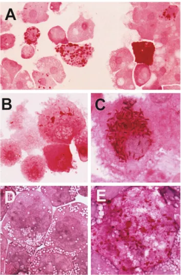
Figure 1. Growth of T. whippelii, isolated from heart valve biopsies
mL hrIL-10 (ED50Å 2 ng/mL in MC-9 costimulation assay; Pepro
of patients (TWZ1 or TWZ2), in cultured human macrophages and
monoblasts. A, Periodic acid – Schiff (PAS) – positive diastase-resis- Tech, Rocky Hill, NJ), and 2.51 1007M dexamethasone (Sigma
tant inclusions in deactivated human macrophages 10 days after inocu- Chemie, Buchs, Switzerland) in an incubator (SteriCult; Forma lation with ground heart valve material (TWZ1, 1st passage). B, PAS- Scientific, Marietta, OH) at 377C, 5% CO2, and 98% humidity. positive diastase-resistant inclusions and slender PAS-positive rods No antibiotics were added.
indicative for multiplication of T. whippelii in deactivated human
For primary infection of cell cultures, homogenized material
macrophages 9 days after inoculation with infected macrophages
fromÇ1 mg of heart valve tissue was suspended in 500 mL of
(TWZ1, 2nd passage) shown in A. C, Formation of numerous
PAS-0.9% NaCl. Aliquots (2 mL) of this suspension were added to the
positive rods in macrophages deactivated with IL-4 alone (TWZ2,
wells of the 6-well plates with deactivated primary phagocytes or
5th passage). D and E, Propagation of passaged T. whippelii in human
monoblast cell line deactivated with IL-4. D, Uninfected SigM5 monoblast cells in a volume of 2 mL of complete medium. Infected
monoblasts. E, Infected, large, foamy monoblast showing numerous cultures were further incubated for 8 – 10 days under the conditions
PAS-positive inclusions and rods (TWZ1, 3rd passage). described above without replacement of the medium. Then cells
were collected together with the culture medium (2 mL), and ali-quots of these cell suspensions were analyzed for T. whippelii by diastase/PAS staining of cytocentrifuge preparations (100-mL specimen obtained after 6 weeks of antibiotic therapy was negative
aliquots), electron microscopy (1.5-mL aliquots), and PCR (300-for PAS-positive inclusions, but his heart valve was loaded with
mL aliquots) or were used as secondary inocula (200 mL/2 mL/ PAS-positive, acid fast – negative bacilli. In both patients, the
diag-well) for further passages of T. whippelii in cultured cells as de-nosis of T. whippelii infection of the heart valves was established
scribed above. by applying a eubacterial broad-range PCR, followed by
sequenc-For expansion of T. whippelii in a large volume of cells, 1 mL ing of the amplicon as described [12] and by T. whippelii – specific
of infected cell suspension after the second passage was inoculated PCR.
into 50 mL of deactivated SigM5 cells at a density of 21 106 For patient TWZ1, a stretch of 384 bp was sequenced with 2
cells/mL on a 250-cm2
growth area. This culture was further incu-mismatches in comparison with the reference sequence of T.
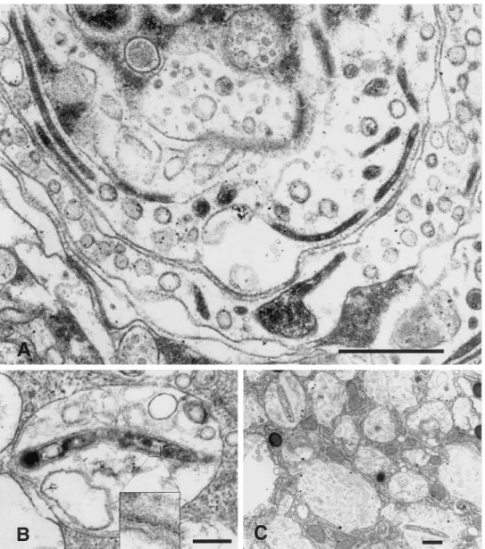
Figure 2. Electron micrographs
showing Whipple bacilli cultured in deactivated human macrophages and SigM5 monoblast cells. A, Macro-phage deactivated with IL-4 and in-fected with passaged inoculum (TWZ2, 3rd passage) showing nu-merous slender bacteria in longitudi-nal and transverse sections. BarÅ 450 nm; original magnification, 112,000. B, SigM5 monoblast deac-tivated with IL-4, IL-10, and dexa-methasone and infected with TWZ2 inoculum (2nd passage) showing ba-cillus within vesicular cellular struc-ture. Insert: Magnification of part of cell wall with trilaminar structure unique to cell wall of T. whippelii. BarÅ 300 nm; original magnifica-tion,19050. C, Deactivated human macrophage infected with TWZ1 in-oculum (5th passage). Bacilli range from normal appearance to advanced stages of digestion; latter consists of cell wall remnants clustered in large vesicles, referred to as Whipple bod-ies, in light microscopy of periodic acid – Shiff – stained cells. BarÅ 450 nm; original magnification,14200.
staining, electron microscopy, and PCR as described above or PCR using primers pW3FE and pW2RB (corresponding to our TW-1 and TW-3 primers) [4, 12, 13].
frozen at0707C in 1-mL aliquots and used for further experiments.
Electron microscopy. For electron microscopy, cell suspen- Statistical analysis. For statistical analysis, the t test with
Dunnett’s posttest analysis for the comparison of several groups sions collected 9 days after challenge were fixed in 3%
glutaralde-hyde and processed using standard methods [10]. with one control was performed with a commercial software pack-age (Instat; Graphpad, San Diego).
Molecular analyses. For PCR analysis, 300-mL aliquots of cell
suspensions collected 9 days after infection were centrifuged, and the DNA was extracted by digesting the samples for 3 h at 557C in 70 mL of 50 mM TRIS-HCl (pH 8.5), 1 mM EDTA, 0.5% SDS,
Results and 200 mg/mL proteinase K. After proteinase K was inactivated
at 957C for 12 min, 2 mL was directly amplified in a PCR mix In a first series of isolation attempts with intestinal biopsy containing 2% Tween 20 and primer pair TW-1/TW-3 or TW-1/
material from a patient with Whipple’s disease, we failed be-TW-2 as described [12], resulting in fragments of 141 and 267 bp,
cause of inevitable bacterial overgrowth. We then were able to respectively. The number of cycles was always 40. After Southern
obtain infected heart valve material from 2 patients undergoing blotting to a nylon membrane, hybridization was done using the
heart surgery for endocarditis. Crushed tissue samples from internal digoxigenin-labeled oligonucleotide TW-4 (5
*-CACGGT-each patient (TWZ1 or TWZ2) were inoculated into cultures TGTCGTCAGCTCGT-3*) at 507C overnight. The positive control
of human monocytes deactivated with a combination of dexa-was intestinal biopsy material from a patient with confirmed
subse-Figure 4. Time-kinetics of growth of T. whippelii in deactivated
human macrophages. Macrophages plated in 24-well tissue culture plates were deactivated with IL-4 and IL-10 for 48 h prior to infection with 100mL of freshly defrosted aliquot of large-volume culture of TWZ1 (3rd passage) in SigM5 cells. At indicated times, cells were collected, cytocentrifuge preparations were stained with
diastase/peri-Figure 3. Specific detection of T. whippelii DNA in cell cultures
odic acid – Schiff, and infected cells were counted. Total barÅ before by polymerase chain reaction (PCR). Top, agarose gel electrophoresis
diastase treatment, hatched areaÅ after diastase digestion (diastase-and ethidium bromide staining; bottom, Southern blot hybridized with
resistant). Data are mean{ SD of duplicate preparations from tripli-digoxigenin-labeled internal oligonucleotide TW-4. Lanes M:
molec-cate wells. ** Põ .001. ular weight marker (pBR322 DNA digested with MspI), fragment
size (from top), 622, 527, 404, 307, and 240 bp down toõ50 bp. Lane 1: human blood – derived macrophages inoculated with heart valve material of patient TWZ1. Lane 2: uninfected macrophages.
remained infective for deactivated monocytes and were used
Lane 3: 2nd passage of isolate TWZ1 in SigM5 cells (monoblast cell
line described in figure 1). Lane 4: uninfected SigM5 cells. Lane 5: for further experiments.
3rd passage of isolate TWZ1 in large-volume cultures of SigM5 cells. Kinetics growth studies. Next we turned to kinetics growth Lane 6: uninfected SigM5 cells from large-volume cultures. Lanes 7 studies for demonstration of multiplication of a defined inocu-and 8: small bowel biopsies from patients without Whipple’s disease.
Lane 9: PCR reagents only (no DNA added). Lane 10: Clinical speci-men (intestinal biopsy) known to contain T. whippelii DNA (supplied by M. Maiwald, Germany) [13].
quently used to further passage T. whippelii in deactivated human macrophages and monoblast cells. Within 8 – 10 days after infection, macrophages appeared filled with coarse, dia-stase-resistant, positive conglomerates and slender PAS-positive rods (figure 1) that were both acid fast – negative (not shown). Heating of inocula at 657C for 2 h precluded the ap-pearance of PAS-positive inclusions (not shown).
By electron microscopy, it was confirmed that these inclu-sions corresponded to intact and degenerating bacteria (figure 2) described in biopsy material from patients with Whipple’s disease [1, 14]. In contrast to findings with tissue specimens, however, extracellular bacteria were never seen by any method we tested, indicating the cellular environment is required for bacterial growth, and PCR analyses of culture supernatants remained negative. PCR studies with specific primers con-firmed that the isolated bacteria from both patients were indeed T. whippelii (figure 3 for patient TWZ1). To establish stable propagation of T. whippelii, we expanded the bacterial
popula-tion of both isolates after two passages in large-volume cultures Figure 5. Microphotographs of cytospin preparations correspond-of deactivated SigM5 monoblasts (figure 1D – E; figure 3, lane ing to figure 4 showing time-dependent increase of periodic acid –
Schiff – positive inclusions. A, 1 h. B, 8 h. C, 24 h. D, 48 h.
Figure 6. Comparative polymer- with IL-4. The method chosen for the support of growth of
ase chain reaction analysis of con- this bacillus was proven to be reproducible with infected mate-secutive passages of original heart
rial from 2 patients. Growth-kinetics studies of passaged
iso-valve inoculum TWZ1 (B, D) and
lates revealed a time-dependent increase of diastase/PAS –
posi-corresponding dilutions of
uncul-tured inoculum (A, C). A and B tive inclusions within macrophages. Multiplication of T.
compare inoculum (A) with culture whippelii in our culture system was further corroborated by passages (B) using primer pair TW- comparative PCR analysis of passaged isolates with corre-1/TW-3, yielding 141 bp. C and D
sponding dilutions of the original (uncultured) inoculum.
compare inoculum (C) with culture
The novel, yet simple strategy used for isolation of this
passages (D) using primer pair
TW-1/TW-2, yielding 267 bp. Lanes 1: uncultured microorganism should also be tried for the isolation
molecular size markers pBR322 of other uncultured or unidentified microorganisms, such as M. DNA digested with MspI (for sizes, leprae, or even suspected agents in diseases, such as Crohn’s see figure 3). Lanes 2, negative
con-disease, sarcoidosis, Wegener’s granulomatosis, or SAPHO
trol (DNA from uninfected human
(synovitis, acne, palmoplantar pustulosis, hyperostosis, osteitis)
macrophages). Lanes 3 – 8:
dilu-tions of original inoculum corre- syndrome.
sponding to dilutions during con- In contrast to observations in histologic studies of tissues secutive passages (by lane): infected with T. whippelii [1], intracellular bacteria were ob-1:10,000 (3), 1:500,000 (4), 1:51
served only in cytospin preparations in our study, indicating
106
(5), 1:2 1 107
(6), 1:81 107
that T. whippelii is an intracellular pathogen, and PCR analysis
(7), 1:3.2 x 10⁸
(8). Comparison of
corresponding dilutions infers mul- of supernatants with both primer sets produced no amplicons.
tiplication by factor of at least 100 – The requirement of IL-4, an immunoregulatory cytokine pro-1000 during 4 passages of original duced by various immune cells, notably Th2 cells [15], indi-inoculum in human phagocytes.
cates a probable contribution by host factors to the pathogenesis of Whipple’s disease, such as an imbalance in Th1-Th2 im-mune response. While the target for IL-4 in the mononuclear lum in phagocytes (figures 4, 5). These experiments showed a
time-dependent augmentation of acid fast – negative, diastase- phagocyte has not yet been elucidated [10, 11], IL-4 clearly renders monocytes permissive for intracellular growth of mi-resistant, PAS-positive inclusions in deactivated macrophages
(figures 4, 5). To further confirm the multiplication of T. whip-pelii in consecutive passages of the original inoculum, passaged isolates of 1 patient (TWZ1) were compared with the corre-sponding dilutions of the original heart valve material without incubation. As shown by PCR analysis (figure 6), the different yields of copies in the cultured versus uncultured inoculum were already clearly visible at passage 3, corresponding to a dilution of 1:500,000 (figure 6, lanes 4). Higher dilutions of the original inoculum no longer yielded amplified products (figure 6A, C), while the corresponding passages were still positive (figure 6B, D).
Requirement of macrophage deactivation for intracellular growth. We then studied the individual effects of the three deactivating signals initially used in combination for the isolation of T. whippelii (figure 7). We found that IL-4, previously shown to be the most effective deactivating agent in the Listeria model [10], was the critical deactivating signal that promoted
intracellu-Figure 7. Effects of single and combined deactivating agents (IL-4,
lar multiplication of T. whippelii. In contrast to results from
IL-10, dexamethasone) on formation of periodic acid – Schiff (PAS) –
studies with L. monocytogenes [10], IL-10 and dexamethasone
positive inclusions corresponding to growth of T. whippelii in human
did not significantly promote intracellular multiplication. To the macrophages infected with TWZ2 inoculum, which was passed twice contrary, dexamethasone interfered with the propagation of T. through macrophages deactivated with combination of all 3 signals.
Results are % of macrophages showing typical PAS-positive
inclu-whippelii in macrophages (figure 7). Identical observations were
sions after incubation for 9 days in continuous presence of
deactivat-made with the monocytic human cell line (not shown).
ing agents. Only IL-4 (2 ng/mL) had significant deactivating effect (Põ .001); IL-10 (5 ng/mL) had no effect (P ú .1). Dexamethasone
Discussion
(2.51 1007
M), with exception of combination with IL-4, suppressed
We report here the first isolation and propagation of T. whip- propagation of T. whippelii. Mean{ SE from 3 independent
experi-ments with phagocytes from 3 donors.
8. Schaffner A, Schaffner T. Glucocorticoid-induced impairment of
macro-croorganisms in vitro [10, 11] and in vivo leads to an increased
phage antimicrobial activity: mechanisms and dependence on the state
susceptibility of cells to intracellular pathogens such as Listeria
of activation. Rev Infect Dis1987; 9(suppl):S620 – 9.
[15] and Leishmania organisms [16]. 9. Schaffner A, Rellstab P. Gamma-interferon restores listericidal activity In any event, the isolation and culture of T. whippelii de- and concurrently enhances release of reactive oxygen metabolites in dexamethasone-treated human monocytes. J Clin Invest1988; 82:913 – scribed herein will permit further studies on the pathogenesis,
9.
immunology, and therapy of this intriguing infection.
10. Blauer F, Groscurth P, Schneemann M, Schoedon G, Schaffner A. Modula-tion of the antilisterial activity of human macrophages by activating and deactivating cytokines. J Interferon Cytokine Res1995; 15:105 –
14.
11. Meier-Osusky I, Schoedon G, Blauer F, Schneemann M, Schaffner A. References
Comparison of the antimicrobial activity of deactivated human macro-phages challenged with Aspergillus fumigatus and Listeria monocyto-1. Dobbins WO. Whipple’s disease. In: Dobbins WO, ed. Whipple’s disease.
genes. J Infect Dis1996; 174:651 – 4. Springfield, IL: CC Thomas,1987:1 – 227.
12. Altwegg M, Fleisch-Marx A, Goldenberger D, Hailemariam S, Schaffner 2. Cohen AS, Schimmel EM, Holt PR, Isselbacher KJ. Ultrastructural
abnor-A, Kissling R. Spondylodiscitis caused by Tropheryma whippelii. malities in Whipple’s disease. Proc Soc Exp Biol Med1960; 105:411 –
Schweiz Med Wochenschr1996; 126:1495 – 9. 4.
13. Meier-Willersen HJ, Maiwald M, von Herbay A. Morbus Whipple in 3. Wilson KH, Blitchington R, Frothingham R, Wilson JA. Phylogeny of the
Assoziation mit opportunistischen Infektionen. Dtsch Med Wochenschr Whipple’s-disease – associated bacterium. Lancet1991; 338:474 – 5.
1993; 118:854 – 60. 4. Relman, DA, Schmidt TM, MacDermott RP, Falkow S. Identification of
14. Silva MT, Macedo PM, Moura-Nunes JF. Ultrastructure of bacilli and the the uncultured bacillus of Whipple’s disease. N Engl J Med1992; 327:
bacillary origin of the macrophagic inclusions in Whipple’s disease. J
293 – 301. Gen Microbiol1985; 131:1001 – 13.
5. Sieracki JC. Whipple’s disease: observation of systemic involvement. Arch 15. Nakane A, Nishikawa S, Sasaki S, et al. Endogenous interleukin-4, but Pathol1958; 66:464 – 7. not interleukin-10, is involved in suppression of host resistance against 6. Dobbins WO. Is there an immune deficit in Whipple’s disease? Dig Dis Listeria monocytogenes infection in interferon-depleted mice. Infect
Sci1981; 26:247 – 52. Immun1996; 64:1252 – 8.
7. Bjerknes R, Laerum OD, Odegaard S. Impaired bacterial degradation by 16. Leal LM, Moss DW, Kuhn R, Muller W, Liew FY. Interleukin-4 transgenic monocytes and macrophages from a patient with Whipple’s disease. mice of resistant background are susceptible to Leishmania major
infec-tion. Eur J Immunol1993; 23:566 – 9. Gastroenterology1985; 89:1139 – 46.