Original article
Microsatellite alterations and TP53 mutations in plasma DNA of small-cell
lung cancer patients: Follow-up study and prognostic significance
R. Gonzalez,
1J. M. Silva,
1A. Sanchez,
1G. Dominguez,
1J. M. Garcia,
1X. qi Chen,
2M. Stroun,
2M. Provencio,
1P. Espana,
1P. Anker
2& F. Bonilla
1'Department of Medical Oncology, Chnica Puerto de Hierro, Madrid, Spain; 2 Laboratory of Plant Biochemistry and Physiology, Faculty of Science of Geneva University, Pavilion des Isotopes, Geneva, Switzerland
Summary
Background: Small-cell lung cancer (SCLC), one of the major
types of lung cancer, is associated with many different somatic
molecular genetic changes. These alterations, observed in
tumor DNA, have also been identified in the plasma DNA of
patients. We undertook the present study to make a prospective
investigation into the correlation between abnormal plasma
DNA and patient survival.
Patients and methods: Thirty-five patients with SCLC were
selected after histological diagnosis. Polymorphic markers
(ACTBP2, UT762 and AR) were chosen for their reported
high rate of alterations in SCLC and analyzed in tumor tissue,
normal blood cells and plasma DNA. Furthermore, we looked
for mutations of the TP53 gene in tumor and plasma DNA.
Results: In 25 patients (71%) at least one molecular change
precisely matching that of the primary tumor was detected in
the plasma DNA. No difference in survival was observed
between patients with aberrant plasma DNA and patients
without plasma DNA alterations. However, patients with
microsatellite modifications and TP53 mutations
concomi-tantly, showed a significant difference (P = 0.02) in survival
compared with patients bearing only one of these molecular
changes. In 15 cases it was possible to find a correlation either
between tumor response and disappearance of abnormal
plasma DNA, or tumor progression and persistence of plasma
DNA alterations.
Conclusions: Free plasma DNA with molecular alterations
is present to a high degree in plasma DNA of SCLC patients
and may have a role as a prognostic factor.
Key words: circulating DNA, plasma DNA alterations,
prog-nostic factor, SCLC, tumor DNA alterations
Introduction
Small-cell lung cancer (SCLC) represents about 20% of
all primary lung cancers [1, 2]. The predominant risk
factor by far is cigarette smoking [3]. The tumor arises
from basal neuroendocrine cells, infiltrating the
sub-mucosa in the early stages of the disease [4]. Despite the
diversity and intensity of current therapies, survival is
usually short. In SCLC several clinical parameters have
been established as prognostic factors. Long-term
sur-vival has been associated to age, 65 years old or less at
diagnosis; performance status, ECOG 0-1; and limited
disease (LD) [5]. The overall survival rate of patients
treated with combination chemotherapy, with or
with-out chest irradiation, is abwith-out 7% at two years, ranging
from 2% for patients with extensive disease (ED) to 13%
with LD [5]. More recently, concurrent chemotherapy
and radiotherapy has been reported, in the case of LD,
to allow a 44% survival at two years [6].
SCLC is associated with different types of molecular
gene aberrations including somatic mutations of
onco-genes such as c-myc [7] or mutations of tumor suppressor
genes such as TP53 and RB [8, 9] as well as allelic loss
and microsatellite alterations in several chromosomal
regions [2, 10-14]. Detection of these molecular changes
may help define histological diagnosis or be used as a
prognostic factor.
Increased quantities of DNA have been found in the
plasma of patients suffering from different malignancies
[15-17]. This circulating extracellular DNA exhibits tumor
related alterations such as decreased strand stability [17],
ras orTP53 mutations [18-24], microsatellite alterations
[25-30] or aberrant promoter hypermethylation of tumor
suppressor genes [31-33].
Several studies seem to indicate that aberrations in
plasma/serum DNA may be used as a prognostic factor.
All patients suffering from head and neck cancer with
microsatellite alterations in the serum matching those of
the primary tumor had advanced disease [27]. Two
reports on pancreatic carcinoma provide evidence that
K-ras mutations in plasma DNA are linked to overall
survival [19, 34]. Nakamori and his group found that
patients showing ras mutations in plasma were more
likely to respond poorly to treatment, and those who
still harbored a mutation in the plasma, even after
seemingly successful surgery, had a poor prognosis
[34]. However, no prospective studies have been
de-signed to analyze the value of free plasma DNA during
patient follow-up and its real implication as a predictive
factor.
In relation to lung cancer, quantitative studies show a
definite correlation between plasma DNA levels and
prognosis [35, 36]. Plasma DNA levels were increased in
patients with advanced disease and correlated with other
enzymatic tumor markers [35].
Moreover, microsatellite instability, expressed either
as a new allele or the loss of one allele [26], or aberrant
DNA methylation [31] have been shown to occur in
serum DNA of patients suffering from non-small-cell
lung cancer (NSCLC). In SCLC, 76% of the patients
presented a microsatellite alteration in the tumor DNA
and 71% in plasma DNA [25].
The purpose of this prospective phase II study was to
identify plasma DNA with similar features to tumor
DNA in SCLC patients, using microsatellite alterations
and TP53 mutations as molecular markers, and
corre-late them with patient survival, trying to investigate the
predictive value in the outcome of aberrant plasma
DNA.
Patients and methods
centrifuged at 14000 rpm for 30 min, after which the clear supernatant was collected (25). Proteinase K (20 mg/ml) (Boehringer Mannheim, Mannheim, Germany) and buffer AL (Qiagen Inc.) were added in a 1:10 proportion with respect to the collected supernatant, and incu-bated overnight at 55 °C.
Clinical characteristics of the patients
Clinical staging procedures included chest X-rays, blood cell counts and biochemical profiles, computed tomography of chest and abdo-men, and finally bone scintigraphy. Computed tomography of the brain was performed only when there was clinical suspicion of central nervous system involvement. This study was designed as a prospective phase II study with all patients undergoing the same treatment. Patients with ED received four chemotherapy (CT) cycles based on carboplatin 300 mg/m2 intravenously on day 1 plus etoposide 100 mg/m2, intravenously on day 1 and orally on days 2,3,4 and 5. Patients with LD were submitted to the same treatment followed by chest irradiation and two additional series of the same CT. Following treat-ment, which usually lasted 28 weeks, clinical follow-up took place every 12 weeks. Plasma was collected and the DNA analyzed on each occasion. A median of 4 plasma samples (range 1-10) were examined for each patient. Disease recurrence was treated with CT (cyclophos-phamide, 1000 mg/m2 i.v. on day 1; adnamycin, 45 mg/m2 i.v. on day 1; and vincristine 2 mg/m2 iv on day 1) until a second complete response or disease progression was observed.
Patient population
Between May 1997 and January 1999, 35 patients with histologically confirmed diagnosis of SCLC were recruited for this phase II study. Informed consent was obtained from all participants after explaining the nature of the study, which was approved by the Research Ethics Board of our hospital. The median age of the patients was 61 years (range 38-79 years). Thirty-one were male and four female. The disease was classified as limited disease (LD) when tumor was restricted to one hemithorax, including ipsilateral supraclavicular fossa, and any wider disease including distant metastasis was classified as extensive disease (ED). The 113-week follow-up period lasted from May 1997 until June 1999; the median follow-up time for all patients was 57 weeks. Patient survival was calculated as the interval between the date of diagnosis and date of death or date of the last follow-up visit, and was the major clinical endpoint.
Tissue samples
A blood sample was collected from each patient with histological diagnosis of SCLC on the day prior to the first chemotherapy course. The diagnosis of SCLC was based on histological analysis of tumor samples collected at bronchoscopy or on cytological examination of sputum. None of the patients underwent surgical extirpation of the primary tumor or metastasis. Sixteen bronchial biopsy specimens were available for molecular study. These were snap-frozen in liquid nitrogen until processed. In addition. 13 healthy controls with no history of cancer or other known disease were recruited from blood donor volunteers.
DNA extraction
Blood cell and plasma DNA was extracted immediately. Tumor tissue and blood cell DNA was purified by a nonorganic method (S-4520 Kit. Oncor Inc., Gaithersburg. Maryland). Plasma DNA was extracted using Qiagen columns (Qiamp Blood Kit, Qiagen Inc., Hilden, Ger-many) according to the protocol for blood and body fluids with the following modifications. Up to 12 ml of plasma previously heated on a heat block to 99 °C for 5 min were used. The heated sample was then
Microsatellite analysis and PCR conditions
Three microsatellite markers were used to determine loss of heterpzygosity (LOH) or microsatellite shift on chromosomes 5 (ACTBP2), 12 (UT762) and X(AR). These markers were previously reported to show a high rate of microsatellite alterations in SCLC patients [10]. PCR was performed in 25 ul volumes using 0.75 units of Amph Taq Gold DNApolymerase (Perkin-Elmer, Roche Molecular Systems, Inc, Branchburg, New Jersey), 2.5 ul of 10X PCR buffer, 200 uM dNTP, each primer at 0.6 uM with different concentrations of MgCl2 depending on the polymorphic marker. A 35-cycle amplifica-tion was performed in a thermal cycler (Perkin-Elmer, Cetus, Foster City, California). The annealing temperatures were 59 °C, 62 °C and 57 °C, respectively. The alleles were separated by mixing 25 ul of PCR products with a 10 ul volume of loading buffer (total volume 35 ul), 0.02% xylene cyanol and 0.02% bromophenol blue. Electrophoresis was run on nondenaturing 8%-12% polyacrylamide gels for 12-15 h at 500 v. After gel electrophoresis, the allelic band intensity was detected by a nonradioisotopic technique using a commercially available silver staining method [37]. We analyzed the allelic intensities by densitometry. The gel image was captured by a GS-690 Imaging Densitometer (Bio-Rad laboratories, Hercules, California) digitized in 400 dpi, and analyzed by using Multi-Analyst/PC (Bio-Rad Labora-tories, Hercules, California).
Ten of the results obtained were checked by laser fluorescence. One primer of each set was labeled with a fluorescent dye at the 5' end and all primers were purified by HPLC. Ten ng and 20 ng of plasma DNA, 20 ng of blood cell and tumor DNA were used as a template in a hot-start PCR in a 25 ul reaction mixture: 1.5 mM MgCl2, 20 uM dNTP,
0.6 u Taq polymerase, 2-10 pmol of each primer (depending on the primer). Each PCR reaction was carried out in the same conditions as described above. PCR products were separated electrophoretically on 8% polyacrylamide gels and detected by laser fluorescence using an automated gene sequencer (Alfexpress, Amersham-Pharmacia). Fluo-rescent gel data was analyzed with the Allele-link 2 program. PCR products from lymphocytes, corresponding blood plasma and tumor tissue were analyzed on the same gel. The size in base pairs of the microsatellite alleles was automatically calculated using internal size markers. Automatic analysis of peak areas allowed relative
quantifica-tion of PCR products and determinaquantifica-tion of allelic ratios. LOH was scored if the allelic ratios of tumor or plasma PCR products and corresponding lymphocyte PCR products were below a cut-off value of 70%. Results indicating LOH were repeated at least twice.
Point mutations of thepS3 gene
Since up to 50% of SCLC patients were reported to exhibit TP53 mutations we also looked for the presence of point mutations in exons 5, 6, 7, and 8 of TP53 in the plasma DNA of SCLC patients. PCR-SSCP analysis was performed according to a modification of the method reported by Orita et al. [38]. The primers used for amplifica-tion of the different exons were: 5-TCCTTCCTCTTCCTACAG and 5-ACCCTGGGCAACCAGCCCTGT for exon 5; 5-ACAGGGCTG-GTTGCCCAGGGT, and 5-AGTTGCAAACCAGACCTCAGGCG for exon 6; 5-TCCTAGGTTGGCTCTGACTGT and 5-AGTGGCCCT-GACCTGGAGTCT for exon 7; and 5-GGGACAGGTAGGACC-TGATTTCCTT and 5-ATCTGAAGGCATAACTGCACCCTTGG for exon 8 (Source: J. Weissenbach, Genethon,Whitehead Institute Center for Genome Research). The annealing temperatures were 65 °C, 67 °C, 62 °C and 68 °C, respectively. PCR was performed under standard conditions in 25 ul that contained 2 ul (100 ng) of DNA template (tumor, normal or plasma DNA); 2.5 ul of 10 X PCR buffer and 0.75 U of Ampli Taq Gold (Perkin-Elmer, Roche Molecular Systems Inc., Branchburg, New Jersey); 200 uM dideoxynucleoside triphosphate (dNTP) mix, each primer at 0.6 uM, different concentrations of magnesium chloride depending on the primer, and distilled H2O
needed to reach the total volume. For PCR amplification, the samples underwent 40 cycles at 94 °C for 1 minute, and were then subjected to different annealing temperatures depending on the primer, and 70 °C for 1 minute. The amplified products were denatured by mixing with 15 ul of denaturing stop solution that contained 98% formamide, 10 mmol/1 edathamil (pH 8.0), 0.02% xylene cyanol and 0.02% bromo-phenol blue, heated to 95 °C for 5 minutes and rapidly cooled on ice. Electrophoresis was run on nondenaturing 8%-12% polyacrylamide gels for 12-15 hours at 250 v. The allelic band intensity on the gels was detected by a nonradioisotopic method using a commercially available silver staining method [37]. The specimens that showed a differential band at SSCP were amplified to obtain templates for DNA sequencing. These amplifications were independent from those used for SSCP analysis. Amplified DNA fragments were purified from 0.9% agarose gels using a Geneclean Kit (Bio-101, Inc., La Jolla, California), and used for direct DNA sequencing with the dNTP method with a Sequenase Kit (United States Biochemical Corp. Cleveland, Ohio).
Statistical analysis
Given the sample size of the study, it was considered that the statistical power of the study was higher than 75%. The statistical study was performed using SPSS version 7.5 for Windows (SPSS, Inc., Chicago, Illinois) and included Kaplan-Meier for estimation of survival curves. Differences between two curves were examined by the log-rank and Breslow tests. The level of significance was set at < 0.05.
Results
Molecular alterations detected
We detected at least one molecular alteration, LOH or
shift, with microsatellite markers UT762, ACTBP2 and
AR (X) (Figures 1-3), or a mutation on TP53 gene
(Figure 4) in the plasma DNA of 25 of 35 patients
(71%). Twenty-two showed molecular alteration at the
time of diagnosis, for the remaining three it appeared
later during follow-up. In eight cases (23%),
microsatel-L T P1 P2 P3 P4
AR(x)
ACTBP
UT762
Figure I. Photograph of the gels taken under normal light after
staining with a (NO3)Ag method showing microsatellite alterations at the AR(X) marker in lymphocyte (L), tumor (T) and plasma (P) DNA of patient # 2 2 . The sequence of plasma determinations (Pl-4) demon-strates the persistence of microsatellite shift at the AR(X) marker at the time of diagnosis (PI), after 4 courses of chemotherapy (P2) and after combined treatment chemotherapy plus radiotherapy (P3), and the disappearance of the microsatellite shift at 52 weeks (P4) coinci-dent with complete disease remission. No alterations were detected at markers ACTBP and UT762 throughout follow-up.
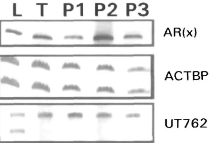
L T P1 P2 P3
AR(x)
ACTBP
UT762
Figure 2. Representative photograph of the gels taken under normal
light after staining with a (NO3)Ag method, showing microsatellite alterations at the UT762 marker in lymphocyte (L), tumor (T) and plasma (P) DNA of patient # 11. A loss of heterozygosity was observed at marker UT762 throughout follow-up of the patient, from diagnosis (PI) until death, 28 weeks later (P3) with no response to treatment. There were no changes at markers AR(X) (homozygous in male patients) or ACTBP.
lite alterations coincided with a TP53 mutation. Twelve
patients (34%) exhibited only microsatellite alterations,
and TP53 mutations alone were detected in five (14%).
Out of 16 available tumor samples, 12 (75%) presented
microsatellite alterations and/or TP53 mutations, 10 of
which (83%) showed similar molecular changes in the
plasma (Table 1). Analysis of microsatellite patterns by
laser fluorescence using an automated gene sequencer
(Figure 3) confirmed the results obtained with the silver
stained gels.
No difference in DNA pattern was detected between
plasma DNA samples and normal blood cell DNA from
13 healthy controls.
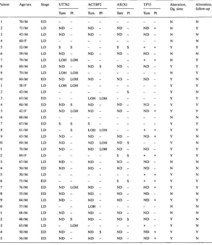
Table 1. Clinical features and microsatellite alterations and TP53 gene mutations in plasma and tumor DNA in SCLC patients. Patient 1 2 3 4 5 6 7 8 9 10 11 12 13 14 15 16 17 18 19 20 21 22 23 24 25 26 27 28 29 30 31 32 33 34 35 Age/sex 70/M 72/M 45/M 60/F 52/M 59/M 79/M 60/M 70/M 60/M 38/F 42/M 63/M 60/M 42/F 68/M 67/M 61/M 65/M 69/M 70/M 69/F 67/M 50/M 50/M 75/M 76/M 55/M 64/M 57/M 68/M 48/M 65/M 50/M 56/M Stage ED LD LD LD LD LD LD LD LD ED LD LD ED ED LD LD ED LD LD LD LD LD LD ED LD ED ED ED LD LD LD LD LD ED ED UT762 Turn -N D N D -S N D LOH N D LOH N D LOH -N D N D -S -N D N D N D -N D N D -N D N D N D -N D N D -N D N D PI -S -LOH -LOH LOH LOH -S LOH -S
s
-LOH -S LOH -ACTBP2 Turn -ND ND -N D -ND -ND -LOH ND N D -S LOH ND ND N D -N D ND -N D N D ND LOH N D N D -N D N D PI -S -LOH -LOH -LOH LOH -S -AR(X) Turn -ND ND -S ND -ND -ND -ND ND -ND ND ND S ND ND -S ND ND ND -ND ND -ND ND PI -S
-s
-s
-s
-s
-s
-TP53 Turn PI -ND + ND -+ -+ ND -+ -+ ND ND ND -ND + -+ -+ ND + ND -+ -+ ND ND -+ -+ + + ND + ND -ND + ND ND -+ ND + ND + Alteration, Dg. time N N N N Y N N Y N Y Y Y Y Y Y N Y Y Y Y Y Y N N Y Y Y N Y N N Y Y Y Y Alteration, follow-up N Y N N Y N Y Y Y N Y N Y Y Y N Y Y N N Y Y N N N Y Y N Y N N N N Y N
Abbreviations: F - female; M - male; ND - not done; N - absence of molecular alterations at diagnosis time or during the follow-up; Y - Presence of molecular alterations at diagnosis time or during the follow-up; - - absence of S, LOH orTP53 mutations; + - presence of TP53 mutations; Turn - tumor DNA; PI - plasma DNA.
Survival data
According to stage at diagnosis, 24 patients had LD and
11 patients ED, with an equivalent proportion of LD or
ED patients having abnormal plasma DNA in both
subgroups. The overall median survival at the end of the
follow-up period was 42 weeks (range 4-97 weeks). Nine
patients remained alive, with a median follow-up of
thirty-three weeks (range 24-73 weeks), all of them
displayed molecular alterations in their plasma DNA,
and twenty-six patients died after a median survival time
of forty-three weeks (range 4-97 weeks). The 10 patients
Full Stale Ftl • Time [Minute) | Plasma DNA 30-20 • 10-220 230 240
J
2M 2tu
V> 270I.
210 Blood 290 Cell 300 DNA 3020 -10 • 220 230 2J0I
LOH1
Tumor DNAm
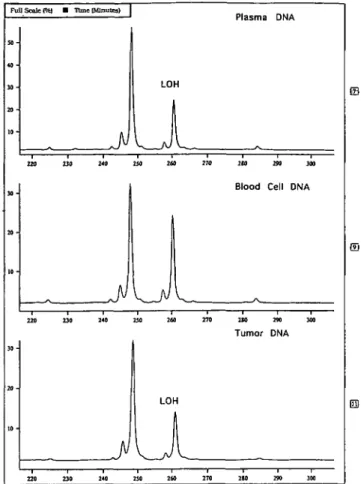
ElFigure 3. Original tracings from microsatellite analysis from plasma,
blood cell and tumor DNA of patient 9, with marker UT762. Plasma and tumor DNA reveal LOH.
3 '
A C G T A C G T
it t
B
G
T
G
C - G
G
T
G
G
A
G
Figure 4. Sequencing analysis in plasma DNA (B) of patient #29
shows a nucleotide change at codon 273 (C to G) in the direct sequencing in exon 8 of the TP53 gene. This nucleotide change causes an amino acid change (Arg-Gly). Panel (A) control sequence.
that showed no aberrant plasma DNA were dead at the
end of the study. All deaths were tumor related and
non-toxic deaths were registered.
Statistical analysis of survival in the different
sub-groups revealed the following results: (a), the
compar-ison between patients with aberrant plasma DNA at the
time of diagnosis (n = 22; median survival 57 weeks)
versus those patients with no DNA alterations (n = 10;
median survival 46 weeks), showed no statistically
sig-Survival time in weeks
Figure 5. Kaplan-Meier survival curves for patients with only
micro-satellites changes, or TP53 gene mutations, versus patients with both type of alterations concomitantly, in free plasma DNA.
nificant difference (P - 0.4); (b), patients in whom
abnormal plasma DNA disappeared following
treat-ment (n = 11; median survival 33 weeks) and patients
with persistence of plasma DNA changes (n = 10;
median survival 57 weeks) did not differ significantly
(P = 0.5); (c), the survival of patients with only
micro-satellite alterations (n = 12; median survival 28 weeks)
or TP53 gene mutations (n = 5; median survival 59
weeks), did exhibit a statistically significant difference
{P = 0.02) regarding patients presenting both types of
DNA changes concomitantly (n = 8; median survival 70
weeks) (Figure 5).
Correlation between clinical development and molecular
events
In 15 patients (60%) of the 25 with abnormal plasma
DNA, a correlation was observed between the clinical
outcome and the presence of plasma DNA changes
(Table 2). Among these cases, three different subgroups
were distinguished: the first included four patients
(patients 5, 15, 29, 34) with partial or complete response
of their tumor to treatment and concomitantly the
disappearance of the plasma DNA molecular changes,
subsequently, with the persistence of partial response or
disease recurrence, new appearance of abnormal plasma
DNA was observed. The second group consisted of eight
patients (patients 8, 11, 13, 14, 17, 21, 26, 27) with no
response to treatment and no clearance of the plasma
DNA molecular changes. The third group, involved
three patients (patients 2, 7, 9) with no molecular
alter-ations at diagnosis and late appearance of plasma DNA
changes coincident with disease recurrence. In one of
these cases, the presence of de novo TP53 gene mutation
was detected 12 weeks prior to clinical relapse.
Discussion
Our study confirms the presence of microsatellite
alter-ations similar to those found in tumors in the plasma
DNA of SCLC patients [25]. However, it is the first
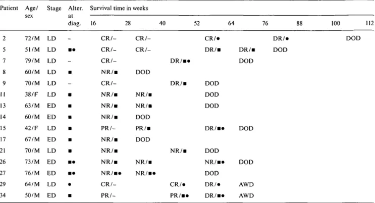
Table 2. Clinical and molecular profiles of the 15 patients that showed a correlation between plasma DNA alterations and clinical outcome.
Patient Age/ Stage Alter. Survival time in weeks sex at diag. 16 28 40 52 64 76 100 112 2 5 7 8 9 11 13 14 15 17 21 26 27 29 34 72/M 51/M 79/M 60/M 70/M 38/F 63/M 60/M 42/F 67/M 70/M 73/M 76/M 64/M 50/M LD LD • • LD LD • LD LD • ED • ED • LD • ED • LD • ED • • ED • • LD • ED • C R / C R / C R / -N R / B C R / -N R / B N R / B N R / i PR/-N R / B N R / B N R / B N R / B » C R / - PR/- CR/-DOD NR/B NR/B DOD PR/B DOD NR/B N R / i « D R / n D R / B N R / B CR/* PR/B* CR/* D R / B DOD DOD DOD D R / B * DOD NR/B* DOD DR/* D R / B * D R / B DOD DOD DOD AWD AWD DR/* DOD DOD
Abbreviations: AWD - alive with disease; DOD - died of the disease; ED - extensive disease; LD - limited disease; a - microsatellite alterations; NED - no evidence of disease; NR - no response; CR - complete response; PR - partial response; DR - disease recurrence; M - male; F - female; * - TP53 mutations.
report of SCLC patients where TP53 mutations were
detected in the same patients studied for microsatellite
alterations. The presence or absence of plasma DNA
with either microsatellite alterations alone, or the
pres-ence or abspres-ence of TP53 alone, had no influpres-ence on
patient survival. Only when microsatellite alterations
and TP53 were present together was the median survival
statistically higher than for patients with no plasma
DNA changes.
Although based on relatively small numbers, this
apparently paradoxical finding could be analogous to
the phenomenon observed on hereditary non-polyposis
colorectal cancer, where it was shown that patients with
replication errors (RER) had a better prognosis and
survival rate than those without RER [39]. A similar
correlation between better prognosis and RER positive
cancers has also been found in sporadic colorectal
carci-noma [40, 41], gastric [42, 43], endometrial carcicarci-noma
[44], or cancer in the papilla of Vater [45]. Microsatellite
alterations and similarly TP53 mutations, especially in
exon 7 or 8, have been found to be associated with poor
prognosis in NSCLC [46-49] but to our knowledge
no report has been published on joint studies of p53
mutations and microsatellite instability or LOH on other
chromosomes. It does not seem illogical to assume that
cells with several genetic defects may be weaker and
more easily destroyed by, or be more accessible to,
chemotherapy [50]. In conclusion, the role of this
asso-ciation, which demonstrated a statistically significant
difference, as a factor for prognosis remains to be
established.
Regarding the correlation between the clinical
out-come of patients and the variations of aberrant plasma
DNA, in 60% of patients the type of response to
treat-ment was in parallel with persistence or disappearance
of abnormal plasma DNA, and moreover, in one patient
the reappearance of plasma DNA alterations preceded
the recurrence of the disease by 12 weeks. The predictive
value of a molecular marker as a possible early
recur-rence indicator that might be of some use in monitoring
the response to treatment, also of use in post-treatment
follow-up and complete response, is an important aspect
to consider.
Our study in small-cell lung cancer shows that
aber-rations in plasma DNA could sometimes be used as a
prognostic factor, an event which has been observed in
other malignancies [19, 34], however, larger studies
should be made to assess the real value of this fact.
Acknowledgements
We are indebted to the patients who participated in this
study; to M. Rosado and Z. Garcia for help with
collec-tion of blood samples; to M. Messman and M.
Hadley-Adams for the revision and preparation of the
manu-script and to I. Millan for the statistical study.
Sup-ported by grants from: Fundacion Caja Madrid; FIS
no. 98/0847; Aventis Pharma; and La Ligue Suisse
contre le Cancer.
References
1. Ries L, Hankey B, Miller B et al. Cancer Statistics Review 1973— 1988. Bethesda: National Cancer Institute 1991 (Table VI-21). 2. Bonfill X. Moreno C. Prada G. Lung cancer mortality among
males of Catalonia and Spain compared with other European countries between 1975-1977 and 1987-1989. Int J Cancer 1996; 65; 751-4.
3. El-Torky M, El-Zeky F, Hall JC. Significant changes in the distribution of histologic types of lung cancer. Cancer 1999; 65: 2361-8.
4. Devesa SS, Shaw GL, Blot WJ. Changing patterns of lung cancer incidence by histological type. Cancer Epidemiol Biomark Prev 1999, 1: 29-37.
5. Ihde DC, Pass HI, Glatstein E. Small-cell lung cancer. In de Vita VT, Hellman S, Rosenberg SA (eds): Cancer: Principles and Practice of Oncology. Philadelphia: Lippincott 1997; 911-43. 6. Turrisi AT III, Kim K, Blum R et al. Twice-daily compared with
once-daily thoracic radiotherapy in limited small-cell lung cancer treated concurrently with cisplatin and etoposide. N Engl J Med 1999; 340: 265-71.
7. Gazzeri S, Brambilla E, Caron de Fromentel C et al. p53 genetic abnormalities and myc activation in human lung carcinoma. Int J Cancer 1994; 58: 24-32.
8. Przygodzki RM, Finkelstein SD, Langer JC et al. Analysis o(p53, K-ras-2, and C-raf-1 in pulmonary neuroendocrine tumors. Cor-relation with histological subtype and clinical outcome. Am J Pathol 1996; 148: 1531-41.
9 Salgia R, Skarin AT. Molecular abnormalities in lung cancer. J Clin Oncol 1998; 16: 1207-17.
10. Mao L, Lee DJ, Tockman MS et al. Microsatellite alterations as clonal markers for the detection of human cancer. Proc Natl Acad Sci USA 1999; 91: 9871-5.
11. Kim SK, Ro JY, Kemp BL et al. Identification of three distinct tumor suppressor loci on the short arm of chromosome 9 in small-cell lung cancer. Cancer Res 1997; 57: 400-3.
12. Merlo A, Gabnelson E, Mabry M et al. Homozygous deletion on chromosome 9p and loss of heterozygosity on 9p, 6p, and 6q in primary human small-cell lung cancer. Cancer Res 1994; 54: 2322-6.
13. Shivapurkar N, Virmani AK, Wistuba II et al. Deletions of chromosome 4 at multiple sites are frequent in malignant meso-thelioma and small-cell lung carcinoma. Clin Cancer Res 1999; 5:
17-23.
14. Kim SK, Ro JY, Kemp BL et al. Identification of two distinct tumor-suppressor loci on the long arm of chromosome 10 in small-cell lung cancer. Oncogene 1998; 17: 1749-53.
15. Shapiro B, Chakrabarty M, Cohn EM, Leon SA. Determination of circulating DNA levels in patients with benign or malignant gastrointestinal disease. Cancer 1983; 51: 2116-20.
16. Leon SA, Shapiro B, Sklaroff DM, Yaros MJ. Free DNA in the serum of cancer patients and the effect of therapy. Cancer Res 1977; 37: 646-50.
17. Stroun M, Anker P, Maurice Pet al. Neoplastic characteristics of the DNA found in the plasma of cancer patients. Oncology 1989; 46: 318-22.
18. Mulcahy HE, Lyautey J, Lederrey C et al. A prospective study of K-ras mutations in the plasma of pancreatic cancer patients. Clin Cancer Res 1998; 4: 271-5.
19. Castells A, Puig P, Mora J et al. K-ras mutations in DNA extracted from the plasma of patients with pancreatic carcinoma: Diagnostic utility and prognostic significance. J Clin Oncol 1999; 17: 578-84.
20. Anker P, Lefort F, Vasioukhin V et al. K-ras mutations are found in DNA extracted from the plasma of patients with colorectal cancer. Gastroenterology 1997; 112: 1114-20.
21. Hibi K, Robinson CR, Booker S et al. Molecular detection of genetic alterations in serum of colorectal cancer patients. Cancer Res 1998; 58: 1405-7.
22. Kopreski MS, Benko FA, Kwee C et al. Detection of mutant
K-ras DNA in plasma or serum of patients with colorectal cancer. Br J Cancer 1997: 76: 1293-9.
23. Vasioukhin V, Anker P, Maurice P et al. Point mutations of the N-ras gene in the blood plasma DNA of patients with myelodysplastic syndrome or acute myelogenous leukaemia. Br J Haematol 1994; 86: 774-9.
24. Silva JM, Gonzalez R, Dominguez G et al. TP53 gene mutations in plasma DNA of cancer patients. Genes Chromosomes Cancer
1999; 24: 160-1.
25. Chen XQ, Stroun M, Magnenat JL et al. Microsatellite alter-ations in plasma DNA of small cell lung cancer patients. Nature Med 1996; 2: 1033-5.
26. Sanchez-Cespedes M, Monzo M, Rosell R et al. Detection of chromosome 3p alterations in serum DNA of non-small-cell lung cancer patients. Ann Oncol 1998; 9: 113-6.
27. Nawroz H, Koch W, Anker P et al. Microsatellite alterations in serum DNA of head and neck cancer patients. Nature Med 1996; 2: 1035-7.
28. Goessl C, Heicapell R, Miinker R et al. Microsatellite analysis of plasma DNA from patients with clear-cell renal carcinoma. Cancer Res 1998; 58: 4728-32.
29. Silva JM, Dominguez G, Garcia JM et al. Presence of tumor DNA in plasma of breast cancer patients: Clinicopathological correlations. Cancer Res 1999; 59: 3251-6.
30. Chen XQ, Bonnefoi H, Diebold-Berger S et al. Detecting tumor-related alterations in plasma or serum DNA of patients diagnosed with breast cancer. Clin Cancer Res 1999; 5: 2297-303.
31. Esteller M, Sanchez-Cespedes M, Rosell R et al. Detection of aberrant promoter hypermethylation of tumor suppressor genes in serum DNA from non-small-cell lung cancer patients. Cancer Res 1999; 59: 67-70.
32. Wong IHN, Lo YMD, Zhang J et al. Detection of aberrant pl6 methylation in the plasma and serum of liver cancer patients. Cancer Res 1999; 59: 71-3.
33. Silva JM, Dominguez G, Villanueva JM et al. Aberrant DNA methylation of tha pl6INK4a gene in plasma DNA of breast cancer patients. Br J Cancer 1999; 80: 1262-4.
34. Yamada T, Nakamori S, Ohzato H et al. Circulating DNA K-ras mutation in pancreatic adenocarcinoma. Clin Cancer Res 1998; 4:1527-32.
35. Fournie GJ, Courtin JP, Laval F. Plasma DNA as a marker of cancerous cell death. Investigation in patients suffering from lung cancer and in nude mice bearing human tumour. Cancer Lett 1995; 2: 221-7.
36. Maebo A. Plasma DNA level as a tumor marker in primary lung cancer. Jap J Thoracic Dis 1990; 28: 1085-91.
37. Oto M, Miyake S, Yuasa Y. Optimization of nonradioisotopic single strand conformation polymorphism analysis with a con-ventional minislab gel electrophoresis apparatus. Anal Biochem 1993; 213: 19-22.
38. Orita M, Suzuki Y, Sekiya T, Hayashi K. Rapid and sensitive detection of point mutations and DNA polymorphisms using the polymerase chain reaction. Genomics 1989; 5: 874-9.
39. Jass JR, Pokos V, Arnold JL et al. Colorectal neoplasms detected colonoscopically in at-risk members of colorectal cancer families stratified by the demonstration of DNA microsatellite instability. J Mol Med 1996; 74: 547-52.
40. Iacopette BJ, Welch J, Soong R et al. Mutation of the trans-forming growth factor-beta type II receptor gene in right-sided colorectal cancer: Relationship to clinicopathological features and genetic alterations. J Pathol 1998; 184: 390-405.
41. Lukish JR, Muro K, DeNobile J et al. Prognostic significance of DNA replication errrors in young patients with colorectal cancer. Ann Surg 1998; 227: 52-6.
42. dos Santos NR, Seruca R, Constancia M et al. Microsatellite instability at multiple loci in gastric carcinoma: Clinicopathologic implications and prognosis. Gastroenterology 1996; 110: 38-44. 43. Seruca R, Santos NR, David L et al. Sporadic gastric carcinomas
with microsatellite instability display a particular clinicopatho-logic profile. Int J Cancer 1995; 64: 32-6.
44. Kobayashi K, Sagae S, Kudo R et al. Microsatellite instability in endometrial carcinomas: Frequent replication errors in tumors of early onset and/or of poorly differentiated type. Genes Chromosomes Cancer 1995; 14: 128-32.
45. Achille A, Biasi MO, Zamboni G et al. Cancers of the papilla of vater: Mutator phenotype is associated with good prognosis. Clin Cancer Res 1997; 3: 1841-7.
46. Rosell R, Pifarre A, Monzo M et al. Reduces survival in patients with stage-I non-small-cell lung cancer associated with DNA-replication errors. Int J Cancer 1997; 74: 330-4.
47. Mitsudomi T, OyamaT, Nishida K et al. Loss of heterozygosity at 3p in non-small-cell lung cancer and is prognostic implication. Clin Cancer Res 1996; 2: 1185-9.
48. Huang C, Taki T, Adachi M et al. Mutations in exon 7 and 8 of
p53 as poor prognostic factors in patients with non-small-cell
lung cancer. Oncogene 1998; 16: 2469-77.
49. Fong KM, Kida Y, Zimmerman PVet al. Loss of heterozygosity
frequently affects chromosome 17q in non-small-cell lung cancer. Cancer Res 1995; 55: 4268-72.
50. Claij N, de Riele H. Microsatellite instability in human cancer: A prognostic marker for chemotherapy? Exp Cell Res 1999; 246: 1-10.
Received 30 March 2000; accepted 28 June 2000.
Correspondence to:
F. Bonilla, MD Molecular Genetics Unit Department of Medical Oncology Clinica Puerta de Hierro C/San Martin de Porres, 4 28035 Madrid
Spain