Supplementary Information
TORC1 Coordinates the Conversion of Sic1 from a
Target to an Inhibitor of Cyclin-CDK-Cks1
Marta Moreno-Torres, Malika Jaquenoud, Marie-Pierre Péli-Gulli, Raffaele Nicastro, and
Claudio De Virgilio
I
NVENTORY OFS
UPPLEMENTARYI
NFORMATIONSupplementary Figures
Figure S1 In vivo and in vitro specificity of the anti-Sic1-pThr
5/-pThr
33antibodies.
Figure S2 Sic1
T173A-expressing cdc4-2
tscells are not defective in the clearance of Cln1, Cln2 or
Clb5 when treated with rapamycin.
Figure S3 Cln2-HA
3and Clb5-HA
3interact with Cdc28 in vivo.
Figure S4 Proliferating, Sic1
T173A-expressing cells exhibit enhanced levels of Cln-/Clb-CDK
activity.
Supplementary Tables
Table S1. Strains used in this study
Table S2. Plasmids used in this study
Table S3. Antibodies used in this study
Supplementary Figures
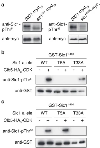
Figure S1 In vivo and in vitro specificity of the anti-Sic1-pThr5/-pThr33 antibodies. (a) Sic1-pThr5 and Sic1-pThr33 levels were determined by immunoblot analyses using respective phospho-specific antibodies and extract of exponentially growing cells that expressed plasmid-encoded versions of myc13-tagged Sic1 and Sic1T5A (left panels), or myc13-tagged Sic1 and Sic1T33A (right panels). The levels of the Sic1-myc13 variants were determined by using polyclonal anti-myc antibodies. The asterisk denotes an unspecific band. (b, c) Clb5-HA3-CDK immunocomplexes from exponentially growing yeast cells were used for in vitro kinase assays in which the bacterially-purified, N-terminal parts (encompassing the first 100 amino acids) of Sic1 (WT), Sic1T5A (T5A), and Sic1T33A (T33A) served as substrates. Sic1-pThr5 (b) and Sic1-pThr33 (c) levels were determined by immunoblot analyses using respective phospho-specific antibodies and the indicated GST-Sic11-100 variants that have been phosphorylated (+), or not (-), by Clb5-HA
3-CDK. The input levels of the GST-Sic11-100 variants were determined by using polyclonal anti-GST antibodies.
SIC1-myc 13 sic1 T33A -myc 13 sic1 T5A-myc 13 anti-Sic1-pThr33 anti-Sic1-pThr5 anti-myc SIC1-myc 13 anti-myc *
a
b
c
anti-Sic1-pThr5 anti-GST GST-Sic11-100 Clb5-HA3-CDK-
+
-
+
-
+
Sic1 allele WT T5A T33A
anti-Sic1-pThr33
anti-GST
GST-Sic11-100
Clb5-HA3-CDK
-
+
-
+
-
+
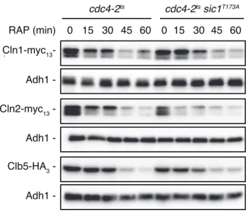
Figure S2 Sic1T173A-expressing cdc4-2ts cells are not defective in the clearance of Cln1, Cln2 or Clb5 when treated with rapamycin. cdc4-2ts and cdc4-2ts sic1T173A strains expressing Cln1-myc
13, Cln2-myc13, or Clb5-HA3 were grown as in Fig. 1A. the levels of the tagged proteins were determined by immunoblot analyses using monoclonal anti-myc or anti-HA antibodies. Adh1 levels served as loading control.
Figure S3 Cln2-HA3 and Clb5-HA3 interact with Cdc28 in vivo. Plasmid-expressed, HA3-tagged Cln2 or Clb5 were immunoprecipitated (IPed) from extracts of exponentially growing wild-type cells. Cell lysates (Input) and anti-HA immunoprecipitates (IP: anti-HA) were analyzed by immunoblotting with anti-HA (top panels), anti-Cdc28 (panels in the middle), and anti-Sic1 (bottom panels) antibodies. Please note that both Cln2-HA3
21
18
20
17
16
19
Cln1-myc13 Adh1 Adh1 -Cln2-myc13 Adh1 -Clb5-HA3 -RAP (min) 0 15 30 45 60 0 15 30 45 60 cdc4-2ts cdc4-2ts sic1T173ASupplementary Figure 2
Cln2-HA3 Cdc28 - CLN2-HA3 Sic1 -Clb5-HA3-Input IP: anti-HA
CLB5-HA3 - CLN2-HA3 CLB5-HA3
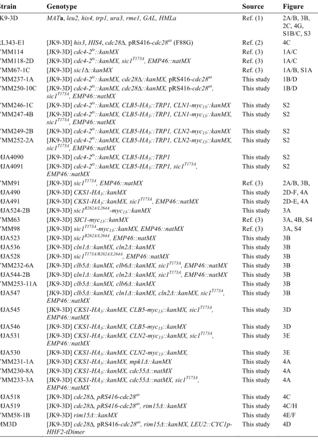
-Figure S4 Proliferating, Sic1T173A-expressing cells exhibit enhanced levels of Cln-/Clb5/6-CDK activity. For synchronization, exponentially growing WT cells expressing genomically-tagged Sic1-myc13 or Sic1T173A -myc13 were treated for 2 h with a-factor (5 µg ml-1). Following a-factor release, samples were collected at the indicated time points. The phosphorylation levels of Thr5 in Sic1 and Sic1T173A were determined by using phosphospecific anti-Sic1-pThr5 antibodies and normalized with respect to the total levels (quantified by using anti-myc antibodies) of Sic1-myc13 and Sic1T173A-myc13, respectively.
Sic1-pThr 5/Sic1-myc 13 Sic1 T173A -pThr 5/Sic1 T173A -myc 13 10 20 30 40 50 60 70 80 90 0 100 0.2 0.3 0.4 0.5 0.6 0.7 0.1 min
Supplementary Tables
Table S1. Strains used in this study
Strain Genotype Source Figure
JK9-3D MATa, leu2, his4, trp1, ura3, rme1, GAL, HMLa Ref. (1) 2A/B, 3B,
2C, 4G, S1B/C, S3
RL343-E1 [JK9-3D] his3, HIS4, cdc28∆, pRS416-cdc28as (F88G) Ref. (2) 4C
YMM114 [JK9-3D] cdc4-2ts::kanMX Ref. (3) 1A/C
YMM118-2D [JK9-3D] cdc4-2ts::kanMX, sic1T173A, EMP46::natMX Ref. (3) 1A/C
YMM67-1C [JK9-3D] sic1∆::kanMX Ref. (3) 1A/B, S1A
YMM237-1A [JK9-3D] cdc4-2ts::kanMX, cdc28∆::kanMX, pRS416-cdc28as This study 1B/D
YMM250-10C [JK9-3D] cdc4-2ts::kanMX, cdc28∆::kanMX, pRS416-cdc28as,
sic1T173A, EMP46::natMX This study 1B/D
YMM246-1C [JK9-3D] cdc4-2ts::kanMX, CLB5-HA
3::TRP1, CLN1-myc13::kanMX This study S2
YMM247-4B [JK9-3D] cdc4-2ts::kanMX, CLB5-HA
3::TRP1, CLN1-myc13::kanMX,
sic1T173A, EMP46::natMX This study S2
YMM249-2B [JK9-3D] cdc4-2ts::kanMX, CLB5-HA
3::TRP1, CLN2-myc13::kanMX This study S2
YMM252-2A [JK9-3D] cdc4-2ts::kanMX, CLB5-HA3::TRP1, CLN2-myc13::kanMX,
sic1T173A, EMP46::natMX
This study S2
MJA4090 [JK9-3D] cdc4-2ts::kanMX, CLB5-HA
3::TRP1 This study S2
MJA4091 [JK9-3D] cdc4-2ts::kanMX, CLB5-HA3::TRP1, sic1T173A,
EMP46::natMX This study S2
YMM91 [JK9-3D] sic1T173A, EMP46::natMX Ref. (3) 2A/B, 3B,
MJA490 [JK9-3D] CKS1-HA3::kanMX This study 2D-F, 4A
MJA491 [JK9-3D] CKS1-HA3::kanMX, sic1T173A, EMP46::natMX This study 2D-E, 4A
MJA524-2B [JK9-3D] sic1R262A/L264A-myc
13::kanMX This study 3A
YMM63 [JK9-3D] SIC1-myc13::kanMX Ref. (3) 3A, 4B, S4
YMM98 [JK9-3D] sic1T173A-myc
13::kanMX, EMP46::natMX Ref. (3) 3A, S4
MJA523 [JK9-3D] sic1R262A/L264A, EMP46::natMX This study 3B
MJA536 [JK9-3D] cln1Δ::kanMX, cln2Δ::kanMX This study 3B
MJA528 [JK9-3D] sic1T173A/R262A/L264A, EMP46::natMX This study 3B
YMM232-6A [JK9-3D] clb5Δ::kanMX, clb6Δ::kanMX, sic1T173A, EMP46::natMX This study 3B
MJA544-2B [JK9-3D] cln1Δ::kanMX, cln2Δ::kanMX, sic1T173A, EMP46::natMX This study 3B
YMM253-11A [JK9-3D] clb5Δ::kanMX, clb6Δ::kanMX This study 3B
MJA547 [JK9-3D] clb5Δ::kanMX, cln1Δ::kanMX, cln2Δ::kanMX, sic1T173A,
EMP46::natMX This study 3B
MJA545 [JK9-3D] CKS1-HA3::kanMX, CLB5-myc13::kanMX, sic1T173A,
EMP46::natMX This study 3D
MJA546 [JK9-3D] CKS1-HA3::kanMX, CLB5-myc13::kanMX This study 3D
MJA531 [JK9-3D] CKS1-HA3::kanMX, CLN2-myc13::kanMX, sic1T173A,
EMP46::natMX
This study 3E
MJA530 [JK9-3D] CKS1-HA3::kanMX, CLN2-myc13::kanMX, This study 3E
Table S2. Plasmids used in this study
Plasmid Genotype Source Figure
pRS415 CEN, LEU2 Ref. (5)
pMJA2881 [pRS415] ADH1p-SIC1-myc13 This study S1A
pMJA3173 [pRS415] ADH1p-sic1T5A-myc
13 This study S1A
pMJA3174 [pRS415] ADH1p-sic1T33A-myc
13 This study S1A
pMJA2995 [pRS415] ADH1p-SIC1150- 285-myc
13 This study 2F
pMJA2996 [pRS415] ADH1p-sic1150-285 - T173A-myc
13 This study 2F
pRS416 CEN, URA3 Ref. (5) S3
pMJA3038 [pRS416] ADH1p-CLB5-HA3 This study 2B/C, 3A, 4F/G, S3, S1B/C, S3
YCplac33 CEN, URA3 Ref. (6)
JCE456 [YCplac33] ADH1p-CLN2-HA3 Ref. (7) 2A/C, 4D, 4F/G, S3
pAS2654 [YCplac33] ADH1p-LST7-HA3 Ref. (8) 4F
pGEX GST Ref. (9) 4G, S1B/C
pMMT2629 [pGEX] GST-SIC1 This study 2C
pMMT2630 [pGEX] GST-sic1T173A This study 2C
pMJA3029 [pGEX] GST-sic1R262A/L264A This study 2C
pMJA3037 [pGEX] GST-SIC11-100 This study 2C, 3A, 4G, S1B/C
pMJA3219 [pGEX] GST-sic1T5A(1-100) This study S1B/C
pMJA3220 [pGEX] GST-sic1T33A(1-100) This study S1B/C
pVW995 [pGEX] GST-RIM15944-1149 Ref. (10) 4G
pVW827 CEN, LEU2, ADH1p-GST-RIM15 Ref. (10) 4F
pVW904 2µ, LEU2, TDH3p-RIM15-myc13 Ref. (10) 4H
pVW910 2µ, LEU2, TDH3p-rim15 T1075A-myc
13 Ref. (10) 4H
pFD1008 CEN, TRP1, ADH1p-rim15K823Y-GFP Ref. (10) 4D/E
Table S3. Antibodies used in this study
Name Dilution Source
Anti-Sic1 1:1’000 sc-50441 Santa Cruz
Anti-Adh1 1:200’000 Calbiochem
Anti-Sic1-pThr173 1:1’000 GenScript
Anti-Sic1-pThr5 1:1’000 GenScript
Anti-Sic1-pThr33 1:1’000 GenScript
Anti-myc 1:3’000 9E10; sc-40; Santa Cruz
Anti-HA 1:1’000 Enzo Life Sciences
Anti-GST 1:3’000 Bethyl Laboratories
Anti-Igo1-pSer64 1:1’000 GenScript
Anti-Igo1 1:1’000 Eurogentec
Anti-Cln2 1:1’000 Santa Cruz
Anti-Cdc28 1:300 Santa Cruz
Anti-Clb5 1:1’000 Santa Cruz
Anti-Sch9-pThr737 1:1’0000 GenScript
Anti-Sch9 1:1’000 GenScript
Anti-Rim15-pThr1075 1:10’000 Eurogentec
Goat anti-rabbit IgG HRP 1:3’000 Biorad
Goat anti-mouse HRP 1:3’000 Biorad
Donkey anti-goat HRP 1:5’000 Abcam
Goat anti-mouse IgG-Fcy HRP 1:5’000 Jackson Immunoresearch Goat anti-mouse IgG, light chain HRP 1:5’000 Jackson Immunoresearch
Supplementary References
1. Heitman J, Movva NR, & Hall MN (1991) Targets for cell cycle arrest by the immunosuppressant rapamycin in yeast. Science 253(5022):905-909.
2. Bodenmiller B, et al. (2010) Phosphoproteomic analysis reveals interconnected system-wide responses to perturbations of kinases and phosphatases in yeast. Sci. Signal. 3(153):rs4.
3. Moreno-Torres M, Jaquenoud M, & De Virgilio C (2015) TORC1 controls G1-S cell cycle transition in yeast via Mpk1 and the greatwall kinase pathway. Nat. Commun. 6:8256.
4. Pedruzzi I, et al. (2003) TOR and PKA signaling pathways converge on the protein kinase Rim15 to control entry into G0. Mol. Cell 12(6):1607-1613.
5. Brachmann CB, et al. (1998) Designer deletion strains derived from Saccharomyces cerevisiae S288C: a useful set of strains and plasmids for PCR-mediated gene disruption and other applications. Yeast 14(2):115-132.
6. Gietz RD & Sugino A (1988) New yeast-Escherichia coli shuttle vectors constructed with in vitro mutagenized yeast genes lacking six-base pair restriction sites. Gene 74(2):527-534.
7. Quilis I & Igual JC (2012) Molecular basis of the functional distinction between Cln1 and Cln2 cyclins.
Cell Cycle 11(16):3117-3131.
8. Péli-Gulli MP, Sardu A, Panchaud N, Raucci S, & De Virgilio C (2015) Amino Acids Stimulate TORC1 through Lst4-Lst7, a GTPase-Activating Protein Complex for the Rag Family GTPase Gtr2.
Cell Rep 13(1):1-7.
9. Smith DB & Johnson KS (1988) Single-step purification of polypeptides expressed in Escherichia coli as fusions with glutathione S-transferase. Gene 67(1):31-40.
10. Wanke V, Pedruzzi I, Cameroni E, Dubouloz F, & De Virgilio C (2005) Regulation of G0 entry by the Pho80-Pho85 cyclin-CDK complex. EMBO J. 24(24):4271-4278.