Clinical cardiac electrophysiologic evaluation of the
positive inotropic agent, DPI 201-106
G. S. BUTROUS, N. M. G. DEBBAS, J. ERWIN, D. W. D A VIES, H. P. KELLER*, M. W. LUNNONI, A. W. NATHAN AND A. J. CAMM
Department of Cardiology, St Bartholomew's Hospital, London, U.K., * Biopharmaceutical Dept. Sandoz AG. Basle, Switzerland, and f Clinical Research Unit London, Sandoz Ltd., Feltham, U.K.
KEY WORDS: DPI, cardiac electrophysiology, QT interval.
DPI 201-106 is a new positive inotropic agent. The cardiac electrophysiology of 16 patients was studied before and during DPI 201-106 administration (loading dose of intravenous DPI 201-106,1-8 mg kg~l h~l admin-isteredover 10 min, followed by a maintenance dose of 0-2 mg kg~l h~x). DPI 201-106 hadno effect on the sinus node. The AH interval during fixed-rate atrial pacing became prolonged during DPI 201-106 infusion. There was a significant prolongation of the QT interval [QT (corrected), 417 ±22 to 502 ±35 ms, P<0-05; QT (atrialpacing at 600 ms), 374 ±17 to 419 ± 23 ms, P<005;QT(ventricular pacing at 600 ms), 409 ±37 to 449±30 ms, P<0-05J. The ventricular effective refractory period significantly prolonged during DPI 201-106 administration (242 ±21 to 287 ±56 ms, P < 005), but the supernormal-period duration decreased.
The atrial effective refractory period was shortened in four patients and prolonged in one (261 ± 67 to 240 ±53 ms, NS). The corrected atrial repolarization time (PTtc) shortened significantly during DPI 210-106 infusion (479 ±26 to 445 ±22 ms at 20 min of the maintenance dose, P<0-05). Atrial fibrillation was initiated in five patients during DPI infusion, but no ventricular arrhythmia was provoked. These findings suggest that DPI 201-106 has novel differential electrophysiological effects on atria and ventricles.
Introduction the regulation of Na + channels, probably by inter-DPI 201-106 is a diphenylmethyl-piperazinyl- f e r i n8 w i t h o n e o f i t s ^activators'6'. DPI 201-106 indole derivative with cardiotonic effect which has c a u s e s l a n i n c r e a s e i n mtracellular calcium but does been demonstrated in vitro"* and in vivo™ in van- ™l e ni T « ^ l m * " ' £ through slow channels'7!, ous animals. Its potent inotropic action has also The effect ofDPI 201-106 on.the Na channehmay been shown in man". DPI 201-106 has a novel i n c ^ M the mtracellularNa load which interferes mechanism of action which is not related to cAMP, * " * lJe dynamics of the Na /Ca exchange™ to beta-adrenergic or histaminergic receptor stimu- fnd' J ^ ' i n c r e a s e s t h e 'ntracellular calcium lation, or to the liberation of catecholamines or to a l o a d-T i a s may consequently prolong phase 2 of the glycoside-likeaction™. There is increasing evidence **™ P°ften,tial <th,e Pl a t e a u> a"d i n c r e a s e ^e a v a i 1" that this agent modulates Na+ channels and hence ability of calcium for contractile activity. The pro-affects phase 0 and phase 2 of the cardiac cell action longauon of cardiac action potential ,s, therefore, potential.Kohlhardt«a/.l«"foundthatDPI201-106 associated with positive inotropic action". Action prolongedtheconductanceoftheNa+channels,i.e. potential prolongationis reflected by the significant prolonging the open state. Tetrodotoxin abolishes Prolongation of the QT interval which has been the effect of DPI 201-106™, hence the inotropic noticed during intravenous administration of DPI effect of DPI 201-106 is due to its interaction with . , • - . •
The proposed mechanism of action may, there-o ._• J r ,., ,o. , « , J J r -,* fore, influence other electrophysiological par-Submiued for publication on 19 June 1987 and in revised form 26 ' . . . .
October 1987. ameters of cardiac tissue, as suggested by some
animal experiments'21. The aim of this paper is to
Address for correspondence: D r G. S. Butrous, Department o f . , , . . c •• • • _•• , . . Cardiological Science?* George's Hospital and Medial School, report the results of a clinical Cardiac eleCtrophySlO-Cranmer Terrace, London SWI70RE, U.K. logic Study of D P I 201-106.
PATIENTS
Sixteen patients (six females), age range 18-76 years (mean 52±15 years), were included in this study. Of 10 patients, the study was performed as a part of routine clinical cardiac electrophysiological evaluation for palpitations in eight, and syncope in two. The other six patients had 2:1 or complete heart block and had already been fitted with dual-chamber pacemakers. None of the patients were on amio-darone. Other antiarrhy thmic medications were dis-continued for at least 72 h prior to the study in all patients. The aims and procedure of the study were fully explained to each patient and written consent was obtained. The study was approved by the Ethics Committee of St Bartholomew's Hospital, London.
ADMINISTRATION OF DPI 201-106
DPI 201-106 was given intravenously according to the following method: a loading dose of l-8mgkg~1h~1 was given intravenously over lOmin (except for the first five patients to whom 0-8—1-5 m g g "1 h "1 were given according to recommendation of the Ethics Committee). This was followed immediately by constant infusion of DPI 201-106 at a dose of 0-2 mg k g " ' h~' for the entire duration of the study (maintenance dose).
PROTOCOLS
Three protocols were included in this study:
Measurement of intracardiac intervals and refractoriness
USCI electrode catheters were introduced via the femoral vein under local anaesthesia (approxi-mately 10 ml of 1 % lignocaine) and placed to record and stimulate the right atrium and right ventricular apex. A tripolar electrode was positioned adjacent to the His bundle. The intracardiac signals and four surface electrocardiographic leads (I, aVF, VI and V6) were recorded simultaneously on a Siemens Elema mingograph 1600 ink-jet recorder at a paper speed of 100 mm s"1. Programmed stimulation at twice the diastolic threshold was performed using a Bloom stimulator.
Basic intervals were measured, during the load-ing and maintenance doses in sinus rhythm and dur-ing constant rate atrial and ventricular pacdur-ing (600, 500 and 400 ms) to ± 5 ms accuracy. These intervals included the following: (a) RR interval, measured between two consecutive high right atrial electro-grams; (b) AH interval, between the first rapid deflection of the low right atrial electrogram and the
measured from the onset of the His bundle electro-gram to the earliest ventricular activation on the surface electrocardiogram; (d) QRS duration, measured between the earliest and last deflection on the surface electrocardiogram; and (e) QT interval, from the earliest deflection of the QRS in the surface electrocardiogram to the end of the T wave191. The measured QT intervals during sinus rhythm were corrected for rate using Bazett's formula'10':
where QTC is the corrected QT interval, QTm is the measured QT interval, and RR is the RR interval.
The QT intervals were also measured during fixed-rate atrial pacing (QTJ and fixed fixed-rate ventricular pacing (QTV).
Refractory periods were assessed by pacing (at twice the diastolic threshold) the high right atrium (or right ventricular apex) at a constant rate for eight beats followed by an extra atrial (or ventricu-lar) paced beat at a decreasing coupling interval. The following parameters were assessed'"': (a) the functional atrioventricular nodal refractory period, which was defined as the shortest distance between the last His bundle deflection of the atrial driven cycle and the His deflection following the extra-stimulus; (b) the effective atrioventricular nodal refractory period which was defined as the longest interval between the last atrial deflection of the driven cycle length and the atrial deflection of any extra-stimulus beat which failed to result in a His bundle depolarization; (c) the atrial refractory period, was the longest interval between the last atrial driven stimulus and any atrial extra-stimulus which did not result in atrail depolarization; (d) the effective ventriculo-atrial refractory period was assessed by ventricular programmed stimulation — it was the longest interval between the last ventricu-lar deflection of the driven cycle length nd the ven-tricular deflection of any extra-stimulus beat which failed to result in retrograde conduction to the atria; and (e) The effective ventricular refractory period which was defined as the longest interval between the last ventricular driven stimulus and any extra-stimulus spike that resulted in no ventricular depolarization.
Anterograde atrioventricular and retrograde ventriculo-atrial Wenckebach cycle lengths were determined by gradually increasing atrial or ven-tricular pacing rates, respectively until a beat failed to conduct anterogradely or retrogradely.
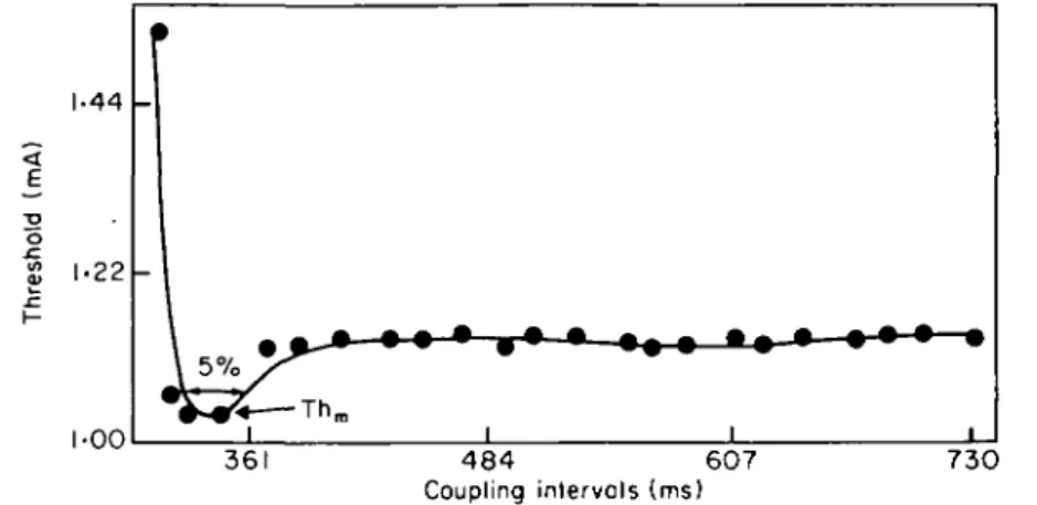
< E 144 122 -1-00 361 484 607 Coupling intervols (ms) 7 3 0
Figure 1 The cathodal strength interval curve, showing the measurements of the super-normal period in this study. The level of the 5% supersuper-normal-period duration is also shown. Thm, minimal threshold during supernormal period.
Sinus node function was assessed by pacing the high right atrium with pacing cycle lengths of 600, 500 and 400 ms, each for 1 min. The longest pause from the last paced atrial depolarization to the first sinus return cycle was considered to be the maxi-mum sinus node recovery time. The maximaxi-mum corrected sinus node recovery time was defined as the longest interval derived by subtracting the prepacing sinus cycle length from the sinus node recovery time1'21.
Assessment of ventricular supernormal period
In six patients, cathodal ventricular strength interval curves were assessed before and during the maintenance dose of DPI 201-106. The distal pole of the electrode was used for cathodal stimulation and the indifferent electrode was placed on the skin. The high right atrium was paced at a constant rate of 600 ms for eight beats followed by one ventricu-lar extra-stimulus. The threshold of the ventricuventricu-lar extra-stimulus was assessed with 0-01 mA accuracy using Bloom stimulator at each coupling interval. The coupling interval of the ventricular extra-stimulus was decreased until the ventricular pacing threshold was > 10 mA.
The supernormal period was measured as that part of the strength interval curve where there was a 5% or more decrease in the threshold below the diastolic threshold. The period of supernormality in the cardiac cycle was expressed in relation to the QT interval (taking Q as 0% and the end of the T wave as 100%). The minimum threshold during the super-normal period (SNP-Tm) was assessed relative to the diastolic threshold (Fig. 1).
Assessment of atrial repolarization
Six patients with 2:1 or complete heart block and who had been fitted with dual chamber pacemakers were included in this study to assess the atrial repolarization time (PT.). Standard bipolar unpaced surface ECG leads I, II and III were recorded at high fidelity (0-6-30 Hz) and high gain (x 1000- x 10000) on 0-5 inch magnetic tape before and during administration of DPI 201-106. The recording signals were then transferred onto the computer memory via a 12-bit analogue/digital converter. The signals were filtered using specially adapted software1'31. The duration of the P and the T waves of the atrium (TJ were measured from the displayed complexes using a high-resolution cross-hair cursor on a Tectronix 4010 VDU before and during the loading dose (5 min and 10 min) and during the maintenance dose (10, 20 and 30 min). The atrial repolarization duration (PTJ was corrected according to the following equation1'3':
PT.C = PT.+ 0-29 (1000-RR)
where PT.C is the corrected atrial repolarization time, PT. is the measured atrial repolarization time, and RR is the sinus cycle length.
ASSESSMENT OF PLASMA LEVEL
Blood samples (10 ml) were taken immediately after the loading dose and at 5-10, 15-20 and 25—35 min intervals during the maintenance dose. Unchanged DPI 201-106 was extracted from alkalinized plasma into a methyltertbutyl ether/ isopropranol n-hexane mixture and back extracted
injected directly into a 100 x 21 mm spheri-5 RP-8 column and isocratically eluted with 0-005 M phos-phoric acid/acetonitrile 35:65 (pH 2-5). Detection was in the UV range at 228 nm (Smith HT, unpubl.).
The assay confidence limits (/> = 0-95) over the whole study concentration range were approxi-mately ±20% of the expected concentrations as estimated from calibration and control samples which had been included in the analysis. Most of the samples were analyzed twice showing a mean inter-assay coefficient of variation of less than 10%. All sample concentrations were above the limit of detection (10 ng ml)"'.
ANALYSIS OF THE DATA
Mean + SD values were used to express the dispersion of the data. For statistical inference, Student's /-test or Wilcoxon's sign ranked test were used as appropriate. Significance was defined as />< 0 0 5 .
Results
PLASMA LEVEL OF DPI 201-106
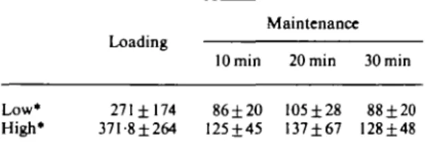
The plasma levels of DPI 201-106 in this study are shown in Table 1. The plasma concentration was steady during the maintenance infusion.
Table I
Low* High*
Plasma level o/DPI201-l(X
Loading 271 ±174 37I-8±264 lOmin 86 ±20 125±45 i (in ngml ) Maintenance 20min 105 ±28 137±67 30min 88 ±20 128±48 *'Low' denotes those patients who had a lower loading dose (0-8-1-5 mg kg"1 h"1) in compliance with the Ethics Com-mittee recommendation; 'high' denotes a loading dose of
EFFECT ON BLOOD PRESSURE
There was an increase in the supine systolic blood pressures during the loading dose of DPI 201-106 (systolic 127±24 to 146±16-3mmHg, f < 0 0 1 ) . Diastolic blood pressures did not show significant changes (74-4 ± 11 to 88 ± 9 mmHg, NS). 200 180 - 160 E \ 140 03 C 5 120 100 80 -_ -- t 3 -1 '' i 1 600 500 400 At rial pacing cycle length (ms)
Figure 2 Changes in the AH interval during different atrial pacing rates before ( • ) and during (D) the maintenance dose of DPI 201-106. These changes were statistically significant (P < 005). The values are represented as mean ± SD.
EFFECT ON SINUS NODE FUNCTION
There was a slight, but statistically insignificant, increase in the RR intervals after the loading dose of DPI 201-106 (795±166 to 850±211ms). The maximum sinus node recovery time changed from 1106 ±28 lms to 948 ± 242 ms (NS), and the corrected sinus node recovery time decreased from 301±123to231±156ms(NS).
EFFECT ON THE ATRIOVENTRICULAR NODE
There were no changes in the AH intervals in sinus rhythm during the loading or maintenance dose of DPI 201-106 (72-5 ± 14 ms). On the other hand, the AH intervals at constant rate atrial pacing showed a significant prolongation (P < 005) during the maintenance dose of DPI 201-106 compared with the control (Fig. 2).
There was a slight increase in the functional atrio-ventricular nodal refractory period during the maintenance dose in four out of six patients in whom these measurements were possible (413 ±78 to 437 ± 79 ms, NS). It was difficult to assess the change in atrio-ventricular nodal effective tory periods because in four patients these refrac-tory periods were limited by atrial refractoriness.
B D
Figure 3 Changes in the atrial effective refractory period
before (B) and during (D) and maintenance dose of DPI 201— 106. These changes were statistically insignificant. The values are represented as mean ± SD.
In one of the remaining two patients no changes occurred, but in the other an increase was seen. It was also difficult to assess the effective refractory period of ventriculo-atrial conduction because in two patients it was limited by the ventricular refrac-tory period and in an additional two patients poor or no retrograde conduction could be seen. In only one patient was there a noticeable increase in the
ventriculo-atrial refractory period at the atrio-ventricular nodal level (480 to 600 ms).
The anterograde Wenckebach cycle length showed no significant change (377 ±98 to 383 ± 163 ms), neither did the retrograde Wenckebach cycle length (508 ± 170 ms to 548 ± 147 ms).
EFFECT ON ATRIAL ELECTROPHYSIOLOGY Refractory period
In four out of six patients who underwent electro-physiological studies, the atrial refractory period decreased during the maintenance dose of DPI 201-106. One patient had a marked increase in the atrial refractory period. The mean value, however, showed no statistical difference (261 ±67 to 240 ± 53 ms) (Fig. 3).
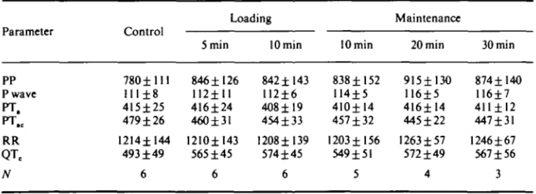
Atrial PT duration
In six patients who had complete heart block the corrected atrial repolarization time (PT.C) interval showed a significant decrease during the mainten-ance dose particularly after 20 and 30 min (/"<0-05). These changes were mainly due to changes in the atrial T-wave duration, as P-wave duration showed no significant changes (Table 2). In these patients the corresponding QTC interval showed a noticeable prolongation (see below).
EFFECT ON VENTRICULAR ELECTROPHYSIOLOGY HV interval and QRS duration
The HV interval and QRS duration remained unchanged during both the loading and mainten-ance infusion of DPI 201-106 at 45 ± 8 ms and 91 ± 12 ms, respectively.
Table 2 Changes (ms) in the surface electrocardiograms of patients with complete heart block before and during DPI 201-106 administration*
Parameter PP P wave PT. PT.C RR QTe N Control 780 ±111 111±8 415±25 479 ±26 I214±144 493 ± 4 9 6 Loading 5 min 846±126 112±11 416±24 460±31 12I0±143 565 ±45 6 lOmin 842±143 U2±6 4O8±19 454 ±33 1208 ±139 574 ±45 6 lOmin 838 ±152 114±5 410±14 457 ±32 1203 ±156 549±51 5 Maintenance 20 min 915±13O 116±5 416± 14 445 ±22 1263 ±57 572 ±49 4 30 min 874±140 116±7 411 ± 12 447±31 1246 ± 67 567 ±56 3 *For abbreviations see text.
400 380 360 340 1 320 Q_ ir ^ 300 £ 280 260 240 220 2 0 0
ft
BFigure 4 Changes in the ventricular effective refractory
period before (B) and during (D) the maintenance dose of DPI 201-106a. These changes were statistically significant
(P < 005). The values are represented as mean ± SD.
Ventricular effect refractory period
There was a significant increase in the ventricular effective refractory period in six out of nine patients studied during the maintenance infusion of DPI 201-106 as estimated by extra-stimulus technique (242 ± 21 ms to 287 ± 56 ms, P < 005). This increase was also seen in all six patients in whom the cathodal strength interval curve was assessed (300±25msto333±31ms/><001)(Fig. 4).
QT interval
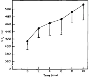
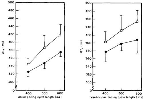
Corrected QT intervals showed a significant in-crease after the loading dose of DPI 201-106 (417±22msto502±35ms, ^ < 0 0 5 ) ( F i g . 5). This increase was also evident during the constant rate atrial or ventricular pacing (Fig. 6).
500 480 460 -g 440 400 -380 360 0f - 1 1 1 B 4 6 Time (min) 8 10
Figure 5 Changes in QTC during the loading dose of DPI 201-106. These changes were statistically significant (/><005). B, before drug administration. The values are represented as mean ± SD.
Effect on ventricular supernormal period
DPI 201-106 had no effect on the ventricular diastolic cathodal threshold. The supernormal period could be demonstrated in five of six patients during DPI 201-106 infusion. The minimum thres-hold during the supernormal period (SNP-Tm) sig-nificantly increased (/><005) (Table 3); but the supernormal-period duration decreased (/)<005, Table 3) during DPI 201-106 infusion. The relation of the supernormal to the atrially paced QT interval showed that, during DPI 201-106 infusion, there was a significant shift in the position (/)<0-05, Table 3) away from the peak of the T wave. The upper limit shifted from 133±8% to 121 ±19%, but these were not significant (Table 3).
EFFECT ON CARDIAC ARRHYTHMIAS
There was no ventricular proarrhythmic effect of DPI 201-106. In one patient with ventricular tachy-cardia, the tachycardia was still provokable during the infusion. In five patients, however, DPI 201-106 appeared to facilitate the provocation of atrial fibrillation. In three of these patients the atrial fibrillation was provoked by atrial extra-stimulus near the atrial refractory period. Two of these had a history of paroxysmal atrial fibrillation. In two patients, atrial fibrillation was spontaneous and lasted for 2—4 h: one had a history of paroxysmal atrial fibrillation and the other a congenital atrio-ventricular block.
500 480 4 6 0 4 4 0 4 2 0 - 400 E ,_• 380 o 360 340 3 2 0 3 0 0 o _ -r • !i
- v ^
- /^C
-
i
-i -i -i 4 0 0 500 6 0 0 Atriol pacing cycle length (mi)
400 500 600
Ventricular pacing cycle length (ms)
Figure 6 Changes in the atrially paced QT interval (QTJ and ventricularly paced QT interval (QT,) before ( • ) and during (D) the maintenance dose of DPI 201-106. The differences were significant (P < 005). The values are represented as mean ± SD.
Table 3 Cathodal supernormal period*
Parameter Control During DPI
Diastolic threshold (mA) SNP-Tm (%) 5% duration (ms) 5% position (%) 0-49 ±0-30 16±10 155±76 85±11-133±8 0-46±0-27 9±9 79 ±42 97± 14-121 ± 19 •For abbreviations and details see text.
Discussion
DPI 201-106 demonstrates a novel spectrum of
electrophysiological effects which may reflect on its mechanism of action. The most remarkable effects were on ventricular electrophysiological par-ameters. The marked prolongation of the QT inter-val and the increase in ventricular refractory period, as documented in this and other studies'21, may be explained by the prolongation of action-potential duration produced by DPI 201-106121, possibly due to the agonistic effect of DPI 201-106 on the Na + channel as explained above. The effect of DPI 201—
106 on the supernormal period is of particular interest. There are, to our knowledge, no studies on
the effect of DPI 201-106 on the cellular action potential supernormal period. The clinically assessed supernormal period of the ventricle is thought to be dependent on the dispersion of the action-potential duration'141. The decrease in the duration of the supernormal period during DPI 201-106 infusion in this study may be due to pro-longation of the cellular action potentials leading to a decrease in their dispersion. The fact that super-normal periods were shifted towards the end of the T waves supports this explanation.
The effect of DPI 201-106 on atrial electro-physiology is of particular interest. Contrary to the effect on the ventricles, there was a decrease in the atrial effective refractory period in four out of six patients studied. This observation was consoli-dated by the observation that the corrected atrial repolarization time (PT,C) was also shortened. The measurement of PT,C in patients with heart block has been described previously1'31 and, in general, the PT.C behaves in a similar way to the ventricular QTC interval. DPI 201-106 prolongs the action potential duration in the isolated rabbit atria'11. It is, there-fore, difficult to attribute the findings of this study to a direct effect of DPI 201-106. A reflex mechan-ism may be an alternative explanation. Cholinergic
lation may decrease the effective atrial refractory period1'61. The increase in systolic blood pressure during DPI 201-106 infusion may trigger baro-receptor activity and, thereby, increase vagal activity. The high incidence of induced atrial fibril-lation in this study may also be explained by a possible increase in vagal tone1'71. Some support for this view come from Walker et al.[lg] who noticed that DPI 201-106 significantly prolonged the sinus cycle length, sinus node recovery time, atrio-ventricular nodal functional refractory period, right ventricular functional refractory period and the QT interval. These findings could not be reproduced in cardiac transplanted dogs.
DPI 201-106 does not show a significant effect on the sinus node. The slight slowing of the sinus rate did reach statistical significance. This finding is not consistent with the hypothesis of increased vagal tone. If this hypothesis is correct, then DPI 201-106 must decrease the sensitivity of the sinus node to vagal effects. The atrio-ventricular nodal conduc-tion during sinus rhythm did not alter during DPI 201-106 infusion; however, there was a slight, but significant, prolongation during fixed-rate atrial pacing. The atrio-ventricular nodal functional refractory period also increased slightly. These findings are consistent with the hypothesis of increased vagal activity. The agonostic effect of DPI 201-106 on the Na+ channel and its consequent influence on the action potential duration may not be applied to atrio-ventricular nodal tissues because they depend mainly on the Ca2 + channel. Recently, it was found that DPI 201-106 has a calcium antagonistic effect with prolonged treatment1"1. It is therefore not clear whether the electrophysiological effects seen in this study will be applicable to long-term treatment.
It should be mentioned that two different loading doses were used in this study. This was at the request of the Ethics Committee of St Bartholomew's Hospital. The recommendation was that the first four patients should receive a lower loading dose before proceeding to the loading dose of l-Smgkg"1 h"1 as originally intended. However, the same effects were observed in the first four patients as in the rest of the patients. Because of the number of the patients, the pattern of the study and the doses' used, it was not possible to assess dose response. Recently, it has been suggested that DPI 201-106 has some antiairhythmic effects'201. This study was not designed to assess this possibility. On
ation of atrial fibrillation in five patients. In one patient with ventricular tachycardia, DPI 201-106 did not change the electrophysiological pattern of that arrhythmia.
In conclusion, DPI 201-106 has an interesting and novel spectrum of electrophysiological effects which may be important during the routine clinical use of this agent.
References
[1] Herzig JW, Quast U. Increase in C a+ + sensitivity of
myocardial contractile structure by DPI 201-106 (Abstr). J Mol Cell Cardiol 1984; 16 (Suppl 3). 6. [2] Scholtysik G, Salzmann R, Berthold R. Herzig JW,
Quast U, Markstein R. DPI 201-106, a novel cardio-active agent. Combination of cAMP-independent posi-tive inotropic, negaposi-tive chronotopic, action potential prolonging and coronary dilatory properties. Nauny-Schmiedeberg's Arch Pharmacol 1985; 329: 316-25. [3] Gloor HO, Kappenberger LJ. DPI 201-106: a positive
inotropic agent with prolongation of the QT-interval (Abstr) Circulation 1985; 72 (Suppl III): 235.
[4] Thormann J, Kramer W, Kindler M, Neuss H, Schlepper M. Comparative efficacy of the new cardio-tonic agent DPI 201-106 versus dopamine in dilated cardiomyopathy: analysis by serial pressure and volume relations and 'one-line' MV02 assessment. J Cardiovas Pharmacol 1986; 8: 749-57.
[5] Petein M, From AHL, Francis GS, Pierpont GL. DPI 201—106: a new non-adrenergic inotropic agent with possible beta adrenergic blocking activity (Abstr). Circulation 1985; 72 (Suppl III): 304.
[6] Kohlhardt M, Frobe U, Herzig JW. Modification of single cardiac N a+ channels by DPI 201-106. J Membr
Biol 1985; 89: 163-72.
[7] Buggisch D, Isenberg G, Ravens U, Scholtysik G. The role of sodium channels in the effects of the cardiotonic compound DPI 201-106 on the contractility and mem-brane potentials in isolated mammalian heart prep-arations. Eur J Pharmacol 1985; 118:303-11.
[8] Capman RA. Control of cardiac contractility at the cellular level. Am J Physiol 1983; 245: H535-52. [9] Lepeschkm E, Surawicz B. The measurement of the QT
interval of the electrocardiogram. Circulation 1952; 6: 378-88.
[10] Bazett HD. An analysis of the time-relations of electro-cardiograms. Heart 1920; 7: 353-70.
[11] Josephson ME, Seides SE. Clinical cardiac electro-physiology. Philadelphia: Lea and Febiger, 1979; 23-59. [12] Narula OS, Samet P Sinus node recovery in man. Fed
Proc 1971; 30: 554.
[13] Debbas NMG, Nathan AW, Cochrane T, Levy AM, Camm AJ. Characterization of atrial repolarization from the surface ECG-effect of antiarrhythmic drugs and atrial pacing (Abstr). PACE 1984; 7:458.
[14] Butrous GS, Davies W, Debbas N, Nathan AW, Camm AJ. Assessment of ventricular supernormal period in man: is it a useful new clinical parameter? (Abstr). Eur Heart J 1985; 6 (Suppl 1): 974.
[15] Randell WC, Armour JA. Gross and microscopic anatomy of the cardiac innervation. In: Randell WC, ed. Neuronal regulation of the heart. Oxford: Oxford University Press, 1977: 13-41.
[16] Prystowsky EW, Naccarelli GV, Jackman WM, Rinkenberger RL, Hegar JJ, Zipes DP. Enhanced para-sympathetic tone shortens atrial refractoriness in man. Am J Cardiol 1983; 51: 96-100.
[17] Coumel Ph, Leclerq J, Attuel P, Lavallee J, Flammang D. Autonomic influences in the genesis of atrial arrhyth-mias: atrial flutter and fibrillation of vagal origin. In:
Narula OS, ed. Cardiac arrhythmias. Maryland, U.S.A.: The Williams and Wilkins Co. 1979: 243-55.
[18] Walker MJ, Tuna IC, Gornick CC, Almquist A, Benditt DG. Magnitude and temporal sequence of cardiac elec-trophysiologic effects of DPI 201-106: a new inotropic agent (Abstr). Circulation 1986; 74:11-267.
[19] Hof RP, Hof A. Mechanism of the vasodilator effects of the cardiotonic agent DPI 201-106. J Cardiovasc Pharmacol 1985; 7: 1188-92.
[20] Scholtysik G, Williams F. Antiarrhythmic effects of DPI 201-106 (Abstr). BrJ Pharmacol 1986; 87: 75P.