News on microenvironmental physioxia to revisit skin cells targeting approaches
C. Grillon1, A. Matejuk1, M. Nadim1,2, N. Lamerant-Fayel1 and C. Kieda1
1 Centre de Biophysique Moléculaire, CNRS, 45071 Orléans, Cedex2, France.
2 LibraGen, 3, rue des satellites, Bat. Canal Biotech, 31400 Toulouse, France.
Corresponding author : Catherine Grillon, Centre de Biophysique Moléculaire, CNRS, 45071 Orléans, Cedex2, France; phone: 00 33 238 25 78 04 ; FAX: 00 33 238 25 54 59, e- mail : [email protected]; web site http://www.cnrs-orleans.fr.
Short title: physioxia in skin cell targeting
Abbreviations: AMP: antimicrobial and antimycotic peptides; DC: dendritic cells; EC: endothelial cells; ECM: extracellular matrix; GAGs: glycosaminoglycans; MMP: matrix metalloproteinase; pO2: oxygen partial pressure; ROS: reactive oxygen species
3973 words
Abstract:
The skin is a multifunctional organ and a first line of defence actively protecting from environmental stress caused by injury, microbial treat, UV irradiation and environmental toxins. Diverse cutaneous cell types together with extracellular matrix elements and factors create a dynamic scene for cellular communication crucial in vital processes such as wound healing, inflammation, angiogenesis, immune response. Direct functional success of skin equilibrium depends on its microenvironment settings and particularly the local oxygen tension. Indeed, skin entire milieu is characterized by and highly dependent on its low oxygen tension called physioxia as emphasized in this review. In the context of skin physioxia, we review and propose here new approaches to minimize age-related changes in skin state and function. We particularly emphasize carbohydrate-mediated interactions and new 3D models of engineered skin substitutes. We highlight newly emerged tools and targets including stem cells, miRNAs, MMPs, mitochondria and natural antioxidants that are promising in prevention of skin aging and disease restraint. In the era of advanced dermatology, new attempts are bringing us closer to “well being” perception.
Keywords:
Introduction
In skin, as the major protective organ of the body, cell-cell interactions are continuously and dynamically operating to achieve the protecting challenge against all aggressive stresses that lead to aging and lower protection allowing damages as inflammatory reactions, poor wound healing and carcinogenic mutations.
Cutaneous microvasculature is the most important player involved in linking skin to the whole organism. Endothelial cells (EC) constituting the microvessel walls allow the entry of nutrients inside the skin but also the recruitment of immune cells when necessary. Moreover ECs contribute to maintain skin homeostasis by partial oxygen pressure (pO2) regulation inside the skin which appears to be very low in physiological conditions (1). This issue is a new and crucial parameter which has been hardly taken into account in research in general and dermatology particularly.
Natural delivery of molecules can occur between distant cells using the circulatory systems, as cells do, in order to achieve inflammatory and immune reactions. Consequently the knowledge of the skin circulatory system and its specific biology is a key development of these last years’ dermatology research. Skin endothelial cells are thus not only a barrier but a specific and very precise portal for cells and molecules (2).
They also control the expression of a harmoniously orchestrated panel of adhesion molecules and ligands that permit the recognition of recruited cells according to the skin tissue physiology (3). A fundamental part is played by lectins recognizing their glycoconjugate counterpart (4, 5).
Understanding such signaling requires the knowledge of sugar specific receptors, their expression and regulation. These receptors, “endogenous lectins”, have been taken advantage of for many years without naming their molecular mechanism of action. As Molière’s «Bourgeois Gentleman»
Monsieur Jourdain was saying : «Par ma foi ! Il y a plus de quarante ans que je dis de la prose sans que j'en susse rien » - Good gracious! It has been more than fourty years I have been speaking prose
without being aware of it -, dermo-cosmetics was using refined glycoconjugates addressing them to previously described receptors that were long neglected. Skin glycobiology is now an active research topic by which the delivery of specific molecules opens ways to carry active molecules that are often brought via vesicles mimicking the natural vesicles delivery.
This review will concentrate on these aspects of skin biology in reaction to stress-induced aging process and its consequences, revisiting them in light of the added new parameters that the concept of physioxia brings in.
I. Importance of skin microenvironmental physioxia What does skin physioxia mean?
Skin microenvironment is constituted by molecules released by various cells, allowing skin structure, organization and function, but also by physicochemical conditions (temperature, oxygen partial pressure….) which are determinants for skin equilibrium.
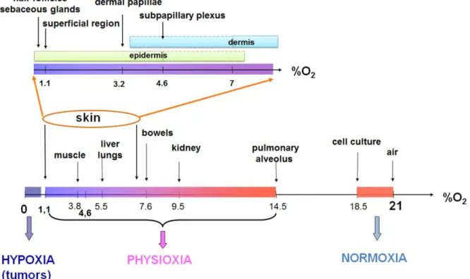
Cutaneous microvessels, bringing oxygenated blood from the whole organism to the skin, are located in the dermal part, suggesting a variable oxygen delivery according to the various skin layers. In addition, atmospheric oxygen participates in skin oxygenation by passive diffusion through epithelium and supply the upper skin layers up to 0.25 – 0.4 mm depth (6). Due to the difficulties to properly assess oxygen level in a so thin organ, only few studies reported pO2 measurements inside the skin (Table 1). Wang et al (7) described spatial variations of local pO2 in human skin from fingers covered by a layer of paraffin oil to avoid O2 delivery by atmospheric air, using microelectrodes. pO2 decreases from the deeper region (sub-papillary plexus, pO2 = 4.6 ± 1.1 %) to the more superficial region of the skin (pO2 = 1.1 ± 0.4 %). In addition to oxygen distribution according to skin depth, imaging using a hypoxia marker (8) allowed to identify a very low pO2 in specific skin dermal structures such as sebaceous glands (pO2 = 0.1 ± 1.3%) or hair follicles (pO2 = 0.1 ± 0.8%). Consequently, skin physiological pO2, what we called «physioxia», is one of the lowest among human organs (Figure 1), differs from atmospheric (1) and is not homogenous, depending on the skin layer depth and structure.
Relevance in respecting physioxia in skin studies
Despite skin physiological pO2 very low value, most of the in vitro studies using skin cells, even 3D skin reconstituting models, are performed at pO2 = 18.55% (actual pO2 inside an incubator
maintaining 5% CO2). Yet, skin physiological pO2 is critical for several functions. More and more results such as the modulation of keratinocyte proliferation and attachment according to oxygen tension (9), the influence of low oxygen pressure (2%) on the cellular cross-talk of dermal fibroblast (10) strengthen the importance of taking into account skin physioxia. Particularly, we reported, in a recent review, the regulation by pO2 of adhesion molecule expression on endothelial skin cells and of soluble factors released by distinct cutaneous cells (1).
As the first factor involved in reaction to low oxygen tension, HIF-1 has been shown to be constitutively expressed in skin (11) and to play a physiological role in preventing skin aging by regulating apoptosis, adhesion molecule expression and consequently wound healing (12). It was also shown to be increased by UVB-induced photoaging in keratinocyte and to regulate skin homeostasis by inducing the expression of proteins involved in DNA repair (13). More generally, HIF-1 was suggested to participate in the control of the organism response to oxygen (14) and also in maintaining skin immunity (15). Additionally, consequently to the sudden change in oxygen tension occurring at birth, HIF-1 is suggested to play an important role in the adaptation of baby skin, particularly in the skin barrier maturation, during the neonatal period (16).
Here, we will focus on another regulation by oxygen level: microRNAs (miRs). Several miRs were reported to be modulated by hypoxia but very few studies were performed in physioxia. Expression of miR-98 which regulates High Mobility Group A2 protein expression in head and neck squamous carcinoma cells increases when pO2 decreases pointing to an intermediate expression level at physiological pO2 of 5% (17). We also found that miR-210, which was largely described as associated with hypoxia (18, 19), is already increased in physioxia (3.5 ± 0.2 fold increase for keratinocytes incubated 30h at pO2 = 3%) as compared to so-called normoxia (personal results). This is consistent with miR-210 involvement in keratinocytes proliferation control (20). This may occur by acting on its FGFRl1 target, arresting cell cycle in G0/G1 (21). Keratinocyte differentiation was shown to be
dependent on oxygen level (22) in association with miR-210 regulation (23). These data suggested a physiological role for miR-210 as well as for other oxygen-dependent miRs.
As skin important functions depend on oxygen level, variations in pO2 have been used as therapy. In fact, hyperoxia treatments were shown to decrease apoptosis and inflammation by reducing HIF-1 and p53 expression in an ischemic wound model (24) but also to attenuate UVB-induced skin angiogenesis and wrinkle formation (25). The administration of external supplemental oxygen by using a topical oxygen emulsion was also reported to accelerate wound healing especially in the situation of extended oxygen limitation such as second degree burns (26). Inversely, a decrease in ambient oxygen (10% versus 21%) was recently reported to reduce oxidative stress and chemical skin carcinogenesis (27).
In several diseases and mainly in cancer, a local decrease in skin physiological pO2, called hypoxia, was reported and has a strong influence on the immune cell recruitment in skin pathologies and repair processes as described in part III.
As physioxia is able to modulate molecule expression such as HIF or miRs, crucial for cell adaptation, it should, in consequence, modify cell communication mediated by molecule interactions such as lectin-carbohydrate.
II. Carbohydrate interactions in skin functions and cell-cell communication
Numerous cells such as keratinocytes, melanocytes, fibroblasts, endothelial cells, lymphatic cells, Langerhans’ cells, Merkel cells are present in the skin and are differently distributed according to the epidermis, dermis and hypodermis layers. Communication between these cells is essential for skin homeostasis and repair. The discovery of specific lectins on skin cells and the presence of numerous
proteoglycans in skin extracellular matrix gave us new insight into the understanding of cell-cell communication in the light of glycobiology.
The “rhamnose story” is a good example. Rhamnose-specific receptors, which were found on keratinocytes (28) and likely on fibroblasts (29), have been reported to be involved in skin cell functions such as cell proliferation, collagen biosynthesis, protection of hyaluronic acid from radicals-induced degradation (29, 30). Rhamnose-rich oligosaccharides have been shown to induce cell signaling by calcium increase and to lead to modulation of various extracellular matrix components and growth factors (31).
Another example is given by the skin specific mechanism of melanin transfer, a fundamental process in skin protection. Such lectin-sugar interaction is involved in the transfer of melanosomes from melanocytes to keratinocytes, as lectins or neoglycoproteins are able to inhibit this transfer (32-34). Moreover, melanosomes exhibit sugar receptors specific for 6-phospho--D-galactosides which are poorly expressed on the producing melanocytes (32).
Cell communication is also crucial in skin wound healing which restores the barrier function of skin by homeostasis, matrix deposition/remodeling, re-epithelialization, vascularization, and contraction. These processes are not sequentially activated, but closely interact through multidirectional crosstalk between cellular players: platelets, inflammatory cells, (myo)fibroblasts, keratinocytes, EC, and smooth muscle cells (35-37). Not only do these cells secrete factors for fine-tuning the healing process, they also produce and modulate the extracellular matrix (ECM) using proteolytic tools [e.g., matrix metalloproteinases (MMP)] (36, 38). Consequently, some carbohydrate structures present in the ECM are degraded into smaller fragments that are able to activate many biological processes. Indeed, low molecular weight hyaluronan participates to cellular proliferation, migration and differentiation, extracellular matrix degradation, angiogenesis and pro-inflammatory cytokines and chemokines expression (39). This is why interactions between cell and extracellular matrix are also
fundamental (40). This is also illustrated by glycosaminoglycans (GAGs), linear polysaccharides found on the cell surface or in ECM, which influence skin physiology by interacting with chemokines (41), but also with other proteins such as cytokines, growth factors or enzymes (42, 43).
As hypoxia (versus normoxia) was reported to modulate the production of various partners involved in these interactions such as cell adhesion glycoproteins (44), MMP-9 (45), MMP-1 (46), proteoglycans (47, 48) and GAGs (personal results), what about such recognition mechanisms in physioxia? An exemple is given by the demonstration that low oxygen tension has a strong influence on sugar-binding properties by lectins and, in case of galectin-1 (10), enhances carbohydrate binding.
Modifications of lectin-carbohydrate interactions by pO2 lead to consequences at the cellular level, particularly in skin immune response.
III. Skin immune response
For many years, skin was envisioned only as static armour separating from external environment. With accumulating data, the skin immune organization gained the promotion to skin-associated lymphoid tissue and this concept has been further extended to peripheral lymphoid organ (49). Skin immune specialized cells, such as various dendritic cell (DC) subpopulations including Langerhans cells, macrophages, mast cells and several T cell types, participate in both adaptive and innate immune responses. Skin immune response is also dependent on pO2 (see part I). Low oxygen tension inversely regulates the innate and adaptive immunity promoting survival, recruitment and activation of innate immune cells and inhibiting effector lymphocyte functions. For example, hypoxic microenvironment endorses discerning pressure on DCs to assume pro-inflammatory and anti-microbial characteristics. The keratinocyte antimicrobial and antimycotic peptides (AMP) production is tightly regulated by HIF-1 which is critical for their function (15), therefore regulated by hypoxia and likely by physioxia. Lymphocytes lectins (50) are means for
homing which is particularly important for inflammatory cells recruitment. Galectin-1, regulated by low oxygen level, diminishes IL-17+ T cells and increases IL-4+ and IL-10+ T cells in human skin-resident T cells (51) and galectin-2 may represent a new therapeutic target for the treatment of CD8-mediated contact allergy (52). The role of lectins is imperious in immune recognition’s mechanisms. For example, the expression of rhamnose- or beta-galactose 6-phosphate- binding proteins defines two distinct T cell populations, respectively suppressor and helper T cells, these receptors playing an extreme role in modulating adaptive immune responses by altering function and fate of T cells (53, 54). Langerin, a C-type lectin, constitutes a main characteristic for different subpopulations of skin DCs including Langerhans cells and a common antigen/pathogen uptake receptor (55). All skin-resident DC subsets have redundant functions and promote distinct antigen-specific responses (56, 57). A high number of antigen presenting cells makes the skin a common route for cancer vaccines (58, 59). As compared to dermal DC, Langerhans cells ignore bacteria but initiate effective CD70-mediated CD8+ T cells in response to virus (60). By activating NKT cells (61) and antigen-specific regulatory T cells in IL-10-dependent way (62), Langerhans cells are necessary for UV-induced immune suppression (63). Unexpectedly, the tolerogenic properties by induction of Foxp3+ regulatory T cells were found for dermis-derived CD103-DC that constitutively produce retinoic acid (64). Keratinocytes play, a key role in innate and adaptive immunity. They participate in innate immunity via AMP production (65) and toll-like receptors (TLRs) (66, 67). Aberrant AMP production via recently identified cholinergic signalling pathway (68) influences the susceptibility to microbial infections and predisposes skin to atopic dermatitis (69, 70). Vitamin D is critical for AMPs production and its TX527 analogue inhibits effector T cell reactivity, induces regulatory T cells and homing to inflammation sites (71). Keratinocytes link innate and adaptive immunity by producing innate inflammatory molecules IL-1 and IL-8, which in turn induce adaptive immunity-related cytokines and chemokines such as IFNs, IL-15, IL-23R, CCL-20 (72). Recently, new cytokines in skin were discovered. IL-28/IL-29 play an important role in
viral and microbial infections clearance and tumor removal (73), whereas IL-13 coordinates the immune cells interaction (74). Lately a new population of self-renewing resident dermal T cells has been described (75). The upper dermal part of the skin is occupied by mast cells. Recent studies prove their remarkable internal and external plasticity and critical role in wound healing, skin inflammation, angiogenesis, cancer and immune response including induction of tolerance (76). Mast proteases besides augmenting allergic inflammation display protective and anti-inflammatory function (77). Basophils high-affinity IgE receptor and chemical mediators participate in healing and protect the skin barrier (78). All stages of skin wound healing are orchestrated by macrophages which stimulate endothelial cells, keratinocytes and fibroblasts to complete extracellular matrix formation. Macrophages dysfunction results in ulcers, chronic wounds, hypertrophic scars and keloids (79). Immune responses are strongly age-dependent and recent developments in the understanding of skin age-related immune changes, regulated under low oxygen tension are crucial to our perception and treatments of skin disorders and new discoveries in dermo-cosmetology.
IV. New concepts in skin aging prevention
Revisiting the main molecular mechanisms involved in skin aging
Skin, as all organs, is affected by a natural time-dependant process, chronological aging. Besides this physiological event mainly caused by genetic factors and shortening of telomeres, skin undergoes premature aging primarily due to UV irradiation, called photoaging. Best described molecular pathways for skin aging are DNA damaging, reactive oxygen species (ROS) production but more recently translation-control by microRNAs, matrix metalloproteinases regulation, GAGs expression and mitochondria involvement are bringing new targets to cure and/or prevent skin aging (Figure 2).
UV-induced ROS inflict serious cell damages. Depending on the wavelength, different species are produced: superoxide (O2.-) by UVA and UVB through the activation of NADPH oxidase and
respiratory chain reactions, but also 1O2 by UVA through a photosensitizing reaction with chromophores, such as porphyrins from bacterial flora living in the skin (80). Besides this external stress, ROS have an inherent origin, a consequence of cellular respiration. Therefore, ROS are implicated in both chronological and photo-aging but differences in oxidative stress products towards lipids and proteins were shown in old compared to irradiated skin (81). Thus, the consequences of both processes must be taken into account to prevent cutaneous aging.
Mitochondria are a major source of ROS and the target for their deleterious effects according to Harman’s free radical theory of aging (82). Indeed, ROS not only damage nuclear DNA but also mtDNA and repetitive UV exposure of human skin were shown to induce mtDNA mutations (83). These organelles are currently searched as a target against skin aging.
ECM is also one of such targets, as it maintains skin structure and its alteration leads to damages such as wrinkling, loss of elasticity, and sagging. Proteoglycans, which are responsible for the assembly of extracellular matrix components (84, 85), decrease (lumican, fibromodulin, syndecan-2, decorin) or increase (syndecan-1) upon UV treatments (86). Furthermore, GAGs structure is affected by aging. Versican and decorin of human skin show age-related differences, primarily in the size of their GAGs for both but also in their sulfation pattern for versican (87).
UV radiations increase the expression of proteins critical in skin aging as matrix metalloproteinases (MMPs), responsible for cleavage of collagen and other dermal ECM macromolecules, therefore are directly involved in skin structural changes. Recent studies using either laser capture microdissection of in vivo UV-irradiated skin coupled with real-time qPCR or in situ zymography (88) pointed out the relative contributions of epidermis and dermis to UV irradiation-induced MMPs. UV elevated collagenase (MMP-1), stromelysin-1 (MMP-3) and 92kDa gelatinase (MMP-9), preferentially in the
dermal part, whereas MMP-14, which exact role remains to be determined, was strongly reduced. Moreover, the various molecular mechanisms induced by skin aging seem to interplay as MMP-1 upregulation was suggested to occur through a ROS-dependent signaling pathway, stimulating subsequently the extracellular-regulated kinase ERK (89).
Contribution of 3D models in studying skin aging
In the past 25 years, substitutes mimicking human skin (90, 91) were challenging. Engineered skin substitutes are critical for medical application to patients with extensive burn wounds. They have also many applications for skin biology research as alternative methods to animal experimentation for investigations of cell–cell and cell–extracellular-matrix interactions, skin barrier penetration, wound healing, angiogenesis, regulation of pigmentation, skin contraction and skin diseases such as melanoma invasion, psoriasis and skin blistering disorders.
To date, three types of skin substitutes were developed. Stratified, differentiated keratinocytes, simulate only the epidermis. The second type is a "dermis" with fibroblasts embedded in scaffolds (full-thickness skin substitutes) and an "epidermis” separated by a functional basement membrane. This model is more relevant allowing the dermis influence on keratinocyte (92-96). Completed by distinct cell types addition as melanocytes to reconstitute pigmentation (97-99) or melanoma cells to study invasion (100-102) and investigate the impaired photo protection of low compared to high phototype individuals (103). Recently, a third skin substitute including the hypodermis was developed (104), allowing studying adipocytes influence on skin homeostasis and also deep dermal/connective layers injuries/defects.
Stem cells were recently introduced in skin substitutes. Skin is a powerful reservoir of adult stem cells where they undergo self-renewal or differentiation into more than 25 specific cell lineages, as required for epidermal replenishment, hair follicle growth or repair. Niches of stem cells have been identified in the basal epidermal layer, the sebaceous gland, hair follicle bulge or fat tissue (for
reviews (105-108)). These stem cells have a special interest in clinical applications to assure the persistence and function of the regenerated tissue. Vollmers and collaborators described the successful establishment of an in vitro 3D stem cell culture model developed from keratinocyte lines derived from neonatal mice (109). Adipose-derived stem cells were successfully used to substitute dermal fibroblasts in an in vitro skin reconstruction model (104). They were also used as a source of endothelial cells in the reconstruction of endothelialized skin equivalents (110). Adipose tissue is a source of cells for amplification and faster production of skin substitutes for burnt patients.
Despite considerable progresses in skin knowledge, models are still not perfect. Important cells are missing, like endothelial and inflammatory cells. Vascularization is fundamental for wound healing and incorporation of endothelial cells in human dermal fibroblast sheet not only improves vascularisation, epithelial coverage and matrix organization but also prevents excessive wound contraction (37). Without vascularisation, no model can depict the relation of skin with the organism.
Moreover, these skin substitutes are prepared under normoxic conditions (i.e., atmospheric oxygen levels) whereas, as we noticed previously, the physiologic oxygen level in the skin is much lower and greatly influences the cells’ biology. Oxygen influences proliferation and metabolism of adipose derived adult stem cells and low oxygen level enhanced skin regenerative potential (111, 112). Now, new cell culture incubation systems and glove chambers allow maintaining define low oxygen tension for incubating and handling cells and bring essential tools to perform experiments in conditions close to the physiological/ pathological one.
New developments in skin aging prevention: skin cell targeting
For specific skin targeting, the lectin-carbohydrate interactions represent a way of choice. The discovery of rhamnose-specific receptors on keratinocytes led us to take advantage of such lectins in order to target specifically keratinocytes. This proof of concept was demonstrated as
liposomes/microbeads bearing rhamnosyl residues were shown to bind selectively to keratinocytes in culture or to the superficial epidermis of skin sections (28, 113).
Among molecules used to prevent oxidative stress linked to aging, natural antioxidants as polyphenols are widely used and are good candidates to be targeted towards distinct skin cells to improve efficacy. This was investigated in an in vitro model of UV-induced photoaging. Indeed, the addition of a glucose moiety to the well-known polyphenol EGCG allows getting a better antioxidant effect when compared with the natural molecule and also a specific effect on keratinocytes when compared to other skin cells (114).
The recent discovery of miRNA, their modulation by oxygen level, their specific panel of expression in skin (115) and their dysregulation in skin aging constitute another strategy for skin aging prevention. MicroRNAs, small non coding RNAs with approximately 22 nucleotides length, control gene expression either by degrading mRNA in a sequence specific manner or by repressing translation. Although, discovered over a decade ago, only recent studies bring evidences that microRNAs expression profile is modified during skin aging. Members of the miR-17–92 cluster are commonly downregulated in several human replicative and organismal aging models (116) and in human aged skin fibroblasts (117). It is still unclear, how and why miR-17–92 cluster is downregulated during aging and senescence but, in any case, members of this cluster might represent novel biomarkers of aging and the link between miR-17–92 cluster and AKT/mTOR via PTEN might provide a novel regulatory loop of life span modulation.
Interestingly, UV irradiations (photoaging) alter miR expression (118). UVA radiations increase miR-21, miR-203, and miR-205 expression whereas UVB radiations decrease the expression of miR-205. The diverging effects of UVA and UVB radiations on miR-205 expression underline different mechanisms of cell damage (119).
MiR-434-5p was identified as an inhibitor of tyrosinase expression, a melanocytic membrane-bound glycoprotein critical for melanin biosynthesis in skin and hairs. Thus, it may be a good candidate for skin whitening and lightening against hyperpigmentation and aging (120).
Modifying protein expression by small RNA was illustrated in skin treatments: siRNA (“similar” to microRNAs) have been formulated in creams to inhibit selectively osteopontin or CD86 expression, respectively in arthritis (121) and in allergic skin diseases (122).
Interesting studies reported the ability of several antioxidant molecules such as EGCG (123), curcumin (124), resveratrol (125) to modulate miRNA levels (cluster miR 17-92, as an example), highlighting new protective mechanisms and bringing novel strategy to prevent skin aging (126). The direct compensation of aging induced-miRNA dysregulation to restore a physiological level of protein expression is a new concept in therapy. We illustrated previously in this review some examples of microRNAs which are deregulated during skin cell senescence, making them very promising targets to prevent skin aging. This can be achieved by either bringing the missing miRNAs using targeting strategy or regulating their level using exogenous molecules as it has been shown with polyphenols.
Conclusion
Skin aging is a multifactorial process which affects all skin cell types, altering both skin structure and functions. Recent findings allow identifying major parameters from the microenvironment, as physioxia, and from molecular mechanisms, like glycobiology or skin immunology, opening thus new ways for strategies to prevent or cure skin aging.
Acknowledgments
The authors thank Induchem for its financial support and Dr Gérard Redziniak for his advises.
Part of the reported work was funded by the Pole of competitiveness EPACA research
program.
C.G. and M.N. designed the research studies, performed the research and analyzed the data.
C.G., M.N., A.M., L.N., C.K. participated in bibliographical research and wrote the paper
Table 1: Oxygen partial pressure in various regions of the skin
Skin regions Oxygen partial pressure references
mmHg % Dermis > 54 > 7 (8) Sub-papillary plexus (100-120 m) 35 ± 8 4.6 ± 1.1 (7) Dermal papillae (45-65 m) 24 ± 6 3.2 ± 0.8 (7) Epidermis 2 - 61 0.2 – 8 (8) Superficial region (5-10 m) 8 ± 3 1.1 ± 0.4 (7) Sebaceous glands 1 – 10 0.1 – 1.3 (8) Hair follicles 1 – 6 0.1 – 0.8 (8)
Figures
Figure 1 : Skin physioxia
Oxygen partial pressure in various human organs expressed as percentage of oxygen.
Adapted from (1)
Figure 2 :
Influence of physioxia on major effectors of skin photoaging
Healthy skin (on the left) cells: keratinocytes, melanocytes, Langerhans cells in the epidermis, and, fibroblasts, dendritic cells, macrophages, endothelial cells in the dermis. Physioxia is dependent on two oxygen sources: mainly microvascularization and, to a lower extend, atmospheric oxygen.
Skin photoaging (on the right) induces molecular dysregulations affecting ROS level, miR expression, MMP overexpression, GAGs modulation, ECM degradation, EC activation, chemokine production and immune response-cell recruitment.
Oxygen partial pressure regulates the expression of fundamental molecules such as miRs, HIF-1 , ROS. At the cellular level, pO2 is involved in keratinocyte proliferation and attachment, crosstalk with dermal fibroblasts but also cell communication by regulating adhesion molecules, soluble factors, lectins, glycosaminoglycans, etc…. It affects therefore skin functions such as wound healing, skin protection against UV, skin immunity…
References
1. Carreau A, El Hafny-Rahbi B, Matejuk A, Grillon C, Kieda C. Why is the partial oxygen pressure of human tissues a crucial parameter? Small molecules and hypoxia. J Cell Mol Med 2011: 15: 1239-1253.
2. Huggenberger R, Detmar M. The cutaneous vascular system in chronic skin inflammation. J Investig Dermatol Symp Proc 2011: 15: 24-32.
3. Fuhlbrigge R C, Weishaupt C. Adhesion molecules in cutaneous immunity. Semin Immunopathol 2007: 29: 45-57.
4. Larsen L, Chen H Y, Saegusa J, Liu F T. Galectin-3 and the skin. J Dermatol Sci 2011: 64: 85-91. 5. Grailer J J, Kodera M, Steeber D A. L-selectin: role in regulating homeostasis and cutaneous inflammation. J Dermatol Sci 2009: 56: 141-147.
6. Stucker M, Struk A, Altmeyer P, Herde M, Baumgartl H, Lubbers D W. The cutaneous uptake of atmospheric oxygen contributes significantly to the oxygen supply of human dermis and
epidermis. J Physiol 2002: 538: 985-994.
7. Wang W, Winlove C P, Michel C C. Oxygen partial pressure in outer layers of skin of human finger nail folds. J Physiol 2003: 549: 855-863.
8. Evans S M, Schrlau A E, Chalian A A, Zhang P, Koch C J. Oxygen levels in normal and previously irradiated human skin as assessed by EF5 binding. J Invest Dermatol 2006: 126: 2596-2606.
9. Horikoshi T, Balin A K, Carter D M. Effect of oxygen on the growth of human epidermal keratinocytes. J Invest Dermatol 1986: 86: 424-427.
10. Boraldi F, Annovi G, Carraro F, et al. Hypoxia influences the cellular cross-talk of human dermal fibroblasts. A proteomic approach. Biochim Biophys Acta 2007: 1774: 1402-1413.
11. Bedogni B, Welford S M, Cassarino D S, Nickoloff B J, Giaccia A J, Powell M B. The hypoxic microenvironment of the skin contributes to Akt-mediated melanocyte transformation. Cancer Cell 2005: 8: 443-454.
12. Rezvani H R, Ali N, Serrano-Sanchez M, et al. Loss of epidermal hypoxia-inducible factor-1alpha accelerates epidermal aging and affects re-epithelialization in human and mouse. J Cell Sci 2011: 124: 4172-4183.
13. Rezvani H R, Mahfouf W, Ali N, et al. Hypoxia-inducible factor-1alpha regulates the expression of nucleotide excision repair proteins in keratinocytes. Nucleic Acids Res 2010: 38: 797-809.
14. Boutin A T, Weidemann A, Fu Z, et al. Epidermal sensing of oxygen is essential for systemic hypoxic response. Cell 2008: 133: 223-234.
15. Peyssonnaux C, Boutin A T, Zinkernagel A S, Datta V, Nizet V, Johnson R S. Critical role of HIF-1alpha in keratinocyte defense against bacterial infection. J Invest Dermatol 2008: 128: 1964-1968. 16. Rezvani H R, Ali N, Nissen L J, et al. HIF-1alpha in epidermis: oxygen sensing, cutaneous angiogenesis, cancer, and non-cancer disorders. J Invest Dermatol 2011: 131: 1793-1805.
17. Hebert C, Norris K, Scheper M A, Nikitakis N, Sauk J J. High mobility group A2 is a target for miRNA-98 in head and neck squamous cell carcinoma. Mol Cancer 2007: 6: 5.
18. Chan S Y, Loscalzo J. MicroRNA-210: a unique and pleiotropic hypoxamir. Cell Cycle 2010: 9: 1072-1083.
19. Huang X, Le Q T, Giaccia A J. MiR-210--micromanager of the hypoxia pathway. Trends Mol Med 2010: 16: 230-237.
20. Biswas S, Roy S, Banerjee J, et al. Hypoxia inducible microRNA 210 attenuates keratinocyte proliferation and impairs closure in a murine model of ischemic wounds. Proc Natl Acad Sci U S A 2010: 107: 6976-6981.
21. Tsuchiya S, Fujiwara T, Sato F, et al. MicroRNA-210 regulates cancer cell proliferation through targeting fibroblast growth factor receptor-like 1 (FGFRL1). J Biol Chem 2011: 286: 420-428.
22. Ngo M A, Sinitsyna N N, Qin Q, Rice R H. Oxygen-dependent differentiation of human keratinocytes. J Invest Dermatol 2007: 127: 354-361.
23. Hildebrand J, Rutze M, Walz N, et al. A comprehensive analysis of microRNA expression during human keratinocyte differentiation in vitro and in vivo. J Invest Dermatol 2011: 131: 20-29. 24. Zhang Q, Chang Q, Cox R A, Gong X, Gould L J. Hyperbaric oxygen attenuates apoptosis and decreases inflammation in an ischemic wound model. J Invest Dermatol 2008: 128: 2102-2112. 25. Kawada S, Ohtani M, Ishii N. Increased oxygen tension attenuates acute ultraviolet-B-induced skin angiogenesis and wrinkle formation. Am J Physiol Regul Integr Comp Physiol 2010: 299: R694-R701.
26. Davis S C, Cazzaniga A L, Ricotti C, et al. Topical oxygen emulsion: a novel wound therapy. Arch Dermatol 2007: 143: 1252-1256.
27. Sung H J, Ma W, Starost M F, et al. Ambient oxygen promotes tumorigenesis. PLoS One 2011: 6: e19785.
28. Cerdan D, Grillon C, Monsigny M, Redziniak G, Kieda C. Human keratinocyte membrane lectins: characterization and modulation of their expression by cytokines. Biol Cell 1991: 73: 35-42. 29. Andres E, Molinari J, Peterszegi G, et al. Pharmacological properties of rhamnose-rich polysaccharides, potential interest in age-dependent alterations of connectives tissues. Pathol Biol (Paris) 2006: 54: 420-425.
30. Ravelojaona V, Robert A M, Robert L, Renard G. Collagen biosynthesis in cell culture: comparison of corneal keratocytes and skin fibroblasts. Effect of rhamnose-rich oligo- and polysaccharides. Pathol Biol (Paris) 2008: 56: 66-69.
31. Robert L, Molinari J, Ravelojaona V, Andres E, Robert A M. Age- and passage-dependent upregulation of fibroblast elastase-type endopeptidase activity. Role of advanced glycation
endproducts, inhibition by fucose- and rhamnose-rich oligosaccharides. Arch Gerontol Geriatr 2010: 50: 327-331.
32. Cerdan D, Redziniak G, Bourgeois C A, Monsigny M, Kieda C. C32 human melanoma cell endogenous lectins: characterization and implication in vesicle-mediated melanin transfer to keratinocytes. Exp Cell Res 1992: 203: 164-173.
33. Minwalla L, Zhao Y, Cornelius J, et al. Inhibition of melanosome transfer from melanocytes to keratinocytes by lectins and neoglycoproteins in an in vitro model system. Pigment Cell Res 2001: 14: 185-194.
34. Greatens A, Hakozaki T, Koshoffer A, et al. Effective inhibition of melanosome transfer to keratinocytes by lectins and niacinamide is reversible. Exp Dermatol 2005: 14: 498-508.
35. Werner S, Krieg T, Smola H. Keratinocyte-fibroblast interactions in wound healing. J Invest Dermatol 2007: 127: 998-1008.
36. Hughes C C. Endothelial-stromal interactions in angiogenesis. Curr Opin Hematol 2008: 15: 204-209.
37. Hendrickx B, Verdonck K, Van den Berge S, et al. Integration of blood outgrowth endothelial cells in dermal fibroblast sheets promotes full thickness wound healing. Stem Cells 2010: 28: 1165-1177.
38. Wong T, McGrath J A, Navsaria H. The role of fibroblasts in tissue engineering and regeneration. Br J Dermatol 2007: 156: 1149-1155.
39. Noble P W. Hyaluronan and its catabolic products in tissue injury and repair. Matrix Biol 2002: 21: 25-29.
40. Watt F M, Fujiwara H. Cell-extracellular matrix interactions in normal and diseased skin. Cold Spring Harb Perspect Biol 2011: 3.
41. Crola Da Silva C, Lamerant-Fayel N, Paprocka M, et al. Selective human endothelial cell activation by chemokines as a guide to cell homing. Immunology 2009: 126: 394-404.
42. Esko J D, Selleck S B. Order out of chaos: assembly of ligand binding sites in heparan sulfate. Annu Rev Biochem 2002: 71: 435-471.
43. Turnbull J, Powell A, Guimond S. Heparan sulfate: decoding a dynamic multifunctional cell regulator. Trends Cell Biol 2001: 11: 75-82.
44. Carreau A, Kieda C, Grillon C. Nitric oxide modulates the expression of endothelial cell adhesion molecules involved in angiogenesis and leukocyte recruitment. Exp Cell Res 2011: 317: 29-41.
45. O'Toole E A, van Koningsveld R, Chen M, Woodley D T. Hypoxia induces epidermal
keratinocyte matrix metalloproteinase-9 secretion via the protein kinase C pathway. J Cell Physiol 2008: 214: 47-55.
46. Kan C, Abe M, Yamanaka M, Ishikawa O. Hypoxia-induced increase of matrix
metalloproteinase-1 synthesis is not restored by reoxygenation in a three-dimensional culture of human dermal fibroblasts. J Dermatol Sci 2003: 32: 75-82.
47. Asplund A, Ostergren-Lunden G, Camejo G, Stillemark-Billton P, Bondjers G. Hypoxia increases macrophage motility, possibly by decreasing the heparan sulfate proteoglycan biosynthesis. J Leukoc Biol 2009: 86: 381-388.
48. Asplund A, Stillemark-Billton P, Larsson E, et al. Hypoxic regulation of secreted proteoglycans in macrophages. Glycobiology 2010: 20: 33-40.
49. Egawa G K, K. Skin as a peripheral lymphoid organ: revisiting the concept of the skin-associated lymphoid tissues. The Journal of investigative dermatology 2011.
50. Kieda C, Roche A C, Delmotte F, Monsigny M. Lymphocyte membrane lectins. Direct
visualization by the use of fluoresceinyl-glycosylated cytochemical markers. FEBS Lett 1979: 99: 329-332.
51. Cedeno-Laurent F, Barthel S R, Opperman M J, Lee D M, Clark R A, Dimitroff C J. Development of a nascent galectin-1 chimeric molecule for studying the role of leukocyte galectin-1 ligands and immune disease modulation. J Immunol 2010: 185: 4659-4672.
52. Loser K, Sturm A, Voskort M, et al. Galectin-2 suppresses contact allergy by inducing apoptosis in activated CD8+ T cells. J Immunol 2009: 182: 5419-5429.
53. Grillon C, Monsigny M, Kieda C. Changes in the expression of lectins in human T lymphocyte membrane upon mitogenic stimulation. Carbohydr Res 1991: 213: 283-292.
54. Grillon C, Monsigny M, Kieda C. Soluble human lymphocyte sugar binding proteins with immunosuppressive activity. Immunol Lett 1991: 28: 47-55.
55. Stoitzner P, Romani N. Langerin, the "Catcher in the Rye": An important receptor for pathogens on Langerhans cells. European journal of immunology 2011: 41: 2526-2529.
56. Igyarto B Z, Haley K, Ortner D, et al. Skin-resident murine dendritic cell subsets promote distinct and opposing antigen-specific T helper cell responses. Immunity 2011: 35: 260-272.
57. van de Ven R, van den Hout M F, Lindenberg J J, et al. Characterization of four conventional dendritic cell subsets in human skin-draining lymph nodes in relation to T-cell activation. Blood 2011: 118: 2502-2510.
58. Matejuk A, Leng Q, Chou S T, Mixson A J. Vaccines targeting the neovasculature of tumors. Vasc Cell 2011: 3: 7.
59. Romani N, Flacher V, Tripp C H, Sparber F, Ebner S, Stoitzner P. Targeting skin dendritic cells to improve intradermal vaccination. Current topics in microbiology and immunology 2012: 351: 113-138.
60. van der Aar A M, de Groot R, Sanchez-Hernandez M, et al. Cutting Edge: Virus Selectively Primes Human Langerhans Cells for CD70 Expression Promoting CD8+ T Cell Responses. J Immunol 2011: 187: 3488-3492.
61. Fukunaga A, Khaskhely N M, Ma Y, et al. Langerhans cells serve as immunoregulatory cells by activating NKT cells. J Immunol 2010: 185: 4633-4640.
62. Yoshiki R, Kabashima K, Sakabe J, et al. The mandatory role of IL-10-producing and OX40 ligand-expressing mature Langerhans cells in local UVB-induced immunosuppression. J Immunol 2010: 184: 5670-5677.
63. Schwarz A, Noordegraaf M, Maeda A, Torii K, Clausen B E, Schwarz T. Langerhans cells are required for UVR-induced immunosuppression. The Journal of investigative dermatology 2010: 130: 1419-1427.
64. Guilliams M, Crozat K, Henri S, et al. Skin-draining lymph nodes contain dermis-derived CD103(-) dendritic cells that constitutively produce retinoic acid and induce Foxp3(+) regulatory T cells. Blood 2010: 115: 1958-1968.
65. Matejuk A, Leng Q, Begum M D, et al. Peptide-based Antifungal Therapies against Emerging Infections. Drugs Future 2010: 35: 197.
66. Hari A, Flach T L, Shi Y, Mydlarski P R. Toll-like receptors: role in dermatological disease. Mediators of inflammation 2010: 2010: 437246.
67. Terhorst D, Kalali B N, Ollert M, Ring J, Mempel M. The role of toll-like receptors in host defenses and their relevance to dermatologic diseases. American journal of clinical dermatology 2010: 11: 1-10.
68. Curtis B J, Radek K A. Cholinergic Regulation of Keratinocyte Innate Immunity and
Permeability Barrier Integrity: New Perspectives in Epidermal Immunity and Disease. The Journal of investigative dermatology 2011.
69. Wollenberg A, Rawer H C, Schauber J. Innate immunity in atopic dermatitis. Clinical reviews in allergy & immunology 2011: 41: 272-281.
70. Gallo R L, Nakatsuji T. Microbial symbiosis with the innate immune defense system of the skin. J Invest Dermatol 2011: 131: 1974-1980.
71. Baeke F, Korf H, Overbergh L, et al. The vitamin D analog, TX527, promotes a human CD4+CD25highCD127low regulatory T cell profile and induces a migratory signature specific for homing to sites of inflammation. J Immunol 2011: 186: 132-142.
72. Kennedy-Crispin M, Billick E, Mitsui H, et al. Human Keratinocytes' Response to Injury Upregulates CCL20 and Other Genes Linking Innate and Adaptive Immunity. The Journal of investigative dermatology 2011.
73. Wolk K, Witte K, Sabat R. Interleukin-28 and interleukin-29: novel regulators of skin biology. Journal of interferon & cytokine research : the official journal of the International Society for Interferon and Cytokine Research 2010: 30: 617-628.
74. Cornelissen C L-F, J. Baron, J.M. Luscher, B. . Signaling by IL-31 and functional consequences. 2011.
75. Sumaria N, Roediger B, Ng L G, et al. Cutaneous immunosurveillance by self-renewing dermal gammadelta T cells. The Journal of experimental medicine 2011: 208: 505-518.
76. Harvima I T, Nilsson G. Mast Cells as Regulators of Skin Inflammation and Immunity. Acta dermato-venereologica 2011.
77. Caughey G H. Mast cell proteases as protective and inflammatory mediators. Advances in experimental medicine and biology 2011: 716: 212-234.
78. Satoh T. Basophils in skin inflammation. Nihon Rinsho Men'eki Gakkai kaishi = Japanese journal of clinical immunology 2011: 34: 63-69.
79. Delavary B M, van der Veer W M, van Egmond M, Niessen F B, Beelen R H. Macrophages in skin injury and repair. Immunobiology 2011: 216: 753-762.
80. Masaki H. Role of antioxidants in the skin: anti-aging effects. J Dermatol Sci 2010: 58: 85-90. 81. Peres P S, Terra V A, Guarnier F A, Cecchini R, Cecchini A L. Photoaging and chronological aging profile: Understanding oxidation of the skin. J Photochem Photobiol B 2011: 103: 93-97. 82. Harman D. The biologic clock: the mitochondria? J Am Geriatr Soc 1972: 20: 145-147. 83. Berneburg M, Plettenberg H, Medve-Konig K, et al. Induction of the photoaging-associated mitochondrial common deletion in vivo in normal human skin. J Invest Dermatol 2004: 122: 1277-1283.
84. Okamoto O, Fujiwara S. Dermatopontin, a novel player in the biology of the extracellular matrix. Connect Tissue Res 2006: 47: 177-189.
85. Frantz C, Stewart K M, Weaver V M. The extracellular matrix at a glance. J Cell Sci 2010: 123: 4195-4200.
86. Shin J E, Oh J H, Kim Y K, Jung J Y, Chung J H. Transcriptional regulation of proteoglycans and glycosaminoglycan chain-synthesizing glycosyltransferases by UV irradiation in cultured human dermal fibroblasts. J Korean Med Sci 2011: 26: 417-424.
87. Carrino D A, Calabro A, Darr A B, et al. Age-related differences in human skin proteoglycans. Glycobiology 2011: 21: 257-268.
88. Quan T, Qin Z, Xia W, Shao Y, Voorhees J J, Fisher G J. Matrix-degrading metalloproteinases in photoaging. J Investig Dermatol Symp Proc 2009: 14: 20-24.
89. Kim C, Ryu H C, Kim J H. Low-dose UVB irradiation stimulates matrix metalloproteinase-1 expression via a BLT2-linked pathway in HaCaT cells. Exp Mol Med 2010: 42: 833-841.
90. Groeber F, Holeiter M, Hampel M, Hinderer S, Schenke-Layland K. Skin tissue engineering--in vivo and in vitro applications. Adv Drug Deliv Rev 2011: 63: 352-366.
91. MacNeil S. Progress and opportunities for tissue-engineered skin. Nature 2007: 445: 874-880. 92. Boehnke K, Mirancea N, Pavesio A, Fusenig N E, Boukamp P, Stark H J. Effects of fibroblasts and microenvironment on epidermal regeneration and tissue function in long-term skin equivalents. Eur J Cell Biol 2007: 86: 731-746.
93. el-Ghalbzouri A, Gibbs S, Lamme E, Van Blitterswijk C A, Ponec M. Effect of fibroblasts on epidermal regeneration. Br J Dermatol 2002: 147: 230-243.
94. Lee D Y, Cho K H. The effects of epidermal keratinocytes and dermal fibroblasts on the formation of cutaneous basement membrane in three-dimensional culture systems. Arch Dermatol Res 2005: 296: 296-302.
95. Nolte S V, Xu W, Rennekampff H O, Rodemann H P. Diversity of fibroblasts--a review on implications for skin tissue engineering. Cells Tissues Organs 2008: 187: 165-176.
96. Sorrell J M, Caplan A I. Fibroblasts-a diverse population at the center of it all. Int Rev Cell Mol Biol 2009: 276: 161-214.
97. Bertaux B, Morliere P, Moreno G, Courtalon A, Masse J M, Dubertret L. Growth of melanocytes in a skin equivalent model in vitro. Br J Dermatol 1988: 119: 503-512.
98. Bessou S, Surleve-Bazeille J E, Sorbier E, Taieb A. Ex vivo reconstruction of the epidermis with melanocytes and the influence of UVB. Pigment Cell Res 1995: 8: 241-249.
99. Topol B M, Haimes H B, Dubertret L, Bell E. Transfer of melanosomes in a skin equivalent model in vitro. J Invest Dermatol 1986: 87: 642-647.
100. Eves P, Katerinaki E, Simpson C, et al. Melanoma invasion in reconstructed human skin is influenced by skin cells--investigation of the role of proteolytic enzymes. Clin Exp Metastasis 2003: 20: 685-700.
101. Eves P, Layton C, Hedley S, et al. Characterization of an in vitro model of human melanoma invasion based on reconstructed human skin. Br J Dermatol 2000: 142: 210-222.
102. Meier F, Nesbit M, Hsu M Y, et al. Human melanoma progression in skin reconstructs : biological significance of bFGF. Am J Pathol 2000: 156: 193-200.
103. Maresca V, Flori E, Briganti S, et al. UVA-induced modification of catalase charge properties in the epidermis is correlated with the skin phototype. J Invest Dermatol 2006: 126: 182-190.
104. Trottier V, Marceau-Fortier G, Germain L, Vincent C, Fradette J. IFATS collection: Using human adipose-derived stem/stromal cells for the production of new skin substitutes. Stem Cells 2008: 26: 2713-2723.
105. Zouboulis C C, Adjaye J, Akamatsu H, Moe-Behrens G, Niemann C. Human skin stem cells and the ageing process. Exp Gerontol 2008: 43: 986-997.
106. Jeong J H. Adipose stem cells and skin repair. Curr Stem Cell Res Ther 2010: 5: 137-140. 107. Fuchs E. Finding one's niche in the skin. Cell Stem Cell 2009: 4: 499-502.
108. Brouard M, Barrandon Y. Controlling skin morphogenesis: hope and despair. Curr Opin Biotechnol 2003: 14: 520-525.
109. Vollmers A, Wallace L, Fullard N, Hoher T, Alexander M D, Reichelt J. Two- and Three-Dimensional Culture of Keratinocyte Stem and Precursor Cells Derived from Primary Murine Epidermal Cultures. Stem Cell Rev 2011.
110. Auxenfans C, Lequeux C, Perrusel E, Mojallal A, Kinikoglu B, Damour O. Adipose-derived stem cells (ASCs) as a source of endothelial cells in the reconstruction of endothelialized skin equivalents. J Tissue Eng Regen Med 2011.
111. Chung H M, Won C H, Sung J H. Responses of adipose-derived stem cells during hypoxia: enhanced skin-regenerative potential. Expert Opin Biol Ther 2009: 9: 1499-1508.
112. Wang D W, Fermor B, Gimble J M, Awad H A, Guilak F. Influence of oxygen on the
proliferation and metabolism of adipose derived adult stem cells. J Cell Physiol 2005: 204: 184-191. 113. Barragan-Montero V, Winum J Y, Moles J P, Juan E, Clavel C, Montero J L. Synthesis and properties of isocannabinoid and cholesterol derivatized rhamnosurfactants: application to liposomal targeting of keratinocytes and skin. Eur J Med Chem 2005: 40: 1022-1029.
114. Lamerant-Fayel N, Auriol D, Lefevre F, et al. Advantages of glucosylated antioxidant molecules in skin protection against UV-induced aging. Int J Cosmet Sci (in revision) 2012.
115. Holst L M, Kaczkowski B, Gniadecki R. Reproducible pattern of microRNA in normal human skin. Exp Dermatol 2010: 19: e201-205.
116. Grillari J, Hackl M, Grillari-Voglauer R. miR-17-92 cluster: ups and downs in cancer and aging. Biogerontology 2010: 11: 501-506.
117. Hackl M, Brunner S, Fortschegger K, et al. miR-17, miR-19b, miR-20a, and miR-106a are down-regulated in human aging. Aging Cell 2010: 9: 291-296.
118. Guo L, Huang Z X, Chen X W, et al. Differential expression profiles of microRNAs in NIH3T3 cells in response to UVB irradiation. Photochem Photobiol 2009: 85: 765-773.
119. Dziunycz P, Iotzova-Weiss G, Eloranta J J, et al. Squamous cell carcinoma of the skin shows a distinct microRNA profile modulated by UV radiation. J Invest Dermatol 2010: 130: 2686-2689. 120. Wu D, Chen J S, Chang D C, Lin S L. Mir-434-5p mediates skin whitening and lightening. Clin Cosmet Investig Dermatol 2008: 1: 19-35.
121. Takanashi M, Oikawa K, Sudo K, et al. Therapeutic silencing of an endogenous gene by siRNA cream in an arthritis model mouse. Gene Ther 2009: 16: 982-989.
122. Ritprajak P, Hashiguchi M, Azuma M. Topical application of cream-emulsified CD86 siRNA ameliorates allergic skin disease by targeting cutaneous dendritic cells. Mol Ther 2008: 16: 1323-1330.
123. Tsang W P, Kwok T T. Epigallocatechin gallate up-regulation of miR-16 and induction of apoptosis in human cancer cells. J Nutr Biochem 2010: 21: 140-146.
124. Sun M, Estrov Z, Ji Y, Coombes K R, Harris D H, Kurzrock R. Curcumin (diferuloylmethane) alters the expression profiles of microRNAs in human pancreatic cancer cells. Mol Cancer Ther 2008: 7: 464-473.
125. Lukiw W J, Zhao Y, Cui J G. An NF-kappaB-sensitive micro RNA-146a-mediated inflammatory circuit in Alzheimer disease and in stressed human brain cells. J Biol Chem 2008: 283: 31315-31322. 126. Li Y, Kong D, Wang Z, Sarkar F H. Regulation of microRNAs by natural agents: an emerging field in chemoprevention and chemotherapy research. Pharm Res 2010: 27: 1027-1041.