. . . .
. . . .
Post-ischaemic silencing of p66
Shc
reduces
ischaemia/reperfusion brain injury and
its expression correlates to clinical outcome
in stroke
R.D. Spescha
1,2, J. Klohs
3, A. Semerano
4, G. Giacalone
4, R.S. Derungs
5, M.F. Reiner
1,2,
D. Rodriguez Gutierrez
1, N. Mendez-Carmona
1, M. Glanzmann
1,2, G. Savarese
1,2,
N. Kra¨nkel
1,2,6, A. Akhmedov
1,2, S. Keller
1,2, P. Mocharla
1,2, M.R. Kaufmann
2,7,
R.H. Wenger
2,7, J. Vogel
8, L. Kulic
5, R.M. Nitsch
5, J.H. Beer
9, L. Peruzzotti-Jametti
4,
M. Sessa
4, T.F. Lu¨scher
1,2,10, and G.G. Camici
1,2*
1
Center for Molecular Cardiology, University of Zurich, Wagistrasse 12, Schlieren CH-8952, Switzerland;2
Zurich Center for Integrative Human Physiology (ZIHP), University of Zurich, Zurich, Switzerland;3
Institute for Biomedical Engineering, Swiss Federal Institute of Technology Zurich (ETHZ), Zurich, Switzerland;4
Department of Neurology, San Raffaele Scientific Institute, Milan, Italy;5
Division of Psychiatry Research, University of Zurich, Schlieren, Switzerland;6
Department of Cardiology, Charite´ - Universita¨tsmedizin Berlin, Campus Benjamin Franklin, Berlin, Germany;7
Institute of Physiology, University of Zurich, Zurich, Switzerland;8
Institute of Veterinary Physiology, University of Zurich, Zurich, Switzerland;9
Department of Internal Medicine, Cantonal Hospital of Baden, Baden, Switzerland; and10
Cardiology, University Heart Center, University Hospital, Zurich, Switzerland Received 16 February 2015; revised 31 March 2015; accepted 6 April 2015; online publish-ahead-of-print 22 April 2015
See page 1573 for the editorial comment on this article (doi:10.1093/eurheartj/ehv175)
Aim Constitutive genetic deletion of the adaptor protein p66Shcwas shown to protect from ischaemia/reperfusion injury. Here, we aimed at understanding the molecular mechanisms underlying this effect in stroke and studied p66Shcgene regu-lation in human ischaemic stroke.
Methods and results
Ischaemia/reperfusion brain injury was induced by performing a transient middle cerebral artery occlusion surgery on wild-type mice. After the ischaemic episode and upon reperfusion, small interfering RNA targeting p66Shcwas injected intravenously. We observed that post-ischaemic p66Shc knockdown preserved blood – brain barrier integrity that resulted in improved stroke outcome, as identified by smaller lesion volumes, decreased neurological deficits, and increased survival. Experiments on primary human brain microvascular endothelial cells demonstrated that silencing of the adaptor protein p66Shcpreserves claudin-5 protein levels during hypoxia/reoxygenation by reducing nicotinamide adenine dinucleotide phosphate oxidase activity and reactive oxygen species production. Further, we found that in per-ipheral blood monocytes of acute ischaemic stroke patients p66Shcgene expression is transiently increased and that this increase correlates with short-term neurological outcome.
Conclusion Post-ischaemic silencing of p66Shcupon reperfusion improves stroke outcome in mice while the expression of p66Shc
gene correlates with short-term outcome in patients with ischaemic stroke.
-Keywords Stroke † Ischaemia † Reperfusion † Free radicals † Endothelium
Translational perspective
In light of the limited repertoire of therapeutical options available for the treatment of ischaemic stroke, the identification of novel potential targets is vital; in this respect, the present study demonstrates that the adaptor protein p66Shcholds this potential as an adjunct therapy to thrombolysis. Post-ischaemic silencing of p66Shcprotein yielded beneficial effects in a mouse model of I/R brain injury underlying an interesting
*Corresponding author. Tel:+41 44 635 64 68, Fax: +41 44 635 68 27, Email:giovanni.camici@uzh.ch;giovannicamici@hotmail.com
Published on behalf of the European Society of Cardiology. All rights reserved.&The Author 2015. For permissions please email: journals.permissions@oup.com.
translational perspective for this target protein. Further, in proof-of-principle clinical experiments using PBMs, we demonstrate that p66Shc gene expression is transiently increased and that its levels correlate to short-term outcome in ischaemic stroke patients. Although these latter experiments are not directly relevant to the experiments performed in mice and in human endothelial cells, they provide novel important information about p66Shcregulation in stroke patients and set the basis for further investigations aimed at assessing the potential for p66Shcto become a novel therapeutic target as an adjunct of thrombolysis for the management of acute ischaemic stroke.
Introduction
Stroke is associated with major disabilities and mortality.1Although over the last decades several novel experimental neuroprotective strategies have been developed,2their translation into clinical prac-tice has proven difficult.3,4Thus, the search for novel therapeutic targets for ischaemic stroke as an adjunct to thrombolysis remains an unmet clinical need.
Although ischaemic stroke is amenable to thrombolysis in patients presenting early after symptom onset,5 vascular leakage and the ensuing oedema formation during reperfusion contributes import-antly to neurological deficits.6Cerebral microvascular endothelial cells are a main component of the blood – brain barrier (BBB)7 which divides the cerebral circulation from brain tissue. These cells are interconnected by tight and adherens junction proteins7whose integrity is critical for stroke outcome.8Indeed, disruption of the BBB following ischaemia/reperfusion (I/R) leads to vascular leakage and infiltration of plasma components into the brain tissue leading to oedema and further organ damage.9–11Overproduction of reactive oxygen species (ROS) following I/R is considered a key mechanism leading to BBB damage.12 p66Shc, an isoform of the mammalian adaptor protein Shc,13,14is a crucial mediator of ROS production in several disease states15–18thereby leading to cellular apoptosis.19–21 Indeed, much of the vasculoprotective properties observed by genetic deletion of p66Shcin mice are the result of reduced oxidative stress and in turn preserved endothelial function.15–17
In line with the above, we previously demonstrated that mice lacking p66Shcdevelop smaller stroke size following I/R.22However, the clin-ical relevance of this observation remains unknown and the underlying molecular mechanisms poorly understood. To this end, we subjected mice to I/R brain injury and, to mimic real life clinical conditions as it would occur in the context of thrombolysis, we performed p66Shc silencing in vivo using small interfering RNA (siRNA) after the ischaemic episode and upon reperfusion. Additionally, we mimicked I/R condi-tions in primary human brain microvascular endothelial cells (HBMECs) by exposing them to hypoxia/reoxygenation (H/R) with and without silencing of p66Shcand assessed the production of ROS as well as the levels of tight and adherens junction proteins. Finally, we studied p66Shcgene expression in peripheral blood monocytes (PBMs) of patients with acute ischaemic stroke and correlated it to the National Institutes of Health Stroke Scale (NIHSS).
Methods
Patients
Twenty-seven patients admitted to the emergency room of San Raffaele Hospital (OSR, Milan, Italy) with a diagnosis of acute ischaemic stroke presenting within 6 h from symptom onset were enrolled. Five patients presented wake-up stroke and were recruited within 6 h from awakening. The initial diagnosis was based on clinical history, neurological
examination (conducted by certified neurologists), and a brain computed tomography (CT) scan. Nineteen sex- and age-matched healthy volun-teers (either relatives or visitors of in-hospital patients), with a negative history of cardio- and cerebrovascular diseases, were included as con-trols. Patients diagnosed with diabetes, systemic inflammatory diseases, acute infections, and malignancy were excluded, to eliminate potential interference of those disease states on p66Shcexpression.23Blood was
withdrawn from the antecubital vein at 6 and 24 h after initial stroke symptoms (for stroke patients), whereas control subjects donated blood once.
Of the 27 ischaemic stroke patients, 14 received thrombolytic treat-ment within 4.5 h from initial stroke symptoms onset. Ischaemic strokes were clinically classified according to the Oxford Community Stroke Project classification (also known as the Bamford or OXFORD classification).24Stroke aetiology was classified according to the Trial
of ORG 10172 in Acute Stroke Treatment criteria.25Moreover, stroke severity was assessed, using NIHSS on admission and at discharge. Fur-thermore, delta NIHSS was calculated as the difference between the NIHSS presented at discharge and the NIHSS presented at admission (delta NIHSS ¼ NIHSS discharge 2 NIHSS admission); thereby, positive values indicate short-term neurologic worsening while negative values in-dicate neurological improvement.
The study was approved by the local Ethics Committee at San Raffaele Scientific Institute, Milan, Italy. All participants (or their representative relatives) signed a written informed consent to authorize the treatment of their biological and clinical data.
Isolation of peripheral blood monocytes
Peripheral blood monocytes from whole blood were isolated using anti-CD14-coated MicroBeads (Miltenyi Biotec) on a magnetic separator (Miltenyi Biotec), as previously described.26
Animals
All animal experiments were performed on male, 11 – 13-week-old wild-type (wt) (C57/BL6J) mice. Study design and experimental protocols were approved by the Cantonal Veterinary Office of the Canton of Zurich.
Middle cerebral artery occlusion
A transient middle cerebral artery (MCA) occlusion (MCAO) surgery was performed on wt mice to induce I/R brain injury, as described.22In brief, anaesthesia was induced with 3% isoflurane in oxygen-enriched air and mice were kept under 1.5% isoflurane anaesthesia during MCAO surgery. Body temperature was controlled by using a warm water heating pad. Incision site was infiltrated with 0.5% bupivacaine so-lution for pain relief. A 6-0 silicone-coated filament (Doccol Corpor-ation) was advanced into the internal carotid artery (ICA) until the thread occluded the origin of the left MCA to induce unilateral MCAO. After 45 min (60 min for evaluation of long-term effect) of ischaemia, the thread was removed to allow reperfusion of the MCA. After wound care and before returning to their standard cage, mice were kept for 1.5 h in a temperature controlled cage. For sham operation, the filament was advanced into the ICA without occluding the MCA.
In vivo p66
Shcsilencing
In vivo p66Shcsilencing was performed as described.27Briefly, 1.6 nmol of predesigned siRNA targeting p66Shcwas incubated with a mixture of 150 mmol/L NaCl solution-jetPEIw
and injected intravenously into the tail vein of the wt mouse randomized. Scrambled siRNA was used as a negative control. Detailed methods are provided in Supplementary material online.
Magnetic resonance imaging
Lesion development was monitored after MCAO on a Bruker PharmaS-can 47/16 (Bruker BioSpin GmbH) operating at 4.7 T. Anaesthesia was induced using 3% isoflurane (Abbott) in a 4 : 1 air – oxygen mixture. During MRI acquisition, mice were kept under isoflurane anaesthesia (1.5%). During the scan session, body temperature was monitored with a rectal temperature probe (MLT415, ADInstruments) and kept at 36 + 0.558C using a warm water circuit integrated into the animal support (Bruker BioSpin GmbH). Magnetic resonance imaging (MRI) recordings were done in a blinded way by an independent person.
The lesion was determined on maps of the apparent diffusion coeffi-cient (ADC) derived from diffusion-weighted images as areas of sig-nificant reduction of the ADC compared with the unaffected, contralateral side. The lesion in the T2-weighted image was determined as hyperintense areas compared with the contralateral hemisphere. Lesion volumes were quantified blinded by drawing regions of interest around the areas of reduced ADC and hyperintensities in T2-weighted images in five MRI slices using an ROI tool (Paravision, Bruker). Brain infarct volumes were calculated by summing the volumes of each section and correcting for brain swelling, as described.28 Detailed methods are provided in Supplementary material online.
Neurological deficits measurement
Neurological status was assessed using an adapted four-point scale test based on Bederson et al.29and was graded as previously described22: Grade 0: normal neurological function; Grade 1: forelimb flexion; Grade 2: circling; Grade 3: leaning to the contralateral side at rest; Grade 4: no spontaneous motor activity. Motor performance was assessed using the RotaRod test. Mice were placed on a rotating rod with increasing speed (4 – 44 revolutions/min; circumference of the rod: 9 cm) and the latency to fall was measured. The experimental trail was ended if the maximum rotation speed was achieved or if the mouse fell off the rod. Per time point, three test runs per mouse were per-formed and the best run was included in the statistics. Neurological deficit measurements were performed in a blinded way.
Evaluation of long-term effect
Long-term effect of post-ischaemic in vivo p66Shcsilencing on stroke was assessed up to 6 days. The well-being of mice during the experimental period was determined using a score sheet that was approved by the Can-tonal Veterinary Office of the Canton of Zurich. This score sheet was used to define survival/death of an animal. Death events include spontan-eous deaths (4 of 16 for siScr stroke mice and 3 of 15 for sip66Shcstroke mice) and mice which did not fulfil the health evaluation criteria.
Evans blue extravasation
Determination of BBB permeability after MCAO was done by evaluating Evans blue extravasation, as described.30Detailed methods are provided in Supplementary material online.
Immunofluorescence staining
Frozen brains were cut into 6 mm thick slices on a cryostat (Leica Cm 1900). Immunofluorescent analysis was performed as described.31
Briefly, brain slices were fixed in 4% paraformaldehyde and incubated with primary and secondary antibodies. Images were taken using a Leica Dm4000 B microscope. Stained area of claudin-5, vascular endo-thelial (VE)-cadherin, or occludin was measured using ImageJ Software and normalized to the total endothelial cell surface (assessed by isolectin B4staining). Detailed methods are provided in Supplementary material
online.
RNA isolation and reverse transcription
Total RNA isolation and preparation of cDNA was performed as previ-ously described.26Total RNA of PBM, or of MCA, was extracted using TRIzol Reagent (Invitrogen). Two micrograms (MCA) or 1 mg (PBM) of RNA was reverse transcribed using Ready-To-Go You-Prime First-Strand Beads (Amersham Biosciences) and first-strand random cDNA primers pd(N)6 (TaKaRa).
Real-time polymerase chain reaction
Determination of p66Shc gene expression was done as previously described.26Detailed methods are provided in Supplementary material online.
Cell culture experiments
Primary HBMECs (Cell Systems) were cultured in EBM-2 medium sup-plied with EGM-2 bullet kit (Lonza). Adhering cells (passages 5 – 9) were grown to confluence and exposed to hypoxia (0.2% oxygen) for 4 h, followed or not by 4 h of incubation in a normoxic incubator (reox-ygenation). Hypoxia was induced using a gas-controlled glove box (Invivo2 400, Ruskinn Technologies). In certain experiments, cells were pre-incubated with apocynin (0.1 mmol/L) (SAFC) for 1 h. Thereafter, HBMECs were harvested for measuring superoxide anion (O−2)
produc-tion, nitric oxide (NO) bioavailability, nicotinamide adenine dinucleotide phosphate (NADPH) oxidase activity or immunoblot analysis.
Small interfering RNA transfection in human
brain microvascular endothelial cells
Human brain microvascular endothelial cells were incubated with prede-signed siRNA targeting p66Shcat final concentration of 25 nmol/L using lipofectaminew
RNAiMAX Reagent (Invitrogen), as previously described.32 Detailed methods are provided in Supplementary material online.
Immunoblotting
Basilar arteries and cells were lysed in lysis buffer and proteins were sepa-rated using SDS – PAGE, as previously described.32Detailed methods are provided in Supplementary material online.
Measurement of O
2 2production and nitric
oxide bioavailability
Electron spin resonance spectroscopy was applied to determine O−2 pro-duction and NO bioavailability, as described.27,32–34Detailed methods are provided in Supplementary material online.
Nicotinamide adenine dinucleotide phosphate
oxidase activity
Nicotinamide adenine dinucleotide phosphate oxidase activity was determined indirectly by measuring the ratio of NADP/NADPH using a commercially available kit (Abcam), according to the manufacturer’s recommendations.
Figure 1 In vivo silencing of p66Shc. (A) Real-time polymerase chain reaction reveals reduced p66Shclevels in middle cerebral artery homogenates 24 h (n ¼ 5 – 6) and 48 h (n ¼ 7 – 8) after p66Shcsmall interfering RNA injection compared with scrambled small interfering RNA injection (siScr). Data are expressed as mean + s.e.m. *P , 0.05 for sip66Shcvs. siScr. (B) Immunoblot analysis confirms si-lencing of p66Shcin basilar arteries within 48 h after p66Shcsmall interfering RNA injection (representative immunoblot of at least five animals per group). (C – N ) Flow cytometry analysis of brain single cell suspensions 24 h after injection of Alexa546-sip66Shc(upper panel) when compared with negative control (lower panel). Singlets (C and I ) with nuclei (D and J ) were plotted in a dot plot of APC (CD45) vs. AlexaFluor488 (CD31) to distinguish endothelial cells (CD452CD31+), leukocytes (CD45+CD312), as well as other cell types (CD452CD312) (E and K ). Histogramms show Alexa546-fluorescence representing content of Alexa546-sip66Shcin endothelial cells (F and L), leukocytes (G and M ), and other cell types (H and N ).
Po st-ischaemic silencing of p66 Shc reduces ischaemia/reperfu sion brai n injury
1593
Statistical analysis
Statistical analysis for comparison of two groups was performed using two-tailed unpaired Student’s t-test, or Mann – Whitney test, when ap-propriate. For comparison of more than two unmatched groups, one-way analysis of variance (ANOVA) followed by Bonferroni post hoc test, or Kruskal – Wallis test followed by Dunn’s post hoc test, when appropriate, was performed. For comparison of groups with repeated measures, two-way ANOVA followed by Bonferroni post hoc test was applied. Statistical analysis for survival studies was performed using log-rank (Mantel-Cox) test. Pearson’s correlation analysis was used to test the correlation between two quantitative variables, and Fisher’s exact test for comparison of categorical data between study subjects. Two-sided P-values were calculated and P , 0.05 denoted a significant difference. Statistical analysis was performed using GraphPad Prismw
software 5.01.
Results
In vivo post-ischaemic silencing of p66
Shcreduces lesion volumes and improves
functional outcome following I/R brain
injury
To study the effect of post-ischaemic p66Shcsilencing on stroke, a
tran-sient MCAO surgery was performed on wt mice to induce I/R brain
injury. To analyse p66Shcsilencing efficiency in vivo beforehand, p66Shc mRNA and protein levels were quantified in cerebral arteries (MCA and basilar artery, respectively). Intravenous injection of siRNA against p66Shcreduced mRNA and protein p66Shclevels within 48 h after injection compared with siScr injection (Figure1A and B). To local-ize the distribution of the p66ShcsiRNA within brain cerebral arteries, wt mice were injected with fluorescence dye-labelled p66ShcsiRNA (Alexa546-sip66Shc). Flow cytometry with whole brain digests revealed a predominant uptake of the siRNA by the brain endothelium. We observed that 21.2% of brain endothelial cells (CD452CD31+) showed a positive signal for Alexa546-tagged p66Shc siRNA (Figure1F) while only 0.6% of leukocytes (CD45+CD312) (Figure1G) and 0.2% of other nucleated cells (CD452CD312) (Figure1H) were positive for the Alexa546 signal. Negative control was stained with Hoechst 33342 to label nucleated cells (Figure1L–N).
Lesion volumes in sip66Shcand siScr injected stroke mice were quantified with MRI. Diffusion-weighted imaging (DWI) denoted matching baseline lesion sizes in both groups after 45 min of ischae-mia and directly upon reperfusion (Figure2B). At 48 h post-MCAO, both DWI and T2-weighted imaging displayed instead a reduced stroke volume in sip66Shcstroke mice when compared with siScr stroke mice (Figure2B and C ).
Neurological deficits after MCAO were assessed using two differ-ent tests. Pre-MCAO, both sip66Shcand siScr stroke mice showed
Figure 2 Impact of post-ischaemic in vivo p66Shcsilencing on stroke outcome. (A) Schematic of experimental study design. Upon reperfusion, spe-cific small interfering RNA against p66Shcis injected intravenously in wt mice, and development of lesion and functional deficits were characterized. (B and C ) Both, DWI and T2-weighted imaging denote reduced lesions in sip66Shcstroke mice (DWI: n ¼ 5; T2: n ¼ 6) compared with siScr stroke mice (n ¼ 7) at 48 h post-MCAO. (D and E) Evaluation of neurological deficits by using the RotaRod test and a four-point scale test based on Beder-son et al.28Both neurological tests demonstrate less neurological deficits in the sip66Shcstroke group (n ¼ 9) compared with the siScr stroke group (RotaRod: n ¼ 7; Bederson: n ¼ 8). All sham operated animals show normal neurological function throughout the experimental period (n ¼ 5 – 6). (F ) Post-ischaemic silencing of p66Shcincreases survival of mice after stroke (n ¼ 15 – 16). Data are expressed as mean + s.e.m. *P , 0.05, **P , 0.01 for sip66Shcstroke vs. siScr stroke.
comparable performance in the RotaRod test and on the four-point scale test based on Bederson et al.29(Figure2D and E). At 24 h post-MCAO, we observed in both groups a reduced latency to fall in the RotaRod test. However, at 48 h post-MCAO, sip66Shcstroke mice showed a significant higher persistence on the rotating drum compared with siScr stroke mice (Figure2D). In line with that, neuro-logical deficits assessed with the scale test were significantly lower in sip66Shcstroke mice compared with siScr stroke mice at 48 h post-MCAO (Figure 2E). All sham operated animas displayed normal neurological functions throughout the experimental period (Figure2D and data not shown).
By analysing the impact of p66Shcsilencing on stroke up to 6 days, we found an improved survival of sip66Shcstroke mice when com-pared with siScr stroke mice (Figure2F, 31.25% survival for siScr stroke mice vs. 66.66% for sip66Shc stroke mice). Survival was assessed in accordance to the health evaluation criteria approved by the Cantonal Veterinary Office of the Canton of Zurich; all sham operated animals survived and fulfilled the health evaluation cri-teria (data not shown).
In vivo p66
Shcsilencing preserves
blood – brain barrier integrity after I/R
brain injury
The BBB plays a critical role for the outcome of stroke,8and its permeability can be assessed in vivo by quantifying Evans blue extravasation.9,30 We tested whether post-ischaemic p66Shc
silencing preserves BBB permeability after I/R brain injury in mice. Indeed, 48 h post-MCAO p66Shcsilencing reduced Evans blue ex-travasation when compared with mice receiving siScr (Figure3A).
Blood – brain barrier permeability is regulated by tight and adhe-rens junctional proteins connecting cerebral microvascular endothe-lial cells.35Thus, we analysed the integrity of tight and adherens junctions following I/R brain injury focusing on claudin-5, occludin, and VE-cadherin. Immunofluorescence staining of claudin-5 on coronal brain sections of siScr-treated stroke mice revealed decreased claudin-5-positive stained areas normalized to the total endothelial surface (measured by isolectin B4staining31,36) in the
ip-silateral hemisphere compared with the contralateral hemisphere (Figure 3B), while this disruption of claudin-5 integrity was not observed in stroke mice receiving sip66Shc (Figure 3B). Unlike claudin-5, occludin and VE-cadherin-positive stained areas were not changed following I/R brain injury in both experimental groups (data not shown).
Role of p66
Shcin H/R in primary human
brain microvascular endothelial cells
In order to characterize the molecular regulation of claudin-5 by p66Shcand to translate our in vivo murine data to human cells, we exposed HBMECs to hypoxia (H) alone, or hypoxia followed by reoxygenation (H/R). Exposure of HBMECs to hypoxia neither altered phosphorylation of p66Shcat Ser36, a critical step for its pro-apoptotic37and pro-oxidant activity,21nor total p66Shcprotein
Figure 3 p66Shcmediates blood – brain barrier disruption after I/R brain injury. (A) Assessment of blood – brain barrier impairment. p66Shcsilenced mice (n ¼ 6) show less Evans blue extravasation compared with siScr stroke mice (n ¼ 5) at 48 h of reperfusion. (B) Representative fluorescence microscopy images of claudin-5 and isolectin B4(endothelial marker) stained brain sections. Following I/R brain injury, claudin-5-positive stained area
normalized to the total endothelial surface is reduced in the ipsilateral hemisphere compared with the contralateral hemisphere in siScr stroke mice (n ¼ 5), but not in sip66Shcstroke mice (n ¼ 5). Scale bar, 35 mm. Data are presented as mean + s.e.m. *P , 0.05 for siScr stroke ipsilateral vs. siScr stroke contralateral; **P , 0.01 for sip66Shcstroke vs. siScr stroke.
levels significantly, compared with normoxia (Figure4A). In contrast, exposure to H/R increased phosphorylation of p66Shcat Ser36 com-pared with normoxia (Figure 4B). Hypoxia-inducible factor-1a protein stabilization was used as an indicator of effective hypoxic con-ditions (Figure4A). Reactive oxygen species and NO are critical for endothelial function20and have been implicated in vascular perme-ability.12Here, we analysed whether H/R regulates O−2 production and NO bioavailability, and whether p66Shcmediates these effects. Exposure of HBMECs to H/R led to an increased O−2 production (Figure4D) and a decreased NO bioavailability (Figure4E) compared with normoxia. However, endothelial NO synthase (eNOS) phos-phorylation at both sites studied (Ser1177 and Thr495) and eNOS protein expression remained unchanged (Figure 4F and G). Pre-incubation of HBMECs with p66Shc siRNA, but not scrambled siRNA, reduced the increased O−2 production (Figure 4D) and increased the reduced NO bioavailability during H/R (Figure4E). Nicotinamide adenine dinucleotide phosphate oxidase is considered as a major source of vascular O−2,38and is a downstream target of
p66Shc.34,39Moreover, NADPH oxidase was demonstrated to play a dominant role in ROS production in brain endothelial cells exposed to H/R.40 Thus, we investigated whether during H/R NADPH oxidase is a source of O−2 production downstream of p66Shc. Indeed, NADPH oxidase activity was increased after expos-ure to H/R, and returned to control levels after silencing of p66Shc (Figure4H).
Effect of p66
Shcand reactive oxygen
species on claudin-5 in vitro
Human brain microvascular endothelial cells exposed to H/R exhib-ited reduced protein levels of claudin-5 compared with normoxia (Figure 5A). In contrast, occludin and VE-cadherin protein levels were not affected by exposure to H/R (Figure 5B and C ). After both, pre-incubation of HBMECs with p66ShcsiRNA and with the antioxidant apocynin41claudin-5 levels were elevated under H/R condition (Figure5D and E).
Figure 4 p66Shcmediates H/R-induced damage of human brain microvascular endothelial cells. (A) p66Shcactivation remains unchanged after ex-posure of human brain microvascular endothelial cells to hypoxia compared with normoxia (n ¼ 8). Hypoxia-inducible factor-1a stabilization is used as an indicator for effective hypoxic condition. (B) Hypoxia followed by reoxygenation increases phosphorylation of p66Shcat Ser36 compared with normoxia (n ¼ 6). (C ) Representative immunoblot of p66Shcsilencing in vitro. Pre-incubation of human brain microvascular endothelial cells with p66Shcsmall interfering RNA selectively reduces p66Shclevels, without affecting the levels of the two other Shc isoforms p52Shcand p46Shc. (D) H/R leads to an increased O−2 production (n ¼ 12 – 13) that is blunted after silencing of p66
Shc
(n ¼ 7 – 13). (E) Human brain microvascular endo-thelial cells exposed to H/R show a reduced nitric oxide bioavailability (n ¼ 12) that is elevated after p66Shcsilencing (n ¼ 6 – 12). (F and G) H/R does not alter phosphorylation of endothelial NO synthase at both sites studied (Ser1177 and Thr495) and endothelial NO synthase protein expression compared with normoxia (n ¼ 7). (H ) Pre-incubation of human brain microvascular endothelial cells with p66Shcsmall interfering RNA reduces H/R-increased nicotinamide adenine dinucleotide phosphate oxidase activity (n ¼ 11). Data are expressed as mean + s.e.m. *P , 0.05, **P , 0.01, ***P , 0.001 for H/R vs. normoxia;#P , 0.05,##P , 0.01 for H/R with sip66Shcvs. H/R.
p66
Shcgene expression is increased
in patients with ischaemic stroke and
correlates to neurological outcome
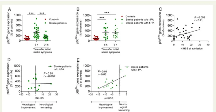
To provide evidence for a role of p66Shcin human ischaemic stroke, we analysed p66Shcexpression in PBMs of patients with ischaemic stroke. A total of 27 ischaemic stroke patients and 19 age- and sex-matched, healthy control subjects were recruited. Clinical character-istics of both groups did not differ statistically (Table1). p66ShcmRNA levels were significantly increased 6 h after initial stroke symptoms (Figure 6A) and returned to control levels 24 h thereafter (Figure6A). Of note, p66Shcgene expression at 6 h was comparable in patients with ischaemic stroke regardless of whether they had received thrombolytic treatment or not (Figure6B). p66Shctranscript levels of all study subjects positively correlated with neurological def-icits at admission, measured according to the NIHSS (Figure6C). Im-portantly, while p66Shcgene expression of stroke patients which did not receive thrombolytic intervention did not correlate with short-term neurological outcome (NIHSS discharge 2 NIHSS admission)
(Figure6D), it positively correlated in stroke patients treated with thrombolysis (Figure6E).
Discussion
In this study, we demonstrate for the first time that post-ischaemic p66Shcsilencing reduces brain injury by preserving BBB integrity by preventing claudin-5 level downregulation. The in vivo findings in the mouse were translated to HBMECs exposed to H/R where p66Shc was phosphorylated at Ser36 leading to the reduction in claudin-5 levels via activation of the NADPH oxidase and increased ROS production. Further, we show that p66Shc expression is increased in PBMs of patients with ischaemic stroke within 6 h from onset of symptoms and that p66Shcgene expression correlates to short-term neurological outcome.
In mice, we recently showed that constitutive genetic deletion of p66Shcreduces early stroke size and neurological deficits following I/R brain injury.22However, the mechanisms of this effect and its clin-ical relevance remained elusive. Furthermore, the use of constitutive
Figure 5 Effect of p66Shcsilencing and reactive oxygen species on H/R-decreased claudin-5 expression. (A) Immunoblot analysis reveals decreased claudin-5 protein levels after exposure of human brain microvascular endothelial cells to H/R (n ¼ 7), but not of occludin (n ¼ 5) (B) and vascular endothelial-cadherin (n ¼ 7) (C ). Pre-incubation with p66Shcsmall interfering RNA (n ¼ 7) (D), or apocynin (0.1 mmol/L) (n ¼ 6) (E), increases claudin-5 levels under H/R condition. Data are presented as mean + s.e.m. **P , 0.01, ***P , 0.001 for H/R vs. normoxia; #P , 0.05, ##
P , 0.01 for H/R with sip66Shcor apocynin vs. H/R.
knockout animals only serves as a proof-of-principle and does not allow delineation of therapeutic time windows as required in the clin-ical setting. In contrast, RNA interference (RNAi)-based strategies allow to modify the expression of a certain target at the post-transcriptional level, thus making it therapeutically interesting.42 Indeed, several RNAi-based strategies are under investigation in clin-ical trials.43Here, we used a clinically relevant experimental setup with siRNA delivery upon reperfusion to reduce p66Shc levels. Reperfusion of an occluded vessel allows reoxygenation of the is-chaemic area which promotes recovery of penumbral areas.44 Nevertheless paradoxically, re-introduction of oxygen in a previous-ly ischaemic area also causes a surge in free radical generation11which interferes with the recovery processes. Indeed, we demonstrate here that silencing of the pro-oxidant p66Shcafter the ischaemic episode and upon reperfusion, as it would be the case in stroke patients pre-senting at the emergency room undergoing thrombolytic treatment,
reduces lesion volume, improves neurological function, and increases survival. These data highlight the potential of p66Shcas novel thera-peutic target in stroke patients undergoing thrombolysis.
Given the key role of BBB permeability in determining stroke outcome,6we characterized its integrity by analysing Evans blue ex-travasation, a known indicator for vascular leakage.9,30,31,45Indeed, and in line with the improved stroke outcome in the sip66Shc stroke mice, BBB disruption was blunted after p66Shcsilencing com-pared with control mice. Cerebral microvascular endothelial cells connected via tight and adherens junctional proteins make up the BBB.7A disruption of this tightly regulated structure is known to occur in I/R brain injury and is responsible for vascular leakage and in turn oedema formation.9–11To investigate the molecular mechan-isms by which p66Shcpreserves BBB integrity after I/R brain injury, we focused in vivo as well as in vitro on claudin-5, occludin and VE-cadherin, three major junctional proteins.7 Consistently, we
. . . .
. . . .
. . . . Table 1 Characteristics of clinical study population
Study population Controls n 5 19 Stroke patients n 5 27 P-value
Age (years) mean (range) 71.8 (63-83) 73.9 (52-90) 0.42
Female, n (%) 10 (52.6%) 15 (55.6%) 1.0
Smoking, n (%) 1 (5.3%) 6 (22.2%) 0.22
Hypertension, n (%) 10 (52.6%) 15 (55.6%) 1.0
Dyslipidaemia, n (%) 2 (10.5%) 4 (14.8%) 1.0
Atrial fibrillation, n (%) 0 4 (14.8%) 0.13
Coronary artery disease, n (%) 0 5 (18.5%) 0.07
Previous stroke/TIA, n (%) 0 4 (14.8%) 0.13
Peripheral artery disease, n (%) 0 4 (14.8%) 0.13
Stroke sub-groups tPA n ¼ 14 (51.9%) w/o tPA n ¼ 13 (48.1%) P-value
Age (years) mean (range) 69.4 (52 – 80) 78.6 (64 – 90) 0.03
Female, n (%) 15 (55.6%) 6 9 0.25 OXFORD classification TACI, n (%) 12 (44.4%) 4 8 0.12 PACI, n (%) 10 (37.0%) 7 3 0.24 LACI, n (%) 5 (18.5%) 3 2 1.0 POCI, n (%) 0 0 0 TOAST classification
Large vessels atherosclerosis, n (%) 5 (18.5%) 2 3 0.65
Card ioembolism, n (%) 13 (48.1%) 6 7 0.71
Small vessels disease, n (%) 1 (3.7%) 1 0 1.0
Undetermined cause, n (%) 5 (18.5%) 3 2 1.0
Other cause, n (%) 3 (11.1%) 2 1 1.0
Stroke severity assessment
Admission NIHSS, mean (SD) 13.3 (6.7) 12.1 (5.0) 14.5 (8.1) 0.38
Discharge NIHSS, mean (SD) 10.1 (9.9) 5.8 (5.3) 14.8 (11.9) 0.046
Early complications
Haemorrhagic transformation, n (%)a 4 (17.4%) 3 1 0.61
Cerebral oedema, n (%)a 6 (26.1%) 1 5 0.05
Clinical characteristics of controls and stroke patients do not differ statistically. Sub-group analysis shows statistically significant differences in age and NIHSS at discharge between patients treated with thrombolysis and patients which did not receive thrombolysis. Age is given as mean (with ranges) and NIHSS is expressed as mean + SD.
TIA, transient ischaemic attack; tPA, tissue plasminogen activator; TACI, total anterior circulation infarct; PACI, partial anterior circulation infarct; LACI, lacunar infarct; POCI, posterior circulation infarct; TOAST, Trial of ORG 10172 in Acute Stroke Treatment; NIHSS, National Institutes of Health Stroke Scale.
a
found less reduction in claudin-5 levels after silencing of p66Shc. In contrast, occludin and VE-cadherin levels remained unaltered in both experimental settings. Our data obtained on murine as well as primary human cells indicate that p66Shcmediates BBB disruption by acting specifically on claudin-5 rather than occludin and VE-cadherin. To characterize the molecular regulation of claudin-5 by p66Shc, we exposed primary HBMECs to H/R, to mimic in vivo settings. Expos-ure of HBMECs to H/R, but not to hypoxia, increased phosphoryl-ation of p66Shcat Ser36 confirming previous data in renal tubular epithelial cells.46 Together with our in vivo data, these results suggest that p66Shcis mainly involved in mediating its deleterious effects during reperfusion, rather than during ischaemia. Endothelial ROS production and NO bioavailability are both critical for I/ R-induced alteration in BBB permeability and stroke outcome12,47 and ROS are known to influence claudin-5 levels in brain endothelial cells.48Here, we demonstrate in vitro evidence that endothelial p66Shc silencing in H/R preserves NO bioavailability, reduces NADPH oxidase activation, and decreases ROS generation thus blunting the reduction in claudin-5 levels. This pathway may be also relevant in vivo. To study p66Shcgene regulation in stroke patients, we performed proof-of-principle experiments assessing its expression in PBMs of acute ischaemic stroke patients. Although it would be of particular interest and relevance to elucidate the role of cerebrovascular
p66Shcin stroke patients, sample collection in humans would prove extremely difficult thus, we selected PBMs since those are easily ob-tainable from whole blood and could still give us interesting insights into gene expression changes. Here we found that p66ShcmRNA levels were increased 6 h after initial stroke symptoms and then returned to basal levels at 24 h. Moreover, p66Shcexpression at 6 h correlated to short-term neurological outcome (delta NIHSS) in stroke patients. Interestingly, delta NIHSS correlated to p66Shc expression only in patients undergoing thrombolysis, but not in those without. Although thrombolysis is known to improve neuro-logical outcome after stroke,5 it is also associated with early reperfusion-induced BBB damage49which is known to be mediated by ROS and affects stroke outcome.6An increased vascular leakage also favours the accumulation of blood circulating cells in brain tissue thus causing tissue damage via production of free radicals.11,50 This could explain why the correlation between neurological outcome and p66Shc expression is found only in thrombolytic patients where monocytic p66Shc-induced ROS production and the consequent damage is likely more pronounced. In contrast, in patients not undergoing thrombolysis, the role of reperfusion-induced ROS-dependent injury is less prominent and thus neuro-logical damage is less likely to be dependent on p66Shc-mediated ROS.
Figure 6 p66Shcgene expression in PBMs of ischaemic stroke patients. (A) Real-time polymerase chain reaction determined increased p66Shc
mRNA levels in stroke patients 6 h (n ¼ 27), but not 24 h (n ¼ 16) after initial stroke symptoms compared with the levels of control subjects (n ¼ 19). Data are expressed as mean + s.e.m. (B) p66Shcgene expression 6 h after initial stroke symptoms was determined in sub-groups of
stroke patients according as they did (t-PA), or did not (w/o t-PA) receive thrombolytic treatment. Values from controls are also shown for compar-isons. p66ShcmRNA levels are not different between both stroke sub-groups (w/o t-PA: n ¼ 13; t-PA: n ¼ 14). Both sub-groups show higher p66Shc
levels compared with controls. Data are expressed as mean + s.e.m. (C ) Correlation between p66Shcgene expression and NIHSS at admission of all study subjects (n ¼ 46). (D and E) Correlation analysis of p66Shctranscripts and delta NIHSS (NIHSS discharge 2 NIHSS admission) in w/o t-PA
patients (n ¼ 13) and in t-PA patients (n ¼ 14). For (C – E) linear regression trend lines are illustrated. ***P , 0.001. NIHSS, National Institutes of Health Stroke Scale; t-PA, tissue plasminogen activator.
In summary, the present study sets the stage for follow-up clinical studies aimed at assessing the potential for p66Shcto become a novel therapeutic target as an adjunct of thrombolysis for the management of acute ischaemic stroke.
Supplementary material
Supplementary Material is available at European Heart Journal online.
Acknowledgements
We thank Prof. Gianvito Martino from the Neuroimmunology Unit, Division of Neuroscience, Institute of Experimental Neurology, San Raffaele Scientific Institute Milan, Italy, for his intellectual and tech-nical contribution.
Funding
This work was supported by the Swiss National Science Foundation (grant number 310030_147017 to G.G.C., 310030-135815 to T.F.L., and 136822 to J.K.) and Helmut-Horten Foundation to G.G.C. Conflict of interest: none declared.
References
1. Nichols M, Townsend N, Scarborough P, Rayner M. Cardiovascular disease in Europe 2014: epidemiological update. Eur Heart J 2014;35:2950 – 2959. 2. O’Collins VE, Macleod MR, Donnan GA, Horky LL, van der Worp BH, Howells DW.
1,026 experimental treatments in acute stroke. Ann Neurol 2006;59:467 – 477. 3. Fisher M, Feuerstein G, Howells DW, Hurn PD, Kent TA, Savitz SI, Lo EH, Group S.
Update of the stroke therapy academic industry roundtable preclinical recommen-dations. Stroke 2009;40:2244 – 2250.
4. Spescha RD, Sessa M, Camici GG. Angiopoietin-like 4 and ischaemic stroke: a prom-ising start. Eur Heart J 2013;34:3603 – 3605.
5. Tissue plasminogen activator for acute ischemic stroke. The national institute of neurological disorders and stroke RT-PA stroke study group. N Engl J Med 1995; 333:1581 – 1587.
6. Obermeier B, Daneman R, Ransohoff RM. Development, maintenance and disrup-tion of the blood-brain barrier. Nat Med 2013;19:1584 – 1596.
7. Zlokovic BV. The blood-brain barrier in health and chronic neurodegenerative dis-orders. Neuron 2008;57:178 – 201.
8. Iadecola C, Anrather J. The immunology of stroke: from mechanisms to translation. Nat Med 2011;17:796 – 808.
9. Su EJ, Fredriksson L, Geyer M, Folestad E, Cale J, Andrae J, Gao Y, Pietras K, Mann K, Yepes M, Strickland DK, Betsholtz C, Eriksson U, Lawrence DA. Activation of PDGF-CC by tissue plasminogen activator impairs blood-brain barrier integrity during ischemic stroke. Nat Med 2008;14:731 – 737.
10. Lo EH, Dalkara T, Moskowitz MA. Mechanisms, challenges and opportunities in stroke. Nat Rev Neurosci 2003;4:399 – 415.
11. Rodrigues SF, Granger DN. Role of blood cells in ischaemia-reperfusion induced endothelial barrier failure. Cardiovasc Res 2010;87:291 – 299.
12. Moskowitz MA, Lo EH, Iadecola C. The science of stroke: mechanisms in search of treatments. Neuron 2010;67:181 – 198.
13. Pelicci G, Lanfrancone L, Grignani F, McGlade J, Cavallo F, Forni G, Nicoletti I, Grignani F, Pawson T, Pelicci PG. A novel transforming protein (SHC) with an SH2 domain is implicated in mitogenic signal transduction. Cell 1992;70:93 – 104. 14. Migliaccio E, Mele S, Salcini AE, Pelicci G, Lai KM, Superti-Furga G, Pawson T, Di
Fiore PP, Lanfrancone L, Pelicci PG. Opposite effects of the p52shc/p46shc and p66shc splicing isoforms on the EGF receptor-MAP kinase-fos signalling pathway. EMBO J 1997;16:706 – 716.
15. Camici GG, Schiavoni M, Francia P, Bachschmid M, Martin-Padura I, Hersberger M, Tanner FC, Pelicci P, Volpe M, Anversa P, Luscher TF, Cosentino F. Genetic deletion of p66(Shc) adaptor protein prevents hyperglycemia-induced endothelial dysfunc-tion and oxidative stress. Proc Natl Acad Sci USA 2007;104:5217 – 5222. 16. Francia P, delli Gatti C, Bachschmid M, Martin-Padura I, Savoia C, Migliaccio E,
Pelicci PG, Schiavoni M, Luscher TF, Volpe M, Cosentino F. Deletion of p66shc gene protects against age-related endothelial dysfunction. Circulation 2004;110: 2889 – 2895.
17. Napoli C, Martin-Padura I, de Nigris F, Giorgio M, Mansueto G, Somma P, Condorelli M, Sica G, De Rosa G, Pelicci P. Deletion of the p66Shc longevity gene
reduces systemic and tissue oxidative stress, vascular cell apoptosis, and early atherogenesis in mice fed a high-fat diet. Proc Natl Acad Sci USA 2003;100:2112 – 2116. 18. Zaccagnini G, Martelli F, Fasanaro P, Magenta A, Gaetano C, Di Carlo A, Biglioli P, Giorgio M, Martin-Padura I, Pelicci PG, Capogrossi MC. p66ShcA modulates tissue response to hindlimb ischemia. Circulation 2004;109:2917 – 2923.
19. Francia P, Cosentino F, Schiavoni M, Huang Y, Perna E, Camici GG, Luscher TF, Volpe M. p66(Shc) protein, oxidative stress, and cardiovascular complications of dia-betes: the missing link. J Mol Med 2009;87:885 – 891.
20. Camici GG, Shi Y, Cosentino F, Francia P, Luscher TF. Anti-aging medicine: molecular basis for endothelial cell-targeted strategies – a mini-review. Gerontology 2011;57: 101 – 108.
21. Cosentino F, Francia P, Camici GG, Pelicci PG, Luscher TF, Volpe M. Final common molecular pathways of aging and cardiovascular disease: role of the p66Shc protein. Arterioscler Thromb Vasc Biol 2008;28:622 – 628.
22. Spescha RD, Shi Y, Wegener S, Keller S, Weber B, Wyss MM, Lauinger N, Tabatabai G, Paneni F, Cosentino F, Hock C, Weller M, Nitsch RM, Luscher TF, Camici GG. Deletion of the ageing gene p66(Shc) reduces early stroke size following ischaemia/reperfusion brain injury. Eur Heart J 2013;34:96 – 103.
23. Pagnin E, Fadini G, de Toni R, Tiengo A, Calo L, Avogaro A. Diabetes induces p66shc gene expression in human peripheral blood mononuclear cells: relationship to oxi-dative stress. J Clin Endocrinol Metab 2005;90:1130 – 1136.
24. Bamford J, Sandercock P, Dennis M, Burn J, Warlow C. Classification and natural history of clinically identifiable subtypes of cerebral infarction. Lancet 1991;337: 1521 – 1526.
25. Adams HP Jr, Bendixen BH, Kappelle LJ, Biller J, Love BB, Gordon DL, Marsh EE 3rd. Classification of subtype of acute ischemic stroke. Definitions for use in a multicenter clinical trial. TOAST. Trial of Org 10172 in Acute Stroke Treatment. Stroke 1993;24: 35 – 41.
26. Franzeck FC, Hof D, Spescha RD, Hasun M, Akhmedov A, Steffel J, Shi Y, Cosentino F, Tanner FC, von Eckardstein A, Maier W, Luscher TF, Wyss CA, Camici GG. Expres-sion of the aging gene p66Shc is increased in peripheral blood monocytes of patients with acute coronary syndrome but not with stable coronary artery disease. Athero-sclerosis 2012;220:282 – 286.
27. Paneni F, Mocharla P, Akhmedov A, Costantino S, Osto E, Volpe M, Luscher TF, Cosentino F. Gene silencing of the mitochondrial adaptor p66(Shc) suppresses vas-cular hyperglycemic memory in diabetes. Circ Res 2012;111:278 – 289.
28. Swanson RA, Morton MT, Tsao-Wu G, Savalos RA, Davidson C, Sharp FR. A semi-automated method for measuring brain infarct volume. J Cereb Blood Flow Metab 1990;10:290 – 293.
29. Bederson JB, Pitts LH, Tsuji M, Nishimura MC, Davis RL, Bartkowski H. Rat middle cerebral artery occlusion: evaluation of the model and development of a neurologic examination. Stroke 1986;17:472 – 476.
30. Gursoy-Ozdemir Y, Bolay H, Saribas O, Dalkara T. Role of endothelial nitric oxide generation and peroxynitrite formation in reperfusion injury after focal cerebral is-chemia. Stroke 2000;31:1974 – 1980; discussion 1981.
31. Bouleti C, Mathivet T, Coqueran B, Serfaty JM, Lesage M, Berland E, Ardidie-Robouant C, Kauffenstein G, Henrion D, Lapergue B, Mazighi M, Duyckaerts C, Thurston G, Valenzuela DM, Murphy AJ, Yancopoulos GD, Monnot C, Margaill I, Germain S. Protective effects of angiopoietin-like 4 on cerebro-vascular and functional damages in ischaemic stroke. Eur Heart J 2013;34: 3657 – 3668.
32. Spescha RD, Glanzmann M, Simic B, Witassek F, Keller S, Akhmedov A, Tanner FC, Luscher TF, Camici GG. Adaptor protein p66Shc mediates hypertension-associated, cyclic stretch-dependent, endothelial damage. Hypertension 2014;64:347 – 353. 33. Besler C, Heinrich K, Rohrer L, Doerries C, Riwanto M, Shih DM, Chroni A,
Yonekawa K, Stein S, Schaefer N, Mueller M, Akhmedov A, Daniil G, Manes C, Templin C, Wyss C, Maier W, Tanner FC, Matter CM, Corti R, Furlong C, Lusis AJ, von Eckardstein A, Fogelman AM, Luscher TF, Landmesser U. Mechanisms underlying adverse effects of HDL on eNOS-activating pathways in patients with coronary artery disease. J Clin Invest 2011;121:2693 – 2708.
34. Shi Y, Cosentino F, Camici GG, Akhmedov A, Vanhoutte PM, Tanner FC, Luscher TF. Oxidized density lipoprotein activates p66Shc via lectin-like oxidized low-density lipoprotein receptor-1, protein kinase C-beta, and c-Jun N-terminal kinase kinase in human endothelial cells. Arterioscler Thromb Vasc Biol 2011;31:2090 – 2097. 35. Cecchelli R, Berezowski V, Lundquist S, Culot M, Renftel M, Dehouck MP, Fenart L. Modelling of the blood-brain barrier in drug discovery and development. Nat Rev Drug Discov 2007;6:650 – 661.
36. Walchli T, Pernet V, Weinmann O, Shiu JY, Guzik-Kornacka A, Decrey G, Yuksel D, Schneider H, Vogel J, Ingber DE, Vogel V, Frei K, Schwab ME. Nogo-A is a negative regulator of CNS angiogenesis. Proc Natl Acad Sci USA 2013;110:E1943 – E1952. 37. Migliaccio E, Giorgio M, Mele S, Pelicci G, Reboldi P, Pandolfi PP, Lanfrancone L,
Pelicci PG. The p66shc adaptor protein controls oxidative stress response and life span in mammals. Nature 1999;402:309 – 313.
38. Drummond GR, Selemidis S, Griendling KK, Sobey CG. Combating oxidative stress in vascular disease: NADPH oxidases as therapeutic targets. Nat Rev Drug Discov 2011;10:453 – 471.
39. Tomilov AA, Bicocca V, Schoenfeld RA, Giorgio M, Migliaccio E, Ramsey JJ, Hagopian K, Pelicci PG, Cortopassi GA. Decreased superoxide production in macrophages of long-lived p66Shc knock-out mice. J Biol Chem 2010;285:1153 – 1165.
40. Zehendner CM, Librizzi L, Hedrich J, Bauer NM, Angamo EA, de Curtis M, Luhmann HJ. Moderate hypoxia followed by reoxygenation results in blood-brain barrier breakdown via oxidative stress-dependent tight-junction protein disruption. PLoS ONE 2013;8:e82823.
41. Heumuller S, Wind S, Barbosa-Sicard E, Schmidt HH, Busse R, Schroder K, Brandes RP. Apocynin is not an inhibitor of vascular NADPH oxidases but an anti-oxidant. Hypertension 2008;51:211 – 217.
42. Mello CC, Conte D Jr. Revealing the world of RNA interference. Nature 2004;431: 338 – 342.
43. Kanasty R, Dorkin JR, Vegas A, Anderson D. Delivery materials for siRNA therapeu-tics. Nat Mater 2013;12:967 – 977.
44. Astrup J, Symon L, Branston NM, Lassen NA. Cortical evoked potential and extra-cellular K+ and H+ at critical levels of brain ischemia. Stroke 1977;8:51–57.
45. Yepes M, Sandkvist M, Moore EG, Bugge TH, Strickland DK, Lawrence DA. Tissue-type plasminogen activator induces opening of the blood-brain barrier via the LDL receptor-related protein. J Clin Invest 2003;112:1533 – 1540.
46. Zhao WY, Han S, Zhang L, Zhu YH, Wang LM, Zeng L. Mitochondria-targeted anti-oxidant peptide SS31 prevents hypoxia/reoxygenation-induced apoptosis by down-regulating p66Shc in renal tubular epithelial cells. Cell Physiol Biochem 2013;32: 591 – 600.
47. Dalkara T, Yoshida T, Irikura K, Moskowitz MA. Dual role of nitric oxide in focal cere-bral ischemia. Neuropharmacology 1994;33:1447 – 1452.
48. Schreibelt G, Kooij G, Reijerkerk A, van Doorn R, Gringhuis SI, van der Pol S, Weksler BB, Romero IA, Couraud PO, Piontek J, Blasig IE, Dijkstra CD, Ronken E, de Vries HE. Reactive oxygen species alter brain endothelial tight junction dynamics via RhoA, PI3 kinase, and PKB signaling. FASEB J 2007;21:3666 – 3676.
49. Kastrup A, Groschel K, Ringer TM, Redecker C, Cordesmeyer R, Witte OW, Terborg C. Early disruption of the blood-brain barrier after thrombolytic therapy predicts hemorrhage in patients with acute stroke. Stroke 2008;39:2385 – 2387. 50. Kochanek PM, Hallenbeck JM. Polymorphonuclear leukocytes and monocytes/
macrophages in the pathogenesis of cerebral ischemia and stroke. Stroke 1992;23: 1367 – 1379.