Exploration of the Neural Correlates of Ticklish Laughter by Functional Magnetic
Resonance Imaging
Elise Wattendorf1, Birgit Westermann2, Klaus Fiedler1, Evangelia Kaza3, Martin Lotze3and Marco R. Celio1 1
Anatomy and Program in Neuroscience, Department of Medicine, University of Fribourg, CH-1700 Fribourg, Switzerland, 2
Department of Neurosurgery, University Hospital, University of Basel, CH-4031 Basel, Switzerland and3Functional Imaging Unit, Diagnostic Radiology, University Hospital, University of Greifswald, D-17489 Greifswald, Germany
Address correspondence to Marco R. Celio, Anatomy and Program in Neuroscience, Department of Medicine, University of Fribourg, Rte A. Gockel 1, CH-1700 Fribourg, Switzerland. Email: [email protected].
The burst of laughter that is evoked by tickling is a primitive form of vocalization. It evolves during an early phase of postnatal life and appears to be independent of higher cortical circuits. Clinicopath-ological observations have led to suspicions that the hypothalamus is directly involved in the production of laughter. In this functional magnetic resonance imaging investigation, healthy participants were 1) tickled on the sole of the right foot with permission to laugh, 2) tickled but asked to stifle laughter, and 3) requested to laugh voluntarily. Tickling that was accompanied by involuntary laughter activated regions in the lateral hypothalamus, parietal operculum, amygdala, and right cerebellum to a consistently greater degree than did the 2 other conditions. Activation of the periaqueductal gray matter was observed during voluntary and involuntary laughter but not when laughter was inhibited. The present findings indicate that hypothalamic activity plays a crucial role in evoking ticklish laughter in healthy individuals. The hypothalamus promotes innate behavioral reactions to stimuli and sends projections to the periaqueductal gray matter, which is itself an important integrative center for the control of vocalization. A comparison of our findings with published data relating to humorous laughter revealed the involvement of a common set of subcortical centers.
Keywords: fMRI, hypothalamus, PAG, tickle, vocalization
Introduction
Tickling involves the unpredictable stimulation (Darwin 1872) of vulnerable parts of the body (armpits, chest, side of the waist, and sole of the foot; Black 1984) by a familiar person. It is an ambivalent stimulus, evoking a mixture of pleasurable and unpleasurable feelings (Plessner 1961) and leads to an in-voluntary stereotyped motor reaction (Provine 2000). Ticklish laughter is associated with sympathetic (Fry 1994; Szameitat et al. 2011) and emotional arousal (Bachorowski and Owren 2003). Tickling evolved from the rough-and-tumble play of animals; it is a primitive form of humor, which was referred to by Darwin as ‘‘protohumor’’ (Darwin 1872; Provine 2000). It provokes stimulus-driven emotionally valenced ‘‘Duchenne’’ laughter, which is classified as a prototypical nonserious social incongruity (Gervais and Wilson 2005).
Darwin (1872) and Hecker (1873) independently advanced the idea that tickling and humor are linked, in so far as both trigger laughter and share common qualities as stimuli (humor being considered as a ‘‘tickling of the mind’’). Later, the tickle reaction was purported to be the primitive building block on which humor developed (Mc Gee 1979; Weisfeld 1993; Provine
2000; Gamble 2001). In support of this theory, Fridlund and Loftis (1990) have observed the existence of a close correlation between self-reported ticklishness and the tendency to laugh, giggle, or smile in everyday life.
However, other authors do not regard ticklish laughter as either a precursor or an imitation of humorous laughter (Gregory 1924; Bergler 1956; Plessner 1961; Harris 1999). According to Harris and Christenfeld (1997), tickling and humor share a common final motor response but not an internal physiological state of mirth.
A wealth of data on humor has been gleaned by functional magnetic resonance imaging (fMRI) (Iwase et al. 2002; Mobbs et al. 2003; Osaka et al. 2003; Wild et al. 2006; Watson et al. 2007; Schwartz et al. 2008), whereas on tickling, only 2 such studies have been conducted (Blakemore et al. 2000; Carlsson et al. 2000). Moreover, in these 2 studies, the activated sensory network, not the vocal response, was evaluated, probably because of the difficulty that is experienced in controlling head movements during the ensuing laughter reaction, which obfuscate localization in the deep brain region (Iwase et al. 2002). Using state-of-the-art fMRI, we have now successfully explored the brain regions that are activated by ticklish laughter. Vocalization involves pathways stemming from both the motor cortex and the limbic system (for a review, see Ju¨rgens 2009), each of which is implicated in laughter (Wild et al. 2003). The limbic pathway arises in the anterior cingulate gyrus and leads through the periaqueductal gray matter (PAG), wherein it receives information concerning the emotional state from the medial thalamus, the amygdala, and the hypothalamus (Ju¨rgens 1998). The motor cortical and the limbic pathways converge at the level of the reticular formation of the pons and the medulla oblongata to interact with phonatory motor neurons. The motor cortical pathway is deemed to subserve the production of learned vocalizations, whereas the limbic one is believed to be critically involved in the production of nonverbal innate vocalizations (Ju¨rgens 2009). The latter tenet is supported by data that has been gleaned from human patients with lesions in the motor cortex. Although speech and song production are compromised in these individuals, non-verbal vocalizations, such as coughing, crying, and even some forms of laughter, are unaffected (Groswasser et al. 1988). Moreover, laughing (gelastic) seizures are observed in young patients developing hamartomas, irritating the lateral hypothal-amus (Valdueza et al. 1994; Delalande and Fohlen 2003); in this situation, speech is not compromised.
Laughter that is evoked by tickling is an ontogenetically precocious form of this emotional manifestation (Leuba 1941; Poeck 1985). As such, it may involve innate mechanisms rather
Ó The Author 2012. Published by Oxford University Press. All rights reserved. doi:10.1093/cercor/bhs0
Advance Access publication April 16, 2012 Cerebral Cortex June 2013;23:1280– 1289
than learned vocal behavior (Scheiner et al. 2006). And apropos of innate mechanisms, a comparison of ticklish laughter in humans with emotional vocalization circuits in other mammals is warranted. A wealth of data has been gleaned from studies with squirrel monkeys (Ju¨rgens 1998). In this species, electrical stimulation of the mediodorsal thalamus, the amyg-dala, the hypothalamus, or other regions of the brain that are implicated in the limbic pathway, corresponds to vocalizations of various emotional states, including expressions of enjoy-ment. And in nonhuman primates, as well as in rats, laughter-like emotional responses have been repeatedly evoked during playful chasing and tickling (Darwin 1872; Matsusaka 2004; Panksepp 2007). Against the background of available evidence, we hypothesize that in humans, ticklish laughter—as an innate and ‘‘primitive’’ form of vocalization—is rooted in the limbic pathway.
In our fMRI experiment, 3 different situations were established: 1) tickling and laughter, which involved tickling healthy participants on the sole of the right foot and allowing them to laugh; 2) tickling and inhibition of laughter; and 3) voluntary laughter. Inhibition of laughter is believed to reduce brain activity in relays that are critical for the neuronal control of vocalization. Voluntary laughter generally lacks the emo-tional features of true laughter. To reveal the regions that are essential for the triggering of ticklish laughter, activation that was registered during tickling and laughter was compared with that elicited under the other 2 conditions and was correlated with the corresponding number of laughter events.
Materials and Methods Subjects
Among the 27 healthy participants, 18 (11 females and 7 males, mean age: 24 years; age range: 21--29 years) were included in this study. Nine subjects were excluded because their head movements consistently exceeded 3 mm in 1 of the 3 evaluated directions using the Statistical Parametric Mapping (SPM) realignment procedure.
The informed consent of all participants was obtained, and the procedure was approved by the Ethical Committee of the University Hospital of Greifswald, Germany.
Behavioral Data
An fMRI-adapted fiber-optic microphone (MR confon, Magdeburg, Germany) was used to record laughter during the scanning procedure. Events of ticklish laughter and voluntary laughter were counted and classified as ‘‘strong’’ (more than one audible articulation) or ‘‘weak’’ (only one audible articulation). Strong vocalizations were awarded a 2-fold--high weighting than weak ones, which permitted a classifica-tion of the vocal response according to intensity as well as frequency.
Experimental fMRI Design
Before the onset of the experiment, both the tickler and the tickled person were instructed on the performance of each condition, and a trial run was conducted. During the scanning session, a friend (for 11 individuals) or the partner (for 7 individuals) of the participant stood in the scanning room and tickled or touched the right foot according to the particular stimulus condition (see Fig. 1). Each of the 3 tested conditions was indicated by a visual stimulus, consisting of a specific ‘‘smiley face’’ which was projected on separate screens for the tickler and for the tickled person. The latter watched the screen in a supine position via a mirror system. During tickling and laughter (T), the participants were manually tickled on the sole of the right foot and were encouraged to produce audible vocalizations. The same stimula-tion as for T was applied during tickling and inhibistimula-tion of laughter (I), but the participants had to prevent themselves from it. During
voluntary laughter (L), the subject was not tickled. Each of the 3 conditions was randomly presented 20 times, each lasting 6.2 s, and alternated with the presentation of a cross-signaled period of rest (11 s). To preserve the unpredictability of tickling, a red bar visible only to the tickler (F in Fig. 1) was randomly presented during T and I, urging stimulation by monotonous foot contact instead of tickling. This procedure ensured that the subjects were not prepared in advance for the real tickling stimulus, which was nevertheless applied 20 times. To minimize head movements and ensuing susceptibility artifacts in the fMRI signal, the participants held a wooden barbecue stick between their teeth during the course of the experiment, which did not interfere with laughter. After the session, the participants were asked to rate the mean sensation of tickling on a visual analogue scale (VAS, 1--10, 1 being the lowest score with no sensation of tickle and 10 the highest score).
Data Acquisition
Imaging was performed on a 3 T Scanner (VERIO, Siemens, Erlangen, Germany) with a 12-channel head coil. Functional images were obtained using a T2*-weighted echo planar imaging (EPI) sequence (repetition time: 2.2 s; echo time: 30 ms; flip angle: 90°), which embraced almost the entire brain in 24 contiguous axial slices (resolution: 3.0 3 3.0 3 3 mm, with a 1-mm gap). Images were additionally tilted by 30° relative to the anterior/posterior commissure (AC-PC line) to minimize susceptibility artifacts. During the functional session, 805 volumes were measured. Thirty-four phase and magnitude images were acquired in the same field of view and slice orientation, using a gradient echo (GRE) sequence with time repetition (TR)=488 ms, time echo, TE(1) = 4.92 ms, TE(2)= 7.38 ms, and a = 60° to calculate a field map in which geometric distortions in the EPI images were nullified. An anatomical T1-weighted, 3D Magnetization Prepared Rapid Gradient Echo image was acquired for each subject. The total number of sagittal anatomical slices amounted to 176 (TR=1900 ms; TE=2.52 ms; a=90°; voxel size=13131 mm3).
Data Analysis
Data were analyzed using SPM5 software (Wellcome Department of Cognitive Neuroscience, London, England), running on Matlab version 7.4 (MathWorks Inc; Natick, MA). Unwrapping of geometrically distorted EPIs was performed in the phase-encoding direction using Figure 1. Stimulus sequence during the fMRI experiment. Different expressions on ‘‘smiley faces’’ denoted the visual stimuli. Tickling and laughter: participants were tickled on the right foot and permitted to laugh (T); tickling and inhibition of laughter: participants were tickled on the right foot but forbidden to laugh (I); voluntary laughter: participants were requested to laugh spontaneously (L). Foot touch: participants were only touched (not tickled) on the right foot (F). (T) and (I) were randomly alternated with (F). The red bar signaling (F) was visible only to the tickler. Hence, during (T) and (I), the participants could not anticipate the ticklish stimulus and prepare to laugh. After each stimulus, the participants were allowed to rest (R) for 11 s (signalized by a ‘‘plus’’ sign).
the FieldMap Toolbox for SPM5. Each individual scan was realigned to the first scan to correct for movement artifacts. EPIs were coregistered with the T1-weighted anatomical image. The coregistered T1-image was segmented and normalized to the Montreal Neurological Institute (MNI) template; the EPIs were resliced at 33333 mm3. The resulting images were smoothed with a 93939 mm3Gaussian Kernel Filter (full-width at half maximum) to increase the signal-to-noise ratio. A temporal high-pass filter (128 s) was applied to eliminate slow-signal drifts. Movement parameters estimated during the realignment pro-cedure were introduced as covariates into the model to control for variance due to head displacements. An event-related analysis was used to separately identify neuronal activity following stimulation for T, I, and L in each subject. The onset of the response to tickling was estimated to be 1 s after the visually presented start signal. This time delay included the presumed mean reaction time for the motor response of the tickler (approximately 200 ms) and the mean delay of the tickled person in discerning the tickling stimulus (approximately 800 ms). Since pain fibers are involved during tickling (Zotterman 1939; Lahuerta et al. 1990), the conduction may include slower pathways than after mere sensory stimulation. The beginning of the ticklish sensation was chosen as the event of interest rather than the onset of audible laughter because we were interested in the specific brain processes that led to the outburst of laughter. The data were thus analyzed by modeling neuronal activity 1 s after the visual presentation of the respective condition. They were then adapted to the hemodynamic response function as supplied by SPM5. This procedure was adopted in the evaluation of all 3 conditions, even L. Voluntary laughter was continuous, and an evaluation of the early event was likely to capture the preparatory activity, which was considered to be most comparable to the T event. A one-sample t-test was implemented for the random-effect analysis at the group level. Main-effect of conditions T, I and L were considered to be statistically significant at a family-wise error rate of P <0.05, using a correction for multiple comparisons across the entire brain volume. To demonstrate activity in the motor cortex, a region of interest (ROI) analysis was applied for T and I. Brain activity that corresponded specifically to ticklish laughter was established by performing a conjunction analysis (global mean) on a full factorial model of the contrast T versus I and L. Furthermore, each participant’s (intensity weighted) number of vocal responses to tickling was correlated with its corresponding fMRI-signal magnitude (as represented by the contrast image) during the condition T. This analysis was calculated by a simple regression (implemented in SPM5) for the whole brain. The thresholds for the 2 latter analyses were set at P <0.001 (uncorrected). Parameter estimates (beta-values) for each experimental condition (T, I, and L) were derived from peaks of activation in the hypothalamus (centered at 3,–12,–15 (x, y, z) for the right, and–6,–6,–15 for the left hemisphere).
Results
Behavioral Results
During L, the vocal response upon request was strong (as defined in the Materials and Methods section), with an average number of 20 laughter events per participant. During T, the average score per subject was 9.75 (‘‘weak’’ laughter: 11.5, weighted as 5.25; ‘‘strong’’ laughter: 4.50). In this situation, each participant laughed at least once during the scanning session. The average tickling stimulus was gauged as moderate with a mean rating of 6.7 on a scale of 1--10 (range: 3.3--9). No statistically significant correlation existed between the partic-ipants’ rating of tickling and the corresponding number of weighted laughter events (r (n) = 0.16; P = 0.52). During L, some of the participants adapted a strategy that evokes emotionally driven laughter, such as imagining a comical situation (2 participants) or recalled true laughter on their attempt to induce voluntary laughter (1 participant). This may have decreased the signal for ticklish laughter relative to that
for voluntary laughter. During I, all participants were able to prevent themselves from laughing.
FMRI Results: Activation during Tickling and Laughter, Tickling and Inhibition of Laughter, and Voluntary Laughter
During T, stimulation of the sole of the right foot was associated with increased activation in the left primary sensory-motor cortex (representing the foot) and bilaterally in the secondary sensory-motor parietal operculum (including SII) and in the supplementary motor area (SMA) (Fig. 2, Table 1). ROI analysis within the S1/M1 revealed bilateral activation in the primary sensory-motor area (representing mimic muscula-ture, larynx, pharynx, and diaphragm). Activity was also recorded in the frontal operculum, the thalamus, the puta-men/pallidum, the hypothalamus (bilaterally), the anterior and posterior lobe of the right cerebellum, and the PAG. Activation in the visual regions was likewise enhanced as a response to the projected ‘‘smiley faces.’’ During I and L, activation occurred in similar regions of the brain to those described for T, with the exceptions that during I none was registered in the midbrain PAG, the cerebellum, the visual cortex, or the auditory cortex. During L, the cerebellum was activated bilaterally, but only in the posterior lobe. In contrast to the irregular laughter that was evoked during T, a vocal event was consistently recorded during L. Consequently, activities in the primary and secondary auditory cortex were recorded only in the latter situation. Neither L nor I activated the hypothalamus.
Increased Activation during Tickling and Laughter To characterize the brain activity that is specifically associated with ticklish laughter the results for T were compared with those for the other 2 conditions: I and L (Fig. 4 and Table 2). A conjunction analysis of the 2 comparisons revealed a network for sensory integration of the right foot stimulation, which included the right anterior cerebellar lobe (lobules I--IV), the bilateral parietal operculum (including the left SII) and the left primary sensory-motor foot area. This analysis also revealed heightened activation for T in brain areas corresponding to the limbic system, namely, in the midbrain substantia nigra, the hippocampus and bilaterally the tuberolateral part of the hypothalamus, and the amygdala.
Correlation Analysis of Tickling and Laughter with Vocalizations
During T, a linear positive correlation between each partic-ipant’s (intensity weighted) number of vocalizations and its corresponding fMRI-signal magnitude occurred in the later-oventral portion of the PAG (Fig. 3 and Table 3, for localization see also Carrive and Morgan 2004). Vocalization-related activity was also observed in the right primary sensory-motor region for facial expression, in the right frontal and the left parietal operculum, in the bilateral medial thalamus, and in the right posterior pallidum. It has to be noted that the observed effect in the above-mentioned regions was specific for T. Namely, at the applied threshold of 0.001 (uncorrected), the number of vocalizations emitted during T did not correlate with fMRI activity during I or L, except in the left primary sensory-motor cortex during I.
Discussion
A comparison of the imaging signals for T with those for I, and L revealed hypothalamic activity exclusively in the former situation. This finding confirms our hypothesis that the limbic pathway of vocalization is critically involved in ticklish laughter. Tickling and laughter is also associated with specific activities in higher-order sensory-motor areas (SII and the cerebellum), possibly paving the way to the deliberate control of the ensuing vocalization. Unexpectedly for such a primitive involuntary reaction, the same primary sensory-motor and premotor regions for the control of vocalization are activated in all 3 situations (T, I, and L). This finding indicates that the spontaneous laughter elicited by tickling is also under the control of the voluntary motor cortical pathway.
Tickling Activates the Motor Cortical Pathway of Vocalization
In all 3 situations (T, I, and L), a common activation of the primary sensory-motor cortex predictably involved brain areas that represent the face, the tongue, larynx and pharynx (Rolandic operculum), and the diaphragm (Maskill et al. 1991; Lotze et al. 2000; Brown et al. 2009). In contrast to previous observations relating to voluntary smiling or laughter (Wild et al. 2003), which were restricted to the somatotopic facial area, our study reveals the broad pattern of effectors that is activated during ticklish laughter. Consistent with the findings of Wild et al. (2003), activity occurred in the SMA and the frontal operculum, which have been previously shown to
encode various other orofacial functions, such as song production (Kleber et al. 2007) and speech (Brown et al. 2008). Taken together, our findings indicate that the motor cortical pathway is involved not only in voluntary but also in involuntary laughter, such as is evoked during tickling, and even in inhibition of laughter. The result for the latter 2 situations deserves a special comment. Inappropriate, un-controllable laughter has been observed in patients with lesions of the motor cortical pathway, thereby implicating this system in inhibitory control of vocalization (Wild et al. 2003). Activation of the motor cortical pathway during T and I signalizes inhibitory control which may regulate the vocal response according to the situational context. Moreover, Table 1 demonstrates that the neuronal response during I is even higher than the one during T: since laughter must be actively inhibited, cortical motor control is augmented. Our data reveal no evidence of prefrontal activity during I (see Table 1), although inhibitory control at the cognitive level is a key function of this region (Konishi et al. 1999; de Zubicaray et al. 2000). In a previous imaging study on humorous smiling and laughter, involvement of the left prefrontal cortex was reported and attributed to the perception and cognitive processing of stimulus characteristics (Wild et al. 2006). Inspection of our data at a lower threshold level (P <0.0001 [uncorrected]) reveals though, activity in the left inferior prefrontal cortex (brain region [BA] 10, MNI coordinates:–39, 45, 12 (x, y, z), z-value=4.25) during I that was not observed during T and L. Hence, we may conclude that, during I, cognitive strategies are adopted to inhibit the reaction to Figure 2. Activation patterns during T, I, and L. Three-dimensional top (first row) and lateral (from the right: R) views (second row) of the brain, and sagittal slices through the right hemisphere (third row). Activity is apparent in the primary sensorimotor areas (S1/M1) representing the diaphragm (D), mimic musculature and larynx (MIM), and the pharynx (Rolandic operculum: RO), as well as in the SMA and in the frontal operculum (FO). Activity in the left-foot area (FOOT) and in the parietal operculum (PO) is observed only during T and I. The auditory cortex (Heschl’s gyrus: HG) is activated only during L. T: P \ 0.0001 (uncorrected); I: P \ 0.0001 (uncorrected); L: P \ 0.05 (FWE-corrected). On each scale, the threshold level (T-value) for observed activity is indicated.
tickling. The absence of activity in the corresponding area of the right hemisphere remains unexplained because older data suggest that naturally arising motor responses to emotional stimuli, notably laughter and smiling, are under the control of the right prefrontal cortex (Shammi and Stuss 1999). Wild et al. (2006) suggested that action of the right inferior prefrontal cortex is essential to restrain laughter but not to effect it. Hence, our results indicate that, during I, the right prefrontal cortex is not involved in the suppression of the vocal response
to tickling; the seat of this inhibitory response lies at other cortical levels.
Finally, activity in the posterior cerebellar lobe occurred only in the 2 conditions that involved laughter (T and L) but not during I. This finding is consistent with the role of the cerebellum in the on-line adjustment of ongoing motor responses rather than in preparatory aspects (during I) relating to these (Bloedel 1992).
Ticklish Laughter Activates Specific Components in the Limbic Pathway of Vocalization
The neuronal equivalent of ticklish laughter was analyzed by correlating the activity during T with its corresponding laughter scores (Table 3 and Fig. 3) and by comparing T with the conditions in which laughter was either forbidden I or voluntary L (Table 2 and Fig. 4). A positive correlation occurred in the midbrain PAG, thereby confirming the role of this region as a crucial relay station in the limbic pathway of vocalization (Graham Brown 1915; Dabby et al. 2004; Ju¨rgens 2009), since the PAG was also activated during the main conditions T and L (see Table 1). Our data support existing evidence of this region’s involvement in initiating and controlling the intensity of all forms of vocal reaction (Larson 1991; Davis et al. 1996).
In contrast, our comparative analysis revealed hypothalamic activity to be specific for ticklish laughter alone. Behavioral and visceral reactions are intrinsically regulated in the hypothala-mus, which is anatomically connected to the PAG (Veazey et al. 1982; Semenenko and Lumb 1992; Saper 2004). Specifically,
Table 1
Main condition of tickling and laughter, voluntary laughter and tickling and inhibition of laughter
Brain region Tickling and laughter Tickling and inhibition of laughter Voluntary laughter
Side MNI Coordinates Z-score L/R MNI Coordinates Z-score L/R MNI-coordinates Z-score Frontal Mimic S1 L 57 21 42 4.39* L/R 54 27 48 5.65 L/R 48 15 42 5.53 M1 L/R 48 9 45 4.10* L/R 42 3 57 5.34 L/R 51 9 45 5.63 Larynx S1 L/R 48 15 39 4.12* L/R 57 21 39 5.05 L/R 39 18 36 5.63 M1 L/R 45 12 39 4.20* L 54 9 39 4.54 L/R 39 18 39 5.82 Diaphragm S1 L/R 39 42 60 4.64 L/R 42 42 63 6.03 L/R 15 30 54 5.47 M1 L/R 15 45 60 5.33* L/R 15 45 60 5.54 L/R 15 30 57 5.68 Foot S1 L 12 45 63 5.48 L 6 33 66 5.48 M1 L 6 27 66 5.34 Rolandic operculum (43/4) L/R 48 3 12 5.07 L/R 54 0 12 5.70 L/R 54 0 12 6.33 Frontal operculum/insula L/R 36 12 3 5.34 L/R 39 9 0 5.59 L/R 42 9 9 6.24 SMA (6) L/R 6 6 54 5.25 R 12 3 60 4.99 L/R 0 3 69 5.82
Anterior cingulate gyrus L/R 6 3 48 5.53 L/R 6 9 42 5.43
Temporal Parietal operculum (43/13/40) L/R 48 27 21 5.96 L/R 66 36 21 6.30 Heschl’s gyrus (41/42) R/L 51 36 15 5.90 Occipital V1/2/3 L/R 6 90 9 4.51 L/R 6 90 3 5.87 Cerebellum Posterior lobe R 33 51 30 5.00 R 33 57 21 6.21 Anterior lobe R 12 36 30 5.31 L 21 63 21 5.91 Subcortical Thalamus L/R 6 18 0 4.95 L 6 24 0 4.54 L/R 9 18 12 5.33
Medial and lateral Medial Lateral
Putam/pallidum L/R 24 9 6 5.31 L/R 21 9 0 4.93 L/R 24 9 3 5.89
Midbrain/PAG L/R 0 27 9 4.69 L/R 12 27 12 4.96
Hypothalamus L/R 6 12 3 4.96
Note: The stereotactic coordinates correspond to the standard laid down by the Montreal Neurological Institute (MNI). The signals in the reported regions exceeded the threshold family-wise error rate (FEW) of P \ 0.05. For primary sensory-motor regions P values of \0.0001 (uncorrected) was applied (denoted with an asterisk; activation maxima exclusively revealed in the premotor area are italicized). From bilateral activations, only the highest value was listed (in bold). S1/M1: primary sensorimotor cortex; PAG: midbrain periaqueductal gray matter.
Table 2
Conjunction analysis of tickling and laughter versus tickling and inhibition of laughter and voluntary laughter
Brain region Side MNI-coordinates Z-score
Superior frontal gyrus R 9 63 3 3.44
L 24 63 12 4.22 M1/S1 foot L 6 18 63 4.43 Parietal operculum R 45 30 27 3.96 L 54 27 21 3.53 SII L 33 21 15 4.39 Amygdala R 24 9 24 3.44 L 21 9 24 3.14 Hippocampus R 27 15 24 3.84 Cerebellum R 15 36 24 5.15 Substantia nigra R 9 15 21 3.36 Hypothalamus R 3 12 15 3.22 L 6 6 15 3.25
Note: Atlas coordinates (in MNI space) and Z-scores of peak activation. The signals in the reported regions exceeded the threshold level of P \ 0.001 (uncorrected). S1/M1: primary sensory-motor cortex.
hypothalamic nuclei comprise the largest input to vocalization areas, and activity stemming therefrom stimulates subregions of the PAG to evoke calls of hedonic or aversive character
(Altafullah et al. 1988; Dujardin and Ju¨rgens 2006). The hypothalamus can also regulate physiological phenomena, such as heart rate that accompany genuine laughter (Kuniecki et al. 2003) and emotional (Bachorowski and Owren 2003) as well as sympathetic arousal (Hecker 1873; Fry 1994). Activation of the hypothalamus coincided with that of the amygdala and the hippocampus (Table 2), which are believed to form part of a network that integrates sensory information and promotes appropriate visceral and behavioral reactions (Price 2005). Both structures project to the hypothalamus and from there to lower brainstem regions, such as the PAG (Leonard and Scott 1971; Poletti et al. 1973). The hypothalamus thus appears to hold a pivotal position between the cortical perception of stimuli and the regulation of behavior (Stearns 1972). It may either facilitate or trigger the stereotyped pattern of ticklish laughter via the PAG, and modulate the physiological bodily changes that are associated with it, if tickling was applied under appropriate consensual conditions.
Interestingly, the increase in hypothalamic activity that was evoked during ticklish laughter was confined to the tubero-lateral portion (Fig. 4), which accords with the findings of several recent studies relating to humor processing in both normal persons (Mobbs et al. 2003; Watson et al. 2007) and narcoleptic/cataplectic patients (Reiss et al. 2008; Schwartz et al. 2008). Since the measured response occurs at an anatomical location that contains the hypocretin/orexin-secreting neurons, their involvement has been suspected (Schwartz et al. 2008).
In this connection, it is worth mentioning that a wealth of neurological data suggests that the hypothalamus generates laughter (Davison and Kelman 1939; Martin 1950). Benign tumors (hamartomas) developing in the tuberal part of the hypothalamus of children, often lead to gelastic (laughing) seizures (Valdueza et al. 1994; Delalande and Fohlen 2003) and also vascular pathologies, such as aneurysm and bleeding in the region of the circle of Willis (Martin et al. 2003). Whilst hamartoma may generate intrinsic epileptic activity (Kuzniecky et al. 1997), vascular pathologies probably trigger laughter via a mechanical irritation or compression of the tuberolateral hypothalamus. This contention is supported by the observation that a swabbing of the floor of the third ventricle or pressing the infundibulum during the resection of ependymal cyst or craniopharyngeoma produced high mood and induced the patient to burst into laughter (Foerster and Gagel 1934).
The increased blood oxygen level--dependent (BOLD)-signal in the tuberolateral part of the hypothalamus could therefore be localized in the hypocretin/orexin cells rich region, explaining the arousal state typically accompanying tickling (Fry 1994). But the signal seems to occur more laterobasally, closer to the lateral tuberal nucleus (LTN), an entity of unknown function particularly well developed in humans (Le Gros Clark 1938), which is target of neuropathological changes in Huntington and Pick disease (Braak and Braak 1998). The human LTN may have its homologous in the rodent PV1, a newly recognized nucleus in the lateral hypothalamus of rodents (Gerig and Celio 2007; Girard et al. 2011; Meszar et al. 2012). In the future, the question of the exact intrahypotha-lamic localization of the BOLD signal could be resolved by high-resolution fMRI using the ticklish-laughter paradigm.
Activation of the anterior cingulate gyrus occurred during L and I, but not during T. Moreover, during L it was coactivated with the PAG (Table 1). fMRI investigations (Liu et al. 2010) Figure 3. Activation during tickling and laughter (first row), and correlation of this
activity with the number of vocal responses to the stimulus (second row). A correlation between the 2 parameters occurred in the PAG. First row: P \ 0.05 (FEW corrected). Second row: P \ 0.005 (uncorrected). L 5 left side. On each scale, the threshold level (T value) for observed activity is indicated.
Table 3
Correlation analysis for tickling and laughter: imaging signal versus vocal responses to the stimulus. Atlas coordinates (in MNI space) and Z-scores of peak activation
Brain region Side MNI-coordinates Z-score
Frontal operculum/insula R 42 33 0 3.32 S1/M1 mimic R 54 18 48 3.23 Parietal operculum L 66 30 12 3.37 Heschl’s gyrus L 54 6 3 3.19 Mediodors thalamus R/L 0 18 3 3.25 PAG R/L 3 27 6 3.38 Putamen/pallidum R 27 6 9 3.40
Note: Atlas coordinates (in MNI space) and Z-scores of peak activation. The signals in the reported regions exceeded the threshold level of P \ 0.001. S1/M1: primary sensory-motor cortex; PAG: midbrain periaqueductal gray.
and case studies in humans (Rubens 1975; Ju¨rgens and Kirzinger 1982), as well as lesioning experiments in monkeys (Sutton et al. 1974), afford evidence that the anterior cingulate gyrus is involved in motivated vocal output. And the findings of another study with monkeys indicate that this region controls vocalization via the PAG (Ju¨rgens and Pratt 1979). The activity in the anterior cingulate gyrus that was observed during L and I reflects the participant’s efforts to control vocalization. Ticklish laughter, on the other hand, appears to be produced in-dependently from these motivational circuits and to involve unconscious pathways relying on the hypothalamus.
Ticklish Laughter Activates Higher-Order Sensory Regions Although tickled persons may enjoy the experience, they typically engage in a defensive reaction, which may include a withdrawal of the tickled body part, a feeling of slight discomfort (Ruggieri and Milizia 1983), and laughing or giggling. The idea of a protective mechanism is supported by the finding that tickle-induced laughter depends partially on an intact pain sensation (Zotterman 1939; Lahuerta et al. 1990). Our comparison of T with the 2 other conditions (I and L) revealed the brain structures that could be involved in such specific processes, for example, the right anterior cerebellar lobe and the left parietal operculum SII (lateralized in accordance to tickling on the right foot, see Table 2). Our findings accord with those of previous imaging studies that focused on the sensory representation of tickling (Blakemore et al. 2000; Carlsson et al. 2000). Increased activity in the
secondary sensory SII has been associated with the conscious perception of both skin contact (Burton et al. 1997) and pain (Coghill et al. 1999). The cerebellum is integrating sensory feedback into an online correction of movement (Bloedel 1992). There are also indications that cerebellar activity is augmented in the presence of a mismatch between predicted and actual sensory perceptions (Restuccia et al. 2007; Bubic et al. 2009) and that it is implicated in the processing and anticipation of pain (Ploghaus et al. 1999). Furthermore, the main input nucleus to the cerebellum—the inferior olive—cannot distinguish between tactile sensory and pain-fibers inputs (Oscarsson and Sjolund 1977), and may thus transmit ambiguous information via the cerebellar climbing fibers during tickling. Additional cortical processing may be necessary to qualify the character of the sensory stimulation. If so, then a functionally relevant mechanism of mismatch detection and resolution could lie within the sequence of actual tickling and the prediction of pain and would entail activity in the cerebellum and the SII. Ticklish laughter would occur when this mechanism was activated to a sufficient degree. Such an idea is reminiscent of models of humor processing which have proposed that the recognition and resolution of cognitive incongruities between stored (anticipated) and actual informa-tion is necessary for the detecinforma-tion of humor and the ensuing burst of laughter (Suls 1972; Attardo 1997).
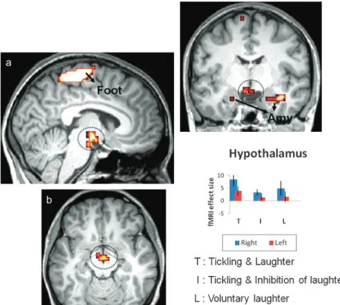
Previous studies relating to humorous laughter have revealed an involvement of the same limbic-related regions that we have observed in the context of ticklish laughter (Mobbs et al. 2003; Figure 4. Conjunction analysis and effect size. Increased significance of hypothalamic activation in response to T versus I and versus L (a: sagittal paramedian; b: axial; c: frontal; P \ 0.005 [uncorrected]). Parameter estimates (beta values) that were derived from the peaks of activity confirmed a higher level in this region during T than during either I or L (bar graph). Activity is also apparent in the amygdala (Amy) and in the somatosensory area representing the foot (Foot). The statistical maps are superimposed on an MNI-normalized image of the brain. Error bars indicate the 90% confidence interval.
Wild et al. 2003; Watson et al. 2007), thereby indicating a tendency to recruit a similar motivational network in both situations. However, the medial prefrontal cortex and the nucleus accumbens are activated only during the processing of humor (Goel and Dolan 2001; Samson et al. 2008; Samson et al. 2009). This network appears to be essential for self-referential processes (Luan Phan et al. 2005; van der Meer et al. 2010). In monkeys, its output feeds the hypothalamus and the PAG (An et al. 1998; Ongur et al. 1998). Hence, humorous stimuli probably generate a subjective sensation of mirth, which contributes to trigger the laughter response that is mediated by the hypothal-amus and the PAG. The tactile stimulation that produces the specific sensation of tickling (possibly in the SII and the cerebellum) would activate the same subcortical centers.
Our study highlights the pattern of brain activity that characterizes ticklish laughter. Several cortical and subcortical centers are implicated, and the pattern of activity resembles that evoked by humorous laughter, excepting an involvement of the medial prefrontal cortex and the nucleus accumbens in the latter situation only. Hence, whilst the subjective feeling of mirth and reward may be relevant in humorous laughter, they play but a subsidiary role in ticklish laughter. Nevertheless, the hypothesis of Darwin (1872) and Hecker (1873), that ticklish laughter forms the primitive building bloc supporting humor-ous laughter, is confirmed by our results.
Funding
Canton of Fribourg (Switzerland).
Notes
Dedicated to Prof. Uwe Ju¨rgens, Go¨ttingen, for his seminal and inspiring contributions to vocalization research. Conflict of Interest : None declared.
References
Altafullah I, Shipley C, Buchwald J. 1988. Voiced calls evoked by hypothalamic-stimulation in the cat. Exp Brain Res. 71:21--32. An X, Bandler R, Ongur D, Price JL. 1998. Prefrontal cortical projections
to longitudinal columns in the midbrain periaqueductal gray in macaque monkeys. J Comp Neurol. 401:455--479.
Attardo S. 1997. The semantic foundations of cognitive theories of humor. Humor. 10:395--420.
Bachorowski J, Owren M. 2003. Sounds of emotion production and perception of affect-related vocal acoustics. Ann N Y Acad Sci. 1000:244--265.
Bergler E. 1956. Laughter and the sense of humor. New York: International Medical Books.
Black D. 1984. Laughter. JAMA. 252:2995--2998.
Blakemore SJ, Wolpert D, Frith C. 2000. Why can’t you tickle yourself? Neuroreport. 11:R11--R16.
Bloedel J. 1992. Functional-heterogeneity with structural homogeneity—how does the cerebellum operate. Behav Brain Sci. 15:666--678.
Braak H, Braak E. 1998. Pick’s disease: cytoskeletal changes in the hypothalamic lateral tuberal nucleus. Brain Res. 802:119--124. Brown S, Laird A, Pfordresher P, Thelen S, Turkeltaub P, Liotti M. 2009.
The somatotopy of speech: phonation and articulation in the human motor cortex. Brain Cogn. 70:31--41.
Brown S, Ngan E, Liotti M. 2008. A larynx area in the human motor cortex. Cereb Cortex. 18:837--845.
Bubic A, von Cramon D, Jacobsen T, Schroger E, Schubotz R. 2009. Violation of expectation: neural correlates reflect bases of pre-diction. J Cogn Neurosci. 21:155--168.
Burton H, Macleod A, Videen T, Raichle M. 1997. Multiple foci in parietal and frontal cortex activated by rubbing embossed grating patterns across fingerpads: a positron emission tomography study in humans. Cereb Cortex. 7:3--17.
Carlsson K, Petrovic P, Skare S, Petersson K, Ingvar M. 2000. Tickling expectations: neural processing in anticipation of a sensory stimulus. J Cogn Neurosci. 12:691--703.
Carrive P, Morgan MM. 2004. Periaqueductal grey. In: Paxinos G, Mai JK, editors. The human nervous system. San Diego (CA): Academic Press. p. 393--423.
Coghill R, Sang C, Maisog J, Iadarola M. 1999. Pain intensity processing within the human brain: a bilateral, distributed mechanism. J Neurophysiol. 82:1934--1943.
Dabby R, Watemberg N, Lampl Y, Eilam A, Rapaport A, Sadeh M. 2004. Pathological laughter as a symptom of midbrain infarction. Behav Neurol. 15:73--76.
Darwin C. 1872. The expressions of the emotions in man and animals. London: John Murray. 200th anniversary edition 2009. Ekman P, editor. London: Harper Perennial Books.
Davis PJ, Zhang SP, Winkworth A, Bandler R. 1996. Neural control of vocalization: respiratory and emotional influences. J Voice. 10:23--38.
Davison C, Kelman H. 1939. Pathologic laughing and crying. Arch Neurol Psychiatry. 42:595--643.
de Zubicaray GI, Andrew C, Zelaya FO, Williams SCR, Dumanoir C. 2000. Motor response suppression and the prepotent tendency to respond: a parametric fMRI study. Neuropsychologia. 38: 1280--1291.
Delalande O, Fohlen M. 2003. Disconnecting surgical treatment of hypothalamic hamartoma in children and adults with refractory epilepsy and proposal of a new classification. Neurol Med Chir. 43:61--68.
Dujardin E, Ju¨rgens U. 2006. Call type-specific differences in vocalization-related afferents to the periaqueductal gray of squirrel monkeys (Saimiri sciureus). Behav Brain Res. 168:23--36.
Foerster O, Gagel O. 1934. A case of ependyma-cysts of the third ventricle. A contribution to the issue of relations of psychic disorders to the brain stem. Z Gesamte Neurol Psychiatr. 149:312--344.
Fridlund AJ, Loftis JM. 1990. Relations between tickling and humorous laughter—preliminary support for the Darwin-Hecker hypothesis. Biol Psychol. 30:141--150.
Fry WF. 1994. The biology of humor. Humor. 7:111--126. Gamble J. 2001. Humor in apes. Humor. 14:163--179.
Gerig AT, Celio MR. 2007. The human lateral tuberal nucleus: immunohistochemical characterization and analogy to the rodent PV1-nucleus. Brain Res. 1139:110--116.
Gervais M, Wilson DS. 2005. The evolution and functions of laughter and humor: a synthetic approach. Q Rev Biol. 80:395--430. Girard F, Meszar Z, Marti C, Davis F, Celio M. 2011. Gene expression
analysis in the parvalbumin-immunoreactive PV1 nucleus of the mouse lateral hypothalamus. Eur J Neurosci. 34:1934--1943. Goel V, Dolan RJ. 2001. The functional anatomy of humor: segregating
cognitive and affective components. Nat Neurosci. 4:237--238. Graham Brown T. 1915. Note on the physiology of the basal ganglia and
mid-brain of the anthropoid ape, especially in reference to the act of laughter. J Physiol. XLIX:195--207.
Gregory JC. 1924. The nature of laughter. London: Kegan Paul, Trench, Trubner & Co.
Groswasser Z, Korn C, Groswasserreider I, Solzi P. 1988. Mutism associated with buccofacial apraxia and bihemispheric lesions. Brain Lang. 34:157--168.
Harris CR. 1999. The mystery of ticklish laughter. Am Sci. 87:344--351. Harris CR, Christenfeld N. 1997. Humour, tickle, and the
Darwin-Hecker hypothesis. Cognit Emot. 11:103--110.
Hecker E. 1873. Die Physiologie und Psychologie des Lachens und des Komischen. Berlin (Germany): Du¨mmler.
Iwase M, Ouchi Y, Okada H, Yokoyama C, Nobezawa S, Yoshikawa E, Tsukada H, Takeda M, Yamashita K, Takeda M, et al. 2002. Neural substrates of human facial expression of pleasant emotion induced by comic films: a PET study. Neuroimage. 17:758--768.
Ju¨rgens U. 2009. The neural control of vocalization in mammals: a review. J Voice. 23:1--10.
Ju¨rgens U. 1998. Neuronal control of mammalian vocalization, with special reference to the squirrel monkey. Naturwissenschaften. 85:376--388.
Ju¨rgens U, Kirzinger A. 1982. The effects of lesions in the cortical face area, supplementary motor cortex and anterior cingulate cortex on vocalization in the monkey. Behav Brain Res. 5:105.
Ju¨rgens U, Pratt R. 1979. Cingular vocalization pathway in the squirrel-monkey. Exp Brain Res. 34:499--510.
Kleber B, Birbaumer N, Veit R, Trevorrow T, Lotze M. 2007. Overt and imagined singing of an Italian aria. Neuroimage. 36:889--900. Konishi S, Nakajima K, Uchida I, Kikyo H, Kameyama M, Miyashita Y.
1999. Common inhibitory mechanism in human inferior prefrontal cortex revealed by event-related functional MRI. Brain. 122: 981--991.
Kuniecki M, Urbanik A, Sobiecka B, Kozub J, Binder M. 2003. Central control of heart rate changes during visual affective processing as revealed by fMRI. Acta Neurobiol Exp. 63:39--48.
Kuzniecky R, Guthrie B, Mountz J, Bebin M, Faught E, Gilliam F, Liu HG. 1997. Intrinsic epileptogenesis of hypothalamic hamartomas in gelastic epilepsy. Ann Neurol. 42:60--67.
Lahuerta J, Bowsher D, Campbell J, Lipton S. 1990. Clinical and instrumental evaluation of sensory function before and after percutaneous anterolateral cordotomy at cervical level in man. Pain. 42:23--30.
Larson CR. 1991. On the relation of pag neurons to laryngeal and respiratory muscles during vocalization in the monkey. Brain Res. 552:77--86.
Le Gros Clark WE. 1938. Morphological aspects of the hypothalamus. In: Le Gros Clark WW, editor. The hypothalamus. Edinburgh (UK) and London: Oliver and Boyd. p. 1--68.
Leonard CM, Scott JW. 1971. Origin and distribution of amygdalofugal pathways in rat—experimental neuroanatomical study. J Comp Neurol. 141:313--329.
Leuba C. 1941. Tickling and laughter two genetic studies. J Genet Psychol. 58:201--209.
Liu H, Hu Z, Guo T, Peng D. 2010. Speaking words in two languages with one brain: neural overlap and dissociation. Brain Res. 1316:75--82. Lotze M, Erb M, Flor H, Huelsmann E, Godde B, Grodd W. 2000. fMRI
evaluation of somatotopic representation in human primary motor cortex. Neuroimage. 11:473--481.
Luan Phan K, Fitzgerald DA, Nathan PJ, Moore GJ, Uhde TW, Tancer ME. 2005. Neural substrates for voluntary suppression of negative affect: a functional magnetic resonance imaging study. Biol Psychiatry. 57:210--219.
Martin DD, Seeger U, Ranke MB, Grodd W. 2003. MR imaging and spectroscopy of a tuber cinereum hamartoma in a patient with growth hormone deficiency and hypogonadotropic hypogonadism. Am J Neuroradiol. 24:1177--1180.
Martin JP. 1950. Fits of laughter (sham mirth) in organic cerebral disease. Brain. 73:453--464.
Maskill D, Murphy K, Mier A, Owen M, Guz A. 1991. Motor cortical representation of the diaphragm in man. J Physiol. 443:105--121. Matsusaka T. 2004. When does play panting occur during social play in
wild chimpanzees? Primates. 45:221--229.
Meszar Z, Girard F, Saper CB, Celio MR. 2012. The lateral hypothalamic parvalbumin-immunoreactive (PV1) nucleus in rodents. J Comp Neurol. 520:798--815.
Mc Gee PE. 1979. Humor: its origin and development. San Francisco (CA): W.H. Freeman.
Mobbs D, Greicius MD, Abdel-Azim E, Menon V, Reiss AL. 2003. Humor modulates the mesolimbic reward centers. Neuron. 40: 1041--1048.
Ongur D, An X, Price JL. 1998. Prefrontal cortical projections to the hypothalamus in macaque monkeys. J Comp Neurol. 401: 480--505.
Osaka N, Osaka M, Kondo H, Morishita M, Fukuyama H, Shibasaki H. 2003. An emotion-based facial expression word activates laughter module in the human brain: a functional magnetic resonance imaging study. Neurosci Lett. 340:127--130.
Oscarsson O, Sjolund B. 1977. Ventral spino-olivocerebellar system in cat .1. Identification of 5 paths and their termination in cerebellar anterior lobe. Exp Brain Res. 28:469--486.
Panksepp J. 2007. Neuroevolutionary sources of laughter and social joy: modeling primal human laughter in laboratory rats. Behav Brain Res. 182:231--244.
Plessner H. 1961. Lachen und Weinen. Eine Untersuchung nach den Grenzen menschlichen Verhaltens. Bern (Switzerland): A. Franke Verlag. Ploghaus A, Tracey I, Gati JS, Clare S, Menon RS, Matthews PM, Rawlins JNP. 1999. Dissociating pain from its anticipation in the human brain. Science. 284:1979--1981.
Poeck K. 1985. Pathological laughter and crying. In: Vinken PJ, Bruyn GW, Klavans HV, editors. Handbook of clinical neurology. Amster-dam (the Netherlands): Elsevier. p. 219--225.
Poletti CE, Kinnard MA, Maclean PD. 1973. Hippocampal influence on unit-activity of hypothalamus, preoptic region, and basal forebrain in awake, sitting squirrel-monkeys. J Neurophysiol. 36:308--324. Price JL. 2005. Free will versus survival: brain systems that underlie
intrinsic constraints on behavior. J Comp Neurol. 493:132--139. Provine RR. 2000. The laughing species. Nat Hist. 109:72--77. Reiss AL, Hoeft F, Tenforde AS, Chen W, Mobbs D, Mignot EJ. 2008.
Anomalous hypothalamic responses to humor in cataplexy. PLoS One. 3:e2225.
Restuccia D, Della Marca G, Valeriani M, Leggio MG, Molinari M. 2007. Cerebellar damage impairs detection of somatosensory input changes. a somatosensory mismatch-negativity study. Brain. 130:276--287. Rubens AB. 1975. Aphasia with infarction in the territory of the anterior
cerebral artery. Cortex. 11:239--250.
Ruggieri V, Milizia M. 1983. Tickle perception as micro-experience of pleasure—its phenomenology on different areas of the body and relation to cerebral-dominance. Percept Mot Skills. 56:903--914. Samson AC, Hempelmann CF, Huber O, Zysset S. 2009. Neural
substrates of incongruity-resolution and nonsense humor. Neuro-psychologia. 47:1023--1033.
Samson AC, Zysset S, Huber O. 2008. Cognitive humor processing: different logical mechanisms in nonverbal cartoons—an fMRI study. Soc Neurosci. 3:125--140.
Saper CB. 2004. The hypothalamus. In: Paxinos G, Mai JK, editors. The human nervous system. San Diego (CA): Academic Press. p. 513--550.
Scheiner E, Hammerschmidt K, Ju¨rgens U, Zwimer P. 2006. Vocal expression of emotions in normally hearing and hearing-impaired infants. J Voice. 20:585--604.
Schwartz S, Ponz A, Poryazova R, Werth E, Boesiger P, Khatami R, Bassetti CL. 2008. Abnormal activity in hypothalamus and amygdala during humour processing in human narcolepsy with cataplexy. Brain. 131:514--522.
Semenenko FM, Lumb BM. 1992. Projections of anterior hypothalamic neurons to the dorsal and ventral periaqueductal gray in the rat. Brain Res. 582:237--245.
Shammi P, Stuss DT. 1999. Humour appreciation: a role of the right frontal lobe. Brain. 122:657--666.
Stearns FR. 1972. Laughing: physiology, pathophysiology, psychology and development. Springfield (IL): CC. Thomas.
Suls J. 1972. A two stage-stage model for the appreciation of jokes and cartoons: an information processing analysis. In: Goldstein J, McGhee P, editors. Psychology of humor. New York: Academic Press. p. 81--100.
Sutton D, Larson C, Lindeman RC. 1974. Neocortical and limbic lesion effects on primate phonation. Brain Res. 71:61--75.
Szameitat DP, Darwin CJ, Wildgruber D, Alter K, Szameitat AJ. 2011. Acoustic correlates of emotional dimensions in laughter: arousal, dominance, and valence. Cognit Emot. 25:599--611.
Valdueza JM, Cristante L, Dammann O, Bentele K, Vortmeyer A, Saeger W, Padberg B, Freitag J, Herrmann HD. 1994. Hypothalamic hamartomas—with special reference to gelastic epilepsy and surgery. Neurosurgery. 34:949--958.
van der Meer L, Costafreda S, Aleman A, David AS. 2010. Self-reflection and the brain: a theoretical review and meta-analysis of neuro-imaging studies with implications for schizophrenia. Neurosci Biobehav Rev. 34:935--946.
Veazey RB, Amaral DG, Cowan WM. 1982. The morphology and connections of the posterior hypothalamus in the cynomolgus monkey (Macaca-Fascicularis). 2. Efferent connections. J Comp Neurol. 207:135--156.
Watson KK, Matthews BJ, Allman JM. 2007. Brain activation during sight gags and language-dependent humor. Cereb Cortex. 17:314--324. Weisfeld GE. 1993. The adaptive value of humor and laughter. Ethol
Sociobiol. 14:141--169.
Wild B, Rodden FA, Rapp A, Erb M, Grodd W, Ruch W. 2006. Humor and smiling—cortical regions selective for cognitive, affective, volitional components. Neurology. 66:887--893.
Wild B, Rodden FA, Grodd W, Ruch W. 2003. Neural correlates of laughter and humour. Brain. 126:2121--2138.
Zotterman Y. 1939. Touch, pain and tickling: an electro-physiological investigation on cutaneous sensory nerves. J Physiol. 95: 1--28.