ChemComm
COMMUNICATION
Received 00th January 20xx, Accepted 00th January 20xx DOI: 10.1039/x0xx00000x www.rsc.org/A Mechano- and Thermoresponsive Luminescent Cyclophane
Yoshimitsu Sagara,*a,b Yoan C. Simon,b,c Nobuyuki Tamaokia and Christoph Weder*bThe first fluorescent cyclophane with mechano- and thermo-responsive solid-state fluorescence characteristics is reported. The new cyclophane is comprised of two 9,10-bis(phenyl-ethynyl)anthracene moieties that are bridged by tetraethylene glycol spacers. It is shown that the solid-state emission color of the compound can be influenced by mechanical and thermal treatments. This stimuli-responsive behavior is based on changes of the molecular assembly.
The last decade has witnessed a continuously increasing number of studies on materials in which the application of mechanical force leads to the controlled cleavage of stable1,2 or dynamic3 covalent
bonds, or the dissociation of supramolecular motifs.4 This general
mechanism has been shown to be useful for the design of stimuli-responsive materials, whose properties can be altered upon application of mechanical forces. If the force-induced separation involves mechanically active motifs (referred to as mechanophores) with extended π-conjugated groups, changes of the material’s absorption and/or photoluminescence properties occur.1,2 Especially, the latter can be easily detected,
making mechanically responsive (luminescent) color-changing materials potentially useful, for example in the in situ monitoring of material failure due to stress fracture or fatigue.5
However, the number of luminophores whose photophysical properties can be altered by mere dissociation of covalent or non-covalent bonds is still limited and few generally applicable design principles have been established.1b-d,h,2
Mechanical forces can also be exploited to induce changes of
molecular arrangements, and a large number of mechano-responsive luminescent (MRL) materials have been reported, which change their emission characteristics upon mechanical stimulation.2,6-8 A most effective strategy to create MRL
materials is to design luminescent molecules so they can assemble in different thermodynamically (meta)stable states, in
which they adopt different conformations and/or intermolecular interactions.6g Organic molecules of various shapes – from
simple rods to more complicated fan or dumbbell shapes – have been demonstrated to form assemblies that show MRL properties.2,6-8 Here we report the first fluorescent cyclophane
with mechano- and thermoresponsive solid-state fluorescence characteristics. While the photophysical properties of cyclic compounds have been extensively investigated in solution,9 little attention has been paid to their stimuli-responsive photophysical properties in the solid state. On a more funda-mental level, our study demonstrates that simple cyclic structures are useful to induce several thermodynamically (meta)stable states, presumably on account of spatial restrictions that prevent the luminophores to assemble in thermodynamically most stable closed-packed structures. We expect that the findings can be generalized and that the MRL feature may be readily imparted to other luminescent motifs by converting into cyclic structures.
Fig.1 Molecular structures of cyclophane 1 and the linear reference compound 2.
Cyclophane 1 (Fig. 1) was designed to feature two 9,10-bis(phenylethynyl)anthracene moieties, as these chromophores (in their non-connected state) are well known to exhibit strong green fluorescence in good solvents.10 Flexible tetraethylene glycol linkers
were chosen to connect the peripheral phenyl rings of two 9,10-bis(phenylethynyl)anthracene moieties to serve as bridge and to enhance the solubility. Because of its cyclic structure, compound 1 was expected to show intramolecular excimer formation and/or exciton coupling between the two luminescent cores in the molecularly isolated state. In the solid state, intra- and intermolecular interactions between the luminophores were expected
to occur, which should independently affect the photophysical properties of the resulting molecular material. The linear mono-9,10-bis(phenylethynyl)anthracene 2 was also prepared and used as a reference compound.
Despite the tetraethylene glycol linkers cyclophane 1 displays a low solubility in common organic solvents. We relate this behavior to the inherently rigid structure as well as to strong π-π interactions between multiple molecules. Gratifyingly, however, NMR spectroscopic measurements were possible because the compound is soluble in hot chloroform. A
comparison of the 1H NMR spectra of 1 and 2 (Fig. S1, ESI†)
reveals a pronounced up-field shift of the resonances of the luminescent cores for cyclophane 1 (0.46 and 0.49 ppm for the outer and inner protons of the anthracene groups, respectively), indicative of the formation of a cyclic structure, which changes the circular current. Thus, together with the mass spectroscopy data, the large difference unequivocally confirms the success of the cyclization reaction (Scheme S1, ESI†).
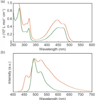
Fig. 2 Absorption (a) and emission (b) spectra of dilute (1·10–5 M) chloroform solutions of cyclophane 1 (orange line) and the linear reference compound 2 (green line). All spectra were measured at room temperature. The emission spectra were recorded with lex = 400 nm and intensities were normalized.
Spectroscopic measurements of cyclophane 1 and model compound 2 were first carried out in dilute (1·10–5 M)
chloroform solutions (Fig. 2). The absorption bands between 400 and 500 nm (Fig. 2a) show similar features, but upon closer inspection subtle differences can be discerned. While the spectrum of cyclophane 1 displays two peaks at 446 and 473 nm, these are observed at 450 and 471 nm in the case of the linear reference compound 2. Moreover, the magnitude of the two transitions is almost identical in 2 (ε = 3.7·104 L·mol–1·cm– 1), whereas in 1 the molar absorption coefficient at 446 nm (ε =
5.7·104 L·mol–1·cm–1) is larger than that at 473 nm (ε = 4.7·104
L·mol–1·cm–1). The difference in spectral shape seen in the
absorption spectra of 1 and 2 and the fact that the extinction coefficients of 1 are only slightly (and not two times) higher than those of 2 are the first indication of intramolecular, ground-state electronic interactions between two 9,10-bis-(phenylethynyl)anthracene moieties in cyclophane 1.
In dilute (1·10–5 M) chloroform solution, the linear reference compound 2 is highly emissive and the emission spectrum reveals a vibronic structure with maxima at 492 and 522 nm
and a weak shoulder around 560 nm (Fig. 2b). These features are characteristic of well-individualized 9,10-bis(phenyl-ethynyl)anthracene molecules in a good solvent.10a,c A much
lower emission intensity was observed when cyclophane 1 was excited under identical conditions (note that the intensities in Fig. 2b are normalized). The emission spectrum (Fig. 2b) shows an extra vibronic band at higher energy (459 nm) and a pro-nounced shoulder that stretches far into the red. The latter is indicative of intramolecular excimer formation,11 which has
also been observed in the case of pyrene- or perylene-based cyclophanes, i.e., in cases where the planar luminophores can form face-to-face stacked structures.12,13
To gain further insight into the difference of the emission characteristics of 1 and 2 in solution, we measured the excited state lifetimes (τ) and photoluminescence quantum yields (ΦPL) in chloroform (Table S1, ESI†). Fig. S2 (ESI†) shows time-resolved emission decay profiles for both 1 and 2. In the case of the linear reference compound 2, the decay curve is well fitted by a single exponential decay function with a lifetime of 2.9 ns (Fig. S2a, ESI†), which is characteristic of fluorescence from well-individualized chromophores. The emission decay of 1 was measured at 495 and 600 nm (Fig. S2b, ESI†). In both cases, multi-exponential decay functions fitted the data better than a single exponential decay function. The decay curve collected at 600 nm comprises a minor component with a lifetime of 8.8 ns that is ascribed to intramolecular excimers. By contrast, the longest emission lifetime observed at 495 nm was 5.8 ns, which can be attributed to monomer emission after dissociation of intramolecular excimers. At both wavelengths, the emission lifetime related to monomer (2.6 ns) is shorter than in the case of 2, on account of the formation of intramolecular excimers. The fastest decay process with emission lifetimes of 0.3 and 0.2 ns at 495 and 600 nm respectively is likely associated with exciton coupling between adjacent luminophores in the cyclophane. Indeed, as shown in Fig. 2a, the molar absorption coefficient at 446 nm is larger than that at 473 nm, which is indicative of exciton coupling with H-type geometry, although the precise conformation of 1 in chloroform is unclear. Aside from minor internal reabsorption features, the emission and absorption spectra of 1 remained unchanged when the concentration in chloroform was reduced from 10-5 to 10-6 (absorption) or 10-7 M (emission) (ESI†, Fig. S3), confirming that the observed exciton coupling is an intramolecular process and does not arise from the aggregation of cyclophane 1. This interpretation is further supported by the fact that previously reported perylene-based cyclophanes also exhibit absorption bands that correspond to
H-type exciton coupling,13 and the additional high-energy
emission peak at 459 nm (vide supra). While radiative relaxation from the higher energy level resulting from the splitting of the lowest unoccupied molecular orbital of the monomer species is forbidden in an ideal H-aggregate geometry, the twisted arrangement of transition dipole moments allows the direct decay process from the higher and forbidden energy level to some extent.14 Our interpretation of the emission decay
profiles is further supported by the results of
photoluminescence quantum yield measurements. Indeed, the photoluminescence quantum yield of 1 in chloroform (ΦPL = 0.07) is one order of magnitude lower than that of reference
compound 2 (ΦPL = 0.90), on account of intramolecular
excimer formation and exciton coupling in the cyclophane. The thermo- and mechanoresponsive luminescent behavior of cyclophane 1 is summarized in Fig. 3. Upon precipitating cyclophane 1 from a hot concentrated chloroform solution into hexane, a yellow-light emitting crystalline powder (YCR-form)
orange-yellow emissive powders (Fig. S4, ESI†). Surprisingly, we found that a transition to another crystalline form that
exhibits reddish-orange photoluminescence (ROCR-form) can
be achieved through thermal treatment; the here-reported
experiments involve heating the YCR-form for 10 min at 250 °C.
While cyclophane 1 displayed thermoresponsive luminescent characteristics, this behavior was not observed for linear reference compound 2 in the solid state. Moreover, cyclophane
1 also shows mechanochromic luminescence behavior.
Grinding of the ROCR-form leads to an amorphous state
showing yellow photoluminescence (YAM1-form). Subsequent
annealing at 250 °C for 10 min largely restored the reddish-orange emission color (ROLO-form), although the molecular
packing appears to be less-ordered (LO) than in the ROCR-form.
Grinding the YCR-form leads to another amorphous state (YAM2
-form), whose emission spectrum is slightly different from that of the YAM1-form (vide infra). However, subsequent annealing
also caused the conversion to ROLO-form. While various
molecular structures have been found to show thermo- or mechanoresponsive luminescence,2,6-8,15 compound 1, to our
knowledge, is the first reported cyclophane exhibiting such behavior.
Fig. 3 Thermo- and mechanoresponsive luminescence behavior of cyclophane 1 with pictures documenting the emission color in each state. All pictures were taken under UV irradiation (lex = 365 nm) on quartz substrates at room temperature.
Fig. 4 Emission spectra of the YCR-form (yellow solid line), ROCR-form (orange solid line), YAM1-form (orange dotted line), YAM2-form (yellow dotted line), ROLO-form obtained from the YAM1-form (orange dash-dotted line), and ROLO-form obtained from the YAM2 -form (yellow dash-dotted line) of cyclophane 1. All spectra were measured at room temperature with lex = 400 nm.
Powder X-ray diffraction (XRD) patterns of compound 1 (Fig. S5, ESI†) clearly reveal that the photoluminescence color changes induced by thermal or mechanical treatment result
from alterations of the molecular packing. The XRD pattern obtained for the YCR-form (Fig. S5a, ESI†) displays many
diffraction peaks that reflect the crystalline nature of this form. The diffractogram changes substantially upon annealing the sample for 10 min at 250 °C (Fig. S5b, ESI†). Thus, the thermal treatment causes a solid-solid phase transition between two different crystal structures, which is responsible for the
emission color change from the yellow YCR-form to the
reddish-orange ROCR-form. The phase transition was confirmed
by differential scanning calorimetry (DSC). The DSC trace of
the YCR-form displays an endothermic peak (DH = 4.4 kJ·mol–
1) at 221 °C (Fig. S6a, ESI†), which we associate with the
tran-sition to the ROCR-form. By contrast, the DSC curve measured
for the ROCR-form (Fig. S6a, ESI†) is void of this and any other
characteristic signals. Thermogravimetric analysis experiments of 1 (Fig. S7, ESI†) show no significant weight loss below 300 °C and confirm that the transition from the YCR-form to the
ROCR-form is not due to the release of trapped solvent. The
linear reference compound 2 could not be converted into a reddish-orange emissive state, indicating that cyclization is the key to the stimuli-responsive behavior of cyclophane 1.
As discussed above, grinding transforms the YCR-form and
ROCR-form into forms that emit yellow light (YAM1-form and
YAM2-form, respectively). The powder XRD patterns of both
amorphous forms are void of any diffraction peaks (Fig. S5c and e, ESI†), suggesting that grinding induces conversions from crystalline to amorphous states. Annealing of both amorphous forms for 10 min at 250 °C caused restauration of some of the peaks in the XRD (ROLO-form, Fig. S5d and f, ESI†), which
appear at similar spacings as observed for the ROCR-form.
Taking into account the fact that the emission spectrum is similar to that of the ROCR-form (vide infra), we conclude that
the thermal treatment restores a molecular assembly that is similar to the ROCR-form. However, the intensities of the peaks
observed in the diffractogram of the ROLO-form are much
lower than observed for ROCR-form, and the DSC trace
acquired upon heating the YAM1-form is featureless (Fig. S6a,
ESI†), indicating that annealing of the amorphous form of 1 leads to assembled structures with limited degree of order. The photophysical properties of 1 were also examined in the solid state and the influence of thermal and mechanical treatments were probed. Fig. 4 shows the steady-state photoluminescence spectra of cyclophane 1. The spectra correspond well with the photoluminescence color observed by the unassisted eye (Fig. 3). The YCR-form and ROCR-form
display broad emission bands with maxima around 555 and 614 nm, respectively. In contrast to the emission spectrum acquired for cyclophane 1 in chloroform solution, which shows well-resolved phonon modes, the solid-state spectra are broad and featureless. The emission band observed for the ROCR-form is
ascribed to static excimer formation of the
9,10-bis(phenylethynyl)anthracene cores. Indeed,
9,10-bis-(phenylethynyl)anthracene moieties forming π-stacked
structures in the condensed state have been reported to exhibit a similar emission band with maximum around 630 nm, which was ascribed to the formation of static excimers.8c Gratifyingly,
emission lifetime measurements support the formation of static
excimers in the ROCR-form. Fig. S8 (ESI†) shows
time-resolved fluorescent decays measured for YCR-form and ROCR
-form at 600 nm. The data are fitted well by a double exponential function, resulting in a long emission lifetime of 12.0 ns (Table S1, ESI†). By contrast, the decay trace of the
YCR-form is dominated by a component with a short lifetime of
0.8 ns, which can be attributed to exciton coupling. In comparison to the ROCR-form, the YCR-form features a much
smaller proportion of excimer emission. The two crystalline states have a similar quantum yield of 0.4 (Table S1, ESI†). The emission spectra of the amorphous YAM1-form and
YAM2-form of cyclophane 1 (Fig. 4, orange and yellow dotted
lines) show maxima that are situated between those of the YCR
-form and the ROCR-form. The small difference between the
emission spectra of the two amorphous forms may be ascribed to a small amount of residual crystalline particles after grinding. In the case of the YAM1-form, this would enable energy transfer
to excimer sites from the parent ROCR-form, leading to the
observed slight red-shift of the emission band compared to that of the YAM2-form. While it is, unfortunately, impossible to
distinguish if the grinding process changes inter- or intramolecular arrangements of the emissive cores of 1, one can speculate that probably a broad range of species with different conformation is present in the two amorphous forms and that the optical properties change on account of alterations of both
these factors. The emission spectrum of the ROLO-form, which
was obtained by annealing the YAM1-form (Fig. 4, orange
dash-dotted line), is very similar to that of ROCR-form, although a
small shoulder appears around 540 nm. The emission spectrum of the ROLO-form obtained from YAM2-form also shows the
same spectral feature (Fig. 4, yellow dash-dotted line). This behavior is consistent with the results obtained from powder XRD measurements and confirms that the annealing procedure does not lead to the complete recovery of the well-ordered molecular assembled structures that were observed for ROCR
-form. However, it appears that in the ROLO-form, energy
transfer from excited states with higher energy to excimer sites is at play, so that the relatively poorly ordered material displays emission characteristics that are reminiscent of the highly ordered ROCR-form. It is noteworthy that compound 2 also
shows a mechanically induced shift of the emission band (Fig. S9, ESI†), which is however much less pronounced than the one observed in the case of cyclophane 1. The comparison clearly shows the benefit of integrating the luminescent motif in a cyclic structure.
In summary, the first mechano- and thermoresponsive luminescent cyclophane has been reported. The compound’s photophysical properties in the solid state depend strongly on the morphology, which in turn can be influenced by mechanical and/or thermal treatment. We have shown that switching between three discrete morphologies (two crystalline and one amorphous structure) is possible. Interpreting the results more broadly, our study demonstrates that the integration of a fluorescent motif into a simple cyclic structure is a promising approach to design stimuli-responsive luminescent molecular materials. We expect our findings to be general and applicable to cyclophanes comprising other luminescent cores and/or spacers. Thus, the present study opens the door to a new suite of stimuli responsive materials that can change their solid-state
properties such as the here-reported luminescence
characteristics and possibly other electronic or magnetic properties, on command.
We thank Prof. T. Nakano for absorption and emission spectroscopic measurements, DSC measurements, and thermogravimetric analysis. We thank Prof. Y. Urano for
emission lifetime measurements and quantum yield
measurements. Y.S. is grateful for financial support from JSPS Postdoctoral Fellowships for Research Abroad. C.W. acknowledges support from the National Center of Competence in Research (NCCR) Bio-Inspired Materials, a research instrument of the Swiss National Science Foundation, the European Research Council (ERC-2011-AdG 291490-MERESPO), and the Adolphe Merkle Foundation.
Notes and references
1 (a) M. M. Caruso, D. A. Davis, Q. Shen, S. A. Odom, N. R. Sottos, S. R. White, J. S. Moore, Chem. Rev., 2009, 109, 5755–5798; (b) D. A. Davis, A. Hamilton, J. Yang, L. D. Cremar, D. Van Gough, S. L. Potisek, M. T. Ong, P. V. Braun, T. J. Martínez, S. R. White, J. S. Moore, N. R. Sottos, Nature, 2009, 459, 68–72; (c) G. R. Gossweiler, G. B. Hewage, G. Soriano, Q. Wang, G. W. Welshofer, X. Zhao, S. L. Craig, Acs Macro Lett., 2014, 3, 216–219; (d) Y. Chen, H. Zhang, X. Fang, Y. Lin, Y. Xu, W. Weng, Acs Macro Lett., 2014, 3, 141–145; (e) H. Zhang, Y. Chen, Y. Lin, X. Fang, Y. Xu, Y. Ruan, W. Weng, Macromolecules, 2014, 47, 6783–6790; (f) K. Imato, A. Irie, T. Kosuge, T. Ohishi, M. Nishihara, A. Takahara and H. Otsuka, Angew. Chem. Int. Ed., 2015, 54, 6168–6172; (g) K. Imato, T. Kanehara, T. Ohishi, M. Nishihara, H. Yajima, M. Ito, A. Takahara and H. Otsuka, ACS Macro Letters, 2015, 4, 1307–1311; (h) R. Göstl and R. P. Sijbesma, Chem. Sci., 2016, 7, 370–375.
2 (a) M. Teng, X. Jia, X. Chen and Y. Wei, Angew. Chem. Int. Ed., 2012, 51, 6398–6401; (b) Z. Ma, M. Teng, Z. Wang, S. Yang and X. Jia, Angew. Chem. Int. Ed., 2013, 52, 12268–12272; (c) Z. Ma, Z. Wang, X. Meng, Z. Ma, Z. Xu, Y. Ma, X. Jia, Angew. Chem. Int. Ed., 2016, 55, 519–522.
3 A. M. Belenguer, G. I. Lampronti, D. J. Wales, J. K. M. Sanders, J. Am. Chem. Soc., 2014, 136, 16156–16166.
4 (a) D. W. R. Balkenende, S. Coulibaly, S. Balog, Y. C. Simon, G. L. Fiore and C. Weder, J. Am. Chem. Soc., 2014, 136, 10493–10498; (b) A. P. Haehnel, Y. Sagara, Y. C. Simon and C. Weder, Top. Curr. Chem., 2015, 369, 345–375.
5 (a) C. Weder, J. Mater. Chem., 2011, 21, 8235–8236; (b) C. Weder, Nature, 2009, 459, 45–46; (c) C. Weder, in Encyclopedia of Polymeric Nanomaterials, eds. S. Kobayashi and K. Müllen, Springer Berlin Heidelberg, Berlin, Heidelberg, 2015, DOI: 10.1007/978-3-642-29648-2_6, pp. 1218–1227.
6 (a) Y. Sagara and T. Kato, Nat. Chem., 2009, 1, 605–610; (b) K. Ariga, T. Mori and J. P. Hill, Adv. Mater., 2012, 24, 158–176; (c) Z. Chi, X. Zhang, B. Xu, X. Zhou, C. Ma, Y. Zhang, S. Liu and J. Xu, Chem. Soc. Rev., 2012, 41, 3878–3896; (d) X. Zhang, Z. Chi, Y. Zhang, S. Liu and J. Xu, J. Mater, Chem. C, 2013, 1, 3376–3390; (e) F. Ciardelli, G. Ruggeri and A. Pucci, Chem. Soc. Rev., 2013, 42, 857–870; (f) Z. Ma, Z. Wang, M. Teng, Z. Xu and X. Jia, ChemPhysChem, 2015, 16, 1811–1828; (g) Y. Sagara, S. Yamane, M. Mitani, C. Weder and T. Kato, Adv. Mater., 2016, 28, 1073–1095. 7 (a) Y. Sagara, T. Mutai, I. Yoshikawa and K. Araki, J. Am. Chem. Soc., 2007, 129, 1520–1521; (b) J. Kunzelman, M. Kinami, B. R. Crenshaw, J. D. Protasiewicz and C. Weder, Adv. Mater., 2008, 20, 119–122; (c) S.-J. Yoon, J. W. Chung, J. Gierschner, K. S. Kim, M.-G. Choi, D. Kim and S. Y. Park, J. Am. Chem. Soc., 2010, 132, 13675–13683; (d) G. Zhang, J. Lu, M. Sabat and C. L. Fraser, J. Am. Chem. Soc., 2010, 132, 2160–2162; (e) Y. Dong, B. Xu, J. Zhang, X. Tan, L. Wang, J. Chen, H. Lv, S. Wen, B. Li, L. Ye, B. Zou and W. Tian, Angew. Chem. Int. Ed., 2012, 51, 10782–10785; (f) X. Luo, W. Zhao, J. Shi, C. Li, Z. Liu, Z. Bo, Y. Q. Dong and B. Z. Tang, J. Phys. Chem. C, 2012, 116, 21967–21972; (g) K. Nagura, S. Saito, H. Yusa, H. Yamawaki, H. Fujihisa, H. Sato, Y. Shimoikeda and S. Yamaguchi, J. Am. Chem. Soc., 2013, 135, 10322–10325; (h) S. Yagai, S. Okamura, Y. Nakano, M. Yamauchi, K. Kishikawa, T. Karatsu, A. Kitamura, A. Ueno, D. Kuzuhara, H. Yamada, T. Seki and H. Ito, Nat. Commun., 2014, 5, 4013; (i) H.-J. Kim, D. R. Whang, J. Gierschner, C. H. Lee and S. Y. Park, Angew. Chem. Int. Ed., 2015, 54, 4330–4333; (j) Y. Sagara, A. Lavrenova, A. Crochet, Y. C. Simon, K. M. Fromm and C. Weder, Chem. Eur. J., in press. DOI:10.1002/chem.201600272
8 (a) Y. Sagara and T. Kato, Angew. Chem. Int. Ed., 2008, 47, 5175– 5178; (b) Y. Sagara, S. Yamane, T. Mutai, K. Araki and T. Kato, Adv. Funct. Mater., 2009, 19, 1869–1875; (c) Y. Sagara and T. Kato, Angew. Chem. Int. Ed., 2011, 50, 9128–9132; (d) H. Li, Z. Chi, B. Xu, X. Zhang, X. Li, S. Liu, Y. Zhang and J. Xu, J. Mater. Chem., 2011, 21, 3760–3767; (e) H. Li, X. Zhang, Z. Chi, B. Xu, W. Zhou, S. Liu, Y. Zhang and J. Xu, Org. Lett., 2011, 13, 556–559; (f) Y. Sagara, T. Komatsu, T. Ueno, K. Hanaoka, T. Kato and T. Nagano, Adv. Funct. Mater., 2013, 23, 5277–5284; (g) Y. Ren, W. H. Kan, V. Thangadurai and T. Baumgartner, Angew. Chem. Int. Ed., 2012, 51, 3964–3968; (h) S. Yamane, Y. Sagara, T. Mutai, K. Araki and T. Kato, J. Mater. Chem. C, 2013, 1, 2648–2656; (i) Y. Sagara, T. Komatsu, T. Ueno, K. Hanaoka, T. Kato and T. Nagano, J. Am. Chem. Soc., 2014, 136, 4273–4280; (j) Y. Sagara, T. Komatsu, T. Terai, T. Ueno, K. Hanaoka, T. Kato and T. Nagano, Chem. Eur. J., 2014, 20, 10397–10403.
9 (a) D. Ramaiah, P. P. Neelakandan, A. K. Nair and R. R. Avirah, Chem. Soc. Rev., 2010, 39, 4158–4168; (b) P. G. Ghasemabadi, T. Yao and G. J. Bodwell, Chem. Soc. Rev., 2015, 44,6494–6518; (c) T. Stangl, S. Bange, D. Schmitz, D. Würsch, S. Höger, J. Vogelsang and J. M. Lupton, J. Am. Chem. Soc., 2013, 135, 78–81; (d) S. Liu, D. Schmitz, S.-S. Jester, N. J. Borys, S. Höger and J. M. Lupton, J. Phys. Chem. B, 2013, 117, 4197–4203.
10 (a) C. A. Heller, R. A. Henry, B. A. McLaughlin and D. E. Bliss, J. Chem. Eng. Data, 1974, 19, 214–219; (b) P. Hanhela and D. B. Paul, Aust. J. Chem., 1984, 37, 553–559; (c) M. Levitus and M. A. Garcia-Garibay, J. Phys. Chem. A, 2000, 104, 8632–8637.
11 L. Qiu, C. Zhu, H. Chen, M. Hu, W. He, Z. Guo, Chem. Commun., 2014, 50, 4631–4634.
12 (a) M. Inouye, K. Fujimoto, M. Furusyo and H. Nakazumi, J. Am. Chem. Soc., 1999, 121, 1452–1458; (b) H. Abe, Y. Mawatari, H. Teraoka, K. Fujimoto and M. Inouye, J. Org. Chem., 2004, 69, 495– 504.
13 (a) J. Feng, Y. Zhang, C. Zhao, R. Li, W. Xu, X. Li and J. Jiang, Chem. Eur. J., 2008, 14, 7000–7010; (b) K. E. Brown, W. A. Salamant, L. E. Shoer, R. M. Young and M. R. Wasielewski, J. Phys. Chem. Lett., 2014, 5, 2588–2593.
14 M. Kasha, H. R. Rawls and M. A. El-Bayoumi, Pure Appl. Chem., 1965, 11, 371–392.
15 A. Seeboth, D. Lötzsch, R. Ruhmann and O. Muehling, Chem. Rev., 2014, 114, 3037–3068.
TOC entry
A Mechano- and Thermoresponsive Luminescent Cyclophane
The first mechano- and thermoresponsive luminescent cyclophane is described in this report.