A N S W E R T O T H E P H O T O Q U I Z
Philip A. Mackowiak, Section Editor
A Well-Preserved Culprit
(See page 704 for the Photo Quiz.)
Diagnosis: reactivated, disseminated histoplasmosis.
Histological examination of laryngeal biopsy specimens showed the presence of budding yeast cells (Figures1and2). Based on this observation, laryngeal histoplasmosis with underlying iatrogenic hypercortisolism, steroid-induced hyper-tension, and diabetes mellitus was suspected. Histopathologi-cal reexamination of the adrenal glands, which had been removed in 1996, also raised suspicion of adrenal histoplasmo-sis instead of the previously suspected culture-negative tuber-culosis. The presence of Histoplasma capsulatum in both the laryngeal and adrenal gland tissues was demonstrated by broad-spectrum polymerase chain reaction (PCR) targeting the fungal internal transcribed spacer (ITS) region, followed by sequence homology analysis of the PCR product [1]. H. capsulatum genotyping using 4 targets of sufficient intra-species variability (arf, h-anti, ole, and tub1) [2,3] provided proof that reactivation, not reinfection, took place. Interesting-ly, tests for specific antibodies remained negative twice, but H. capsulatum antigen detection from a urine sample obtained
after 2 weeks of antifungal therapy was positive. After 6 weeks of incubation,H. capsulatum was also cultivated from the pa-tient’s urine (Figure3).
Treatment with itraconazole was initiated according to the current Infectious Diseases Society of America guidelines [4]. The hoarseness disappeared almost completely, but hospitali-zation became necessary due to progressive asthenia. Cranial computed tomography (CT) scan was not indicative of cere-bral histoplasmosis, but CT scans of the chest and abdomen suggested bilateral pulmonary involvement. The patient, who always had sufficient capacity of discernment, chose to discon-tinue any treatment, including hydrocortisone substitution, and died from hypocortisolism 2 months after the diagnosis of disseminated histoplasmosis was made. The development of diabetes mellitus and immunological changes as a result of ad-vancing age may have contributed to the reemergence of the pathogen 15 years after the initial manifestation.
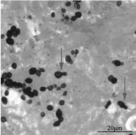
Only <1% of individuals infected with H. capsulatum develop symptomatic disease. The severity of symptoms depends on the degree of exposure and the immune status of Figure 2. Grocott–Gomori methenamine silver stain (magnification ×400) showing blastoconidia of H. capsulatum and the typical narrow-necked budding (arrows).
Figure 1. Yellowish, edematous mucosal changes in the interarytenoid region involving the posterior part of the vocal cords (arrows). Along with Addison’s disease, mucous membrane lesions are a characteristic mani-festation ofH. capsulatum infection.
the host, and can range anywhere from mild pulmonary affec-tion to severe disseminated disease. Disseminated disease mostly occurs in patients with impaired cell-mediated immu-nity [5]. In this context, it is remarkable that invasive fungal infections are not consistently recognized in patients treated with immunosuppressants like corticosteroids and tumor ne-crosis factor–α blockers [6,7].
Disseminated histoplasmosis can affect every organ system. Involvement of mucous membrane tissue in combination with Addison’s disease due to the destruction of both adrenal glands are particularly indicative of histoplasmosis compared to other systemic mycoses. Reactivation of a silent focus can occur decades after the initial exposure [6,8].
H. capsulatum is highly prevalent in the central and south-eastern parts of the United States as well as in Central and South America and parts of Africa. However, our case shows that, due to increasing international travel activity, infection needs to be ruled out in a wide variety of clinical scenarios, not only within endemic regions. In addition to traditional di-agnostic techniques (histopathology, serology, and culture), broad-spectrum PCR targeting the fungal ITS region is a valu-able tool for the detection of this potentially fatal pathogen, not least in settings where reliable histopathological identi fica-tion may be difficult to obtain.
Notes
Acknowledgments. We thank the technicians at the Institute of Medical Microbiology for their technical support.
Financial support. This work was supported in part by the University of Zürich.
Potential conflicts of interest. All authors: No reported conflicts. All authors have submitted the ICMJE Form for Disclosure of Potential Conflicts of Interest. Conflicts that the editors consider relevant to the content of the manuscript have been disclosed.
Florian P. Maurer,1Ewerton Marques Maggio,2Thomas N. Roth,3 Stefan P. Kuster,4and Guido V. Bloemberg1 Institutes of1Medical Microbiology and2Surgical Pathology, Divisions of
3
Otorhinolaryngology, and4Infectious Diseases and Hospital Epidemiology, University Hospital Zürich, Switzerland
References
1. Ciardo DE, Schar G, Altwegg M, Bottger EC, Bosshard PP. Identifica-tion of moulds in the diagnostic laboratory—an algorithm implement-ing molecular and phenotypic methods. Diagn Microbiol Infect Dis 2007; 59:49–60.
2. Kasuga T, Taylor JW, White TJ. Phylogenetic relationships of varieties and geographical groups of the human pathogenic fungusHistoplasma capsulatum Darling. J Clin Microbiol 1999; 37:653–63.
3. Kasuga T, White TJ, Koenig G, et al. Phylogeography of the fungal pathogenHistoplasma capsulatum. Mol Ecol 2003; 12:3383–401. 4. Wheat LJ, Freifeld AG, Kleiman MB, et al. Clinical practice guidelines
for the management of patients with histoplasmosis: 2007 update by the Infectious Diseases Society of America. Clin Infect Dis 2007; 45:807–25.
5. Schnizlein-Bick C, Durkin M, Kohler S, et al. Effects of CD4 and CD8 T lymphocyte depletion on the course of histoplasmosis following pul-monary challenge. Med Mycol2003; 41:189–97.
6. Kauffman CA. Histoplasmosis: a clinical and laboratory update. Clin Microbiol Rev2007; 20:115–32.
7. Administration USFaD. Information for healthcare professionals: Cimzia (certolizumab pegol), Enbrel (etanercept), Humira (adalimu-mab) and Remicade (inflixi(adalimu-mab). Available at: http://www.fda.gov/ Drugs/DrugSafety/PostmarketDrugSafetyInformationforPatientsand Providers/ucm124185.htm. Accessed 22 June.
8. Brandt ME, Warnock DW. Histoplasma, Blastomyces, Coccidioides, and other dimorphic fungi causing systemic mycoses. In: Mandell G, Bennett J, Dolin R, eds. Mandell, Douglas, and Bennett’s principles and practice of infectious diseases. 9 ed. Vol. 2. Elsevier Science, 2007: 1857–73.
Correspondence: Florian P. Maurer, MD, Institute of Medical Microbiology, University of Zürich, Gloriastr. 30/32, 8006 Zürich, Switzerland (fl[email protected]).
Clinical Infectious Diseases 2013;56(5):747–8
© The Author 2013. Published by Oxford University Press on behalf of the Infectious Diseases Society of America. All rights reserved. For Permissions, please e-mail: journals.permissions@ oup.com.
DOI: 10.1093/cid/cis914
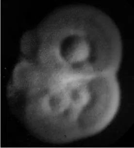
Figure 3. White to buff-brown mould form ofH. capsulatum grown at 25°C on Sabouraud’s dextrose agar. Large numbers of infectious conidia may be released by the mere lifting of the culture plate lid.