Human Reproduction vol.11 no.5 pp. 1043-1048, 1996
Improved fertilization and implantation rates after
non-touch zona pellucida microdrilling of mouse oocytes
with a 1.48 (im diode laser beam
M.Germond1'5, D.Nocera1, A.Senn1, K.Rink2, G.Delacr&az2, T.Pedrazziiii3 and J.P.Hornung4
'Fertility Unit, Department of Gynaecology and Obstetrics, CHUV, Lausanne, 2Applied Optics Laboratory, Swiss Federal Institute of Technology, Lausanne, Switzerland, Division of Hypertension, CHUV, Lausanne, 4Institute of Anatomy, University of Lausanne, Lausanne, Switzerland
5To whom correspondence should be addressed at Unite de St6rilite\ Matemite, CHUV, CH-1011 Lausanne, Switzerland The safety of microdrilling the zona pellucida of moose oocytes with a 1.48 |im diode laser has been investigated by determining the ability of mouse oocytes to fertilize
in vitro and develop in vivo. Mice born after transfer of
control and zona pelludda-microdrilled embryos into foster mothers were submitted to anatomical and immunohisto-chemical investigations, and their aptitude to breed was assessed in two subsequent generations. Decolonization of the oocytes with hyaluronidase induced a reduction of the fertilization and implantation rates, which was attributed to a zona hardening phenomenon. After laser zona pellucida microdrilling, these rates were restored to those obtained with embryos derived from untreated oocyte-cumulus com-plexes. Pups derived from zona pellucida microdrilled embryos were comparable with those obtained from control embryos, confirming the lack of deleterious effects of the laser treatment In conclusion, the 1.48 (un diode laser allows safe microdrilling of the zona pellucida of mouse oocytes after decoronization with hyaluronidase. Based on the health of the F2 generation and the lack of
neuroanatom-ical and neurochemneuroanatom-ical differences, we concluded that this technology may be investigated in the human, particularly when the zona pellucida represents the main impediment for fertilization or embryo hatching.
Key words: assisted reproduction/infrared diode
laser/micro-drilling/mouse/zona pellucida
Introduction
Chemical (Gordon and Talansky, 1986; Garrisi et al, 1990), mechanical (Depypere et al, 1988; Maker and Cohen, 1989; Odawara and Lopata, 1989) or laser methods (Feichtinger
et al, 1992; Laufer et al, 1993; SchUtze et al, 1994, 1995)
have been used to produce holes in the zona pellucida of mammalian oocytes in order to improve fertilization (Gordon, 1988; Gordon et al, 1988; el Danasouri et al, 1993; Ng et al, 1993; Enginsu et al, 1995), facilitate blastocyst hatching (Cohen et al, 1990; Gordon and Dapunt, 1993; Obnica et al.,
1994; Neev et al, 1995; Schiewe et al, 1995) or perform blastomere biopsy (Tarfn and Handyside, 1993). Recently, an infrared 1.48 |im diode laser light, focused through a micro-scope objective, was shown to allow rapid, easy and non-touch microdrilling of mouse and human zona pellucida while maintaining a high degree of accuracy under conventional culture conditions (Rink et al, 1994). The drilling effect was shown to be due to a highly localized heat-dependent disruption of the zona pellucida glycoprotein matrix (Rink et al, 1996), a phenomenon markedly different from the photo-ablation induced by wavelengths close to the UV region (Neev et al., 1992, 1993). Embryos could be maintained in their usual culture dish and medium during the drilling process without requiring special quartz optical equipment as for UV lasers (SchUtze et al, 1994), a change of medium (Blanchet et al, 1992) or micromanipulators as for the 2.9 urn Erbium:YAG laser (Obruca et al, 1994). Contrary to the detrimental effect on precompacted mouse embryos (Schiewe et al, 1995) induced by the 308 nm xenon-chlorine excimer laser (Neev
et al., 1993), the drilling process in the infrared region did not
affect embryo survival in mice (Germond et al, 1995; Rink
et al, 1996) or in humans (Antinori et al, 1994). The purpose
of this study was to obtain additional proof that the infrared 1.48 |im diode laser could be used safely for human applications. For this purpose, the capacity of mouse oocytes to fertilize and implant after laser zona drilling was studied, the reproductive ability of the pups derived from the procedure tested, and the lack of anatomical or immunohistochemical alterations in the progeny checked.
Material and methods
Mouse oocytes and spermatozoa
Female mice (B6D2F1; IFFA, Credo, France) aged 5-8 weeks were stimulated (day 1) with one peritoneal injection (10 IU/0.2 ml) of follicle stimulating hormone (FSH, Folligon; Intervet AG, Pfaffikon, Switzerland), followed on day 3 by a second injection (10 IU/0.2 ml) of human chorionic gonadotrophin (HCG, Pregnyl; Organon, Zurich, Switzerland) to induce ovulation. Females were killed by cervical dislocation 13 h after HCG administration. The swollen ampullae of the oviducts were dissected; the available oocyte-cumulus complexes were isolated under a stereo microscope, suspended in an insemination medium consisting of Whittingham's medium (Whittingham, 1971) supplemented with 3% bovine serum albumin (BSA, Cat. No. A-3311; Sigma, St Louis, MO, USA), and stored immediately under standard incubation conditions (37°C, 5% O2, 5% CO2, 9 0 * N2).
Spermatozoa were squeezed out of the cauda epididymis of adult males (B6D2F1) mated 1 week earlier, and allowed to disperse and capacitate in the insemination medium for 90-120 min.
M.Gtnnond et aL
In-vitro fertilization
The available oocyte-cumulus complexes were obtained from 210 mice and randomly separated into three equally-sized groups: control 1, two or three oocyte-cumulus complexes were placed into 0.9 ml insemination medium in 4-well culture dishes (Nunclon, Gibco, Basel, Switzerland); control 2, two or three oocyte-cumulus complexes were dispersed with hyaluronidase (100 lU/ml) for 1-2 min and the intact metaphase II oocytes washed twice with insemination medium; laser-drilled group, intact hyaluronidase-treated metaphase II oocytes were submitted to the zona pellucida laser drilling procedure. The three groups were inseminated at the same time with suboptimal sperm concentrations of either 25 000 or 50 000 spermatozoa/ml. These concentrations were chosen because they usually give fertilization rates of ~50%, thus enabling external conditions, such as decoroniz-ation or zona pellucida drilling, to measurably affect the fertiliza-tion rates.
Microscopic evaluation of fertilization
The oocytes were washed free of spermatozoa 6-8 h after insemination in INRA Menezo B2 medium (Laboratoire CCD, Paris, France). Oocytes that had extruded the second polar body were isolated under a stereo microscope (X40, Leica, Heerbrugg, Switzerland) and observed under an inverted microscope equipped with Hoffman modulation contrast (X200, Olympus, Zurich, Switzerland) in order to assess the presence of two polar bodies and two pronuclei.
Parthenogenetic activation induced by the decoronization procedure was checked by incubating oocytes sampled from the three groups in presence of heat-immobilized (60°C, 30 s) spermatozoa. After 8 h, eggs that had extruded a second polar body and/or were expressing a nuclear structure were considered to be parthenogenetically activated and were counted.
Experimentally induced parthenogenetic activation
The calcium ionophorc calcimycin (A23187, Sigma) was dissolved in 100% dimethylsulphoxide (DMSO, Sigma) and then diluted with B2 medium to a final concentration of 0.25% DMSO. Decoronized oocytes were incubated for 15 min in Menezo B2 medium, containing 0.25% DMSO with or without 10 uM calcimycin. The oocytes were then washed four times in fresh B2 medium and cultured for 2 more days. Parthenogenetic activations were recorded at 12, 24 and 36 h after exposure to the ionophore.
Embryo transfer
The available fertilized oocytes in the three groups (control 1, control 2 and laser treated) were cultured in Mendzo B2 medium for 16-18 h. A total of 16 2-cell embryos were selected from among the cleaved zygotes in the three groups and transferred into the left oviduct of pseudo-pregnant foster mothers (Hogan
et aL, 1986). The transfers of the three groups of embryos were
performed in parallel in order to avoid interference of uncontrolled experimental conditions. As we were interested primarily in determining the viability and growth of the offspring, all pregnancies were allowed to proceed to term. The deliveries were monitored as closely as possible in order to avoid errors due to the fact that some mothers may eat some of their progeny. In three cases, a Caesarean section was performed when the delivery was delayed by more than 24 h; the pups were then entrusted to adoptive mothers whenever possible. The percentage birth rates were calculated as the number of mice born/embryos transferred.
F2 generation follow-up
The pups were weaned from their mothers 3 weeks after birth and separated according to their phenotype and sex. Two males and two
females were selected from among the pups derived from the three group of embryos. At 8 weeks of age, selected sibling mice from each group were allowed to mate in order to check their reproductive capacity. The F2 generation obtained were grown to the adult stage and again breeding between siblings was allowed to occur.
Anatomical and immunohistochemical evaluation
At the age of 15 weeks, two males and two females were isolated from among the F2 generations deriving from the control 1 and laser-treated groups, from in-vivo fertilized embryos (B6D2F1XB6D2F1) from embryos transferred into foster mothers (control 3) and from spontaneous breeding of the B6D2F1 strain (control 4). These mice were submitted to an anatomical and immunohistochemical evaluation.
Mice were deeply anaesthetized with Nembutal (i.p., 60mg/kg), and perfused transcardially with 100 ml of fresh 4% phosphate-buffered paraformaldehyde. Visceral organs were collected for macro-scopic examination. The brain was removed from the skull, cryopro-tected in 30% glucose and cut coronally on a freezing microtome in alternate series of 50 |im thick sections. One series was stained with Toluidine Blue (cellular stain). Alternate series were reacted with antibodies against the following neuronal specific markers: parvalbu-min, calbindin, S-100(3 and calretinin (Swant, Bellinzona, Switzerland), serotonin (Incstar, Stillwater, MI, USA), tyrosine hydroxylase (Eugene Tech., Allendale, NJ, USA), Y-aminobutyric acid (GABA)-a receptor subtype ccl, a2, p2/3 (gift from Dr J.M. Fritschy, Zurich, Switzerland), alpha-amino-3-hydroxy-5-methylisox-azole-4-proprionic acid (AMPA) glutamate Rl and R2/3 receptors (Chemicon, Temencula, CA, USA), glial fibrillary acidic protein (Dako, Glostrup, Denmark) or were stained histochemically for the localization of acetylcholinesterase activity (Tago et aL, 1986). Primary antibodies were detected with secondary biotinylated antibod-ies (Jackson, Westgrove, PA, USA) and revealed with the Vectastain Elite avidin/horse-radish peroxidase complex (Vector, Burlingame, CA, USA), according to the manufacturers' recommendations. The specificity of the primary antibodies has been previously assessed by the manufacturers. Omitting the primary antibody resulted in complete disappearance of the immunorcaction product
Zona drilling
The set-up used for zona pellucida microdrilling has been described in detail elsewhere (Rink et al., 1994; Germond et aL, 1995). Briefly, a 670 nm diode laser aiming beam and the collimated 1.48 (im cw laser beam (InGaAsP diode laser, Alcatel/Alsthom Research, Paris, France) were fed into an inverted microscope through several mirrors and focused by the microscope objective (X45) within the microscope field in a spot 8 (im in diameter. The power routinely available at the image plane of the objective was 47 mW, corresponding to a maximal power density of 94 kW/cm2.
The oocytes were suspended in groups of 10-15 in a 4-well multidish (Nunclon) in 0.5 ml insemination medium under 100 |il mineral oil (Light mineral oil; Fisher, Fairlawn, USA). Using the X-Y microscope stage, each oocyte was positioned to bring a region of the zona pellucida on the aiming spot and the zona pellucida was exposed to 10-15 ms laser light. Depending on the irradiation time and the temperature of the suspending medium, the diameter of the drilled holes varied between 5-10 |im.
Statistical analysis
Statistical analysis was performed by calculating the j} with a contingency table (StatView Student for Macintosh, Abacus Concept, Berkeley, USA).
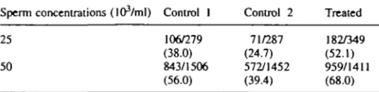
Laser mlcrodrilling of zona pellucida 106/279 (38.0) 843/1506 (56.0) 71/287 (24.7) 572/1452 (39.4) 182/349 (52.1) 959/1411 (68.0)
Table I. Percentage fertilization rates (zygotes/oocytes) observed in the
three groups of inseminated oocytes: control 1, intact oocyte-cumulus complexes; control 2, hyaluronidase decoronized oocytes; treated, hyaluronidase decoronized and zona pellucida laser-drilled oocytes Sperm concentrations (lOVml) Control I Control 2 Treated 25
50
Percentages, compared by contingency table analysis, were statistically different (P < 0.001) when the groups or the concentrations were compared two by two.
Results
Effect of the sperm concentration on fertilization, with or without zona drilling
The early steps of the fertilization process were followed in three groups (control 1, control 2, and laser-drilled) by observ-ing microscopically the inseminated oocytes after 1, 3 and 6 h. In control 1, the initial interaction between the gametes was difficult to observe due to the presence of the cumulus cells, but by the end of the incubation period the oocytes were totally free of corona cells and the fertilization could be assessed without additional mechanical decoronization. After hyaluronidase treatment (control 2, laser-drilled group), sper-matozoa were seen to bind to the zona pellucida in the minutes following insemination. In the zona pellucida microdrilled oocytes, four different situations were observed: (i) no sperma-tozoon in the perivitelline space (PVS), but spermatozoa attached to the zona pellucida; (ii) a single spermatozoon swimming around the oolemma in the PVS; (iii) a spermato-zoon bound to the oolemma close to the hole, and with the tail protruding out of the hole; and (iv) rarely, more than one spermatozoon in the PVS.
In control 1, the fertilization rate was increased from 38.0 to 56.0% when the sperm concentration was increased from 25 000 to 50 000/ml (Table I). Decoronization (control 2) induced a significant (P < 0.001) decrease in fertilization from 38.0 to 24.7% and from 56.0 to 39.4% for both sperm concentrations. When the zona pellucida of decoronized oocytes was drilled, the fertilization rates were dramatically increased (P < 0.001) from 24.7 to 52.1% and from 39.4 to 68.0%, to reach higher values than those observed for intact oocyte-cumulus complexes (control 1, Table I).
Polyploidy was not routinely observed but has been noted occasionally with both sperm concentrations (data not shown). Parthenogenetic activation rates induced by decoronization or drilling procedure remained <7% (Table IT). In contrast, calcimycin produced parthenogenetic activation in both control (59%) and zona pellucida drilled oocytes (55%), while DMSO alone had almost no effect (2%).
Impact of laser zona pellucida drilling on in-vivo development A summary of the embryo transfer procedure is presented in
Table HI. The number of pregnant foster mothers was not statistically different in the three groups. However, a signific-antly lower delivery rate was observed for embryos obtained
Table IL Number of oocytes undergoing parthenogenetic activation (%) per
number of oocytes treated, sampled from the three groups: control 1, intact oocyte-cumulus complexes; control 2, hyaluronidase decoronized oocytes; treated, hyaluronidase decoronized and zona pellucida laser-drilled oocytes
Parthenogenetic activation (%) Groups Without
spermatozoa Heat-inacuvatedspermatozoa (60°C) + 10 (iM calcimycin in DMSO + DMSO Control 1 Control 2 Treated 2/28 (7.1) 2/43 (4.7) 2/40 (5.0) 4/21 (19.0) 3/45 (6.7)1 0/4O (0.0)b 23/39 (59.0)' 27/49 (55.l)b 0/41 (0.0) 1/50 (2.0) DMSO = dimethylsulphoxide.
•^Values with superscripts are significantly different (/> « 0.0001).
Table ffl. Pregnancy and delivery rates (mice bom/embryo transferred) (%)
observed after transfer of embryos derived from the three groups of oocytes: control 1, intact oocyte-cumulus complexes; control 2, hyaluronidase decoronized oocytes; treated, hyaluronidase decoronized and zona pellucida laser-drilled oocytes. The sex ratios were established among the adult live F2 hybrids obtained
Groups Pregnancies/transfer Mice bom per Sex ratios (%) embryo transferred male/female
Control 1 4/5 (80.0) Control 2 5/8 (62.5) Treated 4/6 (66.7) 26/80 (32.5)" 20/128 (15.6)* 28/96 (29.2) 12/14 (46.2) 11/9 (55.0) 13/15 (46.4) •Values were significantly different (/>=£ 0.0001).
from decoronized oocytes (P =£ 0.0001). After laser drilling the rate was restored to that of the untreated oocyte-cumulus complex (control 1) group. The sex ratios were similar in all groups.
F2 generation follow-up
The sex ratios in the F2 generation were similar in the three
groups and close to 50% (Table IH). The reproductive ability of the F2 generation was shown to be similar to that observed
in a previous study with a mean number of 8.5, 7.5 and 8.0 pups obtained in the three groups of embryo treatment.
Anatomical and immunohistochemical evaluation
Upon macroscopic examination, the components of the digest-ive tract and the urinary and reproductdigest-ive systems revealed no morphological alteration in the animals derived from laser microdrilled embryos as compared to those in the control groups 1, 3 and 4. The microscopic examination of the cellular organization of the brain was performed on Nissl-stained (Toluidine Blue) coronal sections of the brain and spinal cord. The laminar structure of the cerebral and cerebellar cortices as well as the size and location of the main nuclei and tracts within the brainstem and spinal cord were investigated. The major cyto-architectonic features of the mouse brain, including the rodent-specific barrelfield of the somatosensory cortex, were preserved in all animals. The immunocytochemical and histochemical analysis of the organization of the major neuro-transmitter systems (glutamatergic, GABAergic, monoamin-ergic and cholinmonoamin-ergic) confirmed that no apparent modification
M.Gennond et aL
of the expression of the specific markers (neurotransmitters, receptors, synthesizing or degrading enzymes, related proteins) occurred in the experimental animals in comparison with the controls. The parameters measured were the dendritic morphology and distribution of the labelled neuronal and glial cells, as well as the distribution and density of the axonal projections. The investigation was primarily centred on the following structures: cerebral cortex, hippocampus, basal fore-brain, thalamus, hypothalamus, monoaminergic brainstem nuclei, cerebellum, and spinal cord.
Discussion
In the experiments described, dispersion of the egg cumulus by hyaluronidase was associated with significantly lower fertilization and implantation rates. It has been postulated that the cumulus-corona radiata complex participates in the selection and acrosome reaction of the spermatozoa, leading them to bind and penetrate the oocytes (Carrell et al, 1993). From this point of view, the reduced fertilization rates observed after cumulus dispersion can be explained. However, this reduction cannot be associated with a decreased ability of the decoronized oocytes to bind the spermatozoa, as no differences were noticed in this respect between control 2 and laser-drilled oocytes. It thus appears that specific modifications of the zona pellucida induced by the hyaluronidase treatment are responsible for the decrease in fertilization.
Such changes, which could be called hardening, have been observed in hamster eggs after hyaluronidase treatment (Drobnis et al, 1988), in mouse and rat eggs during in-vitro ageing (Zhang et al, 1991; Fukuda et al, 1992), after exposure to specific monoclonal antibodies (Garcia Framis et al, 1994) or freeze-thaw procedure (Carroll et al, 1990). Two parameters which can be used to quantify this hardening effect are a reduced fertilization rate and an increased resistance to a-chymotrypsin digestion (Eppig et al, 1992). Our results show that this effect is localized in the zona pellucida, as a fertilization rate comparable to that of untreated oocyte-cumulus complexes is obtained when the spermatozoa are allowed to reach the PVS through the microdrilled hole. With the sperm concentrations used in this study, which are 20-40 times lower than those known to be optimal in the mouse, this phenomenon occurs rapidly after insemination, without a time-dependent increase in the number of spermatozoa inside the PVS. An explanation for this could be that among the cohort of the spermatozoa initially attracted by the zona pellucida, one will reach the oocyte in front of the hole and be selected to freely swim through it. The other spermatozoa remain bound to the zona pellucida and continue to compete for a chance to fertilize. Another possible interpretation would be that the spermatozoa which were able to swim through the hole were acrosome-reacted, which would explain why these spermatozoa are able to fuse with the oolemma.
No attempts were made to increase the number of acrosome-reacted spermatozoa with calcimycin (Kobayashi et al, 1992) for two reasons. Firstly, we wanted to avoid ionophore-induced parthenogenetic activation of the oocytes and secondly, a high fertilization rate (68%) was obtained without it (Table I). 1046
After transferring the in-vitro fertilized oocytes to foster mothers, a significant decrease in the birth rate was observed in the cumulus-dispersed group (control 2). The pregnancy rate in the three groups of eggs was similar, which indicated that the transfer conditions were reproducible. Similar to the situation described above for fertilization, the drilling procedure restored a birth rate close to that of untreated control eggs. Thus, the hyaluronidase treatment of metaphase II oocytes seemed to induce a hardening effect of the zona pellucida leading to reduced fertilization and possibly to impaired or delayed mechanical hatching, with the consequence that the embryos fail to implant.
As the birth rates obtained after the transfer of decoronized and laser-drilled, in-vivo fertilized zygotes have been shown to be comparable in a previous study (Germond et al, 1995), one might suspect that the hyaluronidase treatment or some other undefined in-vitro conditions induces zona pellucida hardening in unfertilized metaphase II oocytes and not in zona pellucida-reacted fertilized zygotes. It is not clear to what extent the hyaluronidase treatment of oocytes prior to intracytoplasmic sperm injection (ICSI) also induces zona hardening in human eggs, as shown here in the mouse. The high implantation rates observed for ICSI-derived embryos (Palermo et al, 1993) contradicts this hypothesis, but one should bear in mind that the mechanical piercing of the zona pellucida during injection might assist hatching unintentionally.
The F2 generation follow-up of the mice bom after transfer
did not show noticeable differences among the three tested groups in terms of phenotypes and sex distribution, or of their ability to reproduce. In order to identify possibly more subtle changes induced by the procedure, mice from the control and experimental groups were submitted to anatomical and morphological examination. Numerous behavioural or motor deficits following alteration of brain development are associ-ated with structural modification of the cerebral or cerebellar cortices (Sotelo, 1980; Hoffarth et al, 1995). In addition, modifications in specific neurotransmitter systems have been shown to be related to neurological pathologies [e.g. GABA with epilepsy, catecholamines with motor and cognitive func-tions, acetylcholine with memory (McGeer et al, 1987)]. A trophic role for early expression of neurotransmitters in embryonic brains has also been recently demonstrated (Lauder, 1993; Zhou et al, 1995). The morphological and neurochemical features investigated were selected for their relevance in a number of brain development disorders. Although subtle modifications of the brain could have remained undetected by our investigations, no alteration was found using techniques suitable to identify known morphological and neurochemical changes in other animal models.
Lasers operating close to the UV region are likely to induce undesirable mutagenic side effects (Ashwood-Smith et al, 19%). With the 1.48 |im infrared laser wavelength used in this study, the energy delivered at the zona pellucida of mouse zygotes was shown to have no effect on their development
in vitro and in vivo (Germond et al, 1995). The drilling
mechanism is the result of a thermal effect induced at the focal point due to the absorption of the laser energy by water and/or zona pellucida macromolecules, leading to a confined
Laser microdriUing of zona pelluclda
thermolysis of the zona pellucida matrix (Rink et al, 1996). The risks of invoking direct or indirect genetic damage are likely to be low as metaphase II oocytes, extremely sensitive to external influences, retained their full ability to be fertilized, to cleave, to implant and to give rise to fertile progeny after zona pellucida microdrilling. Despite the fact that normal fertilization rates were obtained with very low sperm concentra-tions in the mouse, the method might not prove as successful in the human, especially when considering the high fertilization rates achieved by ICSI (Palermo et al, 1993). However, further developments of our laser zona pellucida drilling approach associated with cell trapping (Enginsu et al., 1995) and sperm fusion is still an unexplored alternative for mechanical insertion of the spermatozoon inside the cytoplasm, but use of these techniques for human assisted fertilization clearly demands more experimental data showing absence of damage at the nuclear and mitochondrial DNA levels.
In conclusion, the infrared 1.48 (xm laser wavelength allows the drilling of holes in the zona pellucida of mouse metaphase II oocytes without affecting negatively their ability to be fertilized and to develop in vitro and in vivo. The observed enhanced implantation rate after zona pellucida microdrilling in the mouse suggests that the described procedure may be safely applied to a genetically more stable human material, such as the cleaved embryo, in order to assist hatching when the zona pellucida is suspected to interfere negatively with the implantation process.
Acknowlegements
We arc grateful to Dr Kathleen Clark, for her critical comments. D.N. and K.R. are partially supported by grant 32-34137.92 of the Swiss National Science Foundation, Bem and by private grant from Serono S.A., Zug, Switzerland
References
Antinori, S., Versaci, C , Fuhrberg, P. et al. (1994) Seventeen ljve births after the use of an erbium-yttrium aluminium garnet laser in the treatment of male factor infertility. Hum. Repmd., 9, 1891-18%.
Ashwood Smith, MJ. and Edwards, R.G. (19%) DNA repair by oocytes.
MoL Hum. Repmd., 2, 46-51.
Blanchet, G.B., Russel, J.B., Fincher, C.R. and Portmann, M. (1992) Laser micromanipulation in the mouse embryo: a novel approach to zona drilling.
FeniL SteriL, 57, 1337-1341.
Carrell, D.T., Middleton, R.G., Peterson, CM. et aL (1993) Role of the cumulus in the selection of morphologically normal sperm and induction of the acrosome reaction during human in vitro fertilization. Arch. Androl., 31, 133-137.
Carroll, J., Depypere, H. and Matthews, C D . (1990) Freeze-thaw-induced changes of the zona pellucida explains decreased rates of fertilization in frozen-thawed mouse oocytes. /. Reprod. FeniL, 90, 547-553.
Cohen, J., Eisner, C , Kort, H. et al. (1990) Impairment of the hatching process following IVF in the human and improvement of implantation by assisting hatching using micromanipulation. Hum. Reprod., 5, 7-13. Depypere, H.T., McLaughlin, KJ., Seamark, R.F. et aL (1988) Comparison
of zona cutting and zona drilling as techniques for assisted fertilization in the mouse. J. Reprod. FeniL, 84, 205-211.
Drobnis, E.Z., Andrew, J.B. and Katz, D.F. (1988) Biophysical properties of the zona pellucida measured by capillary suction: is zona hardening a mechanical phenomenon? J. Exp. ZooL, 245, 206-219.
el Danasouri, I., Westphal, L.M., Neev, Y. et aL (1993) Zona opening with 308 nm XeCI excimer laser improves fertilization by spermatozoa from long-term vasectomized mice. Hum. Reprod., 8, 464-466.
Enginsu, M.E., SchOtze, K., Bellanca, S. et al. (1995) Micromanipulation of mouse gametes with laser microbeam and optical tweezers. Hum. Reprod., 10, 1761-1764.
Eppig, JJ., Wigglesworth, K. and O'Brian, M J . (1992) Comparison of embryonic developmental competence of mouse oocytes grown with and without serum. Mol. Reprod Dev., 32, 33-40.
Feichtinger, W., Strohmer, H., Fuhrberg, P. et aL (1992) Photoablation of oocyte zona pellucida by erbium-YAG laser for in-vitro fertilisation in severe male infertility. (Letter). Lancet, 339, 811.
Fukuda, A., Roudebusch, W. and Thatcher, S. (1992) Influences of in vitro oocyte aging on microfertilization in the mouse with reference to zona hardening. J. Assist. Reprod. Genet., 9, 378-383.
Garcia Framis, V., Calafell, J.M., Santalo, J. et aL (1994) Effect of anti-human sperm monoclonal antibodies on mouse in vitro fertilization. Immunol.
Invest., 23, 15-24.
Garrisi, GJ., Talansky, B.E., Grunfeld, L. et al. (1990) Clinical evaluation of three approaches to micromanipulation-assisted fertilization. Fertil. SteriL,
54, 671-677.
Germond, M., Nocera, D., Senn, A. et al. (1995) Microdissection of mouse and human zona pellucida using a 1.48 nm diode laser beam: efficacy and safety of the procedure. FertiL SteriL, 64, 604-611.
Gordon, J.W. (1988) Use of micromanipulation for increasing the efficiency of mammalian fertilization in vitro. Ann. N.Y. Acad. Set, 541, 601-613. Gordon, J.W. and Dapunt, U. (1993) Restoration of normal implantation rates
in mouse embryos with a hatching impairment by use of a new method of assisted hatching. FeniL SteriL, 59, 1302-1307.
Gordon, J.W. and Talansky, B.E. (1986) Assisted fertilization by zona drilling: a mouse model for correction of oligospermia- J. Exp. ZooL, 239, 347-354. Gordon, J.W., Grunfeld, L., Garrisi, G J . et aL (1988) Fertilization of human oocytes by sperm from infertile males after zona pellucida drilling. FeniL
SteriL, 50, 68-73.
Hoffarth, R.M., Johnson, J.G., Krushel, L.A. and van der Kooy, D. (1995) The mouse mutation reeler causes increased adhesion within a subpopulation of early postmitotic cortical neurons. J. NeuroscL, 15, 4838-4850. Hogan, B., Costantini, F. and Lacy, E. (1986) Recovery, culture and transfer
of embryos. In Manipulating the Mouse Embryo. Cold Spring Harbor Laboratory, Cold Spring Harbor, USA, pp. 91-152.
Kobayashi, K., Okuyama, M., Fujimoto, G. etaL (1992) Subzonal insemination with a single spermatozoon using manipulation assisted sperm adhesion onto the ooplasmic membrane in mouse ova. Mol. Reprod. Dev., 31,223—229. Lauder, J.M. (1993) Neurotransmitters as growth regulatory signals: role of
receptors and second messengers. Trends Neurosci., 16, 233—240. Laufer, N., Palanker, D., Shufaro, Y. et al. (1993) The efficacy and safety of
zona pellucida drilling by a 193-nm excimer laser. Fertil. Sleril., 59, 889-895.
Malter, H.E. and Cohen, J. (1989) Partial zona dissection of the human oocyte: a nontraumatic method using micromanipulation to assist zona pellucida penetration. FeniL SteriL, 51, 139-148.
McGeer, PL., Eccles, J. and McGeer, E.G. (1987) Molecular Neurobiology
of the Mammalian Brain. Plenum Press, New York.
Neev, J., Tadir, Y, Ho, P. et al. (1992) Microscope-delivered ultraviolet laser zona dissection: principles and practices. J. Assist. Reprod. Genet., 9, 513-523.
Neev, J., Gonzales, A., Lucciardi, F. et aL (1993) A contact-free microscope delivered laser ablation system for assisted hatching of the mouse embryo without the use of a micromanipulator. Hum. Reprod., 8, 939-944. Neev, J., Schiewe, M.C., Sung, V.W. et aL (1995) Assisted hatching in mouse
embryos using a noncontact Ho:YSGG laser system. J. Assist. Reprod.
Genet., 12, 288-293.
Ng, S.C., Liow, S.L., Schatze, K. et aL (1993) The use of ultra-violet microbeam laser for zona dissection in the mouse. J. Assist. Reprod. Genet., 10 (Suppl.), 204.
Obruca, A., Strohmer, H., Sakkas, D. et aL (1994) Use of lasers in assisted fertilization and hatching. Hum. Reprod., 9, 1723-1726.
Odawara, Y. and Lopata, A. (1989) A zona opening procedure for improving
in vitro fertilization at low sperm concentrations: a mouse model. Fenil. SteriL, 51, 699-704.
Palermo, G., Joris, H., Derde, M.P. et aL (1993) Sperm characteristics and outcome of human assisted fertilization by subzonal insemination and intracytoplasmic sperm injection. FeniL SteriL, 59, 826-835.
Rink, K., Delacretaz, G., Salathi, R.P. et aL (1994) 1.48 urn diode laser microdissection of the zona pellucida of mouse zygotes. Proceedings SPIE,
M.Germood et aL
Rink, K., Delacrctaz, G., Salathi, R.P. et aL (1996) Non-contact microdrilling of mouse zona pellucida with an objective-delivered 1.48 fim diode laser.
Lasers Surg. Med., 18, 52-62.
Schiewe, M.C., Neev, J., Hazeleger, N.L. et al. (1995) Developmental competence of mouse embryos following zona drilling using a non-contact holmiumryttrium scandian gallium gamet (Ho:YSGG) laser system. Hum.
Reprod., 10, 1821-1824.
SchOtze, K., Clement-Sengewald, A. and Ashkin, A. (1994) Zona drilling and sperm insertion with combined laser microbeam and optical tweezers.
Fertil. Steril., 61, 783-786.
SchOtze, K., Clement-Sengewald, A. and Berg, F.D. (1995) Non-contact laser micromanipulation of gametes and embryos. Part I: The UV-laser microbeam. Part II: The I.R.-optical tweezers trap. Hum. Reprod. Update, 1 (CD ROM), Item 1, Video.
Sotelo, C. (1980) Mutant mice and the formation of cerebellar circuitry.
Trends Neurosci., 3, 33—36.
Tago, H., Kimura, H. and Maeda, T. (1986) Visualization of detailed acetylcholinesterase fiber and neuron staining in rat brain by a sensitive histochemical procedure. J. Histochem. Cytochem., 34, 1431-1438. Tarin, J J . and Handyside, A.H. (1993) Embryo biopsy strategies for
preimplantation diagnosis. Fertil. Steril., 59, 943-952.
Whittingham, D.G. (1971) Culture of mouse ova. J. Reprod. Fertil, 14, 7-21. Zhang, X., Rutledge, J. and Armstrong, D.T. (1991) Studies on zona hardening in rat oocytes that are matured in vitro in a serum-free medium. MoL
Reprod. Dev., 28, 292-296.
Zhou, Q.Y., Quaife, C J . and Palmiter, R.D. (1995) Targeted disruption of the tyrosine hydroxylase gene reveals that catecholamines are required for mouse fetal development. Nature, 374, 640-643.
Received on October 26, 1995; accepted on February 23, 1996