HAL Id: tel-02954344
https://tel.archives-ouvertes.fr/tel-02954344
Submitted on 1 Oct 2020HAL is a multi-disciplinary open access archive for the deposit and dissemination of sci-entific research documents, whether they are pub-lished or not. The documents may come from teaching and research institutions in France or abroad, or from public or private research centers.
L’archive ouverte pluridisciplinaire HAL, est destinée au dépôt et à la diffusion de documents scientifiques de niveau recherche, publiés ou non, émanant des établissements d’enseignement et de recherche français ou étrangers, des laboratoires publics ou privés.
Adenosine 2A receptor signalling as a key mechanism of
stabilization of newly formed GABAergic synapses
Ferran Gomez Castro
To cite this version:
Ferran Gomez Castro. Adenosine 2A receptor signalling as a key mechanism of stabilization of newly formed GABAergic synapses. Neurons and Cognition [qbio.NC]. Université Pierre et Marie Curie -Paris VI, 2017. English. �NNT : 2017PA066431�. �tel-02954344�
Université Pierre et Marie Curie
Ecole doctorale n°158
- Cerveau Cognition Comportement -
Institut du Fer à Moulin, équipe « Plasticité des réseaux corticaux et épilepsie »
La signalisation du récepteur d’adénosine 2A comme
mécanisme clé de la stabilisation des synapses GABAergiques
nouvellement formées
Adenosine 2A receptor signalling as a key mechanism of
stabilization of newly formed GABAergic synapses
Par Ferran Gomez Castro
Thèse de doctorat de Neurosciences
Dirigée par Dr Sabine Lévi
Présentée et soutenue publiquement le 28 setembre 2017
Devant un jury composé de :
Dr Pascal Legendre
Président du jury
Dr Corette Wierenga
Rapporteur
Dr David Blum
Rapporteur
Dr Cécile Charrier
Examinateur
Dr Eric Boué-Grabot
Examinateur
ABSTRACT
In the adult brain, adenosine signaling facilitates or inhibits neurotransmitter vesicular release mainly through activation of type 2A or 1 adenosine receptors (A2AR or A1R), respectively. However, its role in
development remains to be elucidated. During my PhD, I addressed the role of A2AR-mediated
signalling in GABAergic synaptogenesis in the hippocampus.
We found (i) a larger activity-dependent release of ATP and adenosine during the period of synaptogenesis in the hippocampus, (ii) a peak of expression of the ecto-5’-nucleotidase, the rate-limiting enzyme for the formation of adenosine from extracellular ATP in synapses during this critical period, and (iii) a peak of peri/post-synaptic expression of A2AR concomitant with the period of
synaptogenesis. This developmental expression of the key molecules of the adenosine A2AR signalling
pathway correlated with a role of A2AR in the stabilization of nascent GABA synapses, a regulation
restricted to the period of synaptogenesis. Furthermore, suppressing A2AR with a shRNA approach in
isolated neurons led to a loss of synapses equivalent to that seen upon A2AR activity blockade, reporting
that the A2AR-mediated synapse stabilization is a cell autonomous process that requires A2AR activation
in the postsynaptic cell.
ATP/adenosine can be secreted by both glia and neurons; however, we found that activity-dependent release of neuronal adenosine is sufficient to stabilize newly formed GABA synapses in vitro. Using live cell imaging, we showed adenosine signalling stabilizes active synapses. We then characterized the molecular mechanism downstream postsynaptic A2AR. We report the contribution of adenylyl
cyclase/cyclic adenosine monophosphate/protein kinase A signalling cascade and we identified a key target, the postsynaptic scaffolding molecule gephyrin. We further showed the A2AR-mediated
stabilization of the presynaptic compartment most probably requires the trans-synaptic Slitrk3-PTPδ complex.
Since GABA exerts a similar function during development and GABA and adenosine are co-released at some synapses, I further investigated the interplay between these two pathways. My results support the hypothesis that GABA signalling converge onto the adenosine signalling pathway by potentiating calcium-sensitive adenylyl cyclases through the activation of calmodulin.
Altogether these results let us propose that, during a key developmental period, postsynaptic A2ARs
act as sensors of the activity of GABAergic presynaptic terminals to stabilize active nascent GABAergic synapses. In absence of activity and therefore secretion of adenosine/ATP, synapses will be eliminated.
RESUMÉ
Dans le cerveau adulte, la signalisation liée à l’adénosine facilite ou inhibe la libération vésiculaire de neurotransmetteurs suite à l’activation des récepteurs de l’adénosine de type 2A ou 1 (A2AR ou A1R),
respectivement. Cependant, son rôle dans le développement est mal connu. Au cours de ma thèse, j’ai étudié le rôle de la signalisation adénosine dans la synaptogenèse GABAergiques de l’hippocampe. Nous avons mis en évidence (i) une sécrétion activité-dépendante accrue d’adénosine et d’ATP pendant la période de synaptogenèse, (ii) un pic d’expression de l’enzyme limitant la formation de l’adénosine à partir de l’ATP extracellulaire, l’ecto-5’-nucleotidase, aux synapses pendant cette période critique, et (iii) un pic d’expression péri/post- synaptique du A2AR concomitant de la période de
synaptogenèse. Cette expression développementale des molécules clés de la signalisation adénosine dépendante du A2AR corrélait avec un rôle de ce récepteur dans la stabilisation des synapses
GABAergiques naissantes, une régulation restreinte à la période de synaptogenèse. De plus, la suppression de A2AR par une approche shRNA dans des neurones isolés conduisait à une perte de
synapses GABAergiques équivalente à celle observée après un blocage pharmacologique de l’activité du A2AR, signifiant que la stabilisation synaptique médiée par le A2AR est un processus « cellule
autonome » indépendant de l’activité du réseau neuronal et qu’elle requiert l’activation du A2AR dans
la cellule post-synaptique.
L’ATP et l’adénosine sont secrétés par la glie et les neurones ; cependant, nous avons montré in vitro que la libération neuronale activité-dépendante suffit à stabiliser les synapses GABAergiques naissantes. En utilisant la vidéomicroscopie sur cellules vivantes, nous avons montré que la signalisation adénosine stabilise les synapses actives. Puis, nous avons caractérisé le mécanisme moléculaire sous-jacent. Nous rapportons la contribution de la cascade adénylate cyclase/adénosine monophosphate cyclique/protéine kinase A et nous avons identifié une ciblé clé, la géphrine, la molécule d’ancrage postsynaptique des récepteurs GABAA. Enfin, nous avons mis en évidence que la
stabilisation de l’élément présynaptique requiert probablement le complexe trans-synaptique Slitrk3-PTPδ.
Puisque le GABA exerce une fonction similaire au cours du développement et que le GABA et l’adénosine sont co-libérés à certaines synapses, j’ai étudié l’interaction entre ces deux voies de signalisation. Mes résultats favorisent l’hypothèse que la signalisation GABA, en activant la calcium-calmoduline, converge vers la signalisation adénosine en potentiant les adenylates cyclases sensibles au calcium.
Mon travail m’a permis de proposer que, au cours d’une période clé du développement, les A2ARs
postsynaptiques agissent comme des senseurs de l’activité des terminaisons présynaptiques GABAergiques pour stabiliser les synapses actives. En absence d’activité et donc de libération d’adénosine/ATP, les synapses seraient éliminées.
ACKNOWLEDGEMENTS
My first acknowledgments go to the jury who has accepted to review my PhD manuscript and discuss my results during the defence. I hence warmly thank Dr Pascal Legendre, Dr Corette Wierenga, Dr David Blum, Dr Cécile Charrier and Dr Eric Boué-Grabot. A special thanks thereby to Dr Cécile Charrier who accepted also to follow my PhD project during the annual PhD committes.
Mes plus grands remerciements sont pour toi Sabine. Pour le support continu tout au long de ma thèse, pour ta patience et ta motivation. Dans les moments de découragement de ma thèse je me suis toujours senti soutenu et ta vision positive m’a beaucoup aidée. Merci pour ton investissement et toutes les heures passées ensemble : soit en réunion, soit les plus excitantes au microscope et d’autres moins au Metamorph ! J’espère avoir retourné l’effort et l’implication que j’ai toujours senti de ta part dans ce projet. Merci pour toutes les connaissances que tu m’as appris, des présentations orales à la rigueur scientifique en passant pour des expressions en français.
Un grand merci aussi à Jean Christophe pour apporter sa vision de physiologiste sur mon travail. Merci encore pour chacune des leçons d’électrophysiologie dans les réunions d’équipe et pour toujours essayer de nous ouvrir vers vision plus globale de la science.
À tous les membres de l’équipe Marie, Eric, Marion, Jessica, Sana, Clémence, Yo and Marianne. Merci à tous pour les pauses-détente mais aussi pour la passion que vous éprouvez tous dans vos projets tout comme dans la vie. J’espère vous avoir retourné un peu de cette passion et vous avoir appris un peu d’espagnol! Je ne peux pas oublier non plus le temps passé avec les gens qui sont passés par le 1er
étage et qui ont aussi marqué ma thèse : Sereina, Manuel, Frank, Mágico Salvatore, Kristina et Anna. Merci aussi pour toutes les autres personnes au sein de l’institut qui ont contribué à la bonne atmosphère de l’IFM. Merci à tous qui ont passé même si c’était un peu de temps avec moi en analysant des images, surtout Richard et Emily. Muchas gracias también a Jimmy, Ana y María por tanto apoyo. A Mariano por las asistencias en la ciencia (¡y en el fútbol!).
Merci encore à mes amis à Paris: Mariangela, Stefania, Pietro, Federica, Giulia, Rodrigo, Pedro, Ángel, Ana Ruiz, Dani (gracias muy fuerte). Et courage pour vos thèses aussi! A la gent de casa, per fer que quan torni tot segueixi igual malgrat la distància.
1
TABLE OF CONTENTS
Table of contents ... 1 Table of figures ... 4 Abbreviations ... 5 INTRODUCTION ... 81. The purinergic system ... 8
1.1 Overview on purinergic system ... 8
1.2 Purinergic signalling ... 9
1.2.1 P2 receptors ... 9
1.2.2 Adenosine receptors ... 10
1.2.3 Source and metabolism of ATP and adenosine ... 13
1.3 Multiple functions of adenosine signalling ... 16
1.3.1 Physiology: at the cellular level ... 16
1.3.1.1 Adenosine in neurons ... 16
1.3.1.2 Adenosine in glia ... 18
1.3.2 Physiology: at the behavioural level ... 18
1.3.3 Adenosine in pathology ... 22
1.4 Impacts of caffeine intake during gestation and lactation in the offspring ... 24
1.5 Spatiotemporal expression of A2AR ... 26
1.5.1 Cellular and subcellular expression of A2AR ... 28
1.5.2 Ontogenic development of A2AR ... 29
2. Synapse formation ... 30
2.1 The inhibitory synapse: an overview ... 30
2.2 Introduction to synapse formation ... 35
2.3 Timing of hippocampal synaptogenesis ... 37
2.4 Synapse formation is a dynamic process ... 39
2.5 Building a synapse ... 42
2.5.1 Generation of specificity ... 42
2.5.1.1 Positive cues ... 42
2.5.2.2 Inhibitory cues ... 46
2.5.2.3 Other factors regulating neuronal specificity ... 46
2.5.2 Recruitment of synaptic elements ... 46
2
2.5.2.2 Anterograde organizers ... 49
2.5.2.3 Retrograde organizers ... 50
2.5.2.4 Glia-secreted organizers ... 50
2.5.3 Activity-dependent regulation of synaptic contacts ... 51
2.5.4 Synapse elimination ... 53
2.5.5 Transcription factors regulating synapse formation ... 55
2.6 Current knowledge of mechanism of synaptogenesis at the inhibitory synapse ... 57
2.6.1 Trans-synaptic adhesion proteins ... 57
2.6.2 Secreted factors ... 60
2.6.3 Intracellular submembranous scaffold ... 62
2.6.4 Synapse specificity ... 64
2.7 Defects in synapse formation: implications in pathology ... 66
2.7.1 Epilepsy ... 66
2.7.2 Psychiatric disorders ... 67
MATERIALS AND METHODS ... 70
Neuronal culture ... 70
DNA constructs ... 70
Neuronal transfection ... 70
Pharmacology ... 71
Immunocytochemistry ... 71
Fluorescence image acquisition and analysis ... 72
Immunohistochemistry ... 72
In vivo pentylenetatrazole injection ... 73
Calcium Imaging ... 73
cAMP Imaging ... 74
Statistics... 75
RESULTS ... 78
1. Adenosine A2A receptor: a key sensor of synaptic activity to stabilize/eliminate GABAergic synapses during development ... 78
Abstract ... 81
Introduction ... 82
Results ... 82
References ... 87
3
Materials and methods ... 95
Supplementary material ... 106
2. Adenosine A2A receptor and GABAA receptor signaling pathways converge to stabilize nascent GABAergic synapses... 116
Abstract ... 118
Introduction ... 118
Material and methods ... 119
Results ... 123
Discussion ... 133
References ... 136
DISCUSSION ... 142
1. Spatiotemporal expression and synapse stabilization function of A2AR ... 143
2. A2AR-mediated molecular pathway leading to the stabilization of synapses ... 148
3. Signalling at the developing inhibitory synapse ... 153
4. Pathological implications for the A2AR-mediated stabilisation of synapse ... 156
General conclusions ... 158
ADDITIONAL PUBLICATIONS ... 162
1. GABAA receptor dependent synaptic inhibition rapidly tunes KCC2 activity via the Cl--sensitive WNK1 kinase ... 162
4
TABLE OF FIGURES
Figure 1: Notable purines ... 8
Figure 2: Adenosine receptors can modulate adenylate cyclase activity ... 10
Figure 3: Extracellular metabolism of ATP and adenosine ... 15
Figure 4: Different molecular targets of caffeine ... 19
Figure 5: Roles of adenosine in the brain in physiology and pathology ... 20
Figure 6: A2AR expression in the postnatal hippocampus. ... 27
Figure 7: Ontogenic expression of A2AR ... 29
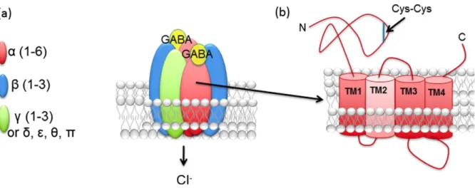
Figure 8: Structure of GABAAR ... 31
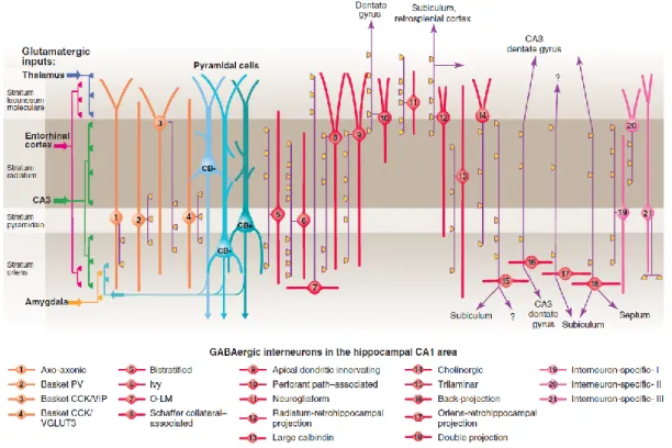
Figure 9: Interneurons in CA1 hippocampus... 36
Figure 10: Development of GABAergic innervation in hippocampus ... 38
Figure 11: Synaptic boutons: persistent and non-persistent ... 40
Figure 12: Inhibitory synapse formation model ... 41
Figure 13: Subregion-specific expression of the LRR proteins ... 45
Figure 14: Different nature of synapse organizers ... 47
Figure 15: Examples of synaptogenic activity by neuroligins and neurexin ... 48
Figure 16: Schematic representation of the trans-synaptic proteins of the GABAergic synapse and their intracellular interactors ... 60
Figure 17: Intracellular scaffolds of the inhibitory synapse ... 64
Figure 18: Inhibitory synapse organizers act on different synapses... 64
Figure 19: Acute A2AR blockade does not affect VGLUT1 and PSD95 clustering ... 145
Figure 20: A2AR is expressed is expressed in the hippocampus, cortex and cerebellum at P16... 146
Figure 21: Acute A1R blockade does not affect VGAT nor GABAARγ2 clustering ... 147
Figure 22: P2 receptors can modulate PSD95 clustering in young neurons ... 148
5
ABBREVIATIONS
A1R Adenosine 1 receptor A2AR Adenosine 2A receptor A2BR Adenosine 2B receptor A3R Adenosine 3 receptor ACh AcetylcholineaCSF Artificial cerebrospinal fluid
ADP Adenosine diphosphate
AIS Axon initial segment
AKAP A-kinase anchoring proteins AKT/PKB Protein kinase B
AMPA α-amino-3-hydroxy-5-methyl-4-isoxazolepropionic acid araC cytosine -D-arabinofuranoside
ASD Autism spectrum disorders
ATP Adenosine triphosphate
BDNF Brain derived neurotrophic factor C. elegans Caenorhabditis elegans
CA1/2/3 Cornu Ammonis 1/2/3
CaM Calmodulin
CAM Cell adhesion molecule
cAMP Cyclic adenosine monophosphate
CB Calbindin
CB1 Cannabinoid receptor type 1
CCK Cholecystokinin
CNS Central nervous system
CNTN Contactin
D2R Dopamine D2 receptors
DG Dentate gyrus
DGC Dystroglican complex
DIV Days in vitro
ENT Equilibrative transporter
ERK Extracellular signal-regulated kinases
GABA γ-aminobutyric acid
GAD Glutamic acid decarboxylase GARLH GABAAR regulatory Lhfpl
GFP Green fluorescent protein
GL Granular layer
GPCR G-protein coupled receptor GSK3β Glycogen synthase kinase 3β
IGHMBP2 Immunoglobulin mu binding protein 2 InSyn1-2 Inhibitory Synapse Protein 1 and 2
IPT Infrapyramidal tract
6 Lhfpl Lipoma HMGIC fusion partner- like
LTP Long term potentiation
MAGI-2 Membrane associated guanylate kinase inverted-2 MAPK Microtubule-associated protein kinases
MDGA MAM domain-containing GPI anchor proteins mEPSC Miniature excitatory postsynaptic current mGluR5 Metabotropic glutamate receptor 5 mIPSC Miniature inhibitory postsynaptic current
ML Molecular layer
NA Noradrenaline
NARP Neuronal activity regulated pentraxin
NMDA N-methyl-D-aspartate
NMJ Neuromuscular junction
NP1 Neuronal pentraxin 1
Npas4 Neuronal PAS domain protein 4
NTD N-terminal domain
NTRK1 Neurotrophic tyrosine kinase receptor type 1
Nxph Neurexophilins
O-LM Oriens lacunosum moleculare PCL Purkinje cell layer
PI-3K Phosphoinositide 3-kinase
PKA Protein kinase A
PKC Protein kinase C
PKG Protein kinase G
PLC Phospholipase C
PNS Peripheral nervous system
PPARγ Peroxisome proliferator-activated receptor PSD95 Postsynaptic density protein 95
PTZ Pentylenetatrazole
PV Parvalbumin
Px Postnatal day
RGC Retinal ganglion cell
Src Sarcoma kinases
SRGAP2 Slit-Robo Rho-GTPase activating protein 2 S-SCAM Synaptic scaffolding molecule
STORM Stochastic Optical Reconstruction Microscopy trkB Tyrosine receptor kinase B
TTX Tetrodotoxin
UDP Uridine diphosphate
UTP Uridine triphosphate
VGAT Vesicular GABAergic transporter VGLUT Vesicular glutamate transporter VIP Vasoactive intestinal polypeptide VNUT Vesicular Nucleotide Transporter
7
INTRODUCTION
8
INTRODUCTION
1. The purinergic system
1.1 Overview on purinergic system
The purinergic system is composed of several types of ligands and its corresponding receptors, as well as enzymes and transporters which are responsible for the production and/or reuptake of purines that mediate intra/intercellular physiological events.
Chemically, a purine is a heterocyclic aromatic organic compound that consists of a pyrimidine ring fused to an imidazole ring (Figure 1). Purines are fundamental molecules of life as they are key components of nucleic acids (RNA and DNA) that constitute the basic forms of life (Burnstock and Verkhratsky, 2012). They include the nucleobases adenine and guanine, with other notable purines being adenosine triphosphate (ATP), adenosine, cyclic adenosine monophosphate (cAMP), and caffeine (Figure 1). Through evolution, a multitude of roles have emerged for purines ranging from energy metabolism, transduction of intracellular signals, fatty acid metabolism, and epigenetic control. Although purines can be found all throughout an organism, the scope of this thesis will cover purinergic signalling in the brain.
Figure 1: Notable purines
9
1.2 Purinergic signalling
Purinergic signalling is a type of extracellular/intracellular signalling mediated by purine nucleotides and nucleosides such as adenosine and ATP. It involves the activation of purinergic receptors thereby regulating cellular functions. The existence of this signalling implies the existence of ligands and receptors. The family of purines is diverse and as such was divided into two primary groups in 1978 by Geoffrey Burnstock (Burnstock, 1978). Based on several criteria he established two types of receptors: P1 and P2 receptors. The P1 receptors, later renamed into adenosine receptors, are responsive to adenosine and are selectively and competitively antagonised by methylxanthines. They modulate the adenylate cyclase activity, resulting in changes in intracellular levels of cAMP (van Calker et al., 1979; Jacobson and Gao, 2006) and downstream activation/inhibition of protein kinase A (PKA). The P2 receptors are responsive to ATP and ADP. They are not antagonised by methylxanthines (Abbracchio and Burnstock, 1994).
1.2.1 P2 receptors
Even before the subdivision of adenosine and P2 receptors, Burnstock proposed ATP as a possible neurotransmitter (Burnstock, 1972) and later it was proposed as a ligand for the P2 receptors. However, it was not until 1994, after the accumulation of many studies that Burnstock and Abbracchio (Abbracchio and Burnstock, 1994) proposed a classification of the P2 receptors based on the structure and function of the receptor: P2X family consisting of ligand-gated cation channels and a P2Y family consisting of G protein-coupled receptors.
P2X receptors are ionotropic receptors. These ligand-gated channels are the only ones in the mammals/human body to be trimeric, the others being tetrameric (glutamate receptors) or pentameric (GABA, glycine, serotonin receptors) (Burnstock and Verkhratsky, 2012). Upon binding of ATP, they open and they are permeable to Na+, K+ and Ca2+. Until now, seven receptor subtypes have been characterized: P2X1 to P2X7 (North, 2002). Every P2 receptor has its own regional and cell-type pattern of expression (Burnstock and Verkhratsky, 2012). All the P2X receptors are present in the hippocampus.
P2Y receptors are metabotropic receptors. They can be sub-classified depending on the G-protein they bind. P2Y1, P2Y2, P2Y4, P2Y6, P2Y11 bind to Gq to activate phospholipase C (PLC) while P2Y11, P2Y12,
P2Y13, P2Y14 bind to Gi to inhibit adenylate cyclase (Abbracchio et al., 2006). P2Y receptors can be
10 diphosphate (UDP), UDP glucose and other nucleotide sugars. Some P2Y receptors show higher affinity for other purines than for ATP itself (Abbracchio et al., 2006). As for P2X receptors, the expression of P2Y is region and cell-type dependent. In the adult hippocampus we can find P2Y1, P2Y2, P2Y4, P2Y6 and P2Y12 receptors (Burnstock and Verkhratsky, 2012).
In total, there are fifteen P2 receptors displaying different pharmacological properties, region and cell specific expression, making the ATP signalling a complex system. ATP signalling has multiple roles in the brain i) acting as neurotransmitter ii) as a paracrine modulator of glial cells iii) as synaptic neuromodulator with an impact in synaptic plasticity iv) controlling inflammation v) regulating oligodendrocyte maturation (Rodrigues et al., 2015; Cunha, 2016).
1.2.2 Adenosine receptors
The first description of adenosine receptors function was performed by van Calker and colleagues when they showed that adenosine could inhibit the accumulation of cAMP (van Calker et al., 1978). They later discovered that other adenosine receptors can increase the intracellular cAMP level (van Calker et al., 1979). This finding, in addition to biochemical, pharmacological and molecular cloning studies showed that there are in fact four subtypes of adenosine receptors: A1, A2A, A2B and A3 receptors (Maenhaut et al., 1990; Libert et al., 1991; Stehle et al., 1992; Zhou et al., 1992). All four subtypes were found to be members of the superfamily of G-protein-coupled receptors (GPCRs). The adenosine receptors share a high degree of homology resulting in all of the receptor subtypes forming a seven transmembrane helices (as all other GPCRs), with the N-terminus facing the extracellular space and the C-terminus in the intracellular milieu (Jacobson, 2009). Classically, A1 (A1R) and A3 (A3R) receptors activate Gi/0 and Gi/q respectively to inhibit adenylate cyclase (AC), while A2A (A2AR) and A2B (A2BR) receptors activate adenylate cyclase via Gs/Golf –A2A receptors- or Gs/Gq –A2B receptors
(Figure 2, Jacobson and Gao, 2006).
Figure 2: Adenosine receptors can modulate adenylate cyclase activity
A1R activate Gi/0 while A3R activate Gi/q to inhibit adenylate cyclase. A2AR and A2BR activate Gs tofurther activate adenylate
11 Regulation of adenylate cyclases
AC activation leads to the production of cAMP signalling modulates a set of developmental processes such as cell differentiation, axon outgrowth and response to guidance molecules (Nicol and Gaspar, 2014). The unique combination of regulations, tissue-specific regulation and subcellular localization of ACs concedes them a key role in cAMP signalling (Cooper and Tabbasum, 2014). To date, ten isoforms of AC have been described. Molecular cloning studies showed that ACs have a highly conserved catalytic domain, however the non-catalytic displays more differences to permit different regulatory features (Willoughby and Cooper, 2007). As observed in Table 1, the regulators of AC are multiple allowing them to integrate the activity of many cellular signalling pathways (Cooper and Tabbasum, 2014).
Regulator AC isoform effect
G-protein Gsα All Stimulation
Giα AC1, AC3, AC5, AC6,AC8, AC9 Inhibition
Gβγ AC1, AC8 Inhibition
Forskolin All Stimulation
Ca2+ AC1, AC8 (via calmodulin) Stimulation
AC9, AC5, AC6 Inhibition
Kinase CaMKII AC3 Inhibition
CaMKIV AC1 Inhibition
PKA AC5, AC6, AC8 Inhibition
PKC AC2, AC4, AC5, AC6, AC7 Stimulation
AC6, AC9 Inhibition
RTK AC1, AC5, AC6 Stimulation
RGS AC3, AC5, AC6 Inhibition
Table 1: Regulation of the adenylate cyclases
Reproduced from (Cooper and Tabbasum, 2014)
Of special interest for my studies are the Ca2+-stimulated ACs. AC1 and AC8 are both stimulated
through calmodulin upon intracellular Ca2+ rise. In hippocampal neurons, AC1 and/or AC8 are
stimulated by Ca2+ entry mediated by NMDAR activation (Chetkovich et al., 1991) or through Ca2+
L-type channels (Ferguson and Storm, 2004). Interestingly, GABAAR activation in young neurons leads to
membrane depolarization inducing Ca2+ influx via the activation of L-type Ca2+ channels (Perrot-Sinal
et al., 2003).
The cAMP responses are different in the different cellular compartments, showing thus that ACs and/or their regulators are targeted to different cell microdomains (Nicol et al., 2011). All Ca2+ sensitive
12 cAMP) are present (Willoughby and Cooper, 2007). Interestingly, A2AR is also in lipid rafts
(Charalambous et al., 2008).
Both AC1 and AC8 are expressed in the brain. The expression of AC1 is higher during embryonic and early postnatal life but decreases in mature neurons (Nicol et al., 2005). Conversely, AC8 expression is low in development but increases in mature neurons. It is mostly expressed in CA1 region (Nicol et al., 2005). This explains why AC1 has been involved in the developmental processes such as axon guidance (Nicol and Gaspar, 2014) and AC8 in modulation of LTP in adult neurons (Wang et al., 2003). However, these ACs have never linked to synaptogenesis.
Multiple pathways activated by adenosine receptors
The concept of adenosine receptors modulating adenylate cyclase only has proved to be oversimplified as later studies have shown that adenosine receptors are capable of activating additional signalling pathways. Apart from the inhibition of adenylyl cyclase, A1R can increase the activity of phospholipase
C (PLC) (Rogel et al., 2005). In cardiac muscle and neurons, A1R can activate K+ channels and inhibit Q,
P- and N-type Ca2+ channels (Jacobson and Gao, 2006).
As mentioned earlier, A2AR activation leads to the activation of adenylate cyclase but can also modulate
other pathways. The complete list of pathways recruited by A2AR can be found in Table 2.
Pathway Reference
Adenylate cyclase (van Calker et al., 1979)
PPARγ (peroxisome proliferator-activated receptor)
(Nayeem et al., 2013) MAPK (microtubule-associated protein kinases) (Canas et al., 2009; Li et al., 2015)
JNK (c-Jun N-terminal kinases) (Genovese et al., 2009) PI-3K (phosphoinositide 3-kinase) (Ahmad et al., 2013)
Protein phosphatases (Murphy et al., 2003)
protein kinase C (PKC) (De Ponti et al., 2007)
Src (acronym for sarcoma) kinases (Che et al., 2007) N- and P- type calcium channels (Gubitz et al., 1996)
Table 2: Summary list of all the pathways activated by A2AR
As A2AR has been linked to several pathways has prompted studies to unravel the reason of this
pleiotropy. First, A2AR can form homo/heterodimers with numerous GPCRs and enzymes (Canals et al., 2004) that can modulate the signalling (Table 3). This is achieved through A2AR long C-terminal tail
13 receptors (Klinger et al., 2002). Second, the lipid microenvironment of the cell is also important as A2AR is enriched in lipid rafts. Its delocalization from these microenvironments by cholesterol depletion selectively hampers their ability to recruit AC rather than MAPK (Charalambous et al., 2008). Third, A2AR can activate different transducing pathways in different brain regions: while A2AR activates AC in
hippocampus; in basal ganglia it mainly activates MAPK pathway. This region-dependent modulation could be caused by the presence/absence of interactors of A2AR in different regions. There are six
G-protein interacting G-proteins that interact with A2AR; actinin, calmodulin, Necab2, translin- associated
protein X, ARNO/cytohesin 2, ubiquitin-specific protease-4, that can bias the signalling in a cell-specific manner (reviewed in Keuerleber et al., 2011).
Molecule Type of molecule Reference
Dopamine D2 receptors (D2R) GPCR (Canals et al., 2003)
A1R GPCR (Ciruela et al., 2006)
A2BR GPCR (Moriyama and Sitkovsky, 2010)
Cannabinoid receptor type 1 (CB1) GPCR (Carriba et al., 2007) Glutamate metabotropic group 5
receptors (mGluR5)
GPCR (Ferre et al., 2002)
G protein-coupled receptor 37 (GPR37)
GPCR (Lopes et al., 2015)
Adenosine deaminase Enzyme (Gracia et al., 2011)
Ecto-5’-nucleotidase Enzyme (Augusto et al., 2013)
Table 3: Proteins forming heterodimers with A2AR
The other two adenosine receptors are also coupled to other pathways than adenylyl cyclase. A2BR is
positively coupled to PLC, and the same is observed for A3R. The latter has also been shown to stimulate release from intracellular calcium stores (Jacobson and Gao, 2006)
1.2.3 Source and metabolism of ATP and adenosine
ATP and adenosine the main ligands for P2 and adenosine receptors respectively, are found in all the cells of the human body. Adenosine comes mostly from the catabolism of ATP (discussed later). The major source of ATP is the mitochondria that keeps the cytosolic ATP in a low millimollar range (3-10mM), while in the extracellular space ATP is in the range of low nanomolar (Burnstock and Verkhratsky, 2012).
14 Release of ATP
ATP release involves two major pathways, regulated vesicular exocytosis and diffusion through various plasmalemmal channels. Both mechanisms can work in isolation or in concert, depending on the physiological context.
ATP can be loaded in synaptic vesicles through Vesicular Nucleotide Transporter (VNUT) (Sawada et al., 2008). Alternatively, ATP may enter vesicles by passive diffusion through non-specific anion channels (Lange and Brandt, 1993). ATP can be co-stored and co-released with glutamate, noradrenaline (NA), γ-aminobutyric acid (GABA), or acetylcholine (ACh) (Pankratov et al., 2003, 2006; Cunha, 2016); however, the presence of ATP-only sets of vesicles in the medial habenula has been suggested (Pankratov et al., 2006). Regarding cell types, ATP can be stored in synaptic vesicles in neurons (Pankratov et al., 2003, 2006) and in astrocytes (Parpura and Zorec, 2010). Exocytic ATP release can occur through lysosomes in astrocytes (Oya et al., 2013) and microglia (Dou et al., 2012), as observed in in vitro preparations. Neuronal lysosomal ATP release has also been observed but only in the peripheral nervous system (Jung et al., 2013).
ATP can also diffuse into the extracellular space via different plasmalemmal channels such as i) anion channels ii) P2X7 receptors (seen in spinal cord astrocytes) iii) connexin-43 hemichannels iv) pannexin channels (Stout et al., 2002; Suadicani et al., 2006; Iglesias et al., 2009; Burnstock and Verkhratsky, 2012). This is an exhaustive list of all the currently described ways of ATP secretion, excluding cell damage that can induce a discontinuity of the plasma membrane leading to release of intracellular content.
Some of these ATP secretion pathways have been observed in pathological scenarios, it is known that physiological ATP release occurs through tonic release and upon electrical stimulation in neurons (Cunha et al., 1996; Lazarowski et al., 2000). Different molecules such as thrombin, lysophosphatidic acid and UTP induce ATP release from astrocytes (Blum et al., 2008; Parpura and Zorec, 2010). ATP can also be secreted in response to mechanical stress from almost all cell types (Darby et al., 2003).
Metabolism of ATP
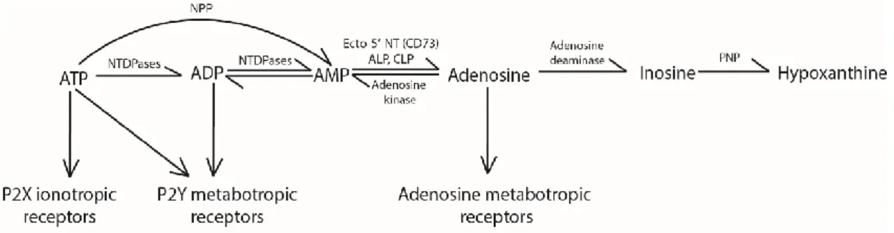
Once in the extracellular space, the ATP concentration is regulated by enzymes called ectonucleotidases that metabolize ATP into other molecules belonging to the purinergic system (Figure 3). There are three families of enzymes that metabolize ATP: nucleoside triphosphate diphosphohydrolases (NTPDases) (Robson et al., 2006); nucleotide pyrophosphatases/phosphodiesterases (NPPs) (Stefan et al., 2006); alkaline and acid phosphatases
15 (ALP and ACP, respectively) (Millán, 2006; Burnstock and Verkhratsky, 2012). At last, the activity of the ecto-5’-nucleotidase (CD73) will hydrolyse AMP into adenosine (Colgan et al., 2006; Kulesskaya et al., 2013).
Figure 3: Extracellular metabolism of ATP and adenosine
ATP can be transformed into adenosine by different pathways, and adenosine can be degraded by adenosine deaminase to terminate its actions on the adenosine receptors. From (Jacboson, 2009)
Source of adenosine
Adenosine can be found in the extracellular milieu after the transformation of ATP by ectonucleotidases, being the main source of extracellular adenosine in the brain (Kulesskaya et al., 2013). Otherwise, the presence of equilibrative transporters (ENTs) permeable to adenosine has also been observed in all the CNS cell types (Parkinson et al., 2011; Wall and Dale, 2013). In physiological conditions, the adenosine concentration gradient is always inward because cytosolic adenosine concentrations are very low (Wall and Dale, 2007). Some authors (Wall and Dale, 2007; Klyuch et al., 2012) suggest a vesicular release of adenosine. In these works, in the cerebellar parallel fibres to Purkinje cell synapses they detected an increase in extracellular adenosine levels by adenosine biosensors, this increase was independent of ENTs and blocked when preventing action potential dependent vesicular release, thus suggesting that there might exist subpopulations of neurons that release adenosine directly (Wall and Dale, 2008). In other cell types and in pathological scenarios, adenosine can be formed from S-Adenosyl-L-homocysteine (SAH), but the contribution of this pathway in the brain is negligible (Latini et al., 1996).
Metabolism of adenosine
The actions of adenosine are terminated upon the action of another ecto-nucleotidase, adenosine deaminase (ADA) that catalyses conversion of adenosine into inosine (Nofech-Mozes et al., 2007). Inosine will be further metabolised to hypoxanthine by yet another ecto-nucleotidase, the purine nucleoside phosphorylase (PNP) (Figure 3, Burnstock and Verkhratsky, 2012).
16
1.3 Multiple functions of adenosine signalling
The different pattern of expression of receptors, ligands and metabolic enzymes related to the purinergic system depicts a complex picture, conferring a wide range of roles in physiology and pathology in different tissues. The P2 receptor-mediated signalling system, expressed all throughout the body is really complex, but more is known regarding the adenosine functions, because the pharmacology of adenosine receptors is more developed. If we focus our attention on the adenosine receptors many efforts have been done to treat pathologies using this system, these were recently reviewed (Jacobson and Gao, 2006; Burnstock et al., 2011). Therapies blocking adenosine receptors, or increasing/decreasing the adenosinergic tone have been tried in cardiovascular disease (arrhythmia, ischemia), renal disorders (ischemia-induced kidney injury and fluid retention disorders), pulmonary disorders (asthma), inflammatory disorders, nervous systems disorders, endocrine disorders and cancer amongst others (Jacobson and Gao, 2006)
I am only going to review the roles regarding adenosine in the brain in this section. Adenosine is present all throughout the brain so its effects are linked to the expression pattern of the receptors. Of the four adenosine receptors it is mostly A1R and A2AR that are responsible for the effects of adenosine
in the brain (Fredholm et al., 2005). Whereas A1R are the most abundant and widespread (they are the
second most abundant metabotropic receptor in the brain), A2AR are more abundant in the basal
ganglia as compared to the rest of the brain (Fredholm et al., 2005). In fact, both A1R and A2AR are
mostly located in synapses and have been found in glutamatergic synapses, GABAergic, cholinergic, dopaminergic, serotoninergic or noradrenergic synapses (Cunha, 2016).
1.3.1 Physiology: at the cellular level
Adenosine is a well-known synaptic modulator but extracellular adenosine can also play multiple physiological roles in the brain parenchyma. Their function will be determined by the subtype of receptor expressed, the cell type and brain region, amongst other factors. To classify the effects of adenosine, I will distinguish between effects on neurons and on glial cells.
1.3.1.1 Adenosine in neurons
The modulatory effect of adenosine in neurons is the best described role for adenosine in the brain. Adenosine can modulate synaptic transmission directly via presynaptic A1R, presynaptic A2AR or
17 A1R -mediated tonic inhibition
Adenosine is considered an inhibitory neuromodulator responsible for a feedback decrease in the activity of excitatory synapses in basal transmission (Cunha, 2016). The increased activity of a glutamatergic synapse leads to an increase in energy consumption, forcing a greater use of ATP, which subsequently leads to greater formation intracellular adenosine that will be released via ENTs. In hippocampal excitatory synapse at steady state adenosine acts on presynaptic A1R to decrease calcium influx through presynaptic voltage-dependent Ca2+ channels (Gundlfinger et al., 2007) therefore
controlling vesicular glutamate release (Barrie and Nicholls, 1993). Postsynaptic activation of A1R decreases the activation of ionotropic glutamate receptors (Klishin et al., 1995) and hyperpolarizes principal neurons through a control of K+ channels (Kim and Johnston, 2015). Interestingly, A
1 R-mediated inhibition becomes less efficient the more intense the recruitment of neuronal circuits is, as we observe a modest effect of A1R in long term plasticity (LTP) paradigms (Costenla et al., 2011).
However, the A1R has been implicated in other plasticity paradigms such as the heterosynaptic
depression (Manzoni et al., 1994; Pascual et al., 2005) and short-term plasticity (George et al., 2016).
A2AR -mediated facilitation
A2AR has an opposite role to A1R, it has a facilitatory role in the glutamatergic neurotransmission.
A2AR has a discrete role in basal neurotransmission but its function is more evident in LTP paradigms
(Costenla et al., 2011; Viana da Silva et al., 2016). The activation of presynaptic A2AR leads to i) increased calcium entry through voltage-sensitive calcium channels thereby increasing the release of glutamate (Gubitz et al., 1996; Matsumoto et al., 2014) ii) enhanced the activity of AMPA receptor by PKA-dependent phosphorylation of serine 845 (Dias et al., 2012), iii) decreased the efficiency of cannabinoid CB1 receptors (CB1Rs) by direct interaction, thereby facilitating synaptic vesicle release (Martire et al., 2011).
The facilitatory effect of A2AR was found at the excitatory glutamatergic Schaffer collateral synapses to
CA1 pyramidal cells, but not on synapses formed on GABAergic inhibitory interneurons (Rombo et al., 2015). Furthermore, A2AR has no effect on GABAergic synapses formed on pyramidal cells, but it boosts
GABAergic inhibitory transmission between CA1 interneurons leading to disinhibition of pyramidal cells (Rombo et al., 2015).
These roles of adenosine in synaptic transmission, i.e. inhibitory in A1R and facilitatory in A2AR, go in
18 to the A2AR facilitation that can be explained by the rapid desensitization of A1R plus the shutdown of
CB1 receptor, shifting the synaptic modulation from inhibitory to facilitatory.
Modulating the effect of neurotrophins
The actions of brain derived neurotrophic factor (BDNF) in the brain are multiple ranging from the survival of differentiated neuron, to synapse formation and maturation, refinement of developing circuits and beyond (Park and Poo, 2013). Activation of A2AR can regulate BDNF production (Tebano et
al., 2008; Jeon et al., 2011), and that A2AR can transactivate the BDNF receptor tyrosine kinase B (trkB)
even in the absence of BDNF (Lee and Chao, 2001). Furthermore, the effect of BDNF on synaptic strengthening (LTP) has been shown to depend on A2AR activation (Diógenes MJ et al. 2004, Tebano et
al., 2008). These evidences show a close link between A2AR and BDNF and raises the possibility that
some effects of BDNF might be mediated by A2AR.
1.3.1.2 Adenosine in glia
The adenosine system is emerging as a master regulator of the glial function. Adenosine can regulate astrocytic metabolism (via A2BR) and frequency of the calcium waves (via A2AR) (Allaman et al., 2003;
Kanno and Nishizaki, 2012). Interestingly, A2AR in astrocytes can modulate the synaptic transmission by inhibiting glutamate uptake and increasing GABA uptake, while A1R blocks GABA uptake (Matos et al., 2012; Cristóvão-Ferreira et al., 2013).
Oligodendrocyte maturation has been shown to be modulated by A1R and A2AR (Coppi et al., 2013,
2015). Proliferation and motility in microglia are also dependent on adenosine receptor function (Orr et al., 2009; George et al., 2015).
The adenosine receptors are present in all cell types in the brain and they have the ability to modulate synaptic transmission. The majority of studies show that A1R activation in neurons and glia has an inhibitory role on synaptic transmission, while A2AR facilitates it.
1.3.2 Physiology: at the behavioural level
The behavioural effects of adenosine have been assessed in animal models. Many studies were performed to examine the effect of caffeine intake, a natural A1R and A2AR antagonist. Caffeine is a
19 compound that can cross both the placental barrier and the blood brain barrier, and has various molecular targets (Fredholm et al., 1999). Caffeine is able to block signalling of A2AR (most potent) and
A1R at micromolar concentrations achieved after a single cup of coffee. At millimolar concentrations,
caffeine can inhibit phosphodiesterases, and at even higher concentrations it can block GABAA receptor
activation and induce intracellular calcium release from calcium stores (Figure 4, Fredholm et al., 1999). Upon the drinking of coffee and/or caffeinated beverages, the only mechanism affected would be the A2AR and A1R (Fredholm, 1995). Subsequent studies using pharmacological agents
blocking/stimulating A1R or A2AR were used to determine if the effects induced by caffeine were A1R
or A2AR -mediated.
Figure 4: Different molecular targets of caffeine
Caffeine blocks adenosine effects on A2AR and A1R receptors at concentrations resulting after the intake of a single cup of
coffee. At higher concentrations it can also block phosphodiesterases, block GABAA receptors and mobilize intracellular
calcium release from calcium stores (Fredholm et al., 1999).
Another tool to study the relation between receptors and behaviour are the genetically modified animals. Two constitutive, global A1R knockout mouse lines (gb-A1R KO) from similar mixed genetic
backgrounds, 129sv/C57BL/6J or 129/ OlaHsd/C57BL backgrounds have been generated. Regarding A2AR, four constitutive, global A2AR knockout mouse lines (gb-A2AR KO) from different genetic
backgrounds, CD1, mixed Sv-129-C57BL/6, Sv-129 or C57BL/6, have been generated (Wei et al., 2011). All the gb-A1R-KO and all the gb-A2AR KO mice lines were viable, without gross anatomic abnormalities,
20 (striatum, cortex, and hippocampus) or striatum only. The animals have no gross anatomic abnormalities, they are viable and fertile. At last, transgenic rats over-expressing human A2ARs have
been generated. An overview of the roles of adenosine in physiology and pathology in shown in Figure 5.
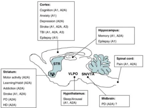
Figure 5: Roles of adenosine in the brain in physiology and pathology
See the implication of adenosine in physiology and pathology linked to a brain region as suggested by pharmacological and knock-out studies. From (Wei et al., 2011).
Learning and memory
The neurophysiological basis for learning and memory involves activity dependent changes in the strength of synaptic transmission mostly in hippocampus and cortex. As adenosine is a neuromodulator expressed in these areas, it was postulated to regulate learning and/or memory processes. It is difficult to draw clear conclusions on the effect of adenosine on learning and memory, due to the multiplicity of types of memory and different paradigms studied. Furthermore, the effects of adenosine could involve different types of memory or memory-related processes (acquisition, consolidation, retention and retrieval).
The evidence points that activation of A1R tends to impair learning and memory function, as it worsens the performance in different memory-related tasks (fear conditioning, 3-panel runway, passive avoidance task amongst others) (Normile and Barraco, 1991; Corodimas and Tomita, 2001; Pereira et al., 2002). On the other hand, acute A2AR blockade improve the learning and memory performance in social recognition memory, eye blink conditioning test (Prediger and Takahashi, 2005)
21 (Fontinha et al., 2009). In line with this evidence, the gb-A2AR -KO show enhanced working memory
(Zhou et al., 2009), while this type of memory was impaired in rats overexpressing A2AR (Gimenez-Llort
et al., 2007). Thus, the stimulation of A1R impairs while the blockade of A2AR improves
memory-related processes.
Sleep and arousal
Regulation of arousal/sleep and circadian rhythms implies the complex regulation of many nucleus in the brain, all of them endowed with adenosine receptors (Burnstock et al., 2011). Adenosine accumulation triggers a response to initiate sleep (Strecker et al., 2000) and mutations increasing the adenosinergic tone lead to increased total sleep time (Rétey et al., 2005); thus adenosine is a somnogenic agent.
It is widely accepted that caffeine intake leads to an arousal state. This caffeine-induced wakefulness is still present in gb-A1R-KO but blunted in gb-A2AR -KO (Huang et al., 2005), but stimulation of A1R in
rat preoptic area increased total sleep time (Ticho and Radulovacki, 1991). Overall, the adenosinergic system can modulate sleep but the direct contribution of A1R and A2AR is probably region-dependent.
Locomotor activity and exploration
The knock-out animals for the adenosine receptors do not have any motor impairment, but regarding spontaneous activity gb-A2AR -KO display hypolocomotion when compared to control littermates or
gb-A1R-KO (Wei et al., 2011a). Surprinsingly, the spontaneous activity of the double knock-out for A1R
and A2AR is even more reduced than in the gb-A2AR -KO (Yang et al., 2009). This suggests that A2AR is
the most important adenosine receptor subtype when it comes to modulation of spontaneous motor activity; but A1R exert an effect that is only revealed when A2ARs are also eliminated.
Feeding
Adenosine receptors can modulate food intake as shown in studies performed after food deprivation: food intake was not changed upon A1R stimulation; reduced upon A1R blockade or A2AR stimulation
and increased upon A2AR blockade (Burnstock et al., 2011).
Anxiety and mood
Adenosine receptors are enriched in the basal ganglia. The basal ganglia connect with the nucleus accumbens, which is highly implicated in mood, motivation and reward-seeking behaviour. This has
22 poised studies that concluded that the A1R stimulation is anxiolytic in different paradigms (Zangrossi
et al., 1992; Florio et al., 1998; Prediger et al., 2004, 2006), which correlates with the elevated anxiety described in the gb-A1R-KO. The pharmacological studies revealed no modulation of anxiety by A2AR,
however, the A2AR -KO mice have been shown to be more anxious (Ledent et al., 1997; Giménez-Llort
et al., 2002). (Prediger et al., 2006). This anxiety level of both gb-A1R-KO and gb-A2AR -KO could lead to
more aggressivity as observed in a resident intruder test (Ledent et al., 1997; Giménez-Llort et al., 2002).
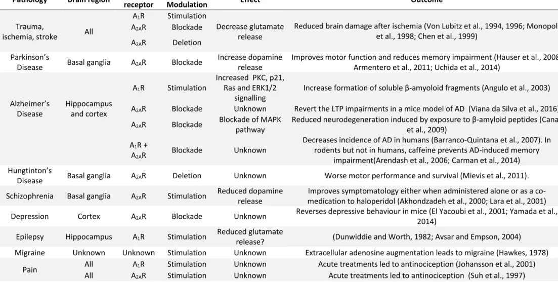
1.3.3 Adenosine in pathology
Adenosine signalling has been implicated in many brain pathologies and many treatments have tried to modulate the adenosine signalling to improve the symptomatology of a given disease. A summary of all the treatments and observations made on different diseases linked to adenosine receptors is found in Table 4.
23 Pathology Brain region Adenosine
receptor
Treatment/
Modulation Effect Outcome
A1R Stimulation
Reduced brain damage after ischemia (Von Lubitz et al., 1994, 1996; Monopoli et al., 1998; Chen et al., 1999)
Trauma,
ischemia, stroke All
A2AR Blockade Decrease glutamate
release A2AR Deletion
Parkinson’s
Disease Basal ganglia A2AR Blockade
Increase dopamine release
Improves motor function and reduces memory impairment (Hauser et al., 2008; Armentero et al., 2011; Uchida et al., 2014)
Alzheimer’s Disease Hippocampus and cortex A1R Stimulation Increased PKC, p21, Ras and ERK1/2
signalling
Increase formation of soluble β-amyoloid fragments (Angulo et al., 2003) A2AR Blockade Unknown Revert the LTP impairments in a mice model of AD (Viana da Silva et al., 2016)
A2AR Blockade Blockade of MAPK
pathway
Reduced neurodegeneration induced by exposure to β-amyloid peptides (Canas et al., 2009)
A1R +
A2AR Blockade Unknown
Decreases incidence of AD in humans (Barranco-Quintana et al., 2007). In rodents but not in humans, caffeine prevents AD-induced memory
impairment(Arendash et al., 2006; Carman et al., 2014) Hungtinton’s
Disease Basal ganglia A2AR Deletion Unknown Worse motor performance and survival (Mievis et al., 2011). Schizophrenia Basal ganglia A2AR Stimulation Reduced dopamine
release
Improves symptomatology either when administered alone or as a co-medication to haloperidol (Akhondzadeh et al., 2000; Lara et al., 2001) Depression Cortex A2AR Blockade Unknown Reverses depressive behaviour in mice (El Yacoubi et al., 2001; Yamada et al.,
2014) Epilepsy Hippocampus A1R Stimulation Reduced glutamate
release? (Dunwiddie and Worth, 1982; Avsar and Empson, 2004)
Migraine Unknown Unknown Stimulation Unknown Extracellular adenosine augmentation leads to migraine (Hawkes, 1978) Pain All A1R Stimulation Unknown Acute treatments led to antinociception (Johansson et al., 2001)
All A2AR Stimulation Unknown Acute treatments led to antinociception (Suh et al., 1997)
24
1.4 Impacts of caffeine intake during gestation and lactation
in the offspring
The roles of adenosine in the adult brain have been described extensively, however the roles of adenosine in the developing brain are starting to be unravelled. Some studies have shown that exposure to drugs (cocaine, nicotine, alcohol amongst others) during pre- and peri-natal development have a deleterious effect for the offspring (Thompson et al., 2009).
The fact that caffeine is the most widely consumed psychoactive drug in the world and is permeable to placental and blood brain barrier (Fredholm et al., 1999), prompted studies about the impact of caffeine on early life development. Upon the drinking of coffee and/or caffeinated beverages, the only mechanism affected would be A2AR and A1R (Figure 4, Fredholm, 1995). It was demonstrated that if a
human mother drinks 3 cups of coffee per day, the caffeine blood levels in umbilical cord are between 0.5-2 mg/L (Nehlig et al., 1992). Similar caffeine levels were found in breast-fed babies of caffeine-consuming mothers (Adén et al., 2000); and in line with these observations it is was observed that a mother who drinks 3 cups of coffee in 1 hour has breast milk caffeine levels of 0.32-1.15 mg/L over 10 hours (Bailey et al., 1982).
Studies in humans show that caffeine consumption (>200mg/day, 3-4 cups of coffee per day) during pregnancy may increase the risk of miscarriage (Weng et al., 2008). Another study points that maternal caffeine intake during pregnancy is not associated with changes in gestational length but with lower birth weight (Sengpiel et al., 2013). Lower weight at birth correlates with higher seizure susceptibility (Sun et al., 2008) and cognitive deficits (Jefferis et al., 2002). High caffeine consumption has been associated with fetal death specially after 20 weeks of pregnancy (Bech et al., 2007);however and although many meta-analysis have been performed with analysis dating from 1970s the studies contemplating moderate doses of caffeine remain inconclusive (de Mérici Domingues Paula et al., 2017).
Studies in rats about the effects of caffeine administered in early life intake (between P2-P6) were reported to upregulate A1R over adulthood (Etzel and Guillet, 1994); however the dose used
(80mg/kg/day) results in caffeine blood levels too high to compare with patterns of human caffeine consumption. This point was raised by a later study in which they administered caffeine during pregnancy and early postnatal life through drinking water (0.3g/L, giving rise to blood concentrations between 0.4-2mg/L) and it did not modify mRNA nor protein level of A2AR, A1R nor GABAAR at any stage
25 of development checked (from E14 to P21) (Adén et al., 2000). To avoid mixing the effects of the different molecular targets of caffeine, I will only describe here the studies that have used concentrations that will block only the A2AR and A1R and thus are relevant in terms of human caffeine
consumption. Most of the studies studying the effect of caffeine on development, they choose to administer the caffeine in the drinking water of the mother (Adén et al., 2000; León et al., 2005; Silva et al., 2013; Mioranzza et al., 2014; Ardais et al., 2016). Concentrations of 0.3g/L mimic correctly the pattern of caffeine human consumption, while 1g/L is too high resulting in blockade of phosphodiesterases (Ardais et al., 2016).
Recently, a study in rats has shown how caffeine intake during pregnancy affects proteins levels in the offspring. It was found an increase in Sonic Hedgehog and a decrease of the axonal marker GAP-43 both in the hippocampus at E18 in offspring whose mother was drinking 0.3g/L of caffeine (Mioranzza et al., 2014). In another study, treating the pregnant dams from gestation until weaning with the same amount of caffeine they report some changes in synaptic proteins (neurotrophin trkB receptor and the glial marker GFAP) of the offspring at adult stages (Ardais et al., 2016). The offspring whose mother was treated with caffeine displayed behaviour changes in recognition memory and in locomotion. This study showed the impact of caffeine on synaptic proteins, memory performance and locomotion and confirms that caffeine exposure during brain development can cause long-term effects in both memory performance and locomotor behaviour (Ardais et al., 2016).
Interestingly, another paper looked at the effects of caffeine intake during gestation and lactation (the first 3 postnatal weeks). Upon treatment with caffeine (0.3g/L) or an A2AR selective antagonist
(KW6002), the migration and insertion of GABA neurons in cortical and hippocampal layers was impaired, leading to anatomical alterations and subsequently to hippocampal hyperactivity accompanied by a shift in the balance between the GABAergic and glutamatergic synaptic inputs in CA3 pyramidal cells. There was also a cell loss observed at 3 months in different hippocampal layers. Behaviourally, the animals were more susceptible to induced epilepsy (at young stages) and showed some cognitive deficits regarding spatial hippocampal-dependent memory in adult life (Silva et al., 2013). In continuing with the described roles of A2AR in development, in young primary cortical cultures
that stimulation of A2AR enhances axonal elongation via 3-kinase (PI3K) , mitogen-activated protein kinase (MAPK) and phospholipase C (PLC) and augments dendritic branching, facilitating the action of BDNF (Ribeiro et al., 2015). The fact that A2AR is implicated in multiple roles during development (interneuron migration, axonal elongation, dendrite branching) questions a possible role in the formation of synapses per se.
26
1.5 Spatiotemporal expression of A
2AR
I studied the role of A2AR on synaptogenesis. It is of crucial importance to assess its spatial and temporal
expression. Both questions are of the same importance to try to better understand the regulation occurring through A2AR.
Early studies studying the brain localization of A2AR were done with autoradiography techniques with
the A2AR selective agonist CGS21680. The first studies in adult rat (Jarvis and Williams, 1989) and adult
human (Martinez-Mir et al., 1991) suggested that the A2AR was restricted to striatum (caudate nucleus,
putamen, nucleus accumbens, olfactory tubercle) and the lateral segment of the globus pallidus.
Later, studies using different techniques demonstrated that A2AR is expressed outside the striatum and
expression of A2AR was observed in the hippocampus (Cunha et al., 1994; Johansson and Fredholm,
1995), cortex (Fink et al., 1992; Johansson and Fredholm, 1995) and midbrain (Fink et al., 1992). Dixon and colleagues (Dixon et al., 1996) performed in situ hybridisation showing expression of A2AR mRNA
in striatum, nucleus accumbens and olfactory tubercle despite the functional evidence for the presence of this receptor in other regions. When using retro-transcriptase-PCR, a technique that is much more sensitive they could show that A2AR is expressed in striatum, olfactory bulb, nucleus
accumbens, hippocampus, hypothalamus, thalamus and cerebellum thus showing that A2AR has a
broader pattern of expression than previously thought. The most comprenhensive study on the expression of A2AR in brain was performed by Rosin and colleagues (Rosin et al., 1998) where they
purified an A2AR antibody and performed immunohistochemistry in 20μm-thin adult rat all throughout
the brain. They described A2AR dense immunoreactivity in striatum, nucleus accumbens, olfactory
tubercles, and areas of amygdala, globus pallidus and nucleus of the solitary tract. Lighter staining was found in the cortex, hippocampus, thalamus, cerebellum, and regions of the hindbrain.
The first observation of A2AR in the hippocampus was done by Rodrigo Cunha (Cunha et al., 1994). In
this study they used adult rats to show that adenosine A2AR mRNA is expressed in the hippocampal
CA1, CA3 and dentate gyrus and they confirm this observation using in situ hybridization to show that A2AR was mainly localized in the pyramidal and granular cells. By autoradiography they show how the
density of [3H]CGS21680 binding was greatest in the stratum radiatum of the CA1 area, followed by
the stratum oriens of the CA1, stratum radiatum of the CA3 area and supra-granular layer of the dentate gyrus. Finally, they confirm its presence by electrophysiological studies at the Schaffer collateral to CA1 pyramidal synapse. This information can be complemented with the one from the study of Rosin and colleagues in which they reported the presence of A2AR in the rostral hippocampus
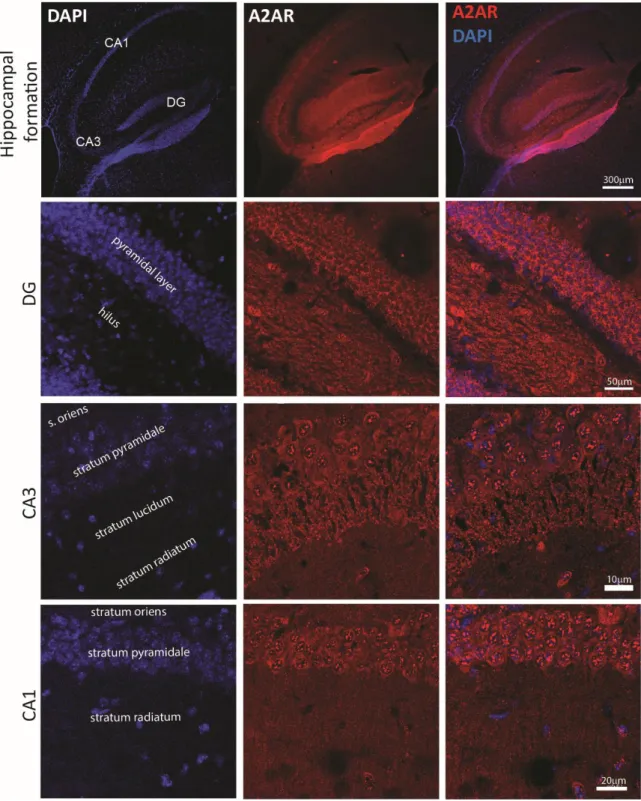
27 and dim staining was observed in CA1, CA2, and CA3 pyramidal cells (Rosin et al., 1998). Images taken in our laboratory by Jessica Pressey show that A2AR is expressed in the adolescent hippocampus of
mice (Figure 6) in dentate gyrus, CA3 and CA1.
Figure 6: A2AR expression in the postnatal hippocampus.
Top, A2AR is expressed in the hippocampal formation at P16. In the dentate gyrus (DG), see the presence of A2AR in the
granule cell layer and in the hilus. In CA3, A2AR is present in pyramidal cells and stratum lucidum. In CA1, A2AR is found in
28
1.5.1 Cellular and subcellular expression of A
2AR
A2AR have been found in adult hippocampal glutamatergic synapses (Cunha et al., 1994; Rebola et al.,
2005b), adult hippocampal GABAergic synapses (Cunha and Ribeiro, 2000; Rombo et al., 2015), adult globus pallidus GABAergic synapses (Shindou et al., 2002), hippocampal cholinergic (Cunha et al., 1995), striatal dopaminergic (Garção et al., 2013) hippocampal sertoninergic (Okada et al., 1999), nucleus tractus solitarius serotoninergic and noradrenergic (Barraco et al., 1995, 1996) or hippocampal noradrenergic synapses (Jackisch et al., 1985). From now on, I will focus my study in the hippocampus, sometimes comparing to the striatum where the A2AR has been mostly studied.
In the hippocampus of adult rats (6-8 weeks) the binding of a radioactive selective antagonist of A2AR
([3H]SCH 58261) is higher in nerve terminals than in total membranes. Subcellular fractionation showed
that A2AR is enriched in the presynaptic active zone in hippocampus (Rebola et al., 2005a). This contrasts with its expression in the striatum where it is predominantly expressed in the postsynaptic density. This suggests different roles of A2AR in striatum (signal processing) and in hippocampus
(control of neurotransmitter release) (Rebola et al., 2005a). Immunocytochemistry on hippocampal synaptosomes from adult rats, revealed that A2AR colocalizes with 29.2±6.2% of the Vesicular
glutamate transporter (VGLUT, marker of excitatory neurons) 1/2-positive terminals. Moreover, they show that in hippocampus A1R and A2AR are expressed by the same neuron in a subpopulation of
pyramidal cells (Rebola et al., 2005b). However, these data must be interpreted with caution, because the sub-synaptic localization of A2AR can vary depending on the synapse. Contrasting with the data of
(Rebola et al. 2005a) which described a presynaptic localization of A2AR in the hippocampus, electronic
microscopy studies showed that A2AR are postsynaptic in the mossy fiber to CA3 pyramidal cells
synapse in adult mice (Rebola et al., 2008).
Less is known about the presence of A2AR in hippocampal interneurons although functional studies
show how A2AR regulates GABA release (Cunha and Ribeiro, 2000) and modulates BDNF effects in these
cells (Fernandes et al., 2008). A2AR is also expressed in glial cells. The presence of A2AR has been
demonstrated in astrocytes (Cristóvão-Ferreira et al., 2013; Orr et al., 2015), oligodendrocytes (Coppi et al., 2015) and microglia (Orr et al., 2009; George et al., 2015).
29
1.5.2 Ontogenic development of A
2AR
The ontogenical expression of A2AR in rat brain has been studied using radioligand and in situ
hidrization assays (Johansson et al., 1997; Adén et al., 2000). The earliest detection of A2AR mRNA was
found at E14 by in situ hibridization, it was diffusely distributed all over the brain. However it was not detected by any of the radioligand assays at this age. From E18, A2AR mRNA was found in the caudate
putamen. The first signal detected by the radioligand assays is at E21 in the caudate putamen. Both signals increase from then on until P14.
Data from the Allen Brain Atlas (Figure 7) show that A2AR is detected at E18.5 as the earliest timepoint
in the midbrain and medullary hindbrain. A2AR is starting to be expressed in the late embryonic stages,
increasing during infancy (P4-P14) and adolescence (P28), then decreasing in adulthood (P56) and then increases again in old animals (P18 months).
Figure 7: Ontogenic expression of A2AR
See the in situ hybridization of A2AR over time (E18.5, P4, P14, P28, P56, 18 months) in the left panel, in the right panel the
expression mask displays those cells with a probability of gene expression (from low/blue to high/red). Data extracted from Allen Brain Atlas
The peak of expression of A2AR in early life is concomitant with the period of synaptogenesis. This
30
2. Synapse formation
2.1 The inhibitory synapse: an overview
In the mammalian central nervous system, GABA is the main inhibitory neurotransmitter. GABA synthesis occurs by the enzyme L-glutamic acid decarboxylase (GAD) that catalyses decarboxylation of glutamic acid to form γ-aminobutiric acid (GABA)(Erlander et al., 1991). After being released from the presynaptic neuron, GABA will be rapidly removed from the synaptic cleft by GABA transporters (GAT1-4) (Minelli et al., 1995). In the synaptic cleft, GABA can bind either GABAA or GABAB receptors.
GABAA receptors (GABAAR) are ionotropic while GABAB are metabotropic receptors. GABAB receptors
are GPCRs that activate second messenger systems such as PLC and AC and activate K+ and Ca2+ ion
channels via G-coupled proteins to hyperpolarize the neuron (Chebib and Johnston, 1999). GABAAR are
ligand-gated ion channels. Upon binding of GABA a conformational change opens the channel leading to chloride influx/efflux depending on the electrochemical gradient of the ion. During early development, GABAAR are depolarizing because the intracellular chloride is high; in adult neurons,
intracellular chloride is lower and GABA is hyperpolarizing (Rivera et al., 2005). In the developing brain, GABA depolarization leads to calcium entry via L-type voltage-gated calcium channels (Perrot-Sinal et al., 2003)which in his turn will activate different pathways, modulating neurite outgrowth and synapse formation (Spoerri, 1988; Barbin and Pollard, 1993; Represa and Ben-Ari, 2005; Oh et al., 2016). In the adult brain, GABAAR are the major source of inhibition (Comenencia-Ortiz et al., 2014).
Molecular structure of GABAAR and subcellular distribution
GABAAR are organized as heteropentamers. To date, twenty-one GABAAR subunits have been
identified and classified into eight classes based on sequence identity: α(1–6), β(1–3), γ(1–3), δ, ε(1– 3), π, θ and ρ(1–3) and π (Olsen and Sieghart, 2009). All the GABAAR subunits present four
transmembrane domains, a long N-terminal extracellular domain and a short C-terminal extracellular domain (Figure 8). The extracellular N-terminal sites are important for the oligomerization of the protein and for subunit– subunit interactions. The intracellular domains contains a critical site for cytoplasmic protein interactions with regulatory and signalling molecules (Jacob et al., 2008). Between the third and fourth transmembrane domains there is a long intracellular loop with multiple sites for post-translational modifications (Jacob et al., 2008).