HAL Id: hal-02430692
https://hal.archives-ouvertes.fr/hal-02430692
Submitted on 7 Jan 2020
HAL is a multi-disciplinary open access archive for the deposit and dissemination of sci-entific research documents, whether they are pub-lished or not. The documents may come from teaching and research institutions in France or abroad, or from public or private research centers.
L’archive ouverte pluridisciplinaire HAL, est destinée au dépôt et à la diffusion de documents scientifiques de niveau recherche, publiés ou non, émanant des établissements d’enseignement et de recherche français ou étrangers, des laboratoires publics ou privés.
Calcium phosphate nanocoatings and nanocomposites,
part 2: thin films for slow drug delivery and
osteomyelitis
Besim Ben-Nissan, Innocent Macha, Sophie Cazalbou, Andy H Choi
To cite this version:
Besim Ben-Nissan, Innocent Macha, Sophie Cazalbou, Andy H Choi. Calcium phosphate nanocoatings and nanocomposites, part 2: thin films for slow drug delivery and osteomyelitis. Nanomedicine, Future Medicine, 2016, 11 (5), pp.531-544. �10.2217/nnm.15.220�. �hal-02430692�
OATAO is an open access repository that collects the work of Toulouse
researchers and makes it freely available over the web where possible
Any correspondence concerning this service should be sent
to the repository administrator: tech-oatao@listes-diff.inp-toulouse.fr
This is an author’s version published in:
http://oatao.univ-toulouse.fr/24456
To cite this version:
Ben-Nissan, Besim and Macha, Innocent and Cazalbou, Sophie
and Choi,
Andy H Calcium phosphate nanocoatings and nanocomposites, part 2: thin
films for slow drug delivery and osteomyelitis. (2016) Nanomedicine, 11 (5).
531-544. ISSN 1743-5889
Calcium phosphate nanocoatings and
nanocomposites, part 2: thin films for slow
drug delivery and osteomyelitis
During the last two decades although many calcium phosphate based nanomaterials have been proposed for both drug delivery, and bone regeneration, their coating applications have been somehow slow due to the problems related to their complicated synthesis methods. ln order to control the efficiency of local drug delivery of a biomaterial the critical pore sizes as well as good control of the chemical composition is pertinent. A variety of calcium phosphate based nanocoated composite drug delivery systems are currently being investigated. This review aims to give an update into the advancements of calcium phosphate nanocoatings and thin film nanolaminates. ln particular recent research on PLA/hydroxyapatite composite thin films and coatings into the slow drug delivery for the possible treatment of osteomyelitis is covered.
Keywords: antibiotics • biofilm • drug delivery • hydroxyapatite • nanocoatings • nanocomposites • nanolaminates • osteomyelitis • thin films
During the last hundred years due to its simi larity to hard tissue, the material of choice for the enhancement of bioactivity has been calcium phosphates (1-6). A number of bio degradable nanocoated biomaterials based on calcium phosphates are currently being examined for applications for slow drug delivery and dispersion of pharmaceuticals and minerais to the targeted area. In these new devices, which comprises a polymeric or ceramic matrix and porous inorganic par ticulate matter, factors such as the chemical composition as well as the critical pore size are the main factors that influence the disso lution rates. Additive particulate sizes which can be incorporated to these films can be from a few nanometers up to microns depen dent on the required thickness and the fonc tion of the films. They are usually designed to contain nano and meso pores for drug loading.
Classical approach of the systemic drug delivery has in the past generated a
num-10.2217/nnm.15.220
ber of problems that can possibly be solved by local or targeted delivery. These include influence of the dissolving drugs to the whole body rather than pin point loca.l delivery. Moreover, aside from reduction of toxicity to healthy cells within the whole environment, targeting these drugs directly to required locations have the possibility of improving drug efficacy and efficiency, resulting in a significant cost savings for the health care system.
During the last decade a number of research groups have been describing the pro duction of nove! nanocoatings and thin film nanolaminates with hydroxyapatite (HAp) and other calcium phosphates for clinical applications. The nano to micro particles of oxide or mixed ceramics, and calcium phosphates are the inorganic components and natural or synthetic polymers such as collagen, chitosan and biodegradable poly mers such as polylactic acid (PLA) are the matrix materials in these new composites.
Besim Ben-Nissan*•1, Innocent Macha1, Sophie Cazalbou2 & Andy H Choi1
1 Faculty of Science. un iversity of Technology Sydney. 15 Broadway. Ultimo NSW 2007. Australia
'Université de Toulouse. C IRI MAT. UMR 5085 UPS-INPT-CNRS. faculté de pharmacie. Toulouse. France •Author for correspondence: b.ben-nissan@uts.edu .au
These nanocomposites can be synthesized by mixing the inorganic particles physically through heat or by solvent introduction into an already existing polymeric matrix material. Evaporation of the solvent aids the formation of the nanocomposite or the thin films.
The development of tissue engineering in the past has been related directly to the types of scaffolding materials used. At present, a number of synthetic bone graft biomaterials such as calcium phosphates are avail-able as options to autogenous bone for augmentation, repair or substitution [3]. New advancements associated with calcium phosphate scaffolds and their improve-ments in microstructure and surface properties have created new opportunities for bone regenerative tech-nologies. These recent developments also make the cal-cium phosphate scaffolds to be thought of as being at least biologically constructive instead of as only osteo-conductive scaffolds specifically with the addition of biologic materials such as bone morphogenic proteins and stem cells [7–9].
Evidence has shown that porous calcium phosphate has a direct influence on the proliferation and differ-entiation of human mesenchymal stem cells (MSCs). Tissue engineering along with new bioactive molecules enhanced the possible applications of calcium phos-phate as scaffolds able to guide the behavior of these cells and the efficiency of bone regeneration as well as being carriers of these cells [7,9].
In this review, we aim to introduce the recent devel-opments of nanocoatings and thin films and compos-ites containing calcium phosphate-based nanoparticles currently being investigated for the delivery of phar-maceutical substances and their use in medicine and specifically for the treatment of osteomyelitis.
Surface modifications & liposomes for drug delivery applications
In bioceramics, the critical pore size and intercon-nectivity can be altered in order to control the ease of delivery and dispersion of a material to the targeted area. Targeting usually achieved with appropriate functionalizing of the surfaces. The pores may range from a few nanometers to micron sizes dependent on the pharmaceuticals used or areas intended to be deliv-ered such as long bones. Delivery systems based on calcium phosphate which is similar at least chemically to the hard tissues, have the potential to increase drug efficacy while at the same time minimizing toxicity to nondiseased cells. Nano drug-delivery systems, embed-ded within a matrix or not, also have the exceptional attribute of being capable of delivering and control-ling dissolution with high precision due to their high surface areas. It is not surprising that the number of research papers covering drug, gene and mineral
deliv-ery of nanoparticles, nanocoatings and composites published during the last decade is very high [9–32].
The appropriate dissolution rates and their con-trol within the human body is the main concern for drug carriers containing nanoparticles and nano thin films [33]. The use of calcium phosphate as a delivery system also broadens its effectiveness as a result of their capacity to locally deliver minerals as well as calcium and phosphate, other active ions and biogenic materi-als such as bone morphogenetic proteins (BMPs) and MSCs if required to be used in the successful treat-ment of bone diseases. In addition the surface modifi-cation approach it can also be used to achieve enhance-ments of stability and long range solubility control of nanocoatings and thin films in aqueous media, as well introduction of new material properties and functions. In principle through the use of a wide range of bio-logical, chemical and/or physical surface modifications methods, the surfaces of nanostructured materials such as nanocoatings can be altered and functional-ized to assist us in slow drug delivery. In the quest for the surface modifications of nanostructured materials, approaches such as macro micro and nanocoating have emerged as the leading strategies resulting in better functionalization of the surfaces of materials and for better osseointegration in the long term.
The biological modification of surfaces of nanocoat-ings is at times essential for the functionality of the devices. Biospecific molecules can be incorporated into the nanocoatings or thin films by using physical or chemical methods, thus presenting biospecific sites for the further immobilization of ligands specific to these molecules. The immobilizations of specific ligands such as antibody–antigen and receptor–ligand can be carried out using biologically specific reactions [34]. Current research work in these areas is very promising.
It is well known that different biomedical applica-tions require different funcapplica-tions and properties of materials. As a result, techniques available to mod-ify nanostructured materials or thin films can vary in order to meet the demands of various biomedical systems. In spite of the advantages offered by nano-coatings and nanoparticle containing composite thin films, such as their small surface pore sizes and load-ing efficiency, a number of issues such as control of the appropriate drug release rates, restricted their use clinical applications.
The targeting ability and efficacy of any drug deliv-ery system are sometimes hindered by the rapid dis-solution of the carrier system within the human body. A good example is their side effects in chemotherapy drug delivery for the cancer patients. The long circula-tion time within the blood is the primary concern for drug carriers of both local and systemic delivery. For
this reason, a number of investigations have been car-ried out to examine ways in which ‘long-circulating-time’ carriers can be designed and engineered. Among these, the surface modification of thin films and nanocoatings with a variety of polymeric macromol-ecules or nonionic surfactant has been demonstrated to be the most effective for maintaining the presence of drug delivery particles in the blood for prolonged periods [35].
The use of surface modification is used in gene ther-apy in an effort to obtain controlled delivery of small interfering RNA and plasmid DNA (pDNA) particu-larly in an acidic pH environment [25–28]. The use of cationic liposomes as transfection vectors has become an ideal choice and most widely employed in the trans-fer of pDNA due to their weak immunogenicity and low toxicity [24]. A study by Zhou et al. [25] has suggested that coating calcium phosphate with liposomes could provide consistently efficient and satisfactory delivery of pDNA. Using mammalian cell culture, their find-ings showed the application of a lipid coating resulted in a tenfold increase in the transfection of pDNA compared with uncoated calcium phosphate [25].
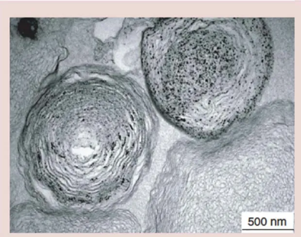
Considered as one of the most clinically recognized thin film nanoscale systems, liposomes consist of a single layer or multiple concentric lipid bilayers that encapsulate an aqueous compartment are currently uti-lized in the delivery of antifungal drugs, vaccines and genes [25,36–41]. The exceptional clinical profile of lipo-some coatings in comparison to other delivery systems is based on their reduced toxicity, biodegradability and capacity for size, and surface manipulations [42]. An improvement in the biocompatibility of liposomes as well as an increase in nanoparticle hydrophilicity and stability in plasma can be achieved through the encap-sulation of nanomaterials such as calcium phosphate within liposomes (Figure 1). In Lewis work multilay-ered liposomes were produced with the incorporation of nano-HAp and other minerals such as strontium, magnesium and zinc [42]. Figure 1 shows a transmission electron microscopy (TEM) image of multilayered liposomes containing nano-HAp particles produced in a calcifying buffer. The figure was obtained in FEI Morgagni 268D transmission electron microscope (Eindhoven, the Netherlands) at 80 kV [42]. The work showed an excellent encapsulation that can help to control drug delivery rates in medical applications such as chemotherapy drug delivery for oncology patients. This observed ease of coating and release delay ability is one of the strong reasons, calcium phosphate based nanoparticle containing thin films and liposome coat-ings are ideal candidates for drug delivery and bone regeneration systems [42–45]. In addition, combinatory therapy–modalities can be accomplished by utilizing
the ability of liposome coatings to carry hydrophobic and hydrophilic moieties as well as their capacity to incorporate therapeutic and diagnostic agents into a single liposome-delivery system [42].
Huang et al. [39] have suggested that the nucleation process for new bone formation could be improved by the presence of negatively charged liposome coatings. In their experiments carried out in miniature swine, artificial bony defects on one side where implanted with liposome-coated tri calcium phosphate while defects on the other side served as controls. They reported that at three weeks postimplantation, dense connective tis-sues surrounded the implant material and new bone formation was visible after 6 weeks.
Using a different strategy, Wang et al. explored the possibility of producing collagen-calcium phosphate scaffolds with the incorporation of liposome thin films for the controlled release of bioactive molecules in bone regeneration and repair [40]. They suggested that bisphosphonate (BP) functionalized liposome encap-sulation could be isolated within mineral-containing scaffolds can be better drug delivery system that local-ize their drug cargo to the directed area. The liposome encapsulation used consisted of cholesterol, distearo-ylphosphodioline, distearoylphosphoethanolamine-poly(ethylene glycol). Based on their observations, the encapsulation of BP within liposomes displayed a strong affinity to the scaffolds and the drugs entrapped within the BP-liposomes showed a slower release rate from the scaffolds as compared with drugs that were un-encapsulated or encapsulated in polyethylene gly-col-liposomes. In a similar study by Chou et al. it was shown that this liposome coating thin films reduced the drug release rates in both BP and antibiotic additions [22,46].
A study by Xu et al. [36] explored the possibility of synthesizing a multifunctional thin film drug car-rier with sustained drug release capability provided by the inner core liposome and osteoconductivity for bone cells supported by the HAp outer layer. The lipo-somes were produced from 1,2-dimyristoyl-sn-glycero-3-phosphate and 1,2-dimyristoyl-sn-glycero-3-phos-phocholine, which is then loaded with the lipophilic drug indomethacin. The release profile of indometh-acin was measured at two different pH levels (4 and 7.4). As expected by coating the liposome with HAp, they reported a reduction in release rate of indometha-cin in comparison to uncoated liposomes. They also reported that without these coatings, the rate of drug release occurred more rapidly at pH 7.4 rather than at pH 4.
It has been reported that the management of pos-sible postoperative infections from bone grafts and prostheses as well as the treatment of bone diseases
Figure 1. Transmission electron microscopy image of multilamellar lipsomes containing nano hydroxyapatite particles and minerais in a calcifying buffer (scale
500 nm).
Reproduced with permission from [42].
such as bone metastases will benefit gready if there is a delivery system which has a high affinity toward bone tissues thereby maximizing its therapeutic effect on bone-related diseases (36,39]. Using this approach,
Anada et al. (37] attempted to develop a calcium phos phate-binding liposome coating for a bone targeting drug delivery system by synthesizing an amphipathic molecule bearing a BP head group to recognize and bind to HAp. Liposomes loaded with the drug doxoru bicin adsorbed onto the surfaces ofHAp were observed to significandy reduce the number of viable human osteosarcoma MG63 cells. Based on these observa tions, they suggested that the system can be excellent coated carriers for anticancer drugs as they specifically target bone tissue (36].
Infection & osteomyelitis
lt is widely accepted in the medical community that wound contamination as well as postoperative infec tions following implantation or during surgical inter vention in orthopedics and maxillofacial surgery can result in serious clinical problems and could jeopardize the osseointegration process. For these reasons, antibi otics either administrated orally or intravenously are often provided as prophylactics.
By far the most frequent complications related to the use of implantable medical devices such as orthopedic or dental prostheses and endotracheal tubes are bacte rial infections. Pseudonomas aeruginosa is regarded as an opportunistic pathogen causing indwelling device related infections especially catheters. P. aeruginosa infection is leading cause of morbidity and mortality in cystic fibrosis patients. On the other hand Staphy
lococcus aureus infection causes serious infectious
complications such as severe sepsis, septic-thrombosis
and/or severe deep-seated infections (endocarditis, osteomyelitis and other metastatic infections). One of the basic hospital and surgical intervention acquired biofilm infections are those associated with S. aureus and Staphylococcus epidermidis mains, including meth icillin-resistant S. aureus (MRSA). A thorough and comprehensive understanding of the molecular bases of biofilm formation and their adhesion may help us to fight biofilm infections.
Osteoarticular infection is frequendy caused by coagulase-negative Staphylococci as main aetiologic agents in lace infections as well as Streptococci, Entero cocci and anaerobes. The number of infections is esti mated to be around 0.5- 2.0% of cases and it increases continuously due to the rise in the need for implants.
The European Center for Disease Prevention and Controll (ECDPAC) reported that approximately 4,100,000 patients are estimated to acquire infections in the European Union every year (47]. The number
of deaths occurring as a direct consequence of these infections is estimated to be at least 37,000 patients and these infections are thought to contribute to an additional 110,000 deaths each year. ln the USA, it was estimated that approximately 1,700,000 patients acquired infections for the year 2002. lt has been esti mated that 5% of patients undergoing clean surgical procedures and up to 20% of patients having intra abdominal surgical procedures develop a surgical site infection. Such infections result in 3.7 million excess hospital days and more than US$1.6-3 billion in excess hospital costs per year.
ln order to mitigate this problem different strate gies have been proposed on either preventing and/or controlling bacterial infections. Modification or devel opment of thin film or nanocoated multilayer devices with surface properties that have an effect against microbial adhesion or viability seems to be a promis
ing approach for the prevention of device-related infec tions. Another strategy is to modify the surface of medical devices biologically, chemically or physically to render the surface free of microbial adhesion. Mul tifunctional thin films or nanocoatings can facilitate this new approach.
ln clinical applications an ideal implant coatings must provide surgeons with several benefits such as providing primarily anchorage with appropriate bioac tivity involving osteoconduction and if possible, osteo
induction. The nanocoating or thin films used should also provide antimicrobial property to prevent implants from developing acute or postoperative infections.
As stated earlier the development of bone infec tion is based on the formation of a bacterial biofilm where the bacteria differentiate from planktonic into sessile forms that protect some bacterial cells that can
be released from the biofilm after antibiotic treatment has ceased.
Bacteria adhesion is a complex phenomenon affected by many factors, including properties of sur-face materials, some characteristics of bacteria itself and the environment where the adhesion takes, such as the presence of serum protein or bactericidal sub-stances [48]. Some of the proposed theory and model of adhesion seem to be limited because they consider physical interaction between the surface and bacteria and neglecting biological aspects of adhesion in which specific bacterial structure responsible for adhesive activities called adhesins that control cell to cell or cell to abiotic surface adhesion. Bacteria may have differ-ent adhesives for differdiffer-ent surfaces (differdiffer-ent acceptor). The ionic strength and pH of the medium in which the adhesion takes place influence the charge of the cell wall and of the substrate (in terms of surface chemistry, charge and hydrophobicity) and therefore affects their interaction.
The antibiotic dosage required to act on bacteria in biofilm conditions can be many folds higher in con-centration of the drug required for treatment of plank-tonic cells. Consequently, it is difficult to completely eradicate active infection by means of ‘systemic anti-biotics’ which in doses active in biofilm can be toxic for a patient.
In orthopedics, osteomyelitis mainly occurs in tis-sues within the infected area, and consequently tar-geted delivery of antimicrobial agent locally is a more appropriate form of treatment. One of the most effec-tive ways of achieving targeted delivery of antibiotics is to use a carrier device that can be placed into the body at a specific site such as coating on an implant, which will release slowly the correct antibiotic dosage. The major challenge associated with the use of antibiotic is ensuring retention of antibiotic release and activity for a prolonged period of time post operation [49].
Treatment of osteomyelitis can be with immobili-zation and antibiotic therapy with a number of drugs including flucloxacillin, gentamicin, tobermycin or vancomycin, and fusidic acid. Surgical drainage and removal of damaged bone, as its presence prevents healing, sequestrum may be possible but recurrence is common. Currently, commercial products in the form of pellets composed of acrylic polymers or ceramics such as calcium sulfate or bioactive glass are available as slow drug delivery devices. However, due to the problems related to their dissolution rates, shape, sizes and chemical composition, they are either nonresorb-able that requires second surgery to remove or quickly resorbed by the body’s natural physiological process, thus limiting their effectiveness. In addition, past investigations have shown that some antibiotics have
been reported to be ototoxic and nephrotoxic at high dosage. For most controlled release systems, the loaded dosages are usually high, and therefore the systemic exposure of antibiotic in blood and urine is the major safety concern.
The concept of pathophysiology of osteomyelitis which has been widely accepted is the infected bone becomes devascularized and the resulting sequestered portions of necrotic cortical bone harbor bacteria. This sequestered, necrotic, infected bone is responsible for the chronicity of osteomyelitis. Pus and granulation tis-sue then surrounds the infected fragments and through increased intraosseous pressure, bacterial toxins and enzymes, further contribute to devascularization of the surrounding bone. The granulation tissue that surrounds the infected area as the infection becomes chronic is replaced by relatively avascular fibrous tis-sue, and stimulation to form new reactive bone referred to as involucrum takes place within the surrounding tissues and in the periosteum permeative mesenchy-mal cells that wraps around the sequestered, necrotic, infected bone. Antibiotics and antibodies must cross this involucrum and relatively avascular fibrous tis-sue to reach the microorganisms once the fibrous and bony encapsulation takes place. Consequently, the very effective effort by the body to quarantine the host from the infection also isolates the microorganism from the defenses of the host. The infection becomes chronic and cannot be eradicated when this pathological stand-off occurs. This condition is the basis for intervention surgery and the excision of necrotic sequestra. Antibi-otic therapy completes the treatment of chronic osteo-myelitis by eradicating the microorganisms once they are no longer isolated [50].
The quest for a more effective means of delivering antibiotics without the complications related to long-term intravenous access and the toxicity of systemic antibiotics has been ongoing. Hence, the most accurate method of assessing diseases is provided by models that utilize eradication of infection as a criterion for suc-cess, the histological findings of new bone formation, inflammation, sequestration and intraosseous bacteria, combined with cultures.
In the past, ceramics as well as other materials incorporated to the thin films and bulk composites have been suggested as potential candidates to be used as biodegradable drug delivery systems. How-ever, manipulating these materials into an appropri-ate shape with adequappropri-ate microporosity in order to be fitted into bone defects of different size and form was found to be difficult. Recently, it has been demon-strated that marine shells, foraminifera and corals incorporated into thin films and nanocoatings with specific microspherical carriers offer desired functions
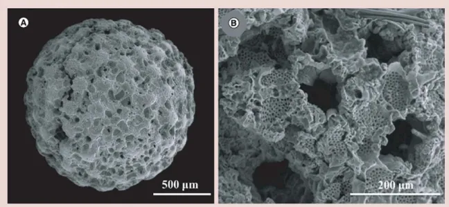
for the delivery ofBP (paminodrate) and an antibiotic (gentamicin) [51-55). This has been possible by virtue of its nano and mesoporous structure and architec ture of the foraminifera shells (Figure 2A) which are difficult if not impossible to produce with our cur rent manufacturing methods [44,45). Foraminifera and coral in addition to their unique interconnected porous structure are made of calcium carbonates that can be easily converted to bone such like phosphate structures [44,45).
Bone repai r drug delivery systems & osteomyelitis
In early 1900s, a calcium phosphate compound (tri calcium phosphate) was first successfully used in bone repaie followed by the first clinical study [!). It took more than six decades until porous calcium phosphate scaffolds were proposed for the treatment of bony defects [2). The reason behind the research and devel opment of calcium phosphate based biomaterials for bone repaie, augmentation and substitution was due to the similarity in composition between biological and synthetic apatites [55).
For bone repaie and slow drug delivery systems, the most appropriate materials are the calcium phos phate based bone substitutes [3). They offer easy pro duction, drug carrying capability and supply both calcium and phosphates during dissolution. They can be easily incorporated within thin films and nanocoat ings [55.56). There have been a large number of stud ies carried out on both experimental and commercial calcium phosphate based drug carriers during the last decade [10-34). Due to their wide areas of applicabiliry, the delivery of antibiotics has become a major focus in
the prevention and treatment against infection during or postoperative surgical interventions.
Biodegradable composite thin films
Biodegradable polymer thin films loaded with gen tamicin have been synthesized to act as a 'composite coatings' for metallic implants and fracture fixation devices in an attempt to prevent implant-associated infections [48.57-59). Due to their tendency to uptake and release pharmaceuticals and minerais as well as the capability to degrade over time, the use of biodegrad able polymer thin films is advantageous. In addition, by controlling the pore sizes and interconnectiviry of these particles, the rates of drug release could be tailored to suit the treatment.
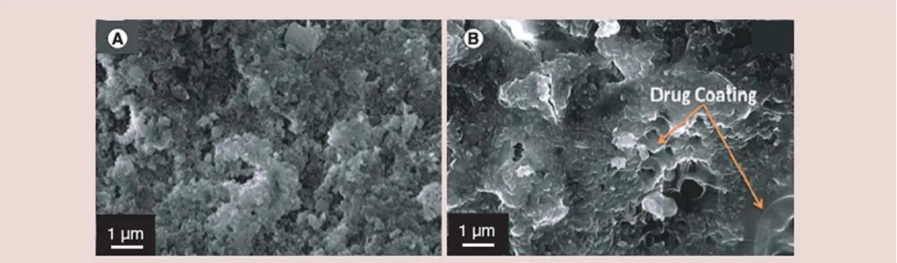
In our experimental work [53-59], hydrothermally converted coralline HAp particles containing nano and mesopores were loaded with medically active sub stances, which cover both the surfaces and intercon nected pores of the particles that ranged from a few hundred nanometers to micron sizes (Figure 3).
The influence of HAp particles within PLA matrix on the relea:se of gentamicin as well as its release kinet ics have been previously examined [59). lt was discov ered that the release kinetics of gentamicin appeared to obey "Power law Korsmeyer Peppas' mode! with mostly diffusional process through a number of different drug transport mechanisms. Statistical analysis revealed a very significant difference on the release of gentamicin between gentamicin-containing PLA (PLAGM) and gentamicin-containing HAp microspheres within PLA matrix (PLAHApGM).
These thin film composite coatings, such as PLA HApGM displays slower release rates than PLA matrix
Figure 2. (A) Foraminifera hydroxyapatite microsphere. (B) Enlarged degraded surface of Foraminifera hydroxyapatite structure within simulated physiological environment.
Figure 3. (A) Hydrothermal converted coralline hydroxyapatite surface. (B) Gentamicin-coated coralline hydroxyapatite surface.
alone. As stated earlier HAp and other calcium phos phates are also the source of Ca2• for the regeneration and repaie of diseased bone tissue. lt was also reported that the release profiles, exhibited an early burst stage and then a steady state release rate with significant antimicrobial activity against S. aureus (SHIO0O) even at high concentration of bacteria. The devices also indicated significant ability to control the growth of bacterial even after 4 weeks of drug release. It was sug gested that clinical release profiles can be easily tuned from drug-HAp physicochemical interactions and deg radation kinetics of polymer matrix. lt was concluded that the developed systems could be applied to prevent microbial adhesion to medical implant surfaces and to treat infections mainly caused by S. aureus in surgery.
lt was reported that the degradation of the poly meric network towards the end of the initial release
will 'favor' the dissolution of any drug residue. Accord ingly, this phenomenon (diffusion and degradation) should take place at the conclusion of surface drug release (when dissolution of drug have created second ary porosity inside the polymeric network and resul tant 'fragility' of it) and/or dependent on factors such has the surface area, dissolution rate and the molecular weight of the PLA film used.
For the coral converted to hydroxyapatite material (coralline HAp) structure contains meso- and nano pores and during the initial drug loading period gen tamicin penetrates the pores of the 'HAp-coral' and coats its surfaces and its porous network. The gentami cin contained in these porosities will be released gradu ally. Hence, the process of polymeric matrix thin film composites containing drug carrying particulate mat ter can be thought to proceed in three stages that can be named as 'progressive dissolution process'.
In the first stage (stage 1), the initial release (burst) of gentamicin can be regarded as the direct dissolution of surface bound drugs in water or the physiologie environ ment due to the exposed outer surface area and concen tration gradient. lt was shown in that the initial burst
took approximately 1 week for gentamicin. At this stage it can be assumed that the release is purely governed by diffusion of drugs from polymeric matrix surface (60,61]. In the second stage the dissolution is driven by the internai diffusion of drugs impregnated within the matrix possibly in the porous part of the matrix generated during preparation. For gentamicin release from PLAHApGM samples, this stage is preceded by 'lag phase' which occurs between 1 and 2 h of release. The presence of HAp loaded with gentamicin could in many ways hinder (or slow down) the release of gentamicin through these micropores. This stage is relatively much slower compared with the previous one due to the drug transporting through mainly from the surface areas and large pores of the particles. This stage proceeds with release from narrow pores of the nano and mesopores of the particles to the matrix and then to the environment. There is no degradation of particles at this stage but dissolution of the drugs only. The third stage involves slow degradation of the polymeric matrix combined with dissolution and deterioration of the particles (Figures 4 & 2B) and slow release of the added drugs or the minerais into the environment. This is terminal release phase or stage for loaded devices. At this stage, there is pore growth due to both mass loss by polymer degradation and pore coalescence (micropores coalescing (or joining) to form larger pores). In recent dissolution studies of BPs, for the device containing HAp (PLAHApBP), the slower dissolution rates observed were reported to be due to the strong bonding of BP to apatite (HAp) as well as encapsulation within the particles. It was ear lier reported that for BP containing calcium phosphate particles have strong affinity to the nanocrystalline apatites with adsorption phenomena that occurs at the surface of apatite crystals (62].
Antibacterial efficacy & biofilm formation Published work states that poor diffusion and penetra tion of antibiotic through the biofilm, contribute to
@
$ 0.97,-_-=pL=-A:---, � �PLAGM � O.S �PLAHAp .5: 0.7 -PLAHApGM u 10.6 � 0.5 -� 0.4.i
E o.3l::
0.2i
.2 0.1 f! o.o-;._--- -�-u.. 0 2 3 4 5 6 7 8 9 lime (weeks)Figure 4. Gentamicin drug delivery device produced from polylactic acid-coralline hydroxyapatite thin film composite. (A) The scanning electron microscopy image of the coralline HAp surface used for drug delivery (only the struts are used as drug delivery vehicles shown in a circled area), and (B) drug release profile of only the gentamicin within PLA (PLAGM) and HAp coralline particles loaded with gentamicin embedded within a PLA matrix (PLHApGM) showing lower amounts of drugs released as well as a longer release rate up to 8 weeks. GM: Gentamicin; HAp: Hydroxyapatite; PLA: Polylactic acid.
the persistence of biofilm infections especially those associated with implanted devices [63-66]. A number of reasons on the microbial resistance to antimicrobial agents have been postulated. An increase in the deple tion of oxygen and nutrients resulting into slow growth of bacteria, adaptive stress responses and formation of persister cells are hypothesized to constitute a multi layered defense. In recent research efforts the focus is directed toward disabling biofilm resistance, which may enhance the ability of existing antibiotics to treat infections involving biofilms [67). lt has been reported that in most cases, biofilm can be prevented aggres sively by antibiotic in their early stages and can also be treated by chronic suppressive therapy. Mah et al. [68)
suggested that the use of traditional antibiotics com bined with drug that interferes with biofilm-specific resistance would be the right approach to render biofilms more susceptible to treatment.
A recent study by our group has suggested that effec tive drug delivery devices with adequate slow release rates can be produced using converted coralline HAp particles loaded with medically active drugs and sub stances embedded within a PLA thin film matrix [48,58-59). To investigate the antibiotic delivery efficiency of
the composite thin films produced and observe the biofilm formation, we carried out a number of tests.
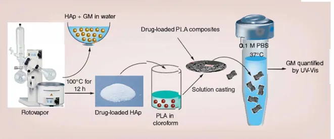
In this current work, we prepared PLA films and film composites based on the previously published work and loaded with gentamicin. Schematic repre sentation of the process is given in Figure 5. The films
were eut inro circular shape and glued on the sterilized cylindrical coupons. They were sterilized using UV for 40 min. Trytpic Soy Broth (TSB) for S. aureus and Mueller Hinton II BR cation adjusted media (MHB) for P. aerugonas were prepared by thoroughly dissolv ing 30 g of TSB powder (BactoTM TSB (BD) and 22 g of MHB powder in I I of polished 18 MQ (MilliQ, Millipore, Victoria, Australia) water, respectively. The solutions were autoclaved to sterilize (121°C, liquid cycle) and stored at room temperature.
Staphylococcus aureus (ATCC 25923) and
P. aeruginosa (ATCC 15692) were used for this study and were cultured in a shaking incubator, 250 rpm at 37°C under anaerobic and aerobic conditions, respec tively. For static biofilm formation, cells were grown in broth medium overnight, and diluted I: 100 into TSB and MHB media, respectively. These cell suspensions were inoculated into a 12-well plate containing tripli cate of samples glued on coupons. The plate was sealed with sterile breathable film (Aeraseal; Excel Scientific, CA, USA) and statically grown at 37°C, 5% C0
2 for 24 h. Biofilm samples were washed with PBS, stained using SYT09 Green fluorescent Nucleic Acid Stain (Life Technologies Corp, CA, USA) and fixed with 4% paraformaldehyde.
The morphological changes of the biofilms were analyzed using confocal laser scanning microscopy (CLSM; Nikon AI, Tokyo, Japan), using oil-immer sion lens (70 Objective lens and numerical aperture of 1.4 with Z-series images taken in 1.0 mm slices)
with NIS Elements Confocal software. A total of eight images were acquired randomly from each specimen.
In biofilm study, four biofilm image features calcu lated by COMSTAT [69) were chosen to characterize biofilm development by S. aureus and P. aeruginosa on
PLA thin film composites. These variables, biomass, average chickness, roughness coefficient and surface to biovolume ratio were selected for interpretation of biological and physical characceristics of biofilm [67] on these surfaces. Biomass represents the overall vol ume of the biofilm, and also provides an estimate of the biomass in the biofilm, average thickness provides a measure of the spatial size of the biofilm, roughness represents a measure of biofilm heterogeneity and sur face to biovolume ratio provided us how large a por tion of the biofilm is exposed to the nutrients flow. Recorded CLSM images were reconstructed using IMARIS imaging system (Bitplane AG, Zurich, Swit
zerland) for biofilm structural quantification in com puter statiscics software COMSTAT and presented as 3D structures.
The preliminary resulcs obcained from
5
days experiments for S. aureus (SHIO00), and Pseudomonas aeroginosa on PLA thin film samples are both strains ac day5
had grown into micro colonies, reflecced by their low surface to volume ratio compared with bio film at day 1. On the other hand, microscopy images of S. aureus on the films showed large and high micro colonies on PLA, PLAHAp and PLAHApGM samples while on PLAGM high single cells and small cell dus cers were observed (Figure 6). Confocal images on the composites without and wich gentamycin shows the intensity of the biofilm formation (Figure 6A & C) and the effect of the antibiotics after24
h (Figure 6B & D).le can be suggested that cells were able to attach on the surface of PLAGM samples but the drug released
from the surface suppresses the ability of bacteria to make biofilm.
Ir
could also be envisaged chat there is possibility under flow-biofilm-growth conditions chat more drugs would be released from the surface and may suppress the attachment of bacteria on the surfaces. Being high-level antimicrobial resistant in Gram-negacive, P. aeroginosa displays similar structurecharacceriscics on the surface of PLA thin films. Pseudomonas aeroginosa showed a stronger cendency
to form microcolonies on the surface of PLA films chan S. aureus, which was indicated by higher rough
ness coefficients. The higher surface to biovolume ratio of P. aeroginosa is an indication of its fiat growth
on the surface compared with S. aureus. This is also consistent with the lower average chickness of the biofilm it forms on the surface. le was also observed chat S. aureus grows faster chan P. aeroginosa on PLA surfaces indicated by lower surface to volume ratio, higher average thickness and hence higher biomass in the biofilm.
The effect of antibiocic in the films (PLAGM and PLAHApGM) against biofilm after
24
h seemed to be minimal for S. aureus and no at ail to P. aerogi nosa. This is possibly due the fact that starie conditions may influence low release rate of drugs from the device and also24
h is not enough cime to realize polymer degradation and significant release of antibiotic.In summary, ail of the PLA and PLA/HAp genta micin loaded chin films under experimental conditions
exhibiced significant ability to prevent bacterial growth
even at high concentration of baccerial. The prolonged ability to release drug from these films was tested by
subjecting films for
4
weeks under the same experimental conditions. Ir was observed that even after releasing
drugs for
4
weeks, films were still able to release enoughgentamicin to prevent microbial accivicies.
HAp + GM in water
O O "" ""' ooooc, Drug-loaded PLA composites Ooc,
Rotovapor Drug-loaded HAp
0 0 0 ooo PLA in cloroform b.1 M PBS GM quantified - byUV-Vis
Figure 5. Schematic representation of the drug loading and release method used in this current work. GM: Gentamycin; HAp: Hydroxyapatite; PBS: Phosphate-buffered saline; PLA: Polylactic acid.
50 100 150 200 250 300 350 400 450 500 50 100 150 200 250 300 350 400 450 500
lntensity map reflecting biofilm thickness (scaled}
50 100 150 200 250 300 350 400 450 500
lntensity map reflecting biofilm thickness (scaled}
50 100 150 200 250 300 350 400 450 500
Figure 6. Confocal microscopy images showing 24 h biofilm growth of Staphylococcus aureus. Biofilm growth on (A) PLAHAp, (B) PLAHApGM films and their intensity map reflecting biofilm thickness in (C &D), respectively, where (B & D) reflects the effectiveness of the gentamicin-loaded composites.
GM: Gentamycin; HAp: Hydroxyapatite; PLA: Polylactic acid. Conclusion & future perspective
At the moment, we are gradually discovering new ways of reproducing structures that are commonly found in nature with desirable properties. The use of biological microstructures is one versatile approach for the repro duction of inorganic structures with identical features. This is achieved by using techniques in biomineral inspired self-repairing materials chemistry. Multifunc tional and multilayered nano coatings and assembly will assise us in this endeavor.
Slow drug delivery and tissue engineering are two common tools chat could assise to help us to solve a number of bone related deficiencies and repaie. Cur rently, the demand is clear for becter tissue engineering scaffolds that possess more natural bioresponsive envi ronments favorable in guiding the natural processes of regeneration. To meet this biological challenge, nove! design and synthesis steps must be incorporated into the new generation scaffolds. We believe that there needs to be a new thinking in tissue scaffold systems
that are responsive in which the synthesized bioma trix evolves in real-time to meet the requirements and optimize for the adaptive growth and regeneration of human tissues, while delivering the right biogene tic materials, pharmaceuticals and minerais. Con sequently, the environments of the scaffolds will be further adjusted as cells proliferate and differentiate. 3D printing methods are currently used for a number of inorganic and metallic materials and stem cells and other biologie structures are incorporated to these new generation 3D materials for organ regeneration and repa1r.
During the last decade atomic force microscopy was used as a powerful platform in nanomedicine for studying and controlling the forces involved in ce!! adhesion and biofilm formation. Atomic force microscopy is well regarded in measuring the small interaction forces between a sharp probe and the sur face of a sample. A relatively more recent equipment the single-cell force spectroscopy (SCFS), a single
cell can be attached on the probe in order to mea-sure the interaction forces between the cell probe and a solid substrate or another cells. SCFS-based tech-niques have recently established as an important tool for understanding how microbial pathogens attach to surfaces and form biofilms [64]. This can be a turn-ing point in our fight against biofilms and infections. SCFS assays will allow us to clarify the specific and nonspecific forces driving cell adhesion on a single-cell basis. Future work in this area will open new ave-nues for the development of new tools, devices, meth-ods and multifunctional coatings capable of detecting and destroying biofilms.
Nevertheless, nanosynthesis based on biological principles of design and assembly is still in its infancy. Better understanding of biofilm formation and the cell forces is pertinent. The use of bio-inspired nanofab-rication techniques including multifunctional multi-layer coatings in nanoscale thicknesses for slow drug delivery and tissue repair is a unique approach. This has enormous potential to improve scaffold or
lab-on-chip designs with the capability to diagnose, identify, stop biofilm formation, self-repair, micro-evolve and osteointegrate fully.
Acknowledgements
The authors would like to give special thanks to B Milthorpe, Faculty of Science UTS and R Cavaliere of iThree Institute, UTS for their assistance on biofilm work and for their continued support in search of the most appropriate biomaterial. Financial & competing interests disclosure
The authors would like to acknowledge and thank the Austra-lian Academy of science-Horizon2020 grant and the Univer-sity of Technology Sydney for the partial financial support of one of the authors during his PhD studies. The authors have no other relevant affiliations or financial involvement with any organization or entity with a financial interest in or financial conflict with the subject matter or materials discussed in the manuscript apart from those disclosed.
No writing assistance was utilized in the production of this manuscript.
Executive summary Background
• Currently, researchers are focusing on the fabrication of new nanocoatings, nanomaterials, nanolaminates and thin film nanocomposites that are appropriate for applications such as drug delivery and tissue engineering.
• Innovative approaches driven by nanotechnology are now available for the production of synthetic bone-like calcium phosphate nanomaterials.
Surface modifications and liposomes for drug delivery applications
• The appropriate circulation time in addition to the dissolution rates within the human body is the main issue for drug carriers containing nanoparticles.
• The surfaces of nanostructured materials such as nanocoatings can be modified and functionalized with several types of reagents via various biological, chemical and/or physical methods.
• The surface modification approach can be used to achieve functional improvements by design, better osteointegration and stability of nanomaterials and nanocoatings in aqueous media.
Infection & osteomyelitis
• Treatment of osteomyelitis can be with immobilization and antibiotic therapy with a number of drugs including gentamicin.
• Modification or development of thin film or nanocoated multilayer devices with surface properties that have an effect against microbial adhesion or viability seems to be a promising approach for the prevention of device-related infections.
• Bacteria adhesion is a complex phenomenon affected by factors such as the properties of surface materials and characteristics of the bacteria.
Bone repair drug delivery systems & osteomyelitis
• The delivery of antibiotics has become a major focus in research for the use in the treatment of bone infections or as prevention against infection during surgical interventions.
• Although not fully satisfactory, the implantation of antibiotic-loaded PMMA microspheres and calcium sulfate based microspheres and powders into the infection site are the approach currently being used in both orthopedics and in maxillofacial surgery.
• Biodegradable calcium phosphate drug delivery system would be better suited to this endeavor as it can also introduce calcium and phosphate ions and range of minerals to assist bone growth.
Biodegradable composite thin films
• The use of biodegradable polymer thin films is advantageous in the fight against bacterial infection. • This is due to their tendency to uptake and release pharmaceuticals and minerals as well as the capability to
References
Papers of special note have been highlighted as: • of interest; •• of considerable interest
1 Albee FH, Morrison HF. Studies in bone growth: triple CaP as a stimulus to osteogenesis. Ann. Surg. 71, 32–39 (1920). 2 Hulbert SF, Young FA, Mathews RS et al. Potential of
ceramic materials as permanently implantable skeletal prostheses. J. Biomed. Mater. Res. 4, 433–456 (1970). 3 Choi AH, Ben-Nissan B. Calcium phosphate nanocoatings
and nanocomposites, part I: recent developments and advancements in tissue engineering and bioimaging. Nanomedicine 10, 2249–2261 (2015).
•• Review discusses the current applications of calcium phosphate nanocoatings and nanocomposite in tissue engineering and bioimaging applications.
4 Choi AH, Cazalbou S, Ben-Nissan B. Nano-biomaterials coatings in dentistry. In: Biomaterials for Oral and Craniomaxillofacial Applications. Deb S (Ed.). Frontiers of Oral Biology Series. Karger Publishing, Basel, Switzerland. 17, 49–61 (2015).
5 Choi AH, Ben-Nissan B, Conway RC et al. Advances in calcium phosphate nanocoatings and nanocomposites. In: Advances in Calcium Phosphate Biomaterials. Ben-Nissan B (Ed.). Springer Series in Biomaterials Science and Engineering (SSBSE), Berlin, Heidelberg, Germany, 485–511 (2013).
6 Choi AH, Ben-Nissan B. Sol-gel production of bioactive nanocoatings for medical applications. Part II: current research and development. Nanomedicine 2, 51–61 (2007). 7 Ben-Nissan B, Choi AH. Sol-gel production of bioactive
nanocoatings for medical applications. Part I: an introduction. Nanomedicine 1, 311–319 (2006). •• Discusses the applications of calcium phosphate
nanocoatings for medical applications.
8 Daculsi G, Fellah BH, Miramond T. The essential role of calcium phosphate bioceramics in bone regeneration. In: Advances in Calcium Phosphate Biomaterials. Ben-Nissan B (Ed.). Springer Series in Biomaterials Science and Engineering (SSBSE), Berlin, Heidelberg, Germany, 71–96 (2013).
9 Gibson IR. Calcium phosphate as scaffolds for mesenchymal stem cell. In: Bimaterials and Stem Cells in Regenerative Medicine. Ramalingam M, Ramakrishina S, Best S (Eds). CRC Press, FL, USA, 219–237 (2012).
10 El-Ghannam A, Ricci K, Malkawi A et al. A ceramic-based anticancer drug delivery system to treat breast cancer. J. Mater. Sci. Mater. Med. 21, 2701–2710 (2010).
11 UskokoviĆ V, Batarni SS, Schweicher J et al. Effect of calcium phosphate particle shape and size on their antibacterial and osteogenic activity in the delivery of antibiotics in vitro. ACS Appl. Mater. Interfaces 5, 2422–2431 (2013).
12 Kester M, Heakal Y, Fox T et al. Calcium phosphate nanocomposite particles for in vitro imaging and
encapsulated chemotherapeutic drug delivery to cancer cells. Nano Lett. 8, 4116–4121 (2008).
13 Bastakoti BP, Hsu YC, Liao SH et al. Inorganic-organic hybrid nanoparticles with biocompatible calcium phosphate
thin shells for fluorescence enhancement. Chem. Asian J. 8, 1301–1305 (2013).
14 Chen Z, Li Z, Lin Y et al. Biomineralization inspired surface engineering of nanocarriers for pH-responsive, targeted drug delivery. Biomaterials 34, 1364–1371 (2013).
15 Mukesh U, Kulkarni V, Tushar R et al. Methotrexate loaded self stabilized calcium phosphate nanoparticles: a novel inorganic carrier for intracellular drug delivery. J. Biomed. Nanotechnol. 5, 99–105 (2009).
16 Rout SR, Behera B, Maiti TK et al. Multifunctional magnetic calcium phosphate nanoparticles for targeted platin delivery. Dalton Trans. 41, 10077–10083 (2012).
17 Zhao XY, Zhu YJ, Chen F et al. Calcium phosphate hybrid nanoparticles: self-assembly formation, characterization, and application as an anticancer drug nanocarrier. Chem. Asian J. 8, 1306–1312 (2013).
18 Liang P, Zhao D, Wang CQ et al. Facile preparation of heparin/CaCO3/CaP hybrid nano-carriers with controllable size for anticancer drug delivery. Colloids Surf. B Biointerfaces 102, 783–788 (2013).
19 Li WM, Chen SY, Liu DM. In situ doxorubicin-CaP shell formation on amphiphilic gelatin-iron oxide core as a multifunctional drug delivery system with improved cytocompatibility, pH-responsive drug release and MR imaging. Acta. Biomater. 9, 5360–5368 (2013). 20 Gonzalez-McQuire R, Green DW, Partridge KA et al.
Coating of human mesenchymal cells in 3D culture with bioinorganic nanoparticles promotes osteoblastic differentiation and gene transfection. Adv. Mater. 19, 2236–2240 (2007).
21 Ahymah Joshy MI, Elayaraja K, Suganthi RV et al. In vitro sustained release of amoxicillin from lanthanum hydroxyapatite nano rods. Curr. Appl. Phys. 11, 1100–1106 (2011).
22 Chou J, Valenzuela SM, Green DW et al. Antibiotic delivery potential of nano and micro porous marine structures derived β-TCP spheres for medical applications. Nanomedicine 9, 1131–1138 (2014).
23 Teller M, Gopp U, Neumann HG et al. Release of gentamicin from bone regenerative materials: an in vitro study. J. Biomed. Mater. Res. B Appl. Biomater. 81, 23–29 (2007).
24 Olton D, Li J, Wilson ME et al. Nanostructured calcium phosphates (NanoCaPs) for non-viral gene delivery: influence of the synthesis parameters on transfection efficiency. Biomaterials 28, 1267–1297 (2007).
25 Zhou C, Yu B, Yang X et al. Lipid-coated nano-calcium-phosphate (LNCP) for gene delivery. Int. J. Pharm. 392, 201–208 (2010).
26 Liu Y, Wang T, He F et al. An efficient calcium phosphate nanoparticle-based nonviral vector for gene delivery. Int. J. Nanomedicine 6, 721–727 (2011).
27 Pittella F, Miyata K, Maeda Y et al. Pancreatic cancer therapy by systemic administration of VEGF siRNA contained in calcium phosphate/charge-conversion polymer hybrid nanoparticles. J. Control. Release 161, 868–874 (2012).
28 Li J, Chen YC, Tseng YC et al. Biodegradable calcium phosphate nanoparticle with lipid coating for systemic siRNA delivery. J. Control. Release 141, 416–421 (2010). 29 Singh RK, Kim TH, Patel KD et al. Development of
biocompatible apatite nanorod-based drug-delivery system with in situ fluorescence imaging capacity. J. Mater. Chem. B 2, 2039–2050 (2014).
30 Hanifi A, Fathi MH, Mir Mohammad Sadeghi H. Effect of strontium ions substitution on gene delivery related properties of calcium phosphate nanoparticles. J. Mater. Sci. Mater. Med. 21, 2601–2609 (2010).
31 Hanifi A, Fathi MH, Sadeshi HM et al. Mg2+ substituted calcium phosphate nano particles synthesis for non viral gene delivery application. J. Mater. Sci. Mater. Med. 21, 2393–2401 (2010).
32 Han JY, Tan TTY, Loo JSC. Utilizing inverse micelles to synthesize calcium phosphate nanoparticles as nano-carriers. J. Nanopart. Res. 13, 3441–3454 (2011).
33 Paul W, Sharma CP. Fatty acid conjugated calcium phosphate nanoparticles for protein delivery. Int. J. Appl. Ceram. Technol. 7, 129–138 (2010).
34 Victor SP, Sharma CP. Calcium phosphates as drug delivery systems. J. Biomater. Tissue Eng. 2, 269–279 (2012). • Discusses the various types of calcium phosphates, their
properties and the different systems that have been used for drug and antibiotic delivery.
35 Xu T, Zhang N, Nichols HL et al. Modification of nanostructured materials for biomedical applications. Mater. Sci. Eng. C 27, 579–594 (2007).
36 Xu Q, Tanaka Y, Czernuszka JT. Encapsulation and release of a hydrophobic drug from hydroxyapatite coated liposomes. Biomaterials 28, 2687–2694 (2007).
37 Anada T, Takeda Y, Honda Y et al. Synthesis of calcium phosphate-binding liposome for drug delivery. Bioorg. Med. Chem. Lett. 19, 4148–4150 (2009).
38 Zhu CT, Xu YQ, Shi J et al. Liposome combined porous β-TCP scaffold: preparation, characterization, and anti-biofilm activity. Drug Deliv. 17, 391–398 (2010). 39 Huang JS, Liu KM, Chen CC et al. Liposomes-coated
hydroxyapatite and tricalcium phosphate implanted in the mandibular bony defect of miniature swine. Kaohsiung J. Med. Sci. 13, 213–228 (1997).
40 Wang G, BabadaĞli ME, UludaĞ H. Bisphosphonate-derivatized liposomes to control drug release from collagen/ hydroxyapatite scaffolds. Mol. Pharm. 8, 1025–1034 (2011).
41 Al-Jamal WT, Kostarelos K. Liposome-nanoparticle hybrids for multimodal diagnostic and therapeutic applications. Nanomedicine 2, 85–98 (2007).
42 Lewis K. The development of liposome encapsulated calcium phosphates for bone regeneration. [PhD thesis]. University of Technology, Sydney, Australia (2010).
43 Ibara A, Miyaji H, Fugetsu B et al. Osteoconductivity and biodegradability of collagen scaffold coated with nano-β-TCP and fibroblast growth factor 2. J. Nanomater. 2013, Article ID 639502 (2013).
44 Ben-Nissan B. Natural bioceramics: from coral to bone and beyond. Curr. Opin. Solid State Mater. Sci. 7, 283–288 (2003).
•• Discusses the production and use of natural bioceramics and calcium phosphate nanocoatings for applications in hard tissue replacement.
45 Milev A, Kannangara GSK, Ben-Nissan B. Morphological stability of plate-like hydroxyapatite. Key Eng. Mater. 240– 242, 484–487 (2003).
46 Choi AH, Ben-Nissan B, Matinlinna JP et al. Current perspectives: calcium phosphate nanocoatings and nanocomposite coatings in dentistry. J. Dent. Res. 92, 853–859 (2013).
47 Healthcare-associated infections. http://ecdc.europa.eu/en/healthtopics
48 An YH, Friedman RJ. Concise review of mechanisms of bacterial adhesion to biomaterial surfaces. J. Biomed. Mater. Res. 43, 338–348 (1998).
49 Kargupta R, Bok S, Darr CM et al. Coatings and surface modifications imparting antimicrobial activity to orthopedic implants. Wiley Interdiscip. Rev. Nanomed. Nanobiotechnol. 6, 475–495 (2014).
50 Evans RP, Nelson CL, Harrison BH. The effect of wound environment on the incidence of acute osteomyelitis. Clin. Orthop. 286, 289–297 (1993).
51 Ben-Nissan B, Green DW. Marine materials in drug delivery and tissue engineering: from natural role models, to bone regeneration and repair and slow delivery of therapeutic drugs, proteins and genes. In: Marine Biomaterials. Kim SK (Ed.). Taylor and Francis, CSR Books, FL, USA, 575–602 (2013). 52 Chou J, Hao J, Ben-Nissan B et al. Coral exoskeletons as a precursor material for the development of calcium phosphate drug delivery system for bone tissue engineering. Biol. Pharm. Bull. 36, 1–4 (2013).
53 Ben-Nissan B, Green DW. Marine structures as templates for biomaterials. In: Advances in Calcium Phosphate Biomaterials. Ben-Nissan B (Ed.). Springer Series in Biomaterials Science and Engineering (SSBSE), Berlin, Heidelberg, Germany, 391–414 (2013).
54 Chou J, Hao J, Hatoyama H et al. Effect of biomimetic zinc-containing tricalcium phosphate (Zn–TCP) on the growth and osteogenic differentiation of mesenchymal stem cells. J. Tissue Eng. Regen. Med. 9(7), 852–858 (2014). 55 Chou J, Ben-Nissan B, Green DW et al. Targeting and
dissolution characteristics of bone forming and antibacterial drugs by harnessing the structure of micro-spherical shells from coral beach sand. Adv. Eng. Mater. 13, 93–99 (2011). 56 LeGeros RZ. Calcium phosphate-based osteoinductive
materials. Chem. Rev. 108, 4742–4753 (2008).
57 Macha IJ, Cazalbou S, Ben-Nissan et al. Marine structure derived calcium phosphate-polymer biocomposites for local antibiotic delivery. Mar. Drugs 13, 666–680 (2015). 58 Macha IJ, Cazalbou S, Ben-Nissan B et al. Development
and dissolution studies of bisphosphonate (clodronate)-containing hydroxyapatite-polylactic acid biocomposites for slow drug delivery. J. Tissue Eng. Regen. Med. doi: 10.1002/ term.2066 (2015) (Epub ahead of print).
59 Macha IJ, Ben-Nissan B, Milthorpe B. Improvement of elongation in nanosurface modified bioglass/PLA thin film composites. Curr. Nanoscience 10, 200–204 (2014). 60 Severino P, Santana MHA, Malmonge SM et al. Polymers
for drug delivery systems formulations. Polimeros Ciencia E Tecnologia 21, 361–368 (2011).
61 Liechty WB, Kryscio DR, Slaughter BV et al. Polymers for drug delivery systems. Annu. Rev. Chem. Biomol. Eng. 1, 149–173 (2010).
62 Pascaud P, Bareille R, Bourget C et al. Interaction between a bisphosphonate, tiludronate and nanocrystalline apatite: in vitro viability and proliferation of HOP and HBMSC cells. Biomed. Mater. 7, 054108 (2012).
63 Stewart PS. Mechanisms of antibiotic resistance in bacterial biofilms. Int. J. Med. Microbiol. 292, 107–113 (2002). 64 Dufrene YF. Understanding forces in biofilms. Nanomedicine
10, 1219–1221 (2015).
65 Hoiby N, Bjarnsholt T, Givskov M et al. Antibiotic resistance of bacterial biofilms. Int. J. Antimicrob. Agents 35, 322–332 (2010).
66 Stigter M, Bezemer J, de Groot K et al. Incorporation of different antibiotics into carbonated hydroxyapatite coatings on titanium implants, release and antibiotic efficacy. J. Control. Release 99, 127–137 (2004).
67 Stigter M, de Groot K, Layrolle P. Incorporation of tobramycin into biomimetic hydroxyapatite coating on titanium. Biomaterials 23, 4143–4153 (2002). 68 Mah TF, Pitts B, Pellock B et al. A genetic basis for
Pseudomonas aeruginosa biofilm antibiotic resistance. Nature 426, 306–310 (2003).
69 Heydorn A, Nielsen AT, Hentzer M et al. Quantification of biofilm structures by the novel computer program COMSTAT. Microbiology 146, 2395–2407 (2000).