Publisher’s version / Version de l'éditeur:
Physical Review Letters, 88, 17, pp. 173903-1-173903-4, 2002-04-16
READ THESE TERMS AND CONDITIONS CAREFULLY BEFORE USING THIS WEBSITE.
https://nrc-publications.canada.ca/eng/copyright
Vous avez des questions? Nous pouvons vous aider. Pour communiquer directement avec un auteur, consultez la première page de la revue dans laquelle son article a été publié afin de trouver ses coordonnées. Si vous n’arrivez pas à les repérer, communiquez avec nous à [email protected].
Questions? Contact the NRC Publications Archive team at
[email protected]. If you wish to email the authors directly, please see the first page of the publication for their contact information.
Archives des publications du CNRC
This publication could be one of several versions: author’s original, accepted manuscript or the publisher’s version. / La version de cette publication peut être l’une des suivantes : la version prépublication de l’auteur, la version acceptée du manuscrit ou la version de l’éditeur.
For the publisher’s version, please access the DOI link below./ Pour consulter la version de l’éditeur, utilisez le lien DOI ci-dessous.
https://doi.org/10.1103/PhysRevLett.88.173903
Access and use of this website and the material on it are subject to the Terms and Conditions set forth at
Attosecond streak camera
Itatani, J.; Quere, F.; Yudin, G. L.; Ivanov, M. Yu; Krausz, F.; Corkum, P. B.
https://publications-cnrc.canada.ca/fra/droits
L’accès à ce site Web et l’utilisation de son contenu sont assujettis aux conditions présentées dans le site LISEZ CES CONDITIONS ATTENTIVEMENT AVANT D’UTILISER CE SITE WEB.
NRC Publications Record / Notice d'Archives des publications de CNRC:
https://nrc-publications.canada.ca/eng/view/object/?id=bc8b5b86-6d18-4ce3-bc22-a04ef044bb3d https://publications-cnrc.canada.ca/fra/voir/objet/?id=bc8b5b86-6d18-4ce3-bc22-a04ef044bb3d
Attosecond Streak Camera
J. Itatani,1F. Quéré,1 G. L. Yudin,1M. Yu. Ivanov,1F. Krausz,2and P. B. Corkum1
1Steacie Institute for Molecular Sciences, National Research Council of Canada, Ottawa, Ontario, Canada K1A 0R6 2Institute für Photonik, Technische Universität Wien, Gusshausstrasse 27, A-1040 Wien, Austria
(Received 29 August 2001; published 16 April 2002)
An electron generated by x-ray photoionization can be deflected by a strong laser field. Its energy and angular distribution depends on the phase of the laser field at the time of ionization. This phase dependence can be used to measure the duration and chirp of single sub100-attosecond x-ray pulses.
DOI: 10.1103/PhysRevLett.88.173903 PACS numbers: 42.65.Ky, 41.50. +h Historically, advances in our ability to measure fast
phe-nomena have led to corresponding scientific advances [1]. It is now possible to use the physics of high harmonic gen-eration for producing single attosecond 共10218 sec, asec兲
pulses in the extreme ultraviolet to soft x-ray regions [2,3]. The next frontier will be to utilize such ultrashort pulses to probe the electronic dynamics of atoms and molecules which occurs on the attosecond time scale [4]. However, one impediment for experimentally demonstrating attosec-ond pulses has been the problem of finding a good method for their characterization [5– 7].
We present a method to determine the duration and chirp of a single attosecond x-ray pulse, based on the ionization of atoms by the x-ray photons in the presence of a strong low-frequency laser field. This method relies on two basic ideas. (i) The subcycle oscillation of the laser electric field is used as a time reference to determine the duration of the x-ray pulse. This can be done only if the x-ray pulse is shorter than the period of the oscillating field. (ii) The photoelectron signal generated by the x-ray pulse is resolved in energy and angle simultaneously. Depending on the laser-field polarization, the information on the pulse duration is contained in the width of the energy spectrum at a given observation angle (linear polarization), or in the width of the angular distribution at a given energy (circular polarization). We show that the resolution limit depends on the photon energy, bandwidth, and chirp of the x-ray pulse. The resolution improves with increasing photon energy, and is on the order of 70 asec at 100 eV for transform-limited pulses.
A group including two of the authors has implemented a closely related configuration, where the photoelectrons are collected in the direction perpendicular to the laser polarization, and successfully characterized x-ray pulses shorter than one oscillation period of the laser field used for their generation [3,8].
The ionization of atoms by x rays in the presence of laser fields has been used for several years to measure the du-ration of x-ray pulses longer than the optical period of the laser field. In these measurements, Auger electrons [9] or x-ray photoelectrons [10– 12] are dressed by the laser field to cross correlate the x-ray pulse and the envelope of the laser pulse. Two effects are used for this cross correlation.
(i) The appearance of sidebands in the photoelectron en-ergy spectrum due to the emission and absorption of laser photons by the x-ray generated photoelectrons. (ii) The ponderomotive shift of these peaks, due to the ac Stark shift of the continuum induced by the laser field.
As shown later, as the pulse duration falls below one period of the laser radiation, the sidebands broaden and merge, and the ponderomotive shift is no longer observ-able. Therefore, these cross-correlation methods cannot be directly extended to subcycle x-ray pulses.
By analyzing the dynamics of the electrons, we will show how the laser field affects the photoelectron dis-tribution in the case of subcycle x-ray pulses, and how this effect can be used to characterize these pulses. The large difference in frequencies between the x-ray pulse VX and the laser pulse vL naturally divides the
two-color ionization process in two steps: absorption of an x-ray photon followed by acceleration in the laser field [13]. The x ray produces an electron with a kinetic energy W0 苷 mey
2
0兾2 苷 ¯hVX 2 Ip, where Ip is the
ionization potential and v0 is the initial velocity. If the Up ¿ ¯hvL and ¯hVX ¿ Ip, classical mechanics can be
used to determine the time-dependent velocity v共t兲 in the field [14,15]. For the laser electric field EL共t兲 苷 E0共t兲兾
p
11 ´2兵cos共v
Lt1 w兲ex1 ´sin共vLt1 w兲ey其 with
ellip-ticity ´, this velocity is expressed as
v共t兲 苷 2 e me A共t兲1 ∑ v0 1 e me A共ti兲 ∏ , (1)
where A共t兲 is the vector potential of the field, EL 苷 2≠A兾≠t, and ti is the time of ionization.
In Eq. (1) the first term describes the electron’s quiver motion and goes to zero as the laser pulse ends. The second term is determined by the initial condition v共ti兲 苷 v0at the
time of ionization. This term is the final drift velocity vf 苷
v01 共e兾me兲A共ti兲 measured after the laser pulse. The fact
that vf is different from v0 is the classical equivalent to
the absorption, emission, and scattering of laser photons by the photoelectron [13,16].
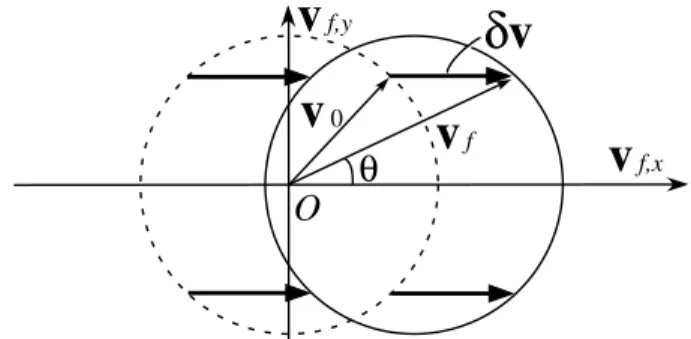
The dashed circle of radius y0in Fig. 1 shows the
elec-tron velocity distribution in the xy plane 共y0,z 苷 0兲 pro-duced by the x rays alone. The solid circle in Fig. 1 represents the drift velocity distribution in the presence of
v
f,yv
f,xv
0v
fδv
O
θ
FIG. 1. Effect of a strong laser field on the photoelectrons
ion-ized by the x-ray pulse at a given phase fi. Dashed circle — drift
velocity distribution 共y0x,y0y兲 of the photoelectrons without the
laser field; solid circle — distribution with the laser field. Note
that neither the uncertainty in v0 due to the bandwidth of the
x-ray pulse nor the angular dependence of the emission proba-bility are represented for simplicity.
the laser field. It is a translation of the dashed circle by dv 苷 共e兾me兲A共ti兲. For linearly polarized light 共´ 苷 0兲,
the translation is parallel to the electric field, and the elec-tron distribution sweeps back and forth along the field di-rection as ti varies. For circularly polarized light 共´ 苷
61兲, the electron distribution is displaced perpendicular to the laser electric field at the moment of birth, and it rotates around the dashed circle as the time of ionization is varied. For all polarizations, electrons that are freed at slightly different times ti will have different final velocities
be-cause of the dependence of vf on the optical phase fi苷 vLti 1 w. It is this phase dependence that offers an
op-portunity for the measurement of subcycle duration. To evaluate the temporal resolution, it is convenient to refor-mulate Eq. (1) in terms of the drift kinetic energy K 苷
meyf2兾2 for electrons moving in the xy plane:
KL 苷 W01 2Upcos 2u sin2fi 6 aL q 8W0Up cosu sinfi, KC 苷 W02 Upcos 2共fi 2 u兲 6 aC q 4W0Up sin共fi 2 u兲 , (2)
aL 苷 兵1 2 共2Up兾W0兲 sin2usin2fi其1兾2, aC 苷 兵1 2 共Up兾2W0兲 2 共Up兾2W0兲 cos关2共fi2 u兲兴其1兾2.
Here KL, KC are the kinetic energies for linear and
cir-cular polarization of the laser field, respectively; Up 苷
e2E02共ti兲兾4mev 2
L is the ponderomotive potential of the
laser field at the time of ionization; u is the angle of ob-servation measured from the x axis as shown in Fig. 1; aL
and aCare correction terms to the expression given in [8],
and deviate from 1 when Up becomes comparable to or
higher than W0. If the field is increased to Up . W0兾2
(linear polarization) or Up . W0 (circular polarization),
some of the photoelectrons are deflected into the reverse direction from v共ti兲 苷 v0. The branches of negative signs
account for these backscattered electrons, and are required only for this high intensity regime. In this case, an-other peak appears on the lower energy side of photo-electron spectra, which makes their interpretation more complex. Hereafter, we will restrict the discussion to
Up , W0兾2 (linear polarization) and Up , W0 (circular
polarization).
There are many possibilities for measurement because the photoelectron spectra depend on the observation angle and the laser polarization. We will concentrate on three configurations. For a linearly polarized field, if we ob-serve only electrons with vf parallel to the laser
polariza-tion, Eq. (2) shows that the drift energy will sweep above and below W0 as the phase of birth varies from 0 to 2p.
If we observe only electrons with vf perpendicular to the
laser polarization, Eq. (2) shows that it will sweep only below W0 twice per laser period. Measurements that are
angle integrated will have a degraded resolution. In cir-cularly polarized light, if we observe only electrons of a given energy, then their direction will sweep. For any po-larization, measurements that are energy integrated [5] will have a degraded resolution.
With a linearly polarized field, the width DE of the angle-resolved photoelectron spectrum is determined by the following factors: (i) the variation of the moment of birth of photoelectrons due to the x-ray pulse duration, and (ii) the bandwidth and the chirp of the x-ray pulse. Thus, the duration and chirp can be determined by comparing the spectra with and without the laser field.
For a transform-limited x-ray pulse, the width of a photoelectron spectrum is given as DE关tX,D共 ¯hVX兲兴 苷
兵共j≠K兾≠fijvLtX兲21 关j≠K兾≠W0jD共 ¯hVX兲兴2其1兾2 provided
that the streaking speed ≠K兾≠fi is constant within
the x-ray pulse. Within one optical period, the best temporal resolution is achieved at the phase that maximizes the resolving parameter bL ⬅
共j≠K兾≠fijvLtX兲兾兵j≠K兾≠W0jD共 ¯hVX兲其. It occurs at fi苷 mp for u 苷 0 or fi苷 共m兾2 1 1兾4兲p for u 苷 p兾2, respectively. bL scales as y0E0共ti兲t 2 X for u 苷 0 and E02共ti兲t 2
X for u 苷 p兾2. Physically, as the x-ray pulse
duration decreases, its bandwidth increases as ~ 1兾tX
and the contribution of sweeping to the photoelectron bandwidth DE decreases as ~ tX.
We set the condition for the resolution limit as bL 苷 1, i.e., the x-ray bandwidth is equal to the broadening by the laser streak. This is the criterion used in traditional streak cameras. For transform-limited x-ray pulses at 100 eV, the resolution limit is 70 asec for u 苷 0 and 100 asec for u 苷 p兾2. Here we assumed helium as an ionizing atom (Ip 苷 24.6 eV), laser wavelength lL 苷 0.8 mm, and the intensity of the laser field set by Up 苷 W0兾2共I 苷 6.3 3
1014 W兾cm2兲.
One drawback of using a linearly polarized laser field is that the streaking speed ≠K兾≠fi varies within the laser
period, as seen in Eq. (2). If the x-ray pulse duration
becomes close to the streaking period, the streaking speed will change within the x-ray pulse, which will make the data interpretation more complicated. This difficulty can be removed by using a circularly polarized laser field.
With circularly polarized light, a photoelectron at a given kinetic energy streaks in angle as shown in Eq. (2). This angular spread Du of photoelectron distribution in the xy plane can be used to measure the duration of x-ray pulses. The main advantage of this configuration is that the streaking speed is constant over the whole optical cy-cle, and is automatically calibrated by the frequency of the laser field. This method generalizes the concept of the at-tosecond streak camera [5], which is based on the measure-ment of the angular distribution of photoelectrons without resolving the kinetic energy. Energy-integrated measure-ments require Up ¿ W0, a condition difficult to achieve
experimentally. The temporal resolution of this configura-tion is again limited by the bandwidth of the x-ray pulse and is similar to that for linear polarization (70 asec for 100 eV photons).
The electron spectrum can also be calculated quantum mechanically [atomic units are used for compactness in Eqs. (3) and (4)]. Following [17], the amplitude of popu-lating a statejv典 with kinetic momentum v at a moment T after the end of both the laser and the x-ray pulses is
av共T兲 苷 2i Z T 2` dt dp共t兲EX共t兲e 2iRT tdt 0共1兾2关p共t0兲兴21I p兲 , (3) where p共t兲 苷 v 1 A共t兲 is the instantaneous kinetic mo-mentum, and dp共t兲 is the dipole transition matrix element
from the ground state to the continuum with kinetic mo-mentum p共t兲. The x-ray field EX共t兲 includes the fast
os-cillations of the carrier, chirp, and the pulse envelope. We assume that the x-ray pulse is a linearly chirped Gaussian, with dimensionless chirp j defined in the spectral domain as ˜EX共V兲 ⬃ exp关2共V 2 VX兲2t2共1 2 ij兲兾2兴. In time domain, positive chirp j . 0 corre-sponds to the instantaneous frequency increasing with time: EX共t兲 ⬃ exp关2共t 2 t0兲2共1 1 ij兲兾2t2共1 1 j2兲 2 iVXt兴, where t0 is the peak of the x-ray pulse and its
duration is tX ⬃ t
p
11 j2. For a sufficiently short x-ray
pulse, Eq. (3) can be approximated using the saddle point method, so we have javj2 ~ jdp共t0兲EX共t0兲j 2 m exp ∑ 2µ p 2共t 0兲 2 2 W0 ∂2 t2 m2 ∏ , m 苷 q 共1 1 hj兲2 1 h2, h 苷 E L共t0兲p共t0兲t2. (4) Equations (3) and (4) apply for all directions of observa-tion and laser polarizaobserva-tions. In the field-free case m 苷 1 and Eq. (4) corresponds to the spectrum of the x-ray pulse shifted by 2Ip. Equation (4) shows that, in the presence
of the laser pulse, the width of the electron spectrum de-pends on EL共t0兲 at the peak of the x-ray pulse. To measure
the x-ray pulse duration, one must produce an observable change in m.
Figure 2a shows photoelectron spectra in the direction parallel to the laser polarization produced by 70 asec (full width at half maximum, FWHM) transform-limited x-ray pulses for the same condition that we used to derive the resolution limit above. The center of the x-ray pulse is set at the maximum of the laser field 共vLt0 1 w 苷 0兲. The solid curve corresponds to the
field-free spectrum. In the presence of the laser field, classical [open circles, from Eq. (2)] and quantum-mechanical [dotted curve, from Eq. (3)] calculations agree well. The laser field broadens the spectrum by p
2 as predicted in the semiclassical analysis above. In the case of a chirped x-ray pulse, substantial modi-fication of the spectrum can be achieved when hj ⬃ 1, which for large chirps allows one to deal with larger band-width DVX ⬃ 1兾t. Physically, if the photoelectrons are
positively streaked 共≠K兾≠fi . 0兲, the width of the
photo-electron spectrum will be larger than the transform-limited case for positive chirp of the x-ray pulse, and narrower for negative chirp. Equation (4) allows us to determine both the sign and the magnitude of the chirp.
Figure 2b shows the photoelectron spectra produced by chirped x-ray pulses (70 asec FWHM, j 苷 6p3). The spectra are calculated using Eq. (3) [Eq. (4) gives almost identical results]. Other conditions are the same as in
1.0
0.5
0.0
Photoelectron spectra (arb. units)
(a)
1.0 0.5 0.0 120 100 80 60 40 20Electron energy (eV)
(b)
FIG. 2. (a) The photoelectron spectra produced by a
transform-limited x-ray pulse (70 asec FWHM). Solid
curve — field-free case; dotted curve —with a laser field 共I 苷
6.33 1014W兾cm2
兲. The classical result is shown by open circles. (b) The photoelectron spectra produced by chirped x-ray
pulses (70 asec FWHM, j 苷 6p3). Solid curve — field-free
case; dashed curve is for the positive chirp; dotted curve is for the negative chirp. Open diamonds are for the transform-limited case (35 asec FWHM, j 苷 0). The field intensity is the same as in (a).
Fig. 2a. The solid line shows the field-free photoelec-tron spectrum. Depending on the sign of the chirp, we can clearly see the broadening or narrowing of the spec-trum. For a transform-limited pulse (35 asec FWHM, j 苷 0), the broadening can hardly be seen (open diamonds in Fig. 2b).
In the above analysis, we have made a number of implicit assumptions that might influence experiments. Shot-to-shot fluctuation of the streaking phase fi will
increase the width of the photoelectron spectra and de-grade the temporal resolution. In high harmonic genera-tion, the most likely source of attosecond pulses, the x-ray pulses are naturally phase locked to the driving laser pulses. Therefore, the combination of these x-ray and laser pulses is ideal for the attosecond streak camera. Fluctua-tion of the field intensity 共~ Up兲 will also degrade the
reso-lution. However, with Ti:sapphire lasers we can expect a good stability of ⬃5%.
The maximum temporal resolution requires the highest feasible intensity of the laser field. Above-threshold ion-ization (ATI) by the laser field alone can produce a sig-nificant number of background electrons which limit the intensity that can be used. There are three ways to mini-mize the influence of ATI electrons. (i) A few-cycle laser pulse reduces the total ionization probability at a given in-tensity [8]. (ii) ATI tends to produce low energy electrons directed along the laser polarization. The energy and angle of observation can be chosen to reduce their influence [8]. (iii) For the linearly polarized case, the small background of rescattered electrons can be suppressed by introducing a slight ellipticity to the field.
Angular dependence of the photoionization cross sec-tion may seem to present a problem for correctly interpret-ing the observed photoelectron spectrum. However, it is technically feasible to cancel this anisotropy by integrat-ing the photoelectron signal while rotatintegrat-ing the polarization of the x-ray pulse with respect to the laser polarization. In the case of high harmonics, this can be done by rotating the polarization direction of the laser pulses used for x-ray generation.
Before concluding, we revisit the multicycle measure-ments from a subcycle perspective. As we have seen, the strong laser field distorts the photoelectron spectrum on the subcycle time scale. This effect corresponds to a frequency modulation of the photoelectron wave function within the laser optical cycle. If the x-ray pulse is longer than the laser period tL, this modulation is repeated identically
ev-ery optical cycle. This leads to a temporal periodicity of the wave function, which is responsible for the appearance of sidebands spaced by ¯hvLin the energy spectrum.
The positions of the sidebands are determined by the phase shift of the wave function that occurs from one opti-cal cycle to the following one. This is analogous to the fre-quency comb spectrum of a pulse train from a mode-locked
laser. The position of the comb is determined by the shift of the carrier-envelope optical phase from pulse to pulse [18,19]. For the electrons, a similar phase shift occurs be-cause an electron born at time t makes one oscillation more in the laser field than one born at t 1 tL. The quantum
phase acquired during this extra oscillation is 2pUp兾 ¯hvL,
and accounts for the ponderomotive shift of the sidebands by 2Up.
In conclusion, the attosecond streak camera will allow resolution &100 asec. To realize this resolution experi-mentally, any phase variation of the laser field with respect to the x-ray pulse must be less than 2p共tX兾tL兲 over the
interaction volume and between laser shots. This criterion appears achievable for attosecond x-ray pulses produced by high harmonic generation.
This work is supported in part by Photonics Research Ontario.
Note added.— Recently isolated attosecond pulses [3] as well as a train of pulses [20] were successfully char-acterized using x-ray photoionization of atoms in a laser field. The former [3] is closely related to our proposed method, but utilizes the broadening of photoelectron spec-tra due to a large angle of collection. The latter [20] is not directly related to our method, but, by using Eq. (3), this experiment can be understood as the interference of photo-electron wave packets with laser-induced phase shifts (see also [21]).
[1] A. H. Zewail, J. Phys. Chem. A 104,5660 (2000).
[2] T. Brabec and F. Krausz, Rev. Mod. Phys. 72,545 (2000),
and references therein.
[3] M. Hentschel et al., Nature (London) 414,509 (2001).
[4] F. Krausz, Phys. World 14,41 (2001).
[5] E. Constant et al., Phys. Rev. A 56,3870 (1997).
[6] Y. Kobayashi et al., Opt. Lett. 23,64 (1998).
[7] A. Scrinzi, M. Geissler, and T. Brabec, Phys. Rev. Lett. 86,
412 (2001).
[8] M. Drescher et al., Science 291,1923 (2001).
[9] J. M. Schins et al., Phys. Rev. Lett. 73,2180 (1994).
[10] T. E. Glover et al., Phys. Rev. Lett. 76,2468 (1996).
[11] A. Bouhal et al., J. Opt. Soc. Am. B 14,950 (1997).
[12] E. S. Toma et al., Phys. Rev. A 62,061801(R) (2000).
[13] A. M. Dykhne and G. L. Yudin, Usp. Fiz. Nauk 121,157
(1977) [Sov. Phys. Usp. 20, 80 (1977)]; Usp. Fiz. Nauk
125,377 (1978) [Sov. Phys. Usp. 21,549 (1978)].
[14] T. F. Gallagher, Phys. Rev. Lett. 61,2304 (1988).
[15] P. B. Corkum, N. H. Burnett, and F. Brunel, Phys. Rev. Lett.
62,1259 (1989).
[16] E. Yablonovitch, Phys. Rev. Lett. 60,795 (1988).
[17] M. Lewenstein et al., Phys. Rev. A 49,2117 (1994).
[18] D. J. Jones et al., Science 288,635 (2000).
[19] A. Apolonski et al., Phys. Rev. Lett. 85,740 (2000).
[20] P. M. Paul et al., Science 292,1689 (2001).
[21] F. Quéré et al. (to be published).