CASE REPORT
Compression of the lateral femoral cutaneous nerve by a fibrous
sartorius strand in a professional female soccer player
Nicole Lindenblatt&Pietro Giovanoli
Received: 20 March 2013 / Accepted: 28 March 2013 / Published online: 3 May 2013 # Springer-Verlag Berlin Heidelberg 2013
Abstract Meralgia paraesthetica represents a neuropathy involving pain, burning, tingling, and numbness in the dis-tribution of the lateral femoral cutaneous nerve (LFCN) of the thigh, which is commonly due to nerve entrapment under the inguinal ligament. We report an unusual case of compression of the LFCN at the anterior surface of the sartorius muscle in a professional female soccer player. Intraoperatively, the LCFN was found to pass without major compression under the inguinal ligament, but was strained over a fibrous thickening of the superficial thigh fascia and the anterior medial border of the sartorius muscle 8 cm caudal of the inguinal ligament. Excision of the fibrous tissue completely resolved the symptoms. In professional athletes, the LFCN should be explored along its entire course on the anterior thigh during operative decompres-sion to rule out a distal muscular compresdecompres-sion site.
Level of evidence: Level V, therapeutic study
Keywords Meralgia paraesthetica . Lateral femoral cutaneous nerve . Entrapment . Soccer . Fibrous . Sartorius
Introduction
Meralgia paraesthetica comprises various sensory symptoms like pain, numbness, or tingling over the anterolateral part of the thigh and was first described by Roth over 100 years ago [1]. It is caused by an irritation or damage to the lateral femoral cutaneous nerve (LFCN) of the thigh which can occur at various levels. The most frequent cause is compres-sion under the inguinal ligament [2].
We report an unusual case of compression of the LFCN at a more distal level at the anterior surface of the sartorius muscle in a professional female soccer player. Written in-formed consent to publish the case was obtained from the patient.
Case report
A female 23-year-old patient was referred to our unit by a general visceral surgeon with chronic pain of the right groin and on the anterior and lateral surface of the right thigh. The pain had been persisting for the past 2 years, and the onset was associated with a common cold by the patient. She described the pain as initially stinging and radiating towards the adductor muscle loge, later on as persisting dull pain that she localized medially and caudally to the anterior superior iliac spine (ASIS). The patient had been investigated intensively with the suspicion of inguinal hernia and osteoarthritis of the hip in external hospitals. Even-tually, no evidence regarding these conditions was found
N. Lindenblatt (*)
:
P. GiovanoliDivision of Plastic and Hand Surgery, Department of Surgery, University Hospital Zurich, Raemistrasse 100,
8091 Zurich, Switzerland
e-mail: niclindenblatt@hotmail.com Eur J Plast Surg (2013) 36:531–534 DOI 10.1007/s00238-013-0838-z
clinically, and groin sonography and arthro-MRI of the right hip showed no definite pathology. The neurological evaluation yielded a tingling sensation in the sensory area of the LCFN without hypo or dysesthesia. Based on the symptomatology, an entrapment of the LCFN was presumed, and a test block with Bupivacaine 0.5 % was performed on five occasions. All these test blocks were capable of relieving the symptoms for 1 to 3 h.
Also, steroids were infiltrated locally with no additional appar-ent benefit.
The patient had been a professional soccer player for the past 5 years and had to give up her career because of the persisting pain problem. At the time of presentation in our office, she quit playing soccer completely and worked full-time as a logistic manager. In this profession, she also was
Fig. 1 Thickened superficial thigh fascia over the sartorius muscle after skin incision
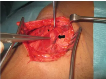
Fig. 2 Attachment of the superficial thigh fascia to the underlying fibrous strand of the sartorius muscle (black arrow) and identification of the LFCN (asterisk)
Fig. 3 Fibrous thickening of the anterior medial border of the sartorius muscle (black arrow) over which the LFCN (asterisk) was strained
Fig. 4 Situs after excision of the fibrous band (white arrow)
considerably impaired, since she was not able to walk more than 15 min at a time due to pain, which only could be alleviated for a short time period by stretching exercises of the right thigh and hip. Anamnestically, she did not report any major injuries during her career; in particular, no reminiscent trauma of the right thigh, hip, or pelvis. Based on the anam-nestic and clinical findings, the patient was scheduled for operative decompression of the LFCN at the level of the right inguinal ligament. The exploration was performed via a 4 cm long slightly curved longitudinal incision 2 cm medially and caudally of the ASIS. A thickening of the superficial thigh fascia was noted (Fig. 1). The fascia was incised, and the LFCN could be located with a subfascial course from craniolateral to caudomedial over the sartorius muscle. Sur-prisingly, no obvious compression of the LFCN under the inguinal ligament was noted. However, the thickened thigh fascia was attached to a fibrous strand within the sartorius muscle 8 cm caudal to the inguinal ligament (Fig.2). As the main cause of nerve compression, we identified this fibrous thickening of the medial border of the sartorius muscle over which the nerve was strained (Fig.3). The strand appeared to be fibrous in nature but not scarred, and no adhesions to the LFCN or the surrounding tissues were present. The fibrous tissue was excised completely (Fig.4). However, the inguinal ligament was incised for 0.5 cm to rule out potential additional compression at this location.
The patient recovered quickly from the operation and reported an amelioration of the pain as soon as on postop-erative day 7. Twenty days postoppostop-erative, she was able to walk 30 min at a time without pain. At 3 months postoper-ative, no more tingling at the right lateral leg was present. At the moment (6 months postoperative), there is complete absence of pain, and the patient is able to walk and run without restriction. She works 100 % in the previous occupation as a logistic manager and plans to resume playing soccer in an amateur league next month.
Discussion
Meralgia paraesthetica is defined as a neuropathy in-volving pain, burning, tingling, and numbness in the distribution of the LFCN, which is commonly due to nerve entrapment under the inguinal ligament [3]. Also, several other causes have been reported such as direct trauma, ischemia, diabetes, pregnancy, and stretch inju-ries [4]. After originating from the lumbar plexus, the nerve passes over the psoas muscle, under the inguinal ligament, and over the sartorius muscle into the thigh, where it divides into an anterior and a posterior branch [5]. Only in a limited number of cases of this condition
have been described in professional athletes [6]. We report a rare case of LFCN compression in a professional female soccer player who never experienced a major injury during her career. Unlike in most cases, where the nerve is compressed during its course under the inguinal ligament, the compression of the LFCN occurred at the medial border of the sartorius muscle. As a main factor, we identified a thickened superficial thigh fascia and a fibrous band within the anterior part of the sartorius muscle 8 cm caudal of the inguinal ligament. Injuries to the groin area and quadriceps muscula-ture are quite common in soccer players [7]. These muscle strains usually involve the rectus femoris muscle [8]. The sartorius muscle lies in close proximity to the quadriceps, but usually is not affected by these injuries. The present findings theoretically could be long-term sequelae of a muscle injury resulting in scarring and subsequent nerve compres-sion. However, no clinical signs of scarring were detected, and the patient negated a direct hit or a distortion trauma to the thigh. Previously, Otoshi et al. reported that in a professional baseball player, the LFCN was found to be unsheathed and compressed in the tedious origin of the sartorius muscle on the ASIS [6]. However, a bowstringing of the nerve over a more distally located fibrotic band in the sartorius muscle has not been described before. This finding goes in line with the fact that the patient experienced short periods of relief from pain after stretching the thigh musculature and subsequently less-ening strain onto the LFCN. A major compression of the nerve at the level of the inguinal ligament was not found in the presented case. From the fact that the patient recovered completely, it can be derived that a sufficient decompression was achieved by excising the fibrotic and constricting tissue at the medial border of the sartorius muscle. The exact nature of this strand cannot be determined in detail. Most likely, it may be a response to excessive muscle stress or tension due to intensive training. Alternatively, it is the result of a not rem-iniscent trauma with subsequent tissue healing.
Concludingly, the LFCN should be explored along its entire course on the anterior thigh during operative decom-pression, particularly in athletes, to rule out a distal muscular compression site.
Conflict of interest None
References
1. Roth VK (1895) Meralgia paraesthetica. Karger, Berlin
2. Aszmann OC, Dellon ES, Dellon AL (1997) Anatomical course of the lateral femoral cutaneous nerve and its susceptibility to compression and injury. Plats Reconstr Surg 100:600e4
3. Turner OA, Taslitz N, Ward S (1990) Lateral femoral cutaneous nerve of the thigh (Meralgia paraesthetica), Handbook of peripheral nerve entrapments. Humana Press, Clifton, pp 143–50
4. Ecker AD, Woltman HW (1938) Meralgia paraesthetica. A report of one hundred fifty cases. JAMA 110:1650–1652
5. Ahmad Z, Devaraj VS, Conn DA (2011) Finding the lateral cuta-neous nerve of the thigh. J Plast Reconstr Aesthet Surg 64:1254– 1255
6. Otoshi K, Itoh Y, Tsujino A, Kikuchi S (2008) Case report: meralgia paraesthetica in a baseball pitcher. Clin Orthop Relat Res 466:2268–2270 7. Wittstein J, Klein S, Garrett WE (2011) Chronic tears of the reflected head of the rectus femoris: results of operative treatment. Am J Sports Med 39:1942–1947
8. Cross TM, Gibbs N, Houang MT, Cameron M (2004) Acute quad-riceps muscle strains: magnetic resonance imaging features and prognosis. Am J Sports Med 32:710–719