ORIGINAL ARTICLE
Tumour assessment in advanced melanoma: value
of FDG-PET/CT in patients with elevated serum S-100B
Klaus Strobel&Jeannine Skalsky&Victor Kalff&
Katrin Baumann&Burkhardt Seifert&
Helen Joller-Jemelka&Reinhard Dummer&
Hans C. Steinert
Received: 20 November 2006 / Accepted: 19 January 2007 / Published online: 28 March 2007 # Springer-Verlag 2007
Abstract
Purpose To evaluate the usefulness of PET/CT in melanoma patients with an elevated serum S-100B tumour marker level. Methods Out of 165 consecutive high-risk melanoma patients referred for PET/CT imaging, 47 had elevated (>0.2 μg/l) S-100B serum levels and a contemporaneous
18
F-FDG PET/CT scan. PET/CT scans were evaluated for the presence of metastases. To produce a composite reference standard, we used cytological, histological, MRI and PET/CT follow-up findings as well as clinical and S-100B follow-up.
Results Among the 47 patients with increased S-100B levels, PET/CT correctly identified metastases in 38 (30 distant metastases and eight lymph node metastases). In one patient with cervical lymph node metastases, PET/CT was
negative. Eight patients had no metastases and PET/CT correctly excluded metastases in all of them. Overall sensitivity for metastases was 97% (38/39), specificity 100% (8/8) and accuracy 98% (46/47). S-100B was significantly higher in patients with distant metastases (mean 1.93μg/l, range 0.3–14.3 μg/l) than in patients with lymph node metastases (mean 0.49μg/l, range 0.3–1.6 μg/l, p=0.003) or patients without metastases (mean 0.625 μg/l, range 0.3–2.6 μg/l, p=0.007). However, 6 of 14 patients with a tumour marker level of 0.3μg/l had no metastases. Conclusion In melanoma patients with elevated S-100B tumour marker levels, FDG-PET/CT accurately identifies lymph node or distant metastases and reliably excludes metastases. Because of the significant number of false positive S-100B tumour marker determinations (17%), we recommend repetition of tumour marker measurements if elevated S-100B levels occur before extensive imaging is used.
Keywords Melanoma . PET/CT . S-100B . Metastases
Introduction
18
F-fluorodeoxyglucose positron emission tomography (FDG-PET) is increasingly used to image patients with melanoma [1]. Recent publications have demonstrated that FDG-PET/CT has a high accuracy for N- and M-staging in melanoma patients and is superior to PET alone, CT alone or conventional imaging methods [2, 3]. Because FDG-PET/CT is still a relatively expensive technique with limited availability, it is not practicable to use PET/CT as a screening tool in all melanoma patients. Thus, it is very important to identify groups of patients and clinical situations where PET/ CT has a diagnostic and therapeutic impact.
DOI 10.1007/s00259-007-0403-8
K. Strobel (*)
:
V. Kalff:
H. C. SteinertDivision of Nuclear Medicine, Department of Medical Radiology, University Hospital Zurich, Raemistrasse 100, 8091 Zurich, Switzerland e-mail: [email protected]
J. Skalsky
:
K. Baumann:
R. DummerDepartment of Dermatology, University Hospital Zurich, Zurich, Switzerland
V. Kalff
Department of Nuclear Medicine, Alfred Hospital Melbourne, Melbourne, Australia
B. Seifert
Institute of Biostatistics, University of Zurich, Zurich, Switzerland
H. Joller-Jemelka
Department of Immunology, University Hospital Zurich, Zurich, Switzerland
Long survival of patients with malignant melanoma has been reported after surgery of regional lymph node metastases or solitary distant metastases. In many institu-tions, patients with multiple distant metastases are included in clinical trials and treated with immuno-chemotherapy [4]. Thus, accurate staging of melanoma patients with exact localisation of metastases is crucial for therapy planning.
Serum S-100B has been reported to be a reliable tumour marker in melanoma, reflecting the tumour burden [5], and it seems to be useful in monitoring therapy [6,7]. Further, it has been shown that S-100B can be used as a prognostic marker in melanoma patients [8–12]. As demonstrated in colon cancer and prostate cancer, PET imaging can be very helpful in localising recurrences in patients with elevated or increasing tumour marker levels [13–16].
The aim of this study was therefore to assess the value of FDG-PET/CT in melanoma patients with elevated (>0.2μg/l) S-100B tumour marker levels.
Materials and methods
Patients
Our institution is a teaching and tertiary care hospital and a major referral site for patients with malignant melanoma.
We received approval from our institutional review board to undertake this study.
One hundred and sixty-five consecutive melanoma patients followed up according to updated Swiss melanoma guidelines [17] were referred for FDG-PET/CT imaging between January 2005 and January 2006. Retrospectively, from among this population, 47 patients (27 women, 20 men; mean age 58.4 years, range 20–83 years) who fulfilled the following criteria were selected: (1) diagnosis of high-risk melanoma (Breslow tumour thickness >4 mm, Clark level III or IV or known resected metastases in the case history); (2) elevated S-100B levels (>0.2μg/l); (3) FDG-PET/CT and S-100B measurement within an interval of not more than 2 weeks; (4) no treatment between PET/CT and tumour marker measurement; (5) no systemic therapy before the PET/CT investigation. Informed consent was obtained from all patients before they were investigated.
Determination of S-100B
The determination of serum S-100B was done with a commercially available immunoassay (ELISA) kit (Sangtec 100 ELISA, Dia Sorin Inc, Stillwater, NM, USA) according to the manufacturer’s instructions. Cut-off was determined to be 0.2μg/l (the 95th percentile of blood donor samples). Values≥0.3 μg/l were considered suspicious for melanoma metastases. The detection limit is 0.03 μl/l (BO+3SD).
Intra-assay and inter-assay precision was estimated by analysis of variance (ANOVA). The within-run and total imprecision is <10%. The evaluation of S-100B was introduced in our clinic in 1992.
PET/CT imaging
All the data were acquired on a combined PET/CT in-line system (Discovery LS or Discovery ST, GE Health Systems, Milwaukee, WI). These dedicated systems integrate a PET scanner (GE Advance Nxi, GE Health Systems, Milwaukee, WI) with a multislice helical CT (LightSpeed plus or Light-speed 16; GE Health Systems, Milwaukee, WI) and permit the acquisition of co-registered CT and PET images in one session. Patients fasted for at least 4 h prior to the scanning, which started 60 min after the injection of 370–400 MBq of
18
F-FDG. All patients were tested for a normal glucose level [range 80–120 mg/dl (4.4–6.7 mmol/l)] before scan-ning. Patients with elevated glucose levels were rescheduled and scanned with normal glucose levels. Oral CT contrast agent (Micropaque Scanner, Guerbet AG, Aulnay-sous-bois, France) was given 15 min before the injection of18F-FDG. Patients were examined in the supine position. No intravenous contrast agent was given. Initially, the CT scan was acquired starting from the level of the head using the following parameters: 40 mAs, 140 kV, 0.5 s/tube rotation, slice thickness 4.25 mm, scan length 867 mm, data acquisition time 22.5 s. The CT scan was acquired during breath holding in the normal expiratory position.
Immediately following the CT acquisition, a PET emis-sion scan was acquired with an acquisition time of 3 min per cradle position with a one-slice overlap in 2D mode (matrix 128×128). The eight to nine cradle positions starting from the head to the knees resulted in an acquisition time of approximately 24–27 min. In the patients with primary tumours of the lower extremities, the scanning of the lower legs was added. The CT data were used for attenuation correction of the PET datasets and the images were reconstructed using a standard iterative algorithm (OSEM). The acquired images were viewed with software providing multiplanar reformatted images of PET alone, CT alone and fused PET/CT with linked cursors using a Xeleris worksta-tion (GE Health Systems, Milwaukee, WI).
PET/CT imaging was performed according to the recently published procedure guideline for tumour imaging with 18F-FDG PET/CT version 1.0 [18].
PET/CT interpretation
The images were reviewed and analysed by two experienced nuclear radiology physicians without knowledge of the results of other imaging studies or the level of serum S-100B. A consensus was reached on every observed lesion. The PET
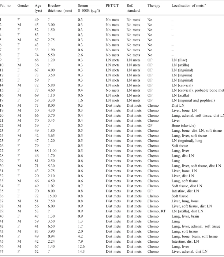
Table 1 Patient characteristics, S-100B values and results of PET/CT imaging
Pat. no. Gender Age
(yrs) Breslow thickness (mm) Serum S-100B (μg/l) PET/CT Ref. standard
Therapy Localisation of mets.a
1 F 49 ? 0.3 No mets No mets No – 2 M 45 3.00 0.3 No mets No mets No – 3 F 52 1.50 0.3 No mets No mets No – 4 F 83 ? 0.3 No mets No mets No – 5 M 67 2.75 0.3 No mets No mets No – 6 F 43 ? 0.3 No mets No mets No – 7 F 33 1.90 0.6 No mets No mets No – 8 F 74 5.50 2.6 No mets No mets No –
9 F 68 1.20 0.3 LN mets LN mets OP LN (iliac)
10 M 36 ? 0.3 LN mets LN mets OP LN (axilla)
11 F 67 4.00 0.3 LN mets LN mets OP LN (inguinal)
12 F 73 3.50 0.3 LN mets LN mets OP LN (inguina)
13 F 59 ? 0.3 LN mets LN mets OP LN (inguinal)
14 M 72 5.80 0.3 LN mets LN mets OP LN (cervical)
15 F 77 4.60 0.4 No mets LN mets OP LN (cervical), probable bone met
16 M 69 1.10 0.6 LN mets LN mets OP LN (axilla)
17 F 58 3.30 1.6 LN mets LN mets OP LN (inguinal and popliteal)
18 M 73 8.00 0.3 Dist mets Dist mets Chemo Dist LN
19 M 50 4.30 0.3 Dist mets Dist mets Chemo Liver, bone, LN
20 M 66 3.70 0.4 Dist mets Dist mets Chemo Lung, adrenal, soft tissue, dist LN
21 M 70 3.45 0.4 Dist mets Dist mets Chemo Liver
22 M 59 ? 0.5 Dist mets Dist mets OP Bone (clavicle)
23 F 49 1.80 0.5 Dist mets Dist mets Chemo Lung, bone, dist LN, soft tissue
24 M 42 3.65 0.5 Dist mets Dist mets Chemo Lung, liver, soft tissue
25 F 47 1.20 0.5 Dist mets Dist mets Chemo LN (inguinal), lung
26 F 79 ? 0.5 Dist mets Dist mets Chemo Soft tissue
27 F 68 11.00 0.5 Dist mets Dist mets Chemo Lung, liver
28 F 46 1.70 0.6 Dist mets Dist mets Chemo Lung, dist LN
29 F 81 2.50 0.6 Dist mets Dist mets Chemo Lung
30 M 71 5.30 0.6 Dist mets Dist mets Chemo Lung, liver, soft tissue, dist LN
31 F 43 2.75 0.6 Dist mets Dist mets Chemo Liver, bone, LN
32 F 20 2.10 0.6 Dist mets Dist mets Chemo Liver, dist LN
33 M 66 4.50 0.6 Dist mets Dist mets Chemo Lung, soft tissue
34 F 49 1.02 0.7 Dist mets Dist mets Chemo Soft tissue, dist LN
35 F 70 8.00 0.8 Dist mets Dist mets OP Intestine, dist LN
36 F 72 15.00 0.8 Dist mets Dist mets Chemo Soft tissue
37 M 51 7.50 0.8 Dist mets Dist mets Chemo Liver, lung, bone
38 M 56 6.00 0.9 Dist mets Dist mets Chemo Liver, soft tissue, dist LN
39 M 53 ? 0.9 Dist mets Dist mets Chemo, RT LN (axilla), dist LN
40 F 47 1.30 0.9 Dist mets Dist mets Chemo Lung, liver, brain
41 M 59 3.50 1.3 Dist mets Dist mets Chemo Lung
42 F 41 6.50 1.7 Dist mets Dist mets Chemo Lung, liver, adrenal, soft tissue
43 M 83 3.90 2.0 Dist mets Dist mets Chemo Lung, soft tissue
44 F 49 0.94 4.2 Dist mets Dist mets Chemo Lung, bone, brain, soft tissue
45 M 42 2.24 7.9 Dist mets Dist mets Chemo Intestine, dist LN
46 M 67 1.40 12.6 Dist mets Dist mets Chemo Lung, liver
47 F 52 ? 14.3 Dist mets Dist mets Chemo Liver, adrenal, dist LN
? Depth of the primary tumour not known, M male, F female, dist mets distant metastases, LN mets regional lymph node metastases, dist LN distant lymph node metastases (no regional LN mets), OP operative resection, Chemo chemotherapy, RT radiotherapy
a
images and the corresponding CT images of the PET/CT study were analysed for the presence and nature of focal lesions with elevated FDG uptake. For all of the patients, the attenuation-corrected PET images were analysed. Lesions were interpreted as metastases if the FDG uptake was clearly greater than background. If a focal FDG-active lesion was detected, the exact anatomical localisation was determined on the fused PET/CT images. Lesions with18F-FDG uptake in physiolog-ical sites or benign variants, e.g. muscles, brown fatty tissue or pulmonary infiltrations, were determined as benign. CT images were additionally analysed concerning FDG-inactive soft tissue dense lesions without calcifications, especially in
the lungs and subcutaneous fatty tissue. Lesions suspicious for metastases according to the established morphological CT criteria known from diagnostic radiology were also diagnosed as metastases.
Reference standard
Lymph node or distant metastases were confirmed by a histopathological or cytological examination or other imaging modalities such as magnetic resonance imaging (MRI), PET/CT follow-up and clinical follow-up for a minimum of 6 months (range 6–18 months in all patients),
Fig. 1 A 77-year-old female patient with high-risk melano-ma and elevated serum S-100B
(0.4μg/l). a MIP image and
b axial images through the neck without evidence of metastases. There is elevated FDG uptake in both shoulders and the right patella (arrows) due to osteoar-thritis. Contamination is present at the injection site of the left elbow (arrowhead). c MIP image 6 months later with increased FDG uptake in the cervical lymph nodes on the left side (arrow) and in the thoracic spine (arrowhead). d Axial images demonstrating that the elevated FDG uptake belongs to a level II lymph node on the left side (arrowhead). The cervical lymph node metastases were resected and confirmed histo-logically. MRI of the spine was inconclusive as it showed a small lesion in the transverse process
including follow-up measurement of the serum S-100B. A false negative PET/CT diagnosis was determined if another imaging method (superior for the investigated region, such as brain MRI) showed metastases or if clinical findings raised the suspicion of metastases which were then proven by histology. A false positive PET/CT diagnosis was determined if histology of the lesion and/or clinical and PET/CT follow-up (complete disappearance of focal active lesion without therapy) ruled out metastases. FDG-negative, non-calcified lesions (for example in the lung) were determined as false positive if there was no change in lesion number or size on the follow-up PET/CT examina-tions 3 or 6 months later and no clinical suspicion of metastases arose >6 months after the scan.
Statistical analysis
The recorded data were entered into a worksheet (Excel; Microsoft, Redmond, Washington). Data analysis was performed on a patient basis. SPSS (version 11; 2002; SPSS Inc.) was used for statistical analysis. Sensitivity, specificity and accuracy are presented with exact 95% confidence intervals (Scientific Tables by Geigy, Volume on statistics, 8th edition, Basle 1980). The Mann-Whitney test with Bonferroni correction was used to assess statistical differences in serum S-100B values in patients without metastases, with regional lymph node metastases and with distant metastases. Thus, p<0.017 was considered to indicate a significant difference.
Results
Patient characteristics are summarised in Table 1. Thirty (64%) of the 47 patients had distant metastases, nine (19%) had regional lymph node metastases and eight (17%) patients had no metastases. Of patients with distant metastases, 20 had their findings confirmed by histology, four by cytology and six by MRI, PET/CT follow-up and clinical follow-up. All nine patients with regional lymph node metastases had their diagnoses confirmed histologi-cally following surgical resection.
PET/CT correctly detected distant metastases in all 30 patients. PET/CT identified lymph node metastases in eight patients and missed multiple level II and III cervical lymph node metastases and a probable vertebral spinal metastasis in one patient (no. 15., Fig. 1). Eight patients had no metastases and PET/CT correctly excluded metastases in all
these patients. Overall sensitivity for metastases was 97% (38/39) [95% CI, 87–100%], specificity 100% (8/8) [63– 100%] and accuracy 98% (46/47) [89–100%]. Serum S-100B was significantly higher in patients with distant metastases (mean 1.93 μg/l, range 0.3–14.3 μg/l) than in patients with regional lymph node metastases (mean 0.49, range 0.3–1.6, p=0.003) or patients without metastases (mean 0.625, range 0.3–2.6, p=0.007); however, there was considerable overlap between each of the subgroups. There was no statistical significant difference in S-100B values between patients with lymph node metastases and patients without metastases (p=0.86).
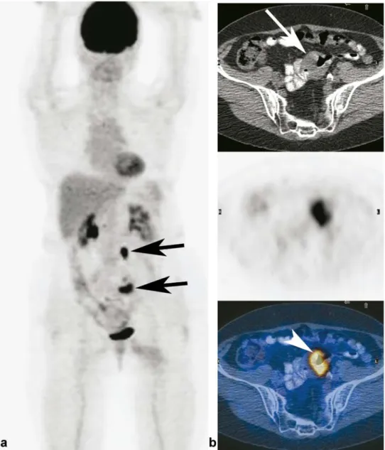
Six (42.9%) of the 14 patients with a weakly elevated tumour marker of 0.3μg/l had no metastases, six (42.9%) had regional lymph node metastases and two (14.3%) had distant metastases. An example of a patient with distant metastases and elevated S-100B is shown in Fig.2.
Fig. 2 A 70-year-old female patient with high-risk melano-ma and elevated S-100B
(0.8μg/l). a MIP image
dem-onstrating two focal FDG-active lesions in the abdomen (arrows). b On the axial images one FDG-active lesion (arrow-head) corresponds to wall thickening (arrow) in the small intestine. The lesion was resected and histology con-firmed the PET/CT diagnosis of small intestine metastases
Eight patients had“false positive” S-100B tumour markers (Table2). In three patients (nos. 5, 6 and 7) with S-100B values of 0.3–0.6 μg/l we found a history of brain operation for melanoma metastases (2 weeks, 6 months and 8 years previously). One patient (no. 8) with a normal S-100B value 6 months prior to the study showed an increase in S-100B to 2.6μg/l without clinical evidence of metastases. PET/CT at the timepoint of the elevated tumour marker and PET/CT follow-up investigations after 2, 6 and 18 months were normal. S-100B levels measured 2 weeks and 6 and 18 months after the pathological value were normal (0.1– 0.2μg/l). Similarly, in the remaining four patients (nos. 1–4) we found no explanation for an elevated S-100B value in the history. The follow-up S-100B measurements, PET/CT scans and clinical investigations remained without evidence of metastases. In one patient (no. 37) with an S-100B of 0.8μg/l, PET/CT only detected a solitary metastasis in a vertebral body of the thoracic spine (Fig.3). MRI and contrast-enhanced CT, performed in the following week, showed disseminated small liver and bone metastases, so that PET/CT as correct in the diagnosis of distant metastasis but severely underesti-mated the extent of metastases. Twelve patients had lung metastases. Lung metastases were FDG active in eight patients and FDG inactive in four patients. In these four patients the lung metastases were only detected by the additional interpretation of the lung window of the CT examination. Two patients had brain metastases. They were detectable in both patients with PET/CT because of elevated FDG uptake compared with normal brain tissue in one patient and because of additional bleeding in the other patient. Additionally in both patients perifocal vasogenic oedema was present in CT images.
Discussion
To our knowledge, this is the first study to describe the value of integrated PET/CT imaging in the follow-up of melanoma patients with elevated S-100B tumour marker levels. The results demonstrate that FDG-PET/CT is very useful in this selected group of melanoma patients with a high risk for the presence of metastases. FDG-PET/CT reliably discriminates between patients with distant metas-tases, which are usually treated with chemotherapy, and patients with regional lymph node metastases, which are often treated by surgery. If an S-100B level of 0.3 μg/l or more is considered pathological, there is a significant number of patients with false positive S-100B. FDG-PET/ CT excluded metastases correctly in all of these patients. One of our study patients with a slightly elevated S-100B level (0.4 μg/l) had a normal PET/CT scan. Cytologically proven cervical lymph metastases were diagnosed 5 months after the FDG-PET/CT examination. This situation was interpreted as a false negative PET/CT diagnosis. It has been shown in several studies that FDG-PET/CT has limitations in the detection of microscopic lymph node metastases. FDG-PET was compared with sentinel node biopsy in a prospective study. The sensitivity of FDG-PET was 16.7% and the specificity, 95.8%. [19]. Thus, sentinel lymph node biopsy remains the standard and is clearly superior to FDG-PET(/CT) in detecting regional lymph node metastases [20,21].
FDG-PET reliably detects lymph node tumour deposits with a volume larger than 80 mm3, but sensitivity falls rapidly below this value because of partial volume effects that reduce the FDG signal [22]. All lymph node metastases
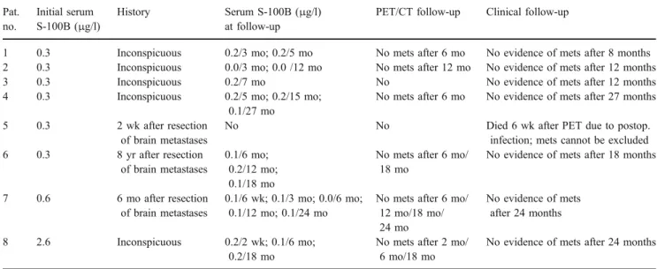
Table 2 History and follow-up of eight patients with false positive S-100B tumour marker levels Pat. no. Initial serum S-100B (μg/l) History Serum S-100B (μg/l) at follow-up
PET/CT follow-up Clinical follow-up
1 0.3 Inconspicuous 0.2/3 mo; 0.2/5 mo No mets after 6 mo No evidence of mets after 8 months
2 0.3 Inconspicuous 0.0/3 mo; 0.0 /12 mo No mets after 12 mo No evidence of mets after 12 months
3 0.3 Inconspicuous 0.2/7 mo No No evidence of mets after 12 months
4 0.3 Inconspicuous 0.2/5 mo; 0.2/15 mo;
0.1/27 mo
No mets after 6 mo No evidence of mets after 27 months
5 0.3 2 wk after resection
of brain metastases
No No Died 6 wk after PET due to postop.
infection; mets cannot be excluded
6 0.3 8 yr after resection
of brain metastases
0.1/6 mo; 0.2/12 mo; 0.1/18 mo
No mets after 6 mo/ 18 mo
No evidence of mets after 18 months
7 0.6 6 mo after resection
of brain metastases
0.1/6 wk; 0.1/3 mo; 0.0/6 mo; 0.1/12 mo; 0.1/24 mo
No mets after 6 mo/ 12 mo/18 mo/ 24 mo No evidence of mets after 24 months 8 2.6 Inconspicuous 0.2/2 wk; 0.1/6 mo; 0.2/18 mo
No mets after 2 mo/ 6 mo/18 mo
No evidence of mets after 24 months
detected with PET/CT in our patients measured more than 6 mm. We know from other studies that some patients with microscopic or occult lymph node metastases have normal S-100B values. Reinhardt et al. reported that 8 of 11 patients with lymph node metastases and no distant metastases had normal S-100B values (all 0.1 μg/l) and only three had elevated levels (0.3, 0.9 and 1.6μg/l). By contrast, all 13 patients with proven distant metastases had elevated S-100B values (range 0.3–23 μg/l) [23]. Bearing in mind the hypothesis that S-100B values are related to total tumour burden, it seems reasonable that if lymph node
metastases lead to an elevated S-100B, the chance is very high that they will be detectable with FDG-PET/CT.
We also found a considerable number of false positive S-100B tumour marker determinations (17%; 95% CI, 6%– 28%). There are various possible reasons for a false positive S-100B value: elevated S-100B levels have been observed after head trauma, subarachnoidal haemorrhage and stroke. It has been reported that S-100B reflects the extent of injury and the outcome of brain-injured patients [24, 25]. Furthermore, it indicates blood-brain barrier dysfunction [26,27]. Molina et al. measured S-100B levels
Fig. 3 A 51-year-old male pa-tient with high-risk melanoma
and elevated S-100B (0.8μg/l).
PET (MIP image, a) and axial fused PET/CT image (b) show a solitary bone lesion in a thoracic vertebral body (arrow). MRI (sagittal contrast-enhanced fat-saturated image, c) shows mul-tiple disseminated enhancing bone metastases (arrows) in the whole spine. CT (axial contrast-enhanced image, d) detects dis-seminated small liver metastases
in healthy people, patients with benign diseases and patients with malignancies, including patients with locoregional disease and advance diseases. All of the healthy people had normal S-100B values. Slightly elevated S-100B concen-trations were found in 25% of individuals with benign diseases. Significantly higher S-100B serum levels were found in patients with liver cirrhosis or renal failure. Pathological S-100B serum levels were found in 22.5% of individuals with malignancies. The highest S-100B concen-trations were found in patients with malignant melanomas. They concluded that“S-100 is a useful marker for melanoma, but abnormal levels of this tumour marker may be found in benign and malignant diseases associated with liver or renal injury” [26]. In three of our eight patients with false positive S-100B values we found brain operations in their past history. In one patient a melanoma brain metastasis had been resected 2 weeks before an S-100B level of 0.3 μg/l was measured. This patient died 6 weeks after the PET/CT due to postoperative infected subdural haematoma. In two other patients the brain operations could not explain the elevated S-100B level because the operations had been performed 6 months and 8 years previously. In the latter two patients no brain or other metastases developed at follow-up. In five of the eight patients with“false positive” tumour marker levels we found no explanation in the history or follow-up.
It is well known that PET has poor sensitivity in detecting brain metastases owing to the high physiological FDG uptake in the normal brain [28]. MRI is the imaging gold standard in the detection of brain metastases [29]. Among our patients we had no case in which PET/CT missed brain metastases; however, in patients with repeat-edly elevated tumour markers and negative PET(/CT), additional brain MRI is strongly recommended.
A recently published study compared FDG-PET/CT imaging for N- and M-staging of 250 consecutive melano-ma patients with PET alone and CT alone. The accuracy of PET/CT for M-staging was significantly higher than that of PET alone and CT alone (98% vs 93% and 84%). A change of treatment according to PET/CT findings occurred in 121 of the patients (48.4%). Interestingly, the authors pointed out that the most significant advantage of PET/CT in comparison to the single modalities PET and CT was observed in the detection of visceral metastases [2]. Performance of PET/CT in this study corresponds quite well with our results (overall accuracy 97.8%). Although we report a 100% specificity in this paper, we want to point out that false positive cases occur in our daily routine outside of this presented patient population.
The effect of chemotherapy in stage IV melanoma patients is still disappointing [30,31]. Many patients with metastases are included in clinical trials where therapy response has to be assessed [32]. The data of Henze et al. support the value of serum S-100B as a clinical marker for
monitoring response of metastatic melanoma during sys-temic therapies [6] but there are clear limitations in its sensitivity and specificity. We think that FDG-PET/CT may be very useful in therapy assessment of melanoma patients in a similar way to its use in patients with oesophageal, lung and head and neck cancers, but data are still very limited. In an ongoing study we plan to evaluate the behaviour of S-100B, standardised uptake value and total lesion glycolysis in stage IV melanoma patients treated with chemotherapy.
Our study has some limitations. As in all studies evaluating the accuracy of staging, the establishment of a standard of reference with which the method can be compared is difficult, because we cannot ethically justify obtaining histological proof of the diagnosis for all lesions identified. Nevertheless, we exercised great caution in establishing our standard of reference by using histopath-ological or cythistopath-ological confirmation of the suspected metastases in 33/39 of patients (24 of 30 patients with distant metastases and all nine patients with lymph node metastases) and by confirming the findings with another structural imaging modality or with PET/CT and clinical follow-up.
We did not investigate whether the outcome of the patients correlated with the S-100B level. We know from other publications that there is a strong association between S-100B level and overall survival and that S-100B can be used as a prognostic marker [8, 9, 11, 12, 33]. Whether FDG-PET/CT can be used as an independent prognostic factor has to be investigated in further studies.
This is a retrospective study and has a selection bias because only patients with elevated S-100B values were included. We chose this selection because we were interested in the value of PET/CT in this particular clinical situation.
To avoid unnecessary PET/CT investigations in patients with false positive S-100B values, like other authors we recommend repeating the S-100B determination after 2–4 weeks if it is initially elevated in the absence of clinical evidence of metastases [34]. Further, we recommend that the S-100B measurements should be performed in the same laboratory always using the same method, thereby avoiding the bias associated with use of different assays [35]. Our results confirm that S-100B is not useful in melanoma patients after brain operation or trauma. Because FDG-PET/CT has been proven to be superior to PET alone or conventional imaging methods like CT or ultrasound [2, 36], we recommend FDG-PET/CT as the first imaging method in patients with repeatedly elevated S-100B.
In conclusion, our results indicate that integrated PET/ CT imaging has a significant value in triaging melanoma patients with elevated S-100B tumour marker levels.
References
1. Schwimmer J, Essner R, Patel A, Jahan SA, Shepherd JE, Park K, et al. A review of the literature for whole-body FDG PET in the
man-agement of patients with melanoma. Q J Nucl Med 2000;44:153–67.
2. Reinhardt MJ, Joe AY, Jaeger U, Huber A, Matthies A, Bucerius
J, et al. Diagnostic performance of whole body dual modality18
F-FDG PET/CT imaging for N- and M-staging of malignant melanoma: experience with 250 consecutive patients. J Clin Oncol 2006;24:1178–87.
3. Fuster D, Chiang S, Johnson G, Schuchter LM, Zhuang H, Alavi A.
Is18F-FDG PET more accurate than standard diagnostic
proce-dures in the detection of suspected recurrent melanoma? J Nucl
Med 2004;45:1323–7.
4. Tsao H, Atkins MB, Sober AJ. Management of cutaneous
melanoma. N Engl J Med 2004;351:998–1012.
5. Schultz ES, Diepgen TL, Von Den Driesch P. Clinical and prognostic relevance of serum S-100 beta protein in malignant
melanoma. Br J Dermatol 1998;138:426–30.
6. Henze G, Dummer R, Joller-Jemelka HI, Boni R, Burg G. Serum
S100—a marker for disease monitoring in metastatic melanoma.
Dermatology 1997;194:208–12.
7. Hauschild A, Engel G, Brenner W, Glaser R, Monig H, Henze E, et al. Predictive value of serum S100B for monitoring patients with metastatic melanoma during chemotherapy and/or immuno-therapy. Br J Dermatol 1999;140:1065–71.
8. Hansson LO, von Schoultz E, Djureen E, Hansson J, Nilsson B, Ringborg U. Prognostic value of serum analyses of S-100 protein beta in malignant melanoma. Anticancer Res 1997;17:3071–3. 9. Buer J, Probst M, Franzke A, Duensing S, Haindl J, Volkenandt
M, et al. Elevated serum levels of S100 and survival in metastatic
malignant melanoma. Br J Cancer 1997;75:1373–6.
10. Abraha HD, Fuller LC, Du Vivier AW, Higgins EM, Sherwood RA. Serum S-100 protein: a potentially useful prognostic marker
in cutaneous melanoma. Br J Dermatol 1997;137:381–5.
11. Andres R, Mayordomo JI, Zaballos P, Rodino J, Isla D, Escudero P, et al. Prognostic value of serum S-100B in malignant melanoma.
Tumori 2004;90:607–10.
12. Domingo-Domenech J, Molina R, Castel T, Montagut C, Puig S, Conill C, et al. Serum protein s-100 predicts clinical outcome in
patients with melanoma treated with adjuvant interferon
—com-parison with tyrosinase rt-PCR. Oncology 2005;68:341–9.
13. Stamey TA, Yang N, Hay AR, McNeal JE, Freiha FS, Redwine E. Prostate-specific antigen as a serum marker for adenocarcinoma of the prostate. N Engl J Med 1987;317:909–16.
14. Libutti SK, Alexander HR Jr, Choyke P, Bartlett DL, Bacharach SL,
Whatley M, et al. A prospective study of 2-[18F] fluoro-2-deoxy-D
-glucose/positron emission tomography scan, 99mTc-labeled
arcitu-momab (CEA-scan), and blind second-look laparotomy for detecting colon cancer recurrence in patients with increasing carcinoembryonic
antigen levels. Ann Surg Oncol 2001;8:779–86.
15. de Jong IJ, Pruim J, Elsinga PH, Vaalburg W, Mensink HJ.11
C-choline positron emission tomography for the evaluation after treatment of localized prostate cancer. Eur Urol 2003;44:32–8, discussion 38–9.
16. Cimitan M, Bortolus R, Morassut S, Canzonieri V, Garbeglio A,
Baresic T, et al. [18F]fluorocholine PET/CT imaging for the detection
of recurrent prostate cancer at PSA relapse: experience in 100
con-secutive patients. Eur J Nucl Med Mol Imaging 2006;33:1387–98.
17. Dummer R, Panizzon R, Bloch PH, Burg G. Updated Swiss guidelines for the treatment and follow-up of cutaneous melanoma.
Dermatology 2005;210:39–44.
18. Delbeke D, Coleman RE, Guiberteau MJ, Brown ML, Royal HD,
Siegel BA, et al. Procedure guideline for tumor imaging with18
F-FDG PET/CT 1.0. J Nucl Med 2006;47:885–95.
19. Wagner JD, Schauwecker D, Davidson D, Coleman JJ 3rd, Saxman S, Hutchins G, et al. Prospective study of fluorodeox-yglucose-positron emission tomography imaging of lymph node basins in melanoma patients undergoing sentinel node biopsy. J
Clin Oncol 1999;17:1508–15.
20. Morton DL, Cochran AJ, Thompson JF, Elashoff R, Essner R, Glass EC, et al. Sentinel node biopsy for early-stage melanoma: accuracy and morbidity in MSLT-I, an international multicenter trial. Ann Surg 2005;242:302–11, discussion 303–11.
21. Acland KM, Healy C, Calonje E, O'Doherty M, Nunan T, Page C, et al. Comparison of positron emission tomography scanning and sentinel node biopsy in the detection of micrometastases of primary cutaneous malignant melanoma. J Clin Oncol 2001;19:2674–8. 22. Wagner JD, Schauwecker DS, Davidson D, Wenck S, Jung SH,
Hutchins G. FDG-PET sensitivity for melanoma lymph node metastases is dependent on tumor volume. J Surg Oncol
2001;77:237–42.
23. Reinhardt MJ, Kensy J, Frohmann JP, Willkomm P, Reinhold U, Grunwald F, et al. Value of tumour marker S-100B in melanoma
patients: a comparison to 18F-FDG PET and clinical data.
Nuklearmedizin 2002;41:143–7.
24. Pleines UE, Morganti-Kossmann MC, Rancan M, Joller H, Trentz O, Kossmann T. S-100 beta reflects the extent of injury and outcome, whereas neuronal specific enolase is a better indicator of neuroinflammation in patients with severe traumatic brain injury. J Neurotrauma 2001;18:491–8.
25. Spinella PC, Dominguez T, Drott HR, Huh J, McCormick L, Rajendra A, et al. S-100beta protein-serum levels in healthy children and its association with outcome in pediatric traumatic brain injury. Crit Care Med 2003;31:939–45.
26. Molina R, Navarro J, Filella X, Castel T, Ballesta AM. S-100 protein serum levels in patients with benign and malignant diseases: false-positive results related to liver and renal function.
Tumour Biol 2002;23:39–44.
27. Banfalvi T, Gergye M, Beczassy E, Gilde K, Otto S. Role of S100B
protein in neoplasms and other diseases. Magy Onkol 2004;48:71–4.
28. Rohren EM, Provenzale JM, Barboriak DP, Coleman RE. Screening for cerebral metastases with FDG PET in patients undergoing whole-body staging of non-central nervous system
malignancy. Radiology 2003;226:181–7.
29. Sze G, Shin J, Krol G, Johnson C, Liu D, Deck MD. Intra-parenchymal brain metastases: MR imaging versus
contrast-enhanced CT. Radiology 1988;168:187–94.
30. Morgan RA, Dudley ME, Wunderlich JR, Hughes MS, Yang JC, Sherry RM, et al. Cancer regression in patients after transfer of genetically engineered lymphocytes. Science 2006;314:126–9. 31. Lens MB, Dawes M. Global perspectives of contemporary
epidemiological trends of cutaneous malignant melanoma. Br J
Dermatol 2004;150:179–85.
32. Eigentler TK, Caroli UM, Radny P, Garbe C. Palliative therapy of disseminated malignant melanoma: a systematic review of 41
randomised clinical trials. Lancet Oncol 2003;4:748–59.
33. Martenson ED, Hansson LO, Nilsson B, von Schoultz E, Mansson Brahme E, Ringborg U, et al. Serum S-100b protein as a prognostic marker in malignant cutaneous melanoma. J Clin
Oncol 2001;19:824–31.
34. Jury CS, McAllister EJ, MacKie RM. Rising levels of serum S100 protein precede other evidence of disease progression in patients
with malignant melanoma. Br J Dermatol 2000;143:269–74.
35. Smit LH, Korse CM, Bonfrer JM. Comparison of four different assays for determination of serum S-100B. Int J Biol Markers
2005;20:34–42.
36. Rinne D, Baum RP, Hor G, Kaufmann R. Primary staging and
follow-up of high risk melanoma patients with whole-body18
F-fluorodeoxyglucose positron emission tomography: results of a prospective study of 100 patients. Cancer 1998;82:1664–71.