Biophysical Origin of Cancer and Other Diseases
The MIT Faculty has made this article openly available. Please share how this access benefits you. Your story matters.Citation Davidson, Robert, Ann Lauritzen, and Stephanie Seneff. “Biological Water Dynamics and Entropy: A Biophysical Origin of Cancer and Other Diseases.” Entropy 15, no. 9 (September 13, 2013): 3822-3876.
As Published http://dx.doi.org/10.3390/e15093822
Publisher MDPI AG
Version Final published version
Citable link http://hdl.handle.net/1721.1/83885
entropy
ISSN 1099-4300
www.mdpi.com/journal/entropy
Review
Biological Water Dynamics and Entropy: A Biophysical Origin
of Cancer and Other Diseases
Robert M. Davidson 1,*, Ann Lauritzen 2 and Stephanie Seneff 3
1 Internal Medicine Group Practice, PhyNet, Inc., 4002 Technology Center, Longview, TX 75605, USA 2 Independent Researcher, Houston, TX 77084, USA; E-Mail: crzdcmst@sbcglobal.net
3 Computer Science and Artificial Intelligence Laboratory, MIT; 32 Vassar Street, Cambridge, MA
02139, USA; E-Mail: Seneff@csail.mit.edu
* Author to whom correspondence should be addressed; E-Mail: patrons99@yahoo.com;
Tel.: +1-903-235-0731; Fax: 903-845-5451.
Received: 3 June 2013; in revised form: 26 August 2013 / Accepted: 30 August 2013 / Published: 13 September 2013
Abstract: This paper postulates that water structure is altered by biomolecules as well as
by disease-enabling entities such as certain solvated ions, and in turn water dynamics and structure affect the function of biomolecular interactions. Although the structural and dynamical alterations are subtle, they perturb a well-balanced system sufficiently to facilitate disease. We propose that the disruption of water dynamics between and within cells underlies many disease conditions. We survey recent advances in magnetobiology, nanobiology, and colloid and interface science that point compellingly to the crucial role played by the unique physical properties of quantum coherent nanomolecular clusters of magnetized water in enabling life at the cellular level by solving the “problems” of thermal diffusion, intracellular crowding, and molecular self-assembly. Interphase water and cellular surface tension, normally maintained by biological sulfates at membrane surfaces, are compromised by exogenous interfacial water stressors such as cationic aluminum, with consequences that include greater local water hydrophobicity, increased water tension, and interphase stretching. The ultimate result is greater “stiffness” in the extracellular matrix and either the “soft” cancerous state or the “soft” neurodegenerative state within cells. Our hypothesis provides a basis for understanding why so many idiopathic diseases of today are highly stereotyped and pluricausal.
Keywords: aluminum; entropy; toxicants; carcinogens; heparan sulfate proteoglycans;
breast cancer; hydrophobic effect; interphase; interfacial water stress; lymphoma; magnetized water; ovarian cancer; pancreatic cancer; lung cancer; water nanoclusters
PACS Codes: 87.19.xj; 87.19.xr; 87.19.xv; 87.19.xw; 87.19.xb; 87.19.xp; 03.75.Hh;
03.75.Kk; 42.50.Gy; 87.15.B; 03.75.Lm; 87.15.N; 75.10.-b; 75.78.-n; 82.70.Uv; 36.40.Wa; 68.35.Rh; 74.25.fc
1. Introduction: Is Biomacromolecular Dysfunction a Cause or Biomarker of Disease?
The vast medical research literature contains extensive documentation of dysfunctional changes in biomacromolecular structure and activity seen in chronic and infectious diseases. Molecular-level phenomena described include inappropriate gene activation and protein synthesis driving uncontrolled division of cancer cells, diversion of this same gene expression process to proliferate infectious viruses, and beta-amyloid protein tangles characteristic of Alzheimer’s disease, to name just a few. Details for individual diseases vary, but the common feature of molecular mechanisms offered for all of them is emphasis on the roles of macromolecules and their non-aqueous substrates or ligands, with little or no attention given to water, the most abundant molecule in the body and the most essential for all forms of life. In this paper, we present an alternative view of disease etiology that places water at the center of the stage. We propose that a major cause of inflammation and disease is disruption of normal water structures between and within cells, which then gives rise to the pathological macromolecular changes reported in the literature. We provide a detailed hypothesis specifying a water-driven route to pathology and review recent advances in magnetobiology, nanobiology, and colloid and interface science that support our ideas.
The disruptions in water and biomolecular structure that we discuss here generally involve increases in entropy, where the word entropy is used in the conventional thermodynamic sense of “disorder” or “energy not available for useful work.” We direct our attention to molecular structural entropy of water and biomolecules, as opposed to systems entropy. Hence, we do not wish to confuse our use of the word entropy with the concept of biosemiotic entropy, as defined by Oller [1], although molecular structural entropy would logically be a component of a nested hierarchical model of biological organization, perhaps providing the means for both energy and information flow.
We begin our “water-based” view of the etiology of disease in Section 2 below, where we briefly discuss key developments in diagnostic and analytical instrumentation that have enabled scientists to measure properties of water essential to life, to obtain evidence for water’s crucial role in determining and maintaining normal macromolecular structure and function, and to detect differences between water structure in normal and diseased tissue. These findings show biological water structure disruptions as causes of pathology. Indeed, in an earlier review, we proposed exogenous interfacial water stress (EIWS), a pathological increase in water tension at biological interfaces such as cell surfaces, as the initial stage in a common pathway to inflammation and thrombohemorrhagic phenomena, including sudden death [2]. Building on our prior review [2], we describe in Section 3 our “central hypothesis” that EIWS causes a sequence of events in extracellular and intracellular space that can lead to a variety of pathological
responses. Sections 4–6 survey the extensive literature that provides strong support for each step in our proposed path to oncologic, infectious, and neurologic disease states. Specifically, Section 4 presents recent findings on the structures and properties of biological water, as maintained by biological sulfates on membrane surfaces and by weak magnetic fields. Section 5 considers the water-disrupting effects of exogenous interfacial water stressors, with emphasis on the aluminum cation, which has been associated with breast cancer and Alzheimer’s disease. This section also deals with the ensuing extracellular and intracellular damage caused by the disconnection between the extracellular matrix and cellular cytoskeleton, as well as additional direct adverse effects of interfacial water stressors on the intracellular environment. Section 6 surveys the application of EIWS to specific diseases, including breast cancer, neurologic disease, and infectious disease. Section 7 contains our concluding remarks.
2. Historical Background: Advances in Measurement of Biologically Relevant Water Properties
The electrical conductivity of aqueous systems in the body such as blood plasma and neurons is a well-accepted phenomenon today. Also, it has been known since the 19th century that electrical currents generate magnetic fields, and, more recently the converse also has been found to be true: moving magnetic fields can give rise to electrical currents in nearby conductors. However, the science of magnetobiology did not gain credibility until the 1960s, when magnetometers of sufficient sensitivity were finally developed to enable measurement of the heart’s magnetic field (about a million times weaker than that of the Earth) and the even weaker magnetic fields of other organs and tissues [3,4]. The most widely-used and sensitive magnetometers today are superconducting quantum interference devices (SQUIDs), which contain Josephson junctions, consisting of two superconductors separated by a thin layer of insulating material through which quantum tunneling can take place. With the help of SQUIDs, researchers have been able to gain insight into how migrating animals navigate [5] and how pulsed electromagnetic therapy can help heal broken bones [3]. SQUIDs have also enabled studies of magnetized biological water as discussed in Section 4 below.
The development of SQUIDs to measure very weak, biologically-relevant magnetic fields has paralleled the development of magnetic resonance imaging (MRI), which utilizes much higher-energy magnetic fields in the radiofrequency range as a medical diagnostic tool. MRI depends on the high concentration of water in body tissues, as water is the main source of the 1H nuclei that align with or against the applied magnetic field. Differences of water protons’ relaxation times and spin density underlie the spatial and contrast resolution of MRI images. Since the invention of MRI in the early 1970’s [6], studies have validated its usefulness in distinguishing tumor cells from non-tumor cells, with water appearing less structured in tumor cells [7–9]. Proton magnetic resonance studies have also revealed significant changes in cell water structure during normal mitosis [10–12]. Despite the widespread use of MRI as a medical diagnostic tool, the changes in biological water structure indicated by the MRI measurements seem to be widely regarded as signs rather than possible causes of the diseases being investigated. This irony has been remarked upon by Oschman [3] but seems to have gone unnoticed by “mainstream” medical researchers.
In addition to the advances in weak magnetic field measurement and MRI diagnostics, recent developments in various spectroscopic techniques have enabled researchers to probe the properties of water molecules close to hydrophilic and hydrophobic surfaces, including inorganic and biological materials.
Inelastic and quasielastic neutron scattering [13,14], sum-frequency generation spectroscopy [15], infrared photodissociation (IRPD) spectroscopy [16], and broadband dielectric spectroscopy [17] comprise a few examples of these newer analytical tools. The major results of these studies, which reveal substantial structural differences between interfacial and bulk liquid water, as well as the abundance of interfacial water in biological systems, are discussed in Section 4 of this paper.
The fourth analytical development worth noting here comprises techniques such as broadband dielectric spectroscopy, various neutron scattering modalities, and kinetic terahertz absorption spectroscopy (KITA) that enable study of biological systems on time scales down to picoseconds and can thus provide insight into the connection between water and biomolecular motions [18–25]. In a recent KITA investigation of peptide substrate binding to a human metalloproteinase, no conclusion was reached as to whether water motions preceded or followed enzyme motions [23]. In contrast, numerous studies of protein folding and of proteins and nucleic acids passing through their glass transition temperatures (the temperature below which the hydrated macromolecule shows highly restricted movement and little or no biological activity) have generally indicated that more-rapid changes in local water structure precede the slower, major conformational changes of the macromolecules [17–36].
Based on the data from the folding and glass transition temperature studies, some researchers have claimed that biological molecule motions are “slaved” to changes in water structure [17,36], although others believe that this term does not sufficiently acknowledge the influence of the macromolecule on the surrounding water [14,37]. The complicated nature and timing of this mutual water-macromolecule interaction is well illustrated in experiments by Fuxreiter et al. [38], who showed that distributions of hydration water near DNA display base sequence-dependent variations, which in turn control the number of water molecules released from a given sequence upon transformation from the loose to the tight complex. However, even the investigators who object to “slaving” as a descriptive term for these phenomena have noted that “there may be no “enslavement”, but the hydration water must be the driving force in the dynamic coupling” [14].
The evidence that changes in water structure drive or determine normal changes in protein and DNA structure leads naturally to the question of whether other changes in water structure could cause the pathological changes observed in these macromolecules during disease development. In the remainder of this paper, we consider the extensive evidence supporting the conclusion that disruption of biological water structure is indeed a cause rather than merely a biomarker of disease.
3. Central Thesis: EIWS Drives Extracellular and Intracellular Changes toward Disease
The central thesis is that exogenous interfacial water stress (EIWS), by disrupting biological water structure, initiates a series of events in extracellular and intracellular space leading toward disorder and disease, such as neuropathologies, infections, cancers, and fatalities. Disruptive changes occur in the aqueous interphase, the zone near a biomacromolecular surface where water structure and properties differ from those of bulk liquid water. (A detailed discussion of the interface/interphase distinction is provided by Geckeler et al. [39].) The proposed cascade toward pathology consists of the following steps:
(a) Life-enabling water structures in the aqueous interphase, normally maintained by weak magnetic fields and the heparan sulfate proteoglycans (HSPGs) that decorate cell membrane surfaces, are
disrupted by exogenous interfacial water stressors such as aluminum cations.
(b) This disruption leads to localized water hydrophobicity, unwetting, increased water tension, and membrane “softening.” In addition, cationic aluminum ties up cell surface HSPGs by charge neutralization and thus breaks up the HSPG-membrane complex that connects extracellular matrix components to the intracellular cytoskeleton.
(c) The resulting disconnection of the cytoskeleton from the plasma membrane has several adverse consequences, including impaired electrical conductivity of the cytoskeleton and microtubules and re-orientation of the cytoskeleton toward the cell nucleus, which can accelerate the pathological mitosis characteristic of cancer.
(d) In addition, penetration of the interfacial water stressor (e.g., aluminum cations) into the cell disrupts intracellular water structure, leading to unfolded protein response, unfolded DNA response, and excess ROS production.
Emphasizing the central role of water, the most abundant molecule in the body, marks a departure from the typical molecular biological enzyme-substrate, protein-receptor, and genetic, Watson-Crick base pairing, “lock and key” approach to understanding cancer and other diseases. The following sections survey the extensive literature showing the special properties of magnetized water as found in biological systems and supporting each step in the proposed sequence from EIWS toward disorders, diseases, cancers, and fatalities.
4. Biological Water Structures in Extracellular and Intracellular Space
The first step in the EIWS journey toward increasingly severe pathologies is to consider recent research pertaining to the structure of biological water. In biological systems, arrangements of water molecules are affected by interaction with small solutes (ions, dissolved gases, small molecules), extended surfaces of large biomolecules or assemblies (proteins, cell membrane surfaces, etc.), and weak electromagnetic fields. To a first approximation, normal water structures are maintained largely by interactions with biomacromolecular surfaces and electromagnetic fields, which enable extended networks for electron and proton conductivity. However, as discussed in Section 5, aluminum cation and many other interfacial water stressors (e.g., mercury, lead, glyphosate, ammonia, formaldehyde, arsenic, fluoride, etc.) are small solutes which can disrupt extended networks for conductivity, so understanding how small solutes impact local water structure is a necessary starting point.
4.1. Interaction with Small Solutes
In 1888, Hofmeister reported the results of his experimental studies on the power of various salts to precipitate or solubilize proteins in aqueous solution [40]. Based on these and subsequent studies, many ions have been classified as “kosmotropes” or “chaotropes” depending on their inferred “structure-making” or “structure-breaking” effects on surrounding water molecules [41–44]. Ions with high charge density and low polarizability, such as Li+ and F−, tend to be kosmotropes, while those with lower charge density and high polarizability, such as Cs+ and SCN−, tend to be chaotropes.
In this paper, we focus on the effects of two strongly kosmotropic ions, SO4−2 and Al3+, on
stressor, are covered extensively in Sections 5 and 6 below. In contrast, as discussed earlier [2], sulfate’s beneficial lowering of surface tension and raising of the zeta potential of suspended molecules and cells in the bloodstream toward more-negative values are maximized at about 0.5 mM, the concentration of sulfate in blood plasma (the zeta potential of a colloidal particle, a quantity closely related to its net surface charge and the amount and types of ions present in the medium in which it is suspended, can be readily determined by measuring the mobility of the particle in the medium under the influence of an applied electric field (electrophoresis) [45]. Most biological colloids have a net negative surface charge; hence, a higher (more negative) zeta potential indicates a greater tendency for the particle to resist coagulation or agglomeration with other, similarly-charged particles).
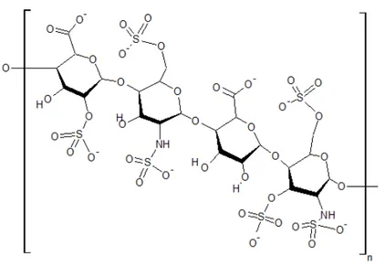
Although Hofmeister ion effects on water structure are currently considered to be confined to the first one or two hydration layers [42–44], recent infrared photodissociation (IRPD) spectroscopy studies suggest that a sulfate ion may “order” up to ca. 36–43 water molecules, equivalent to at least three hydration layers [16]. However, it should be noted that no comparable IRPD data have yet been reported for other biologically-significant kosmotropic anions such as phosphate and carbonate. Organic polysulfates (complex sulfated molecules such as heparan sulfate, as shown in Figure 1) decorate the exterior of nearly all cells in the body, and they are essential to the function of the glycocalyx lining the luminal wall in all blood vessels [46]. The beneficial, longer-range effects of organic polysulfates such as these sulfated glycosaminoglycans on biological water structure are considered below.
Figure 1. Structural formula of a typical heparan sulfate unit.
Like kosmotropic ions, small, nonionic hydrophobic solute molecules can also induce local ordering of surrounding molecules. With the small nonpolar solutes, however, this local water rearrangement involves a significant loss of entropy—with the term “entropy” used here in the classical thermodynamic sense of association with “disorder” or “energy unavailable for useful work.” Loss of entropy is thermodynamically disfavored and thus gives rise to the hydrophobic effect (low solubility of hydrophobes) at scales below ca. 1 nm. However, molecular dynamics simulations with hard spheres and graphene sheets indicate that hydrophobic interactions between larger surfaces are enthalpy- rather than entropy-driven [47,48], where “enthalpy” (another thermodynamic term) refers to the heat energy transferred in a constant-pressure
process, such as a chemical reaction or other type of intermolecular interaction. The hydrophobic effect in biological systems will be discussed further below.
4.2. Interfacial Water: Interaction with Hydrophilic and Hydrophobic Surfaces
In biological systems, liquid water interacts not only with small solutes but also with many larger, extended hydrophilic and hydrophobic surfaces, such as those of proteins, nucleic acids, various organelles, and cell membranes. Results of inelastic incoherent neutron scattering studies of several cell and tissue types suggest that ca. 20%–30% of the total (intracellular plus extracellular) water in these systems is interfacial water, i.e. water located within 1–4 nm of these surfaces, with bulk water comprising the remaining 70%–80% [13]. Not surprisingly, experimental and computational studies reveal different changes in water properties at hydrophilic vs. hydrophobic surfaces at this nanoscale level. Interfacial water near hydrophilic surfaces like hydroxylated diamond or amorphous silica displays viscosity from about 2 up to 106 times greater than that of bulk water, while no significant water viscosity changes were seen near hydrophobic surfaces like methylated silica or hydrogenated diamond [49,50].
Most interestingly, exposure of a hydrophobic hydrogenated nanocrystalline diamond surface to 670 nm laser light gave evidence of hydrogen bond excitation and a resulting density decrease/volume increase in the interfacial water, whereas bulk liquid water is essentially transparent to this wavelength [51]. This result is consistent with predictions of Chandler et al. based on computational studies indicating that water near extended (> ca. 1 nm) hydrophobic surfaces shows less hydrogen bonding and behaves more like water near a liquid-vapor interface than bulk water [47,52–57]. The full significance of this observation and of the Lum-Chandler-Weeks theory for our central thesis of EIWS-driven disease etiology is discussed later in this paper.
Historically, the interaction of water with large biomolecules, especially their nonpolar domains, has been considered mainly from the macromolecule’s point of view and designated as the
hydrophobic effect. Rezus and Bakker describe it as the tendency of apolar groups to associate in
aqueous solution, thereby minimizing the total hydrophobic surface that is exposed to water [58]. Previously, in 2004, Despa, Fernandez, and Berry showed that water constrained by vicinal hydrophobes undergoes a librational dynamics effect that lowers the dielectric susceptibility and induces a “redshift” of the relaxation frequency in the hydration shell [59]. Subsequently, in 2006, Despa described how water in tissues and cells is confined and subject to structural effects not present in its bulk counterpart. Despa added that the structuring effect of confined water in tissues is also a source of polarization fields that contribute to the effective interactions between macromolecules. Dissimilar behavior of water molecules at hydrophilic sites versus hydrophobic sites was said to promote the anisotropy of the hydration shell of proteins. According to Despa, the anisotropy of the hydration shell is essential for enzyme function [60].
No definite consensus has yet been reached regarding the structure of water near the hydrophobic surfaces of biomolecules. While “clathrate-like” (lattices, sometimes layered sheets, or cage-like) structures of hydrophobic hydration have been proposed, studied, and seem reasonable, some studies suggest more-subtle restructuring effects involving the second hydration layer [61–64]. The subject of hydrophobic hydration structure is discussed in an excellent review article on water in cell biology written by Ball in 2008 [65]. Keutsch and Saykally presented a compilation of terahertz laser
vibration-rotation-tunneling spectra and mid-IR laser spectra of several small nanoclusters of water, including a cyclic hexamer of water whose cage structure has a certain clathrate-like quality to its appearance [66]. Molecular dynamics simulations have shown that cyclic pentamers are a dominant topology in liquid water. Csajka and Chandler found that pentamer-like patterns are important in solvation of hydrophobic solutes and in the structures of clathrate hydrates [67]. Significantly, the most stable structure of the water hexamer determined in the gas phase resembles the basic unit in ice [66]. In 1995, Xantheas reported ab initio studies of cyclic water clusters (H2O)n, for n = 1–6 [68]. In 2000,
Nauta and Miller identified a cyclic water hexamer in liquid helium which closely resembled the six membered ring forms found in crystalline ice forms of water [69].
According to Perez et al., the water hexamer is predicted by theory to be the smallest water cluster with a three-dimensional hydrogen-bonding network as its minimum energy structure. Previous experimental work provided evidence for cage, book, and cyclic isomers of hexameric water. Using broadband rotational spectroscopy in a pulsed supersonic expansion, this group unambiguously identified all three coexisting isomers. The cage was found to be the minimum energy structure. Rotational spectra consistent with heptamer and nonamer structures were also reported [70].
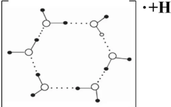
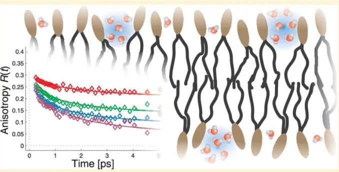
Evidence for water nanocluster formation near lipid surfaces has recently been reported by Piatkowski et al. [71]. Using ultrafast Forster Vibrational Energy Transfer, they found that water hydrating 1,2-dioleoyl-sn-glycero-3-phosphocholine (DOPC) membranes forms nanoclusters at low hydration levels with an average intermolecular distance of 3.4 Å. See Figure 2 below for the putative relative positioning of water molecules. While the density of the water nanoclusters increased with increasing hydration level, the average intermolecular distance did not [71].
Figure 2. The putative relative positioning of water molecules in
1,2-dioleoyl-sn-glycero-3-phosphocholine (DOPC) membranes obtained by measuring the rate of vibrational resonant (Forster) energy transfer between the water hydroxyl stretch vibrations. Reproduced here from Piatkowski et al. [71] with permission of the American Chemical Society.
Like the polar headgroups of lipid membranes, biological polysulfates also present an extended hydrophilic surface that interacts with water. There is even some evidence for a longer-range effect of biological polysulfates on water structure. The findings are particularly relevant to the structure of extracellular water near cell membrane surfaces, which are heavily decorated with sulfate-rich HSPGs arranged in protruding structures called glycocalyces. Observation of diffusion patterns of dyes added to living blood vessels reveals a gel-like, impenetrable layer of water surrounding the interior, HSPG-rich capillary wall [72,73]. This result is reminiscent of in vitro studies reported by Pollack et al. on the behavior of water near the hydrophilic, highly-charged surface of tubes and sheets made of Nafion, a sulfonated fluoropolymer [74–76]. The viscous surface water layer, extending out up to 200-300 µm from the Nafion surface and containing almost no ions or other solutes, has been described by Pollack as an “exclusion zone” (EZ), or a 4th phase of water. EZ thickness can increase by a factor of two to four upon exposure to IR radiation [77]. Whether these empiric in vitro results are relevant to in vivo physiology remains to be determined.
The properties of EZs described by Pollack and coworkers overlap to some extent with those of water “coherence domains” (CDs) proposed by del Giudice et al. based on quantum field theory calculations [78]. A water CD is a ca. 0.1 µm collection of ca. one million liquid water molecules oscillating in tune with a self-trapped electromagnetic field at some well-defined frequency. Evidence for the existence of stable water clusters up to several microns in size, based on electric force microscopy, atomic force microcopy, and infrared and Raman spectroscopic studies of evaporation of very dilute aqueous NaCl solutions at room temperature and pressure, has been reported by Lo and coworkers [79]. EZs may be regarded as longer-range ensembles of CDs, and some researchers use these two terms interchangeably [78]. These CDs/EZs create a negative electrical potential of as much as -150 mV relative to adjacent, “normal” liquid water and a corresponding concentration of protons at the interface with “normal” water. The detailed physics of this type of water, as revealed by the in vitro studies of Pollack et al., is only recently being elucidated, and investigation of potential clinical significance is warranted.
4.3. Interaction with Electric and Magnetic Fields
In this section we survey recent literature relating to the effects of electric and magnetic fields on interfacial water structure and properties. We will also consider evidence pointing to the Ca2+ signaling system as the primary cellular target of magnetic fields, as this has important implications for the uptake of toxic xenobiotics, including interfacial water stressors such as Al3+, into the cell.
The available evidence points to differing effects of electric and magnetic fields on water structure. In 2008, Rai et al. reported results of density functional theory calculations indicating that applied electric field “opened up” circular- or ring-type water clusters to form linear, branched, or netlike structures by making the dipolar water monomers align along the field axis. In general, the number of hydrogen bonds in a cluster decreased with an increase in the electric field strength [80]. In 2011, Acosta-Gutierrez et al. performed additional computational studies of the physical properties of small water clusters in low and moderate electric fields. At low electric field strengths, the hydrogen bonds oriented the water permanent dipoles along the field, whereas larger field strengths induced more
extensive structural reorganization, including hydrogen bond-breaking as the cluster stretched along the field direction, with “the larger clusters (N > 10) usually forming helical structures” [81].
In contrast with the computational studies suggesting that external electric fields break up small water clusters and cause water monomers to line up in the direction of the field, the results of molecular dynamics simulations by Chang and Weng imply that external magnetic fields increase the stability and hydrogen bond strength of supramolecular water clusters while decreasing the self-diffusion of individual water molecules [82]. Moreover, experimental data obtained by Pang et al. on the effects of external magnetic fields on water properties [83–86] support Pang’s earlier hypothesis that such fields promote formation of both linear and closed chains of hydrogen-bonded water molecules [87]. Applying magnetic fields ranging from 2000 to 4400 G (0.20–0.44 T), Pang and Deng found that the infrared and ultraviolet absorptions, Raman scattering and X-ray diffraction of magnetized water were greatly changed relative to those of unexposed water: infrared (IR) peak strengths increased, frequencies of some peaks shifted, and some new peaks occurred after water was magnetized [83,84]. Significant hysteresis effects were observed in the IR absorption spectrum of magnetized water as temperature was increased and then decreased over the range of 25 °C to 70 °C. Importantly, magnetized water displayed a lower contact angle (lower hydrophobicity, or increased ability to solvate hydrophobic surfaces) than non-magnetized water with copper, graphite, and muscovite surfaces. For each surface, the contact angle difference between magnetized and unmagnetized water was small, on the order of 0.4 to 1.4, but still outside the range of experimental error of the instrument. External magnetic fields increased the refractive index, dielectric constant, and electrical conductivity of water while decreasing its viscosity [84]. The longer the magnetization time, the more the viscosity of the magnetized water decreased, until a minimum was reached.
As noted above, the results of these experimental studies of magnetic field effects on water properties [83–86] are consistent with Pang’s earlier proposal that exposure of water to a magnetic field facilitates formation of linear and closed hydrogen-bonded water clusters, the latter of which can become ring electric-current or “molecular electric-current” elements with magnetism due to their proton conductivity under the action of the Lorentz force [87]. This enables magnetic interactions of these “molecular electric-current” elements with each other or with the externally applied magnetic field to change the distribution and features of water molecules and the “magnetization of water” [83].
−Examples of the proposed linear (open) and circular (closed) hydrogen-bonded chains of water
molecules are shown in Figure 3.
The magnetic field strengths of 0.20–0.44 T used by Pang and Deng were several orders of magnitude higher than that of the geomagnetic field at the earth’s surface (ca. 50 µT) and even higher than the ca. 10-10–10-15 T values measured for human organs [3,4]; hence, the relevance of their studies to water in biological systems may legitimately be questioned. However, a complementary mechanism of water magnetization, presented by Mohri [88,89], is based on experimental studies involving a more physiologically-relevant, 6 Hz, 10 µT pulsed magnetic field. Mohri’s hypothesis involves an assumption of cyclotron resonance of protonated water clusters (H3O+(H2O)n).
Figure 3. Illustration of potential linear and circular clusters of hydrogen-bonded water
molecules induced by an external magnetic field as proposed by Pang 2006 [87]. Reproduced here from Pang (2006) [87] with permission of Springer-Verlag Berlin/Heidelberg.
Cyclotron resonance refers to the phenomenon of energy transfer to a charged particle that is moving circularly, normal to the direction of an applied magnetic field, as a manifestation of the Lorentz force; the so-called “cyclotron resonance frequency” of this circular motion depends on the particle’s charge and mass and the strength of the magnetic field. According to Mohri, this cyclotron resonance effect activates proton transport in water under the geo-magnetic field, an effect described as “magneto-protonics” [89]. Formation of a string of such resonating water clusters can give rise to enhanced proton conductivity. This hypothesis is consistent with the decreased electric resistivity of magnetized water reported by Mohri in studies conducted with the weak, pulsed magnetic field described above [89].
In addition to influencing the properties of interfacial water, magnetic fields can also induce changes in the many biological Ca2+ signaling systems. In 2002, Mohri found enhanced phagocytic immune activity and elevated intracellular Ca2+ levels in neutrophils exposed to phosphate buffered
saline (PBS) solution that had been subjected to a milliGauss ultra-low-frequency AC (mg ULF-AC) magnetic field prior to exposing the neutrophils to it [90]. In a subsequent study, Mohri reported a reliable method for decreasing the electric resistivity of highly purified water by applying a small magnetic field of several milliGauss in amplitude and twin cyclotron resonance frequencies of 7.0 Hz and 8.4 Hz to excite hexameric and pentameric hydromolecular clusters, respectively [88].
In 2005, Fukushima et al. reported another extraordinary finding: pure water exposed to a 10 mG, ultra-low frequency (6 Hz) AC magnetic field (generated by a Helmholtz coil under visible light) stimulated firefly luciferin-luciferase luminescence and induced intracellular Ca2+ elevation of Chinese
hamster ovary (CHO) cells in the absence of ATP [91]―suggesting that exposure to the magnetic field increased signaling activity without taxing normal energy sources. Thus, the luciferase-catalyzed luminescence of luciferin, which normally requires ATP in untreated water, occurred without any added ATP in water that had been treated with the magnetic field and light. Indeed, the luminescence activity of the luciferin-luciferase complex in water that was exposed to the magnetic field and light was several-fold higher than that obtained in light-shielded conditions. It should be noted that these experiments were conducted at 40°C, close to the normal human body temperature of 37 °C and near the optimum temperature for most enzymatic reactions. The 6 Hz frequency of the applied magnetic field corresponds to the cyclotron resonance frequency of the protonated hexameric water cluster
H3O+(H2O)5 under the influence of the geomagnetic field (ca. 500 mG) and to the alpha-wave frequency of
the brain [88,91]. The authors speculated that the magnetic energy applied to pure water was stored in a water cluster with stable hydrogen bond resonance and transferred to the luciferin-luciferase complex, with resultant formation of oxy-luciferin and luminescence in the absence of ATP [91].
While the results of Fukushima and coworkers [91] may seem surprising, recent work indicating an ability of low-entropy sunlight to impart long-range order in bulk as well as surface water [92] provides a plausible route by which water CD's may provide energy catalyzing chemical reactions not only at enzymes but indeed near many hydrophobic or hydrophilic surfaces [93,94], as well as for actual diffusion of enzymes through bulk aqueous solution toward areas of high local substrate concentration [95–97].
The results obtained by Fukushima and coworkers [91] are consistent with those of Gartzke et al. (2002), who pointed to the Ca2+ signaling system as the primary cellular target of magnetic fields. Specifically, the ion-conducting actin filament bundle within microvilli was proposed as the cellular target for magnetic fields. This target combines physiological relevance for Ca2+ signaling with unusual electrical properties capable of explaining the effect of low-energy magnetic fields on biological systems [98]. This target was previously shown to exhibit nonlinear, cable-like cation conduction through arrays of condensed ion clouds. Stochastic resonance and/or the Brownian motor hypothesis were employed to explain how the interaction of ion clouds with periodically applied electromagnetic fields results in cation pumping through a cascade of potential barriers within polyelectrolytes [98]. The proposed interaction mechanism was in accord with the postulated extreme sensitivity for excitation by very low field energies within specific amplitude and frequency windows. Thus, instead of a disturbing role, thermal “noise” itself became an essential and necessary signaling component. Microvillar cation transduction by F-actin bundles shielded by a lipid membrane amplify coherent signals on cation transduction and reduce stochastic (thermal) noise. The weak coherent signals are thought to be amplified by thermal noise via stochastic resonance which occurs upon application of a very low energy periodically-applied field, resulting in unidirectional cation transport along F-actin bundles.
An important implication of this proposed systematization is the synergistic action of magnetic fields on the uptake of xenobiotics into the cell. Toxic compounds can enter the cell more readily under the influence of electromagnetic fields, activating the Ca2+ signaling pathway. Lange pointed out that maintenance of intact microvillar surfaces is essential in providing the natural barrier function of epithelial cells [99]. Any disorganization of microvillar surface morphology was shown to severely accelerate the entrance of ionic and lipophilic xenobiotics into the cytoplasm. This point is discussed further in Sections 5 and 6 below when we consider the adverse effects of aluminum cation and other exogenous interfacial water stressors on biological systems.
4.4. Life-Enabling Properties of Water at the Interphase
We propose that the main systems by which structured interfacial water promotes life-enabling biological processes include:
(A) Promoting electrical conductivity at biological interfaces, thereby facilitating metabolism and voltage differences maintained by intracellular organelles;
(B) Absorbing, storing, and emitting electromagnetic energy, enabling storage and transmission of energy and information;
(C) Overcoming the kT or “thermal diffusion” problem; and
(D) Solving the intracellular crowding and molecular self-assembly problems by way of chirality (handedness of molecules) and magnetization.
These interactions are discussed below, along with supporting data. 4.4.1. Promoting Electrical Conductivity at Biological Interfaces
Nanomolecular ensembles of water CDs at the aqueous interphase can provide an extended, long-range, scaffolding for protomeric and electromeric transfer on a mesoscopic, supramolecular scale, which supports energy metabolism in vivo. We have mentioned previously a role for external HSPGs in connecting the cytoskeleton to the plasma membrane. The cytoskeleton also plays a central role in caveolin-based lipid transport between the Golgi apparatus and the plasma membrane [100]. We hypothesize that the cytoskeleton also facilitates the transport of both electrons and protons, taking advantage of the water CDs to induce a magnetic field promoting proton and electron currents, thus sustaining the cell’s membrane voltage gradient. Similar ion transport to and from cytoplasmic organelles such as mitochondria (which must maintain a highly basic pH) and lysosomes (which must maintain a highly acidic pH) is likely also maintained by the cytoskeleton. The actin cytoskeleton has been shown to be integrally linked to both lysosomes [101] and mitochondria [102]. If these organelles are unable to maintain their extreme pH values, they will fail to function and the cell will be disabled.
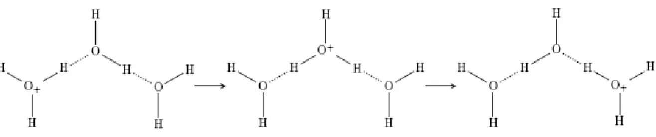
Figure 3 above depicts some possible water nanoclusters that could promote proton conductivity [87]. Three additional proposed water cluster arrangements for enhanced proton transfer are shown in Figures 4–7 [103–106].
Figure 4. The Eigen-Zundel-Eigen (EZE) proton mobility phenomenon [103,104].
Reproduced here from Markovitch et al. (2008) [105] with permission of the American Chemical Society.
Figure 5. Protomeric ensembles acting as substrates for Grotthuss phenomenon.
Reproduced here from Verdel et al. (2011) [106] with permission of MDPI AG.
Verdel et al. [106,107] attributed increased proton transfer deduced from conductivity measurements to the “autothixotropic” phenomenon (weak gel-like behavior) of water, which supposedly develops spontaneously with time, where ions and hydrophilic surfaces seem to play an important role. Voth et al. [105,108,109] have shown that sulfonate groups in the sulfonated fluoropolymer Nafion influence excess proton solvation, as well as the proton hydration structure, by stabilizing a more Zundel-like (H5O2+) structure in their first solvation shells [110]. The sulfonate
groups were also found to affect the proton hopping directions. These findings suggest how biosulfates function in living organisms.
Studies with Nafion, used in proton exchange membrane-based fuel cells [108,109,111], and with carbon nanotubes (CNTs) functionalized with CF3SO3H groups [112], have provided additional
insights. At low water content, the sulfonated side chains of Nafion form isolated hydrophilic regions. As the water content increases, these domains expand and eventually form spanning water channels which are capable of efficiently transmitting protons. It is likely that eukaryotic cells use such a system for efficient proton transport. In ab initio molecular dynamic studies with the fluorosulfonated CNTs, decreasing the distance between sulfonate groups increased proton dissociation and interactions between water molecules. As sulfonate-sulfonate distance increased, connectivity among the water molecules decreased as they formed more isolated clusters around the sulfonate groups. Sulfonate-sulfonate distance and geometry were the most dominant factors in proton dissociation; however, the hydrophobic environment and nanoscale confinement became more important as distance between sulfonate groups increased [112].
Martin Chaplin, a preeminent expert in water structure and properties, has recently argued that both proton and electron delocalization constitute the normal state of affairs in liquid water molecule networks, as illustrated schematically in Figure 6 below [113,114]. Thus, if he is correct, electron (as well as proton) conductivity is enhanced in ensembles of water CDs. Czerlinski and Ypma proposed that electrons move statistically in electromeric domains like a dipole, initiating similar behavior in other domains by resonance [115,116]. When water networks are exposed to ionizing radiation, the structure is modified so as to give mobility to both protons and the hydroxyl radicals [117].
Theoretical physicist, Herbert Frölich, originally proposed in 1968 that coherent electrical polar oscillations and the generation of electromagnetic fields play important roles in living cells, and their disturbances occur in cancer cells [118,119]. Experimental support for Frölich’s ideas continues to accumulate. In 2013, Pokorný et al. reviewed the current biophysical literature pertaining to cancer transformation [120], wherein measurements performed on living cells have disclosed electric and electromagnetic oscillations, including dielectrophoretic forces of the cellular oscillating electric field. The resulting attraction of dielectric particles depends on their permittivity [121]. We refer the reader
to [120] for descriptions of the experimental and theoretical research of the cellular electromagnetic activity, which today, points strongly to microtubules as major sources of electromagnetic interactions. Evidence for a key role of EIWS-induced cytoskeleton disruption in cancer causation is discussed in Section 6.1 below.
An additional route to enhanced electrical conductivity in biological systems could be ion-radical separation converting mesomeric [122] water nanoclusters into superconductors. Being mesomeric enables the delocalization of protons (making these nanoclusters protomeric) as well as free radical electrons (making them also electromeric). Such properties enable catalysis of oxidation-reduction reactions, propagation of electric currents, and generation of magnetic fields. Mesomeric systems, along with other stable water clusters, can theoretically also serve as vehicles for storing incident radiant energy as entropy loss and charge separation, as theorized by Chai, Yoo, and Pollack in 2009 [76]. Figure 7 (below) illustrates a hypothetical cyclic bipolaron―an electromeric and protomeric ensemble of structured water. Such a complex could assist in maintaining membrane potentials and enabling cellular cytoskeletal conduction. Stable cyclic hexamers of water have been studied spectroscopically and theoretically by Saykally [66], Pang [84–87], and Mitsui [123]. The electron movement in these extended coherence domains seems to resemble the free electron movement in metals or even superconductors [115,124–126]. Other models for electron capture in water―for example, electrons in p-orbital-like water cavities―have been proposed and verified experimentally [127].
Figure 6. Neither the protons nor the electrons are pinned to individual molecules. Reproduced
Figure 7. A hypothetical radical-cation cyclic water hexamer accounting for protomerism
and electromerism.
Electrical current which depends on the presence of water has been detected in association with cellulose [128], proteins [129,130], microtubules [131], and DNA [132,133]. In 1987, Careri et al. demonstrated direct current (DC) protonic conductivity of powders of lysozyme for varied levels of hydration [134,135] and suggested that hydration-induced protonic conduction and enzymatic activity corresponds to the formation of a percolation network of absorbed water molecules on the surface of the macromolecule. Computer simulation studies reported in 2006 suggest that hydration water percolation on DNA surfaces drives polymorphic transitions and DNA conductivity [136]. In 2012, Sontz et al. proposed a mechanism for charge transport mediation by duplex DNA [133]. Are these currents dependent on the presence of nanomolecular ensembles of water CDs? The research literature suggests that they are. Czerlinski and Ypma have provided much of the theoretical support for the electromerism (electron conductivity) of nanomolecular water CDs [115,116,137,138]. Given the Josephson effect in physics and the fact that the overlapping base pairs of the double helix have a certain metallic quality, almost like sheets of graphite [126,133], it is not surprising that DNA has been experimentally associated with electrical current.
We propose that dynamical nanomolecular ensembles of water CDs represent structural entropy-consuming [139–143] nano-engines which trap, transduce, and conduct the energy to induce conformational changes in both DNA and proteins. Nanoclusters of magnetized water and DNA, then, may act in concert to provide a supramolecular scaffolding acting to transmit both energy and information over long distances. Empirical evidence already cited shows that interfacial water stress (IWS) provides a supramolecular basis for both the formation and stability of rings of circular DNA [144], microDNAs [145], non-B-conformation DNA [146], and Z-DNA [147]. IWS and nanoclusters of magnetized water also provide a supramolecular basis for modulating DNA structural stability in both health, e.g., normal cell division, and disease, e.g., oncogenesis. This topic is explored in greater depth in Section 6 below.
4.4.2. Absorbing, Storing, and Emitting Electromagnetic Energy
In addition to their ability to enhance electrical conductivity, relevant research and sound theory suggest that water CDs can absorb and emit electromagnetic energy, thus storing and transmitting both information and energy [148–150]. EZ water absorbs light at 270 nm and fluoresces when excited at this wavelength [151]. Based in part on Preparata’s application of quantum electrodynamic field theory [152–154], Marchettini, Del Giudice, Fuchs, Vitiello, and Voeikov proposed in 2010 that water
CDs provide a “redox pile” of “quasi-free electrons” [78,155]. In 1998, Voeikov and Naletov described weak photon emission of non-linear chemical reactions of amino acids and sugars in aqueous solutions which they proposed provides evidence for self-organizing chain reactions with delayed-branching [149]. In 1999, Kobayashi reported spontaneous ultraweak photon emission from a rat’s brain correlated with cerebral energy metabolism and oxidative stress [156]. In 2004, Curtis and Hurtak [157] proposed that biophotonic processes in humans may represent the way biophysical light interacts with the human self-organization of information that may be achieved by means of biomolecular metabolic, or neural communication.
In 2005, Kim et al. demonstrated that spontaneous photon emissions from cancer tissues contrasted with those of normal tissues, and their delayed luminescent properties were investigated [158,159]. Mean values of spontaneous photon emissions from normal tissues and tumor tissues were measured with standard errors at 625 ± 419 counts/minute/cm2 (n = 6) and 982 ± 513 counts/minute/cm2 (n = 14), respectively. Peak values of the intensity of delayed luminescence from normal and cancerous tissues were 63 ± 20 counts/ms (n = 6) and 48 ± 12 counts/ms (n = 14) [158] respectively.
In 2007, Whissell and Persinger showed that prenatal exposure of pregnant Whistar albino rats to extremely weak 7 Hz magnetic fields in the 1, 5, 10, 50, and 500 nT range, caused behavioral deficits in their offspring which persisted into adulthood. These changes were found to be waveform-specific and may involve nitric oxide [160]. Co-administration of the nitric oxide synthase (NOS) inhibitor n-methylarginine appeared to mitigate the behavioral deficits induced by the magnetic fields, to suggest a critical developmental role of NO and the involvement of NO in magnetic field effects [160]. Could these findings show how an external electromagnetic field modulates IWS leading to the unfolded protein response (UPR), perhaps by increasing the hydrophobicity of water? NOS activation requires calcium-binding to calmodulin. As stated previously, calcium has been a proposed cellular target of magnetic fields. In principle, magnetic fields could alter vascular blood flow, by their effects on erythrocytic eNOS and endothelial eNOS. If the ion cyclotron resonance (ICR) frequency of calcium is induced by the magnetic field, this phenomenon may be generalizable to a larger number of functions, given the multifarious roles of calcium in biological signaling pathways [161].
In 2010, Tafur et al. proposed that the detection of biophotons, the production of which is associated with cellular redox state and the generation of ROS, represents a noninvasive redox measure which may be useful in advancing low intensity light therapy [162]. In 2011, Czerlinski described long-lived nanodomains of water that form coherent cooperative aggregates controlled by the geomagnetic field. These domains either slowly emit biophotons or perform specific biochemical work at their target [115,116].
In 2012, Pang [83,84,86] determined from energy spectra that protein molecules can both radiate and absorb bio-photons with wavelengths of < 3 μm and 5–7 μm, consistent with the energy level transitions of the excitons, and consistent with experimental infrared absorption data. Pang’s findings appear to provide support for the controversial experimental results of Gerald Pollack in 2006 [77] wherein large EZs were observed in the vicinity of many types of surfaces, including artificial and natural hydrogels, biological tissues, hydrophilic polymers, monolayers, and ion-exchange beads, as well as with a variety of solutes. Moreover, it was further shown that radiant energy profoundly expands these zones in a reversible, wavelength-dependent manner. Pollack wrote: “It appears that
incident radiant energy may be stored in the water as entropy loss and charge separation” [76].
Whether Pollack’s in vitro results apply to the living state, e.g., cell biology, remains to be determined. 4.4.3. Overcoming the kT or “Thermal Diffusion” Problem
According to Ho (2011), the “thermal threshold” is a fallacy arising from the assumption that living organisms can be described in terms of conventional equilibrium thermodynamics; whereas by general consensus they are open systems meticulously organized and maintained far away from thermodynamic equilibrium [163]. Could water CDs provide a physical basis for overcoming both the kT paradox and the intracellular crowding problem [28,29,31,164–172]? The term kT requirement, where k is the Boltzmann constant and T is temperature (deg K), relates generally to the temperature dependence of chemical reaction rates, and the need to impart enough energy to biological molecules in cells to achieve such reactions at physiological temperatures, without resorting to thermal diffusion (i.e., heating up the reactants).
Through the power to store and amplify electromagnetic energy, water CDs provide a means for achieving biological effects with very weak magnetic fields by ion cyclotron resonance (ICR), as seen in Section 4.3 above, and by temperature-independent means [78,163,166,167,169,173,174]. ICR provides just one of various ways to theoretically account for observed interactions between weak low-frequency electromagnetic fields and biological systems. The ICR hypothesis has been detailed in 2006 by A.R. Liboff [175], and Del Giudice [161] has employed the principles of quantum electrodynamics (QED) in attempting to explain biological ICR, especially the results reported by Zhadin and coworkers, who observed increased ion currents in aqueous glutamic acid solutions exposed simultaneously to weak static and alternating magnetic fields [167,176,177].
4.4.4. Solving the Intracellular Crowding and Molecular Self-Assembly Problems by Way of Chirality and Magnetization
It is clear that the aqueous phase of the cytoplasm is crowded rather than dilute, and that the diffusion and partitioning of macromolecules and vesicles in cytoplasm is highly restricted by steric hindrance as well as by unexpected binding interactions [170,171,178]. In 2001, Aggeli et al. presented a generic statistical mechanical model for the self-assembly of chiral rod-like units, such as beta-sheet-forming peptides, into helical tapes, which, with increasing concentration, associate into twisted ribbons (double tapes), fibrils (twisted stacks of ribbons), and fibers (entwined fibrils) [179]. Results of a recent study of self-assembly behavior of isomers of the hydrophobic tripeptide leu-phe-phe suggest that proteins composed of homochiral amino acids would likely assemble and pack more efficiently than those containing both D- and L-amino acids [180].
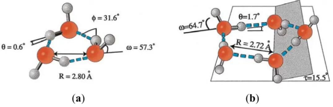
If chirality in biological molecules can promote more-efficient packing of macromolecules in the limited intracellular space, then anything that could induce chirality in water (the most-abundant molecule in the cell), and/or promote more-efficient packing of water molecules by some other means, could also reduce intracellular crowding. Individual water molecules are not chiral, but results of extensive terahertz laser vibration-rotation-tunneling (VRT) and mid-IR laser spectroscopic studies, in conjunction with theoretical calculations, indicate that cyclic water trimers and pentamers are indeed chiral [66], as shown in Figure 8.
Figure 8. Illustration of (a) chiral cyclic water trimer, and (b) chiral cyclic water pentamer.
Reproduced here from Keutsch and Saykally [66], with permission of the publisher, copyright (2001) National Academy of Sciences, USA.
(a) (b)
Thus, at least some of the closed-chain hydrogen-bonded supramolecular water structures proposed by Pang [83,84,86,87] for magnetized water (see discussion in Section 4C above) could be chiral. As suggested by the results of two-dimensional studies of non-chiral, equilateral triangle-shaped polymer particles in aqueous solution, formation of such local chiral “supraparticle” clusters appears to be driven by the increased entropy afforded by increased motion of the monomers within the chiral arrangement relative to that attainable with the more-ordered tight packing of individual particles [181]. Based on these considerations, we propose that formation of chiral supramolecular water clusters, facilitated by magnetic fields, can help to solve the intracellular crowding problem.
Another area in which we think magnetized water could make an essential contribution is in facilitation of biological macromolecular and supramolecular self-assembly. Elegant experiments conducted by Whitesides et al. have provided ample evidence that use of magnetic forces, such as in magnetic levitation, can guide the self-assembly of three-dimensional structures from even diamagnetic components [182,183].
4.5. Tuning the Aqueous Interphase: Modulating Interfacial Water Systems
As noted in Section 2 above, experiments with new spectroscopic techniques that enable study of biological systems on time scales down to picoseconds indicate that large-scale motions of proteins and nucleic acids are determined by fluctuations in the hydration shell, which are controlled by solvent viscosity and hydration, and are absent in a dehydrated protein [14,17–34,36]. If changes in interfacial water structure drive biomacromolecular conformational changes, then biological systems must have means to vary interfacial water structure and properties (within properly-functioning, life-enabling limits) to carry out their wide range of life-sustaining activities. In section 4C above, we surveyed the evidence that ultra-low frequency magnetic fields can reduce water surface tension and hydrophobicity. In addition, magnetic microemulsion formation from anionic magnetic surfactants has been reported [184–186]. These data suggest that interfacial water tension is modulated in vivo by magnetic fields and anionic surfactants. Indeed, results of the molecular dynamics studies of fluorosulfonated carbon nanotubes mentioned above, indicating that the geometry and distance between sulfonate groups played dominant roles in determining proton transfer rates in surrounding water molecules [112], suggest a way that biosulfates in cell membranes may perform a similar function in biological systems, and also suggest a pathology when the sulfation levels are depleted.
Cell membranes generally become more compliant (less “stiff”) when they are depleted of biosulfates, such as cholesterol sulfate (Ch-S) and HSPGs, in their outlet leaflets and glycocalyces, respectively. Cell membranes which are more compliant (less “stiff”) are relatively hydrophobic, i.e. relatively “dewetted” by interfacial water. Ultimately, even phase transitions, such as nanobubble formation, could occur, especially near the triple point where interfacial water is about to freeze [142]. Under the Lum-Chandler-Weeks theory [47], relative hydrophobicity and the molecular theory of capillarity [187–189] are predicted to play major roles in determining cellular compliance (“softness”) at both the intracellular and extracellular aqueous interphase domains near our biomembranes. The speed of capillary waves at the surface can be used to measure surface tension, by how much they scatter light from a laser [189]. Suzuki et al. used dielectric spectroscopy with microwaves to study the hydration of myosin subfragment 1 (S1). The observed changes in S1 hydration were quantitatively consistent with the accompanying large thermodynamic entropy and heat capacity changes estimated by calorimetry, indicating that the protein surface hydrophobicity change plays a crucial role in the enthalpy-entropy compensation effects observed in the steps of S1 ATP hydrolysis [190].
It must be expected, therefore, that many, if not all, sterols are biophysically active as their corresponding sulfates, which have the amphiphilic character needed for bioavailability. Vitamin D3 sulfate,
cholesterol sulfate, DHEA-sulfate, estrone sulfate and the sulfated neurosteroids are examples [2,191,192]. Also, the sulfated neurosteroids appear to be acting “from a distance” within the synapses, as opposed to acting at receptor sites. Hence, the biosulfates are likely to be active by virtue of stabilizing cell membranes and maintaining the CD water in the extracellular space. As noted earlier, enhanced Grotthuss and Josephson effects have been implicated with water CD ensembles [126,148–150].
A number of medium-chain fats and cofactors, e.g., lauric acid, capric acid, ascorbic acid, panthothenic acid, -lipoic acid, and niacin, all share apparent membrane-stabilizing properties; it is plausible to infer that they all act predominantly by lowering interfacial water tension. Bioactive polyphenols and polyketones represent other classes of surfactant mimics, whose bioactivity and bioavailability may be significantly modified by sulfation. Resveratrol, curcumin, ascorbic acid, and the health-promoting phenols in coffee and chocolate can be sulfated, and this may be the key to their biological benefits. Studies on resveratrol metabolism revealed that it is sulfated in the gut prior to absorption [193]. The fact that vitamin C catalyzes the conversion of homocysteine thiolactone to sulfate may also depend on the fact that vitamin C can also be sulfated [194]. In in vitro experiments with chondrocytes, vitamin C was found to induce a 70% increase in the biosynthesis of sulfated proteoglycans [195], which we hypothesize is a result of its ability to carry sulfate [196]. In 1973, Verlangieri and Mumma demonstrated the in vivo sulfation of cholesterol by ascorbic acid 2-sulfate [197]. Such findings suggest that the health benefits of all such molecules may be due to their propensity to participate in sulfate synthesis and transport.
The empirical evidence that biosulfates embedded into membranes can have beneficial effects on interfacial water suggests that phosphorylation signaling may impart a similar kosmotropic anionic feature to membrane-bound molecules. Also, it is known that phosphorylation signaling cascades are triggered by cholesterol depletion in the membrane [198]. A depletion of cholesterol is likely to be at least linearly related to a decrease in cholesterol sulfate in the membrane, and this may trigger the phosphorylation of other membrane biomolecules such as phosphatidylinositol (PI) via phosphatidylinositol 3 kinase (PI3K), a key intermediary in phosphorylation signaling cascades, as a compensatory action.
Three additional phosphate groups can be added to Pi to form phosphatidylnositol phosphate (PIP), phosphatidylinositol bisphosphate (PIP2) and phosphatidyl-inositol trisphosphate (PIP3), collectively called the phosphoinositides. PI3K is a known regulator in angiogenesis and tumor growth [199].
5. Exogenous Interfacial Water Stress and Its Pathological Consequences
In the preceding sections, we have surveyed the research literature relevant to the structure and properties of life-enabling biological water at interfaces in extracellular and intracellular space. We have examined evidence that the interaction of this interfacial water with hydrophobic and hydrophilic surfaces and with electromagnetic fields can create extended networks of coherent, structured forms of water. These extended networks can act as electrical wires or circuits that enable and control life processes (such as macromolecular motions) by enhanced and rapid absorption, storage, emission, and transmission (conductivity) of energy and information. We have also looked at possible ways in which interfacial water structure and tension may be modified by magnetic fields and/or anionic surfactants at membrane surfaces, enabling biological systems to perform their usual life-sustaining activities.
In contrast with the variations in interfacial water structure and tension, induced by endogenous agents such as, for example, Ca+2, Mg2+, Zn2+, Co2+, Mn2+ ions, that enable normal life processes, we use the term “exogenous interfacial water stress” (EIWS) to denote a pathological, perhaps more acute increase in interfacial water tension brought about by a xenobiotic agent. Incremental surface tension values have been reported by Marcus [200]. As discussed more fully in our earlier review [2], potential exogenous interfacial water stressors include kosmotropic cations such as Al3+ and Hg2+, as well as various cationic and nonionic surfactants. If biological water systems are regarded as extended electrical circuits that can store and transmit energy, then exogenous interfacial water stressors are agents that can weaken and unload energy from these networks by creating “leaks,” or by creating short-circuits that provide a lower-resistance path for rapid energy discharge, thus depleting energy and causing collateral damage along the discharge pathway. Examples of such damage would include disruption of membrane and/or protein and/or nucleic acid systems, increased hydrophobicity, protein aggregation, cell-cell aggregation, microbe-cell aggregation, and excess production of reactive oxygen species, to name only a few of the undesirable outcomes.
5.1. Exogenous Interfacial Water Stress as a Short-Circuit, Energy-Unloading Phenomenon Causing Extracellular and/or Intracellular Damage
In extracellular space, the negatively-charged cell membrane surface is particularly vulnerable to short-circuiting by interaction with a cationic kosmotrope or surfactant that can tie up a protruding sulfate, phosphate, or carboxylate group and disrupt local water systems. Consistent with the energy-depletion hypothesis, quite a few recent research papers support an inverse relationship between interfacial water stress and surface energy [201–206]. Interfacial waves have been identified by intravital microscopists [207–209]. Gallez and Coakley showed empirically that the average
number of waves per wavy cell rim decreased when cell surface charge was depleted, and when cationic drugs were present, and increased in the presence of anionic drugs [198,209]. Because of the
that the spatial distribution of wave-like densities noted on scanning electron micrographs may be produced by the interaction (reflection) of electrons with the structured water at the interface.
The likeliest recipient for sudden energy discharge in any short-circuiting caused by interfacial water stress would be the nearby interfacial water. Energy discharge into the interfacial water would disrupt its dynamic balance leading to lowered density and higher volume, as when water near a hydrophobic surface is exposed to 670 nm light [51], as mentioned in Section 4 above. Lower-density water has less power to solubilize hydrophobic surfaces. This trend was demonstrated experimentally with water containing the protein ubiquitin artificially “stretched” at negative pressure in an adaptation from Berthelot in 1850 [35,210,211]. Water density was reduced from 1.00 to 0.95 g/cm3 in a sealed glass nuclear magnetic resonance (NMR) tube. The protein in this “stretched” water became less stable than in normal-density water.
The hydrophobic interaction impacts stabilization of many biological components and plays a decisive role in protein folding [212]. The hydrophobic effect is an entropically-driven phenomenon arising from the difference in density between the open order arrangement of water in the neighborhood of a nonpolar surface and less ordered bulk water [58–60,212]. If EIWS decreases the entropic gain of minimizing the exposed nonpolar surfaces to interfacial water, it must eventually “kill” the hydrophobic interaction, with consequent denaturation of the protein. This inference is supported by the work of Defay and Prigogine, who showed that, at the interphase, the triple-point of water is affected by curvature and surface tension [213], and by the Lum-Chandler-Weeks theory of hydrophobicity [47,53,55,57,171].
It follows that the in vivo, gel-sol transitions are modulated by surface tension and curvature, as originally postulated by Prigogine [213,214]. However, surface tension at the interphase is likely to be affected by such variables as the presence of static and dynamic electromagnetic fields, chirality, pH, and concentration of solutes, including the presence of amphiphilic surfactants. Roughness and curvature of the biomembranes clearly has major impact on such properties as capillarity [189] and capillary blood flow [188]. The anomalous properties of supercooled water and glass formation may have in vivo correlates [26,215–219]. Water at the interphase, under conditions of acute local hydrophobic stress, described as “unwetting” or “stretching” [210], may be followed ultimately by a phase transition, which could be devastating in vivo [142]. Patel et al. [53] used molecular dynamic simulations to show that a large enough hydrophobic surface can induce the formation of a water-vapor-like interface, and as such, the probability of water depletion is enhanced near such a surface. Marked similarities were demonstrated between water-vapor interfaces and water-oil interfaces. It is also well known that purely repulsive hydrophobic surfaces induce a vapor-liquid-like interface [47,220,142].
Damage to a cell membrane caused by interfacial water stress can make it easier for interfacial water stressors to gain access to intracellular space and cause further harm. Depending on the interfacial water stressor and the type of surface encountered, such intracellular damage could include protein unfolding or misfolding, DNA misfolding, and generation of excess reactive oxygen species (ROS), which further disrupt intracellular systems. Specific types of intracellular damage are considered below with respect to toxic actions of the aluminum cation, Al3+, a quintessential exogenous interfacial water stressor that has been linked with breast cancer and neurological disease, as discussed in Section 6 below. An additional pathway by which EIWS can give rise to both extracellular and intracellular damage is suggested by results of a study involving detergent treatment of rat embryo fibroblast cells, which
![Figure 3. Illustration of potential linear and circular clusters of hydrogen-bonded water molecules induced by an external magnetic field as proposed by Pang 2006 [87]](https://thumb-eu.123doks.com/thumbv2/123doknet/14164106.473589/12.892.224.675.210.432/illustration-potential-circular-clusters-hydrogen-molecules-external-magnetic.webp)
![Figure 3 above depicts some possible water nanoclusters that could promote proton conductivity [87]](https://thumb-eu.123doks.com/thumbv2/123doknet/14164106.473589/14.892.104.795.835.1022/figure-above-depicts-possible-nanoclusters-promote-proton-conductivity.webp)
![Figure 6. Neither the protons nor the electrons are pinned to individual molecules. Reproduced here from Chaplin (2013) [113] with permission of the Institute of Science in Society](https://thumb-eu.123doks.com/thumbv2/123doknet/14164106.473589/16.892.206.687.644.1079/electrons-individual-molecules-reproduced-chaplin-permission-institute-science.webp)