Biomaterials-Tissue Interaction of an Injectable Collagen-Genipin Gel in a Rodent
Hemi-Resection Model of Spinal Cord Injury
By
AoSSACHUQE Tr-!: 1NSTrUTE
Daniel J. Macaya 7T E
B.S. Materials Science & Engineering
E
2
0
Ai
Cornell University, 2008
SUBMITTED TO THE HARVARD-MIT PROGRAM IN HEALTH SCIENCES AND
TECHNOLOGY IN PARTIAL FULFILLMENT OF THE REQUIREMENTS FOR THE
DEGREE OF
DOCTOR OF PHILOSOPHY IN MEDICAL ENGINEERING AND MEDICAL PHYSICS
AT THE
MASSACHUSETTS INSTITUTE OF TECHNOLOGY
SEPTEMBER 2013
0 2013 Daniel J. Macaya. All rights reserved
The author hereby grants to MIT permission to reproduce and distribute publically paper and
electronic copies of this thesis document in whole or in part in any medium now known or
hereafter created.
Signature of Author:
Harvard MIT Prcogram in Health Sciences & Technology
June 28, 2013
A
Certified by:
Myron Spector, Ph.D.
Professor of O
opedic urgery (Biomaterials), Harvard Medical School
Senior Lecturer, Department of Mechanical Engineering, MIT
Thesis Supervisor
Accepted by:
Emery Brown, MD, Ph.D.
Director, Harvard-MIT Program in Health Sciences and Technology
Professor of Computational Neuroscience and Health Sciences and Technology
Biomaterials-Tissue Interaction of an Injectable Collagen-Genipin Gel in a Rodent
Hemi-Resection Model of Spinal Cord Injury
By Daniel J. Macaya
Submitted to the Harvard-MIT program in Health Sciences and Technology on June 2 81h, 2013 in Partial Fulfillment of the Requirements for the Degree of Doctor of Philosophy in Medical Engineering and
Medical Physics
ABSTRACT
Spinal cord injury (SCI) is a significant health issue resulting in life-long disability and associated secondary complications, affecting approximately 300,000 individuals in the United States. Primary barriers to functional recovery after SCI include the formation of a growth inhibitory astrocyte scar at the lesion border and a lack of a supportive stroma within the defect allowing for axon regeneration. Interestingly, in animals capable of spinal cord regeneration, astrocytes create a tissue bridge across the injury site to facilitate the regeneration of axons through the defect and thus enable functional recovery.
The overall goal of this thesis was to develop an injectable collagen-genipin (Col-Gen) hydrogel to facilitate the intrinsic regenerative response after SCI by promoting the population of the defect with astrocytes through a provisional scaffold pennissive of astrocyte migration. The specific aims of the thesis involved: 1) development and materials characterization of an injectable collagen hydrogel for neural tissue regeneration, capable of undergoing covalent crosslinking in vivo; 2) evaluation of the permissiveness of Col-Gen gels with and without Fibroblast growth factor-2 (FGF-2), a known astrocyte chemoattractant, incorporated within lipid microtubules (LMTs) to infiltration by primary astrocytes using an in vitro cellular outgrowth assay; 3) evaluation of select formulations of the gel, based on the in
vitro findings, in a standardized hemi-resection defect in the rat spinal cord. Functional, locomotor, and
histopathological outcome measures, recorded up to 4 weeks post-SCI were correlated with each other and with micro MRI studies.
In vivo, the implantation of Col-Gen gels containing FGF-2 LMTs resulted in the enhancement of astrocyte, blood vessel, and laminin infiltration of the defect; increased the amount of spinal cord tissue spared from secondary degeneration; and increased functional recovery, at four weeks post injury as compared to control or Col-Gen treatment groups. Micro MRI was found to be a suitable modality to non-destructively observe the features of the injury in situ. This work commends an injectable, covalently cross-linkable formulation of collagen gel incorporating FGF-2-releasing LMTs for further investigation for the treatment of SCI.
Thesis Supervisor: Myron Spector
Title: Professor of Orthopedic Surgery (Biomaterials), Harvard Medical School. Senior Lecturer, Department of Mechanical Engineering, MIT
Acknowledgements:
This work is dedicated to Randy Duchesneau. Randy, your perseverance to lead a successful and fun-filled life despite your injury has been an inspiration for me throughout my PhD. I hope we are now
one step closer to a brighter future.
I am immensely grateful to my advisor Dr. Myron Spector. Dr. Spector is my role model for what a great mentor and teacher should be. In addition to guiding my project, he has preened me as a scientist, and fully supported my personal and professional development in any way he could. He always made time for me when I needed it and helped me mn workshops aimed at supporting graduate students throughout MIT. I could not have asked for a more dedicated and supportive advisor.
I would like to thank my fellow lab members who I came to know over the years at the VA for their help and support of my work: Karen Ng, Alix Weaver, Paul Elias, Cathal Kearney, Rahmat Cholas, Lily Jeng, Teck Chuan Lim, Toh Wei Seong, Thomas Cheriyan, Wanting Niu. Ravikumar Rajappa, Swetha Rokkappanavar, and Casper Foldager. I am especially grateful to Dr. Hu-Ping Hsu for his
exceptionally skillful hands in performing the animal surgeries and Ravi for his invaluable assistance with animal care. I would also like to thank my thesis committee Dr. Michael Cima, Dr. Zhigang He, and Dr. Ken Arai for their guidance through the process of forming my work, as well as my collaborators who made many aspects of this work possible. Specifically, Dr. Gareth McKinley for use of his rheometer, Michelle Farley and Dave Bennet for their technical expertise with MRI, and Dr. Kazuhide Hayakawa for isolation of the primary astrocytes.
A special thank you to my parents Iris and Joseph Macaya for inspiring the scientist inside of me for as long as I can remember. You fed my thirst for knowledge with books, plastic reptiles/dinosaurs,
and countless trips to the zoo, aquarium, and museum of natural history. You always encouraged me to shoot for the stars and pursue my dreams. To my grandma Pauline Freundlich, thank you for helping to raise me and bring me to where I am today. I know you are proudly looking down at me from above.
To Christine Hsieh: My love, we have seen each other grow and develop as people and as scientists since I first entered graduate school. We talked deeply and intimately about life and we were there for each other through all the highs and lows. You looked out for me as I looked out for you, and you carried me through my toughest moments in grad school. Our adventures, whether they be urban festivals, suburban getaways, or epic hikes have been a cornerstone of my happiness these past few years.
To Ryan Cooper: You have been my best friend since we both embarked on this journey to get our PhDs many years ago. You opened my eyes to the outdoors and have changed my life ever since. I will always remember those first few years when almost every weekend was another adventure. I can trust you with everything and anything.
To the rest of my friends (my buddies from Sidney-Pacific, HST, MIT, and back home in NYC) and family who have helped me along this journey of the PhD, I sincerely appreciate all of your love and support.
The funding for this work was provided by: The National Science Foundation Graduate Research Fellowship Program; The GEM consortium; the Harvard-MIT division of Health Sciences & Technology; U.S. Department of Veterans Affairs, Veterans Health Administration, Rehabilitation Research and Development Service; and the Department of Defense.
Table of Contents
Acknowledgements 3
Table of Contents 4
Chapter 1: Introduction 8
Chapter 2: Background and Motivation 12
2.1. Introduction to SCI 13
2.1.1. Pathology and current treatment
2.1.2. Chronology of pathophysiological changes 2.1.3 Inflammatory and vascular events after SCI 2.1.3. Considerations for regeneration
2.2. Injectable materials for SCI: Overview 21
2.2.1 Current status of injections into the spinal cord 2.2.2. Advantages of injectable materials
2.2.3. Requirements for injectable systems 2.2.3.1 Main functions
2.2.3.2. Design parameters
2.2.4. Considerations for administration in the context of SCI
2.3. Classes of injectable hydrogel materials for SCI 28
2.3.1. Natural vs. synthetic materials 2.3.2. In situ physical gels
2.3.2.1. Thermogels 2.3.2.2. Ionic crosslinking 2.3.2.3. Enzymatic
2.3.2.4. Self-assembling peptide systems 2.3.3. In situ chemical gels
2.3.3.1. Photo-initiated polymerization and crosslinking 2.3.3.2. Molecular/chemical crosslinking
2.4. In vivo studies of injectable materials for SCI 38
2.4.1 Major findings and correlations between in vivo studies
2.4.1.1. Cellular interaction with injectable scaffolds is beneficial for decreasing the astrocytic response and scar formation, and for the promotion of axonal growth after SCI.
2.4.1.2. Cellular infiltration of scaffolds is dependent on scaffold microstructure, degradability, and the presence of soluble or insoluble cues for cell growth
2.4.1.3. Placement of DDS determines release profile and localization of the therapeutics
2.4.1.4. Combinational approaches will be required to achieve significant recovery after SCI
2.4.2 Description of prior studies using collagen as a scaffold for SCI
2.5. Summary 45
2.6. References 47
Chapter 3: Development and characterization of an injectable collagen-genipin gel 55
3.1 Introduction 56
3.2 Background and motivation 56
3.3 Overall goal and hypothesis 58
3.4 Methods 58
3.4.1 Materials
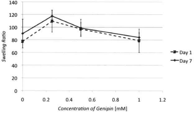
3.4.3 Rheological testing 3.4.4 1-D degradation assay 3.4.5 3-D degradation assay 3.4.6 Swelling ratio
3.4.7 Gel morphology 3.4.8 Stem cell seeding 3.4.9 Cell viability 3.4.10 Cell proliferation
3.5 Results 62
3.5.1 Absorbance and fluorescence 3.5.2 Mechanical and gelation studies 3.5.3 Degradation studies
3.5.4 Swelling ratio and gel morphology 3.5.5 Stem cell seeded collagen gels
3.6 Discussion 79
3.6.1 Cross-linking of collagen and genipin 3.6.2 Absorbance and fluorescence properties 3.6.3 Mechanical and degradation properties
3.6.4 Correlation of fluorescence, mechanical, and degradation properties (long term behavior of collagen-genipin)
3.6.5. Cell-biomaterial interactions
3.7 References 85
Chapter 4: Permissiveness of Collagen-Genipin gels Containing FGF-2 to
Infiltration by Primary Astrocytes using an In Vitro Cellular Outgrowth Assay 88
4.1 Introduction 89
4.2 Background and Motivation 89
4.3 Overall goal and hypothesis 90
4.4 Methods 91 4.4.1 Experimental design 4.4.2 Materials 4.4.3 Collagen gels 4.4.4 Lipid microtubules 4.4.5 Astrocyte culture 4.4.6 Outgrowth assay
4.4.7 Quantification of the number of cells infiltrating the gels, migration distance, and cell morphology
4.4.8 Cell viability and proliferation 4.4.9 Statistical analysis
4.5. Results 96
4.5.1 Number of cells infiltrating into the gels
4.5.2 Infiltration distance and morphology of infiltrating cells 4.5.3 Proliferation and viability
4.6. Discussion 106
4.6.1 Rationale for scaffolding to enable astrocyte infiltration after SCI 4.6.2 Gel permissiveness of astrocyte infiltration
4.6.3 Effects of genipin 4.6.4 Cell phenotype
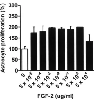
4.6.5 Sustained delivery and dosage of FGF-2 4.6.6 Proliferation
4.7 References: 113
gels in spinal cord injury 116
5.1 Introduction and motivation 117
5.2 Overall goal and hypotheses 117
5.3 Methods 117
5.3.1 Collagen gel fabrication 5.3.2 Animal procedure
5.3.3 Animal sacrifice, transcardial perfusion 5.3.4 Histology and imunohistochemistry
5.4 Results 122
5.4.1 Appearance of the Col-Gen gel via Masson's trichrome staining 5.4.2 Response at one-week Col-Gen treated animals
5.4.3 Response at four-weeks Col-Gen treated animals
5.4.4 Response at one-week Col-Gen LMT-FGF-2 treated animals 5.4.5 Response at four-weeks Col-Gen LMT-FGF-2 treated animals
5.5 Discussion 130
5.5.1 Application of a Col-Gen gel to the hemi-resection defect 5.5.2. Addition LMTs loaded with 1 mg/ml FGF-2 to the Col-Gen gel 5.5.3 Optimization of the procedure and methods for the animal study
5.6 References 134
Chapter 6: Biomaterials-tissue interaction of a second-generation collagen-genipin
gel containing FGF-2 in a rodent hemi-resection model of SCI 135
6.1 Introduction & motivation 136
6.2 Overall goal and hypotheses 136
6.3 Methods 137
6.3.1 Collagen gel fabrication: 6.3.2 Animal procedure
6.3.3 Animal sacrifice, transcardial perfusion 6.3.4 Histology and immunohistochemistry 6.3.5 Image quantification:
6.3.6 Functional Evaluation:
6.3.7 Statistical analysis and sample size determination
6.4 Results 142
6.4.1 Functional Evaluation
6.4.2 Early analysis of the defect at one day and one week post injury 6.4.3 Histomorphologic assessment of the extent of injury and new tissue formation within the defect at 4 weeks
6.4.4 Immunohistochemical analysis of the cellular and extracellular composition of the defect
6.4.4.1 a-SMA 6.4.4.2 Laminin 6.4.4.3 Astrocytes
6.4.4.4 Endothelial cells/angiogenesis
6.4.4.5 Inflammatory response: macrophages
6.4.4.6 Regenerating axons
6.4.4.7 Overview of marker co-localization in animals exhibiting a Type A response to injury
6.5 Discussion 164
6.5.1 Functional evaluation 6.5.2 Histological evaluation
Chapter 7: Pilot ex vivo MRI study of the gel implantation and tissue remodeling
after hemi-resection injury in rodents 174
7.1 Introduction & Motivation 175
7.2 Overall goal and hypotheses 175
7.3 Methods 176
7.3.1 Experimental groups 7.3.2 Surgical procedures 7.3.3 MRI acquisition
7.3.4 Histology and immunohistochemistry
7.4 Results 177
7.4.1 One-day post injury: Col-Gen gel
7.4.2 One-week post injury: Control and Col-Gen gel 7.4.3 Four-weeks post injury: Control, Col-Gen, and Col-Gen LMT FGF-2 gel
7.5 Discussion 187
7.5.1: Early extent of injury and gel localization
7.5.2: Secondary damage and cellular infiltration at one week post injury 7.5.3: Remodeling of the defect by four weeks post injury
7.6 References 191
Chapter 8: Conclusions 192
Chapter 9: Limitations and future directions 196
Chapter 1:
Introduction
Spinal cord injury (SCI) is an extremely debilitating condition often resulting in complete or partial paralysis. In the United States there are approximately 12,000 new cases per year with an
additional 232,000-316,000 people currently living with SCI [1]. Since most SCI patients are young, with an average age at injury of 40.7 years, the lifetime cost to care for these individuals is high, ranging from $300,000 for incomplete motor function at any level to over $4,000,000 for high tetraplegia [1]. Currently, there are no clinically available treatments to restore significant function after injury.
SCI presents a complex regenerative problem due to the multiple facets of growth inhibition that occur following trauma to the cord parenchyma and stroma. Clinically, SCI is further complicated by the heterogeneity in the size, shape, and extent of human injuries. Many of these injuries do not breach the dura mater and have continuous viable axons through the injury site that can later lead to some degree of functional recovery. In these cases, surgical manipulation of the spinal cord by implanting a preformed scaffold or drug delivery device may lead to further damage. Given these circumstances, utilizing in situ-forming scaffolds are an attractive approach for SCI regeneration. These synthetic or natural polymers undergo a rapid transformation from liquid to gel upon injection into the cord tissue,
conforming to the individual lesion site and directly integrating with the host tissue. Injectable materials can be formulated to have mechanical properties that closely match the native spinal cord extracellular
matrix, and this may enhance axonal ingrowth. Such materials can also be loaded with cellular and molecular therapeutics to modulate the wound environment and enhance regeneration.
Overall goal of this thesis is to develop an injectable gel capable of undergoing covalent cross-linking in vivo, which can modulate the native tissue response to spinal cord injury, allowing for the formation of a regenerative template of astrocytes and blood vessels within the defect formed after spinal cord injury.
In order to enhance the infiltration of the gel-filled defect with astrocytes, a known
chemoattractant for these cells, fibroblast growth factor (FGF)-2, was incorporated into the collagen solution prior to its injection. In addition to acting as a chemoattractant for astrocytes, FGF-2 is
commended for use in this study for its ability to: promote revascularization of the injury site and act as a neuroprotective factor for the spared tissue. The gel formulations were evaluated in a hemi-resection defect model in the rat spinal cord.
The specific aims of the thesis involve: 1.) Development of an injectable collagen hydrogel for neural tissue regeneration, capable of undergoing covalent crosslinking in vivo. .2.) Enhancement of astrocyte infiltration into Col-Gen gels through the sustained delivery of fibroblast growth factor 2 (FGF-2) encapsulated within lipid microtubules (LMTs). 3.) In vivo histological and behavioral study of the
acute- early chronic tissue response to injection of Col-Gen gels in a standardized hemi-resection defect in the rat spinal cord.
The overall hypothesis is that by designing a provisional matrix with the proper attractive cues for axon supportive cellular ingrowth (i. e. astrocytes and endothelial cells), a regenerative template will be formed within the spinal cord defect, allowing for the ingrowth of axons.
This thesis consists of several chapters, summarized below, which provide a detailed background, methods, results, and discussion of the present study:
Chapter 2 details the background and motivation underlying the work performed in this thesis. It includes a through literature review of the pathophysiology of SCI, current treatments, the role of injectable therapies in SCI, the design parameters and classes of materials appropriate for use as injectable gels for SCI, and finally an overview of the prior in vivo studies using injectable collagen gels for the treatment of SCI. A majority of the content in this chapter has been published in Biomedical Materials [2].
Chapter 3 characterizes the injectable collagen-genipin gel in terms of its mechanical behavior, cross-linking kinetics (via absorbance and fluorescence measurements), resistance to enzymatic degradation, and cell viability when incorporated into these gels. A majority of the content in this chapter has been published in Advanced Functional Materials [3].
Chapter 4 describes the cellular outgrowth assay used to assess the permissiveness of select collagen-genipin gel formulations containing FGF-2 to astrocyte infiltration. This chapter also provides insight in to the phenotype of the astrocytes in response to FGF-2, the benefits of sustained release of FGF-2 via lipid microtubules (LMTs), the effects of genipin on astrocytes, and the role of proliferation and viability in the observed response. A majority of the content in this chapter has been published in Biomaterials [4]. Chapter 5 describes a pilot in vivo biocompatibility study in a standardized rodent hemi-resection model
of SCI using collagen-genipin gels with and one without FGF-2 LMTs. The formulations used in this study were guided by the experiments in chapters 3 and 4. The animals were assessed at one and four weeks post injury and evaluated histologically and behaviorally.
Chapter 6 describes a full in vivo study of the biomaterials-tissue interaction of collagen-genipin gels in a standardized rodent hemi-resection model of SCI. Histologic and behavioral analysis was performed on three groups: control, collagen-genipin gel, and collagen-genipin gel with FGF-2 LMTs, at four weeks post injury. The formulations of the collagen-genipin gels were determined based on the pilot study in chapter 5.
Chapter 7 details a pilot ex vivo magnetic resonance imaging study of the hemi-resection defect with and without implanted gel at 1 day, 1 week, and 4 weeks post injury. The results correlate features of the
injury, gel localization, and tissue remodeling with histology on the same samples.
Chapter 8 lists the conclusions that are supported by the work presented in this thesis.
Chapter 9 discusses the limitations of the worked performed and identifies areas for future investigation.
Following the main chapters of the thesis is an appendix with detailed protocols for the fabrication and assays performed in the thesis.
References:
[ 1] Center NSCIS. Spinal cord injury facts and figures at a glance. Birmingham Alabama: University of Alabama at Birmingham; February 2012.
[2] Macaya D, Spector M. Injectable hydrogel materials for spinal cord regeneration: a review. Biomed Mater. 2012;7:012001.
[3] Macaya D, Ng KK, Spector M. Injectable Collagen-Genipin Gel for the Treatment of Spinal Cord Injury: In Vitro Studies. Adv Funct Mater. 2011;21:4788-97.
[4] Macaya DJ, Hayakawa K, Arai K, Spector M. Astrocyte infiltration into injectable collagen-based hydrogels containing FGF-2 to treat spinal cord injury. Biomaterials. 2013;34:3591-602.
Chapter 2:
2.1. Introduction to SCI
2.1.1. Pathology and current treatment
An understanding of the pathophysiology of SCI is essential for the rational development of therapeutic strategies. We now have available a wide array of agents which can be employed as
treatments for SCI individually or in combination: biomaterials; exogenous cells of various types; protein regulators of cell function (e.g., growth factors and chemoattractants), and their genes; antagonists of inflammation and nerve growth inhibitors; and enzymes for extracellular matrix (ECM) molecules which interfere with the regenerative process. The informed selection of specific agents and the timing of their administration require an understanding of the intrinsic components of the response to SCI that prevent or interfere with a regenerative response.
The mechanism of SCI is usually a mechanical insult to the spinal cord parenchyma following a fractured vertebra or disk intruding into the spinal canal [1]. This results in various degrees of contusion,
laceration, and in very rare cases a complete transection of the spinal cord tissue [1-3]. Human cases of SCI vary widely with respect to the site and degree of tissue destruction, and may involve multiple lesions with sizes smaller than a single vertebral segment [31 SCI rarely results in a complete disruption of tissue through the lesion site [1]. Approximately 61% of SCIs are classified as incomplete, meaning that there is still a degree of function in segments of the spinal cord innervated below the injury [4]. Complete injuries may also have continuous parenchyma through the lesion, although there is no function in distal segments [1, 3, 5]. Taken together, these studies show that after injury, the protective dural covering of the spinal cord may be intact and there is almost always some amount of viable tissue traversing the site of the lesion. Given these observations, minimally invasive therapies are an attractive treatment option for SCI.
Although our knowledge of the pathology of human SCI is limited, it is believed that most injuries are caused by cord contusion. In one classification system, a contusion/compression type SCI presents with extensive damage and cyst formation in the cord parenchyma with no breach or disruption in the surface anatomy or adhesions to the dura [2]. Another class of SCI, solid cord injury, appears normal grossly but displays tissue that is clearly damaged histologically [2].
One of the challenges in the treatment of SCI is that such injuries may not show gross
morphological changes until hours post injury, but eventually encompass 1-2 vertebral segments proximal and distal to the lesion site [6]. The major clinical manifestations of the injury result from damage and subsequent degeneration of the ascending and descending white matter tracks (axons) [1]. In the case of a contusion injury, viable and intact axons are still present at the injury site but are rendered virtually useless from demyelination due to oligodendrocyte loss, which greatly reduces their conduction velocity
[7]. While some plasticity may be observed, the function of areas innervated by segments below the injury is lost.
Current treatment for SCI addresses 2 features of the injury: the persistent mechanical compression of the cord and the acute inflammatory response. Surgical decompression of the injured segments is immediately followed by the administration of steroids to neutralize acute inflammation and decrease swelling to further reduce compression on any remaining neurons [8, 9].
2.1.2. Chronology ofpathophysiological changes
SCI pathology can be divided into immediate, acute (0-7 days), sub-acute (7-14 days), and chronic (months/years) stages [9]. The microenvironment of the injured spinal cord presents many significant obstacles to regeneration at these various stages (Table 2-1). The initial injury causes necrotic cell death due to direct mechanical trauma and ischemia from vascular disruption and hemorrhage. During the acute phase, a cascade of secondary injury processes occurs that leads to further cell death, scarring, and loss of function. Continued vascular injury, ischemia, inflammation, free-radical
production, and the massive release of excitatory neurotransmitters by the injured cells cause secondary necrosis, apoptosis, and swelling 1-2 segments above and below the original lesion site. Supportive glial astrocytes at the lesion periphery begin to hypertrophy and proliferate into a "reactive" phenotype that contains the injury [10]. The most widely used indicator of the reactive astrocytic response to injury is cellular hypertrophy and the enhanced expression the intermediate filament, glial fibrillary acidic protein (GFAP) [11]. The cytoskeletal intermediate filaments, vimentin and nestin, which are typically found in immature astrocytes, and the following non-cytoskeletal proteins are also expressed in reactive astrocytes: class II histocompatibility antigens, glutamine synthetase, and glutamine transporters [11, 12].
Table 2-1: Key cellular and molecular events after SCI
Event Type Timeframe Underlying Pathological Mechanism Necrosis min-hours Oxidative stress, edema, hemorrhage and
disruption of BBB, ischemia, physical trauma, excitatory neurotransmitters, infiltration of acute
Apoptosis days/week inflammatory cells.
Oxidative stress, limited ability to replace damaged glia & neurons via endogenous progenitors. Activation of apoptotic pathways. Demyelination days-months Oligodendrocyte loss
Glial scar formation days-weeks Reactive astrocytes, ECM remodeling Axon degeneration days-months Loss/block of function (signaling)
Inhibitory/repulsive environment blocks regrowth
In the sub-acute phase, microglia begin to clear the injured area of debris leaving behind a cystic cavity, which is subsequently surrounded by a glial scar consisting of reactive astrocytes. The glial scar is composed of long astrocytic processes that interweave to form a dense mesh and ECM material including chondroitin sulfate proteoglycans (CSPGs) [13, 14]. Macrophages, fibroblasts, and Schwann cells are also known to infiltrate the wound from outside of the CNS, usually from the spinal nerve roots, especially in the case of laceration injuries where the dura is disrupted [3, 15].
Finally, in the chronic phase, the astrocytic scar stabilizes around cysts/syrinxes and extends down the path of degenerated axons [16]. Depending on the extent of fibroblast infiltration, there may also be a dense collagenous scar within the injury site. The fibrous scar formed after SCI can present a greater barrier to regeneration than glial scarring due to its dense framework and ability to bind growth inhibitory molecules [17]. Over the course of the next few months, oligodendrocyte populations continue to undergo apoptosis, which leads to progressive demyelination of axons. Wallerian degeneration, the destruction of the distal end of severed axons, also occurs over the next few months-years leaving behind a trail of myelin debris that further inhibits axon growth [16, 18]. Additionally, Schwannosis, the infiltration of Schwann cells and their associated axons from the peripheral nervous system (PNS), is prevalent in human SCI after approximately four months and is attributed to the loss of astroglial framework at the injury site[3]. This ingrowth by elements of the PNS potentially serves as an
impediment to neurite outgrowth and may cause pain, spasticity, and other abnormal physical responses.
Unlike experimental models of SCI, human injuries are highly heterogeneous and the exact pathology and time course of events will vary depending on the type of injury [1, 2]. It is worth noting that the time course of human SCI is typically extended with respect to rodents [19]. In humans, the loss of myelin in degenerating axon tracts takes approximately 2-3 years, as opposed to months in rodents
[18]. Schwann cell invasion of the lesion is seen as early as I week in rodents but is not noted in humans until 3 weeks [15]. Interestingly, human SCI exhibits less glial scarring and CSPGs are mainly localized to the lesion site. There is also a lower incidence of chronic demyelination in humans when compared to rodents [15, 19]. Such variation in pathology between human and animal models should be considered when designing and testing potential therapies for SCI (Table 2-2) [19].
Table 2-2: Differences in pathophysiology between rodent models and clinical human spinal cord injury. From Hagg et al.
Rodent aUmwan
Degenerative processes
Vascular response Hemorrhage. angiogenesis Hemorrhage angiogenesis
Inflammation Extensive Much less pronounced. despite
similar cytokine expression
Demyelination Yes Yes, but perhaps less pronounced
Axonal degeneration Some die-back and Wallerian Wallerian degeneration much
degeneration more protracted
Glial scar Extensive, with astroglial CSPG Not extensive. CSPGs mostly in blood vessels
Cyst formation Rat yes; mouse no Yes
Schwann cell response Some invasion Extensive Schwannosis
Regenerative processes
Sprouting Yes Yes
Remyelination Yes Yes
Plasticity of uninjured systems Yes Yes
CSPG. chondroitin sulphate proteoglycan.
2.1.3. Inflammatory and vascular events after SCI
The time course of inflammatory cell infiltration after injury is illustrated in Figure 2-1 [20]. Further detail about the contributions of specific inflammatory cells to injury and tissue repair is summarized in Table 2-3 [20].
One specific inflammatory cell type, the macrophage has been extensively studied due to its role as an important modulator of tissue injury and repair after SCI. Macrophages are recruited to the site of injury immediately after SCI, reach a peak density at 14 days post injury, and finally decrease to an elevated by stable value by 3-4 weeks post injury and remain around the injury site indefinitely [20-22]. Interestingly, chondroitin sulfate proteoglycans (CSPGs) most noted for their role in axon inhibition, actually have a pivotal role in spatially and temporally controlling the activity of infiltrating blood-borne monocytes and resident microglia during the acute phase after the injury. Rolls et al. demonstrated that immediate inhibition of CSPG production after SCI in mice caused a dramatic effect on the spatial organization of infiltrating macrophages and an alteration in their cytokine/growth factor release leading to enhanced tissue destruction and decreased functional recovery [23]. In contrast, allowing CSPG synthesis during the first 2 d post injury, followed by inhibition, improved recovery.
Two main phenotypes of macrophages, M I and M2 can be seen after SCI. M
1
macrophages (pro-inflammatory) clear tissue debris from the injury site and areas of Wallerian degeneration but may contribute to the axonal retraction and secondary tissue damage after SCI. M2 macrophages(anti-inflammatory) secrete a variety of neurotropic factors may be responsible for neuroprotection, angiogenesis, and the promotion of axon growth however, an excessive or prolonged presence of M2 macrophages may result in fibrosis and scarring, which could hinder axon regeneration [21]. Therefore, an important balance must be struck between MI and M2 macrophages to achieve the clearance of tissue debris, reduce secondary tissue damage, and promote repair.
Recent work by Kigerl et al. suggests that after injury to the cord there is a transient appearance
I
Tk&rw
rO
y)e
Ira 40 ,4 4-13
7 14 21Days post-injury
S 42Figure 2-1: Time course of inflammatory cell infiltration of the defect site after rodent spinal cord injury with relation to major pathogenic and wound healing events. From Trivedi et al.
of M2 macrophages early (1-7 days) after injury with 40-50% of the macrophages adopting an M2 phenotype [21]. However, by 28 days post injury, fewer than 10% of the macrophages are M2. This work shows that the environment of SCI is conducive to MI macrophage polarization despite the
phagocytosis of red blood cells and cellular debris, which normally promote a M2 phenotype. Within the M2 phenotype there are three subtypes, which modulate different aspects of the inflammatory response, however these have been poorly characterized in the context of SCI.
The vascular events after spinal cord injury play a critical role in the degree of secondary damage, which occurs after the primary injury. Mechanical trauma to the spinal cord (contustion, compression, lasceration) causes vasospasm of superficial vessels and intraparenchymal hemorrhage particularly in the highly vascular central gray matter [24]. There is also a disruption of the blood-spinal cord barrier leading to edema, an alteration of tissue perfusion from the release of vasoactive molecules, and a loss of autoregulation (the ability to maintain blood flow despite differences in perfusion pressure).
Additionally, there may be post-traumatic systemic events, which affect blood flow such as hypotension, bradycardia, and decreased cardiac output [24]. Together, these events lead to ischemia and necrosis within the spinal cord parenchyma. The resulting cell death further initiates a cascade of events such as the release of excitotoxicity neurotransmitters from dead/dying cells and the generation of free radicals from the reperfusion if ischemic tissue or degradation of hemoglobin, which results in additional (secondary) damage to previously viable spinal cord tissue.
Cell Type Type of Immunity Pro-inflammatory molecules Mediators of cell injury/ Pro-regeneration/
__________ Innate (4)_ _ death wound healing events
Neulrophils Innate4) Express receptors for various Produce Phagocytosis
chemokines and cvtokines, metalloproteinases, MMP-9 reactive oxygen and
nitrosyl radicals neutronhil elastase
Dendritic cells Innate and Produce TNF-agr, IL-1, IL-2 Function as antigen Production of adaptive IL-6- IL-12, lL-18. IFN-y presenting cells neurotrophin-3
Monocytes/ Innate Produce TNF-agr;. IL-I. IL-2 Produce reactive oxygen Phagocytosis. production Macrophages IL-6, IL-12, 1L-18 species and nitrosyl of trophic factors. IL-10,
radicals TGF-B
Microglia/ Innate and Produce TNF-agr;, IL-I. IL-2 Function as antigen Phagocytosis, production Macrophages adaptive IL-6, IL-12, IL-1 presenting cells; produce of trophic factors. IL-10,
reactive oxygen species TGF-s
and nitrosyl radicals
B- Adaptive Express receptors for various Produce antibodies Unknown
Ly'phOCyts chemokines and cytokines
T- Adaptive Express receptors for various Pro-inflammatory Production of trophic Lymphocytes chemoknes and cytokmes, cytokines and factors, IL-10, ILA
E
_ __________ _ IFN-. TGF-6 chemokines IL-13Abbreviations: matrix inetalloproteinase-9 -MMP-9; tumor necrosis factor-alpha -TNF-agr; interleukn -IL; interferon-gamma -IFN--y; transforming growth factor-beta -TGF-p;
Table 2-3: Overview of inflammatory cells, their inflammatory mediators, and mechanism of tissue destruction or wound healing. From Trivedi et al.
The hemorrhage which occurs after vascular injury to the spinal cord is greatest at the injury epicenter and can be seen extending into the dorsal columns both rostral and caudal to the injury [24]. Zhang et al, studied the regenerative response within these dorsal column lesions and found they were extensively infiltrated by macrophages and the beginnings of blood vessel growth by 1 week [25]. By 2 weeks, there was a network of blood vessels and clusters of glial cells within the defect and regenerating nerve fibers could be seen at the interface of the lesion and adjacent normal tissue.
1.3. Considerations for regeneration
There are a number of intrinsic and extrinsic factors that lead to regenerative failure in the CNS. Therefore, one may consider multiple targets for intervention (Figure 2-2).
Mylin Node of Ranvier
Grav Matter
A
Microglia Inhibitors of Axonal Growth Figure. 2-2. A) A fluid-filled cystic cavity in a spinal cord resulting from injury, surrounded by a glial scar (white circle) shown in more detail in (B). The glial scar (B) is made up of activated astrocytes and contains inhibitors of axonal growth (depicted as yellow dots).
Illustration C 2011 Edmond Alexander, alexanderandturner.com
Severed Oligodendrocyte Activated Astrocyte Axons
Unlike the CNS, the PNS is capable of spontaneous regeneration. Nerve grafts and guidance tubes have been shown to enable severed axons to grow through large gaps in the PNS and promote recovery [26, 27]. After a PNS injury, the axons at the distal end degenerate while those at the proximal end begin to sprout, elongate, develop growth cones, and eventually reform synapses to nerves/muscles. The major support and myelinating cell of the PNS (Schwann cell) assists the process by re-myelinating axons, secreting growth factors and synthesizing ECM (laminin, fibronectin), and guiding axon growth
[28-30]. There is a 1:1 ratio of axons to Schwann cells in the PNS and after injury cells from the
degenerated portions of axons assist in the regeneration process. In contrast in the CNS, oligodendrocytes myelinate between one and eighteen axons, making the loss of these cells detrimental to a larger
proportion of nerve fibers [31]. Perhaps the most interesting example of the difference between the regenerative potential of the CNS and PNS relates to the dorsal root ganglia (DRG). DRGs are areas beside the spinal cord where the bodies of sensory neurons are located. Although these neurons have axons within the spinal cord and the PNS, they can only regenerate their peripheral process [32]. In the case of complete SCI, Schwann cells infiltrate the spinal cord and associated PNS axons can be seen growing within the former spinal cord parenchyma [15].
As noted above, in the CNS, the major support and myelinating cell, the oligodendrocyte is associated with multiple neurons. Consequently, after injury, many of these cells die; demyelinating their associated neurons and greatly reducing the support for regenerating axons. Degraded myelin contains potent growth inhibitors such as Nogo-A, oligodendrocyte myelin glycoprotein (OMG), and myelin-assisted glycoprotein (MAG) [33-35]. In the PNS these products are cleared by macrophages and Schwann cells, but in the CNS, microglia cells are much slower in clearing this debris which may be present as long as 3 years post injury [18].
The cyst and glial scar formed after injury to the CNS also act as significant physical and chemical barriers to spontaneous regeneration. But the astrocytic response can be beneficial as well as detrimental to regeneration [10, 36]. At early times, it serves limit further damage by: reestablishing the blood brain barrier (BBB) and ionic homeostasis; limiting immune cell infiltration; and containing excitatory neurotransmitters. At later times, however, the dense scar and associated CSPGs are inhibitory towards regeneration. Reactive astrocytes secrete an ECM containing chondroitin sulfate proteoglycans (CSPGs), which have been implicated as a major growth inhibitory molecule for neurons [37-39]. In cases of SCI where there is disruption of the dura, there is also substantial fibrous infiltration of the spinal cord, which leads to an even greater physical and chemical barrier to axon regeneration.[17, 40]
Although there are many barriers to regeneration in the CNS, it has the potential for repair. CNS white matter can support axon regeneration in uninjured as well as degenerating white matter tracts in rodents [41, 42]. The inhibitory factors deposited at sites of chronic SCI do not form absolute boundaries. Early work by David, et al., using a peripheral nerve graft, demonstrated it was possible to achieve
significant axon elongation through a lesion site in the spinal cord by providing a permissive environment for growth similar to the PNS [43]. Axons can regenerate through scars into sites of chronic SCI using diffusible signals such as neurotrophin (NT)-3 from modified mesenchymal stem cells (MSCs) [44]. Numerous others have shown that neurotrophic factors, antibodies against myelin debris receptors, and CSPG degrading enzymes can promote regeneration and axonal sprouting after SCI [45].
The regeneration of a large number of tracts may not be necessary to promote substantial recovery after SCI. Signals can be re-routed to downstream targets over other pathways through neural
plasticity. Significant neurological function may be preserved with as few as 5-10% of the original number of axons present in the white matter [46]. Basso, et al., showed that after a graded contusion by a weight drop, an increase in spared white matter from 5% to 10% allowed rats to go from not supporting their own weight to supporting their own weight and exhibiting consistent stepping [47]. This ability has also been noted in humans. Cordotomy procedures for the relief of pain following cancer showed that the anterior half of the thoracic cord could be cut without any disturbance to motility, and cutting one lateral corticospinal tract (CST) and 85-90% of the opposite tract and the reticulospinal fibers anterior to the tract caused total paralysis of the lower limbs which recovered over 2 months so that the patient eventually walked again [48].
In sum, the local environment after SCI plays a significant role in the regenerative response of neurons. If a minimally invasive therapy is utilized to spare viable axons, reduce the amount of
secondary damage after the insult, and promote axon regeneration and collateral sprouting, a significant amount of functional recovery can potentially occur. By addressing the multiple facets of growth inhibition in SCI, it may be possible to restore a healing response similar to that of the PNS.
2.2. Injectable materials for SCI: Overview
Research into injectable biomaterials holds great promise in the fields of tissue engineering and regenerative medicine. A wide variety of injectable materials have been developed for various tissue specific applications such as bone, cartilage, intervertebral disk, brain, and spinal cord [49-51]. Each tissue poses its own unique set of properties and challenges that must be addressed by the scaffold material. In particular, the CNS presents a complex regenerative obstacle due to the multiple mechanisms of degeneration and growth inhibition that occur after injury. These obstacles require a combinational approach utilizing scaffolds, cellular, and molecular therapies tailored to the specific injury at hand.
2.2.1. Current status of injections into the spinal cord
The injection of substances directly into the spinal cord or surrounding tissue has been used clinically for a variety of scenarios. Intrathecal infusion of medication via catheters has been used for chronic/severe pain relief [52]. For drug delivery applications, mini-pumps and catheters are sub-optimal for sustained delivery as they are prone to blockage and subsequent infection [53]. Currently, there are a few clinical trials are underway for the injection of cells into the spinal cord including autologous macrophages, olfactory ensheathing cells, oligodendrocyte progenitor cells [7, 54, 55].
2.2.2. Advantages of injectable materials
Hydrogels are natural or synthetic hydrophilic polymers capable of binding large percentages (50-99.9% by wt.) of water. Hydrogels are viscoelastic materials resulting from the physical or chemical bonding of molecules in a liquid state: physical gels by van der Waals interactions and hydrogen bonding, and for the purpose of this review, ionic bonding; and chemical gels by covalent crosslinking. The conversion of the injected liquid into a physical or chemical gel in vivo enables the filling of small defects such as the cavities/cysts, which form after SCI or the space between transected parts of the spinal cord. The gel, serving as a scaffold, can eliminate void spaces and form a regenerative template to direct cellular infiltration and matrix deposition towards repair of the injured region.
Injectable materials that form scaffolds in situ can conform to the shape of the defect and create an integrative implant-tissue interface to restore the continuity of the tissue and reduce significant contraction/deformation of the scaffold [56]. This injectable gel property obviates the need to create preformed scaffolds for each individual patient and avoids excising viable tissue at the injury site to accommodate implantation of the preformed scaffold, which could cause further tissue damage and loss of functionality. Poor graft integration is often the cause of graft failure and pathological changes in the host spinal cord including extensive glial-connective scar and cyst formation [57]. For incomplete cases of SCI where the BBB has not been disrupted and the dura mater is intact, injectable therapies will be beneficial since they will not further disrupt the dura. Damage to this important structure has been implicated in scar formation, cellular invasion, and cystic cavity formation after SCI [40].
Injectable materials, in their liquid state, can be uniformly mixed with cells and other therapeutics prior to delivery within the spinal cord defect. Spatial and temporal control over the release of these agents can be tailored through multiple mechanisms including incorporation within secondary release vehicles such as micro- and nano-particles, liposomes, and microtubules [50, 58], covalent tethering to the gel with subsequent enzymatic release [59], or harnessing the ability of macromolecules such as heparin to bind growth factors such as NT-3 and platelet derived growth factor (PDGF) [60].
The mechanical properties of gel scaffolds can more closely match the properties of the native spinal cord tissue, compared to most preformed biomaterial matrices. The implications of this include providing a permissive substrate for neuronal growth and will be discussed in subsequent sections.
For patients, injectable therapies hold the promise of minimally invasive procedures. The prime and most clinically practical window for intervention may not be immediately after injury but delayed until the sub-acute stage (7-14 days post injury), when the initial inflammatory response has subsided [7, 61]. Having flexibility on the time course of administration could enhance the clinical outcome of
patients without risking further damage.
2.2.3. Requirements for injectable systems 2.2.3.1. Main functions
The development of injectable therapies for spinal cord regeneration may be designed with a focus on three broad functions.
1) Scaffolding for cellular infiltration and axonal ingrowth. The gel material itself will serve to bridge the lesion site. Scaffolding acts to reduce cyst formation, inflammation, and the associated secondary cell damage, while accommodating the infiltration of supportive cell populations and the growth of axons into the gel-filled lesion site [62]. The gelation kinetics, final gel microstructure, mechanical properties, degradation rate, and bioactive properties of the scaffold can be tuned to optimize its regenerative potential.
2) Encapsulation of drugs and maintenance of bioactivity throughout gelation and release. Therapeutics such as neurotrophic factors, antibodies against degrading myelin products, chondroitin sulfate-degrading enzymes, and others, can stimulate axonal regeneration through lesion and encourage neural plasticity [63]. Injectable systems can provide a sustained and tunable delivery of these agents locally to the lesion site. This is especially important for therapeutics such as NT-3, which has an in vivo half-life of only 30 minutes, but have a therapeutic effect when delivered over the course of days to weeks
[64, 65].
3) Support of suspended cell populations prior to injection, throughout solidification process, and within in the lesion site. The incorporation of transplanted cell populations have been shown to enhance functional recovery after SCI in animal models by providing necessary trophic factors and extracellular cues for axon regeneration, myelination, and a source to restore lost cell populations [66, 67]. Cellular therapies are more effective when delivered and maintained locally in the injured area as opposed to delivered systemically.
2.2.3.2. Design parameters
It should be noted that in most polymer systems it is difficult to isolate crosslink and macromer concentration-dependent material properties such as mechanical stiffness, mesh or pore size, degradation rate, and bioactive ligand density. Therefore several group have created multi-component systems to study the effects of each variable on neuronal behavior [68-70].
Biocompatibility. The reactants and degradation products of the material must be non-toxic and non-immunogenic to reduce excessive inflammation at the injury site leading to additional cellular
damage. Materials obtained from natural sources should be carefully screened and treated to reduce the risk of disease transmission and immunological responses. Initiators, crosslinkers, or other additives required for scaffold formation should not adversely affect encapsulated cells or cross-react with therapeutics.
Solidification Solidification of the injectable material should occur under mild conditions to avoid additional tissue damage at the lesion site, adversely affecting encapsulated cell populations, or sensitive drug compounds. The speed of gelation must be rapid enough to contain the material within the injection site and maintain the localization of incorporated cells or therapeutic agents. Ideal solidification times are on the order of seconds to minutes depending on the initial viscosity of the injectable material. Factors affecting the gelation time include, temperature, crosslinker concentration, ionic strength, pH, light, surface area, among others [50].
Porosity/mesh size The overall mesh size of nanoporous fibrillar scaffolds, and the porosity, and pore diameter and interconnectivity of microporous scaffolds are crucial for tissue ingrowth, the removal of nutrients and waste, and the diffusion of therapeutic agents from the polymer matrix. A growth-permissive substrate for endogenous/exogenous cell populations may recapitulate the three-dimensional (3-D) organization of the native ECM. In situ-forming scaffolds typically have >99% water content and a typical mesh/pore size on the order of 0.10 Itm. Neurons are 20-30 [tm in length with approximately 1-[tm diameter growth cones [71, 72]. The relative dimensions of the growth cones and mesh opening may hinder cell motility unles the material can be moved or degraded by the cells [68, 73]. For example, agarose gels with decreasing mesh size and increasing stiffness severely inhibited neurite outgrowth below a mesh opening of 150 nm [74]. Additionally, decreasing the mesh size of collagen gels from 7.2 ptm at 0.4 mg/ml to 3.1 [tm at 2.0 mg/ml impeded neurite extension of encapsulated DRG cells [73]. The polymer size and concentration of the monomer/polymer, as well as the initiator and crosslinker
concentrations, can be used to control the mesh or pore size of injectable scaffolds.
Mechanical properties The mechanical properties of the gel should match the native tissue to withstand in vivo forces, maintain structural integrity, and transmit physiological forces until it can be replaced by native tissue. While the spinal cord itself is not a load-bearing structure, injected materials should not collapse under the weight of the surrounding tissue. Additionally, the mechanical properties of substrates have a profound effect on cell behavior especially in the CNS [70, 75, 76]. The soft and viscoelastic nature of native CNS tissue and hydrogels make rheology an ideal technique to study their mechanical properties. In this regard, the shear modulus is often reported as the measure of "stiffness."
Numerous model systems using several materials have been created to investigate the effects of substrate stiffness on neurite behavior. Flanagan, et aL., created a system to isolate the mechanical effects
on primary mouse spinal cord neurons using polyacrylamide gels coated with matrigel to provide a constant mesh size and number of adhesion ligands. Neurons exhibited a high branching density on substrates of 50Pa-350 Pa which is on the order of bovine (50 Pa) or human (200 Pa) spinal cord tissue [70]. In comparison, the branching frequency on 550 Pa gels was reduced, and was similar to glass. Similarly, Leach, et al., used a 2-D polyacrylamide gel with a constant fibronectin density to explore neurite outgrowth and branching of PC12 cells induced to a neuronal phenotype [77]. Unlike Flanagan's experiment, the cells displayed a threshold response to substrate stiffness from a shear modulus of 100
-10,000 Pa. In this stiffness range, a greater percentage of cells expressed neurites, which were also longer and more branched than the softer gels (<0.1 kPa).
Wilits, et al., cultured DRG cells in 3-D collagen gels of varying protein content, ranging in shear modulus from 2.2-17 Pa and found that maximum lengths of neurites occurred in softer gels [73]. It
should be noted that increasing the collagen concentration had multiple effects, which factored into the observed results including a decreased interfiber spacing, increased ligand density, and increased shear modulus. Using a 3-D agarose culture of DRG cells, Balgude, et al., found the average rate of axon elongation was inversely correlated to the concentration of the agarose gel, which ranged in modulus from 5-130 Pa [71]. Banerjee, et al., explored a greater range of gel stiffness -180-20,000 Pa with alginate gels encapsulating neural stem cells (NSCs) and found that the softest gels, which were on the order of magnitude of the stiffness of brain tissue, resulted in significantly higher NSC proliferation and neuronal differentiation (B-tubulin III staining) than stiffer gels [78]. Seidlits, et al., used acrylated high molecular weight hyaluronic acid (HA) gels to investigate ventral midbrain neural progenitor cell (NPC)
differentiation and spinal astrocyte behavior in a 3-D culture environment with tunable mechanical properties [79]. By adjusting the degree of acrylate substitution, the compressive modulus could be modified between 3-10 kPa, which is similar to adult rat spinal cord (8.1 ± 1.1 kPa). After 3 weeks in culture, NPCs on the softest gels were furthest differentiated towards the neuronal phenotype and exhibited 0-III tubulin-positive long branching processes while NPCs on the stiffest gels did not survive the full 3 weeks. On the other hand, spinal astrocytes survived the full 3 weeks in all gels and only showed evidence of migration on the stiffest gel.
Overall, the difference in the observed optimal stiffness for neurite branching among the different studies is due to specific factors, which have an effect on the response of cells to compliant substrates, such as: the cell type (i.e., PC12, DRG, primary neurons); material type and adhesive ligand density; mesh/pore size; and range of modulus investigated. Interestingly, glia, which are implicated in scarring after SCI, had poor survival on soft substrates [70]. Other groups demonstrated that DRG neurons tend to elongate processes down gradients in stiffness and fibroblasts, implicated in mesenchymal scarring,
preferentially migrate back towards stiffer areas of patterned hydrogels [80-82].
Degradation rate The degradation rate of injectable scaffolds should proceed on the time scale of tissue formation so that axons are provided with a constant stable platform for growth. If the scaffold degrades prematurely, the injury site will also be subject to compressive stresses that would cause further inflammation and glial scaring [83], and there will be lack of sufficient stromal support for the ingrowth of axons. If the scaffolding degrades too slowly, it will impinge on new tissue formation and hinder axonal growth. Mahoney, et al., investigated the process of extension of neural tissue into polyethylene glycol (PEG)-polylactic acid (PLA) gel matrices with controllable degradation rates. The group was able to control the time course of neurite extension from 1-3 weeks depending on the degradation rate of the polymer and concomitant change in the matrix mesh size [68]. Subsequent work by this group showed a relationship between the degradable macromer content of the PEG-PLA matrix and the cellular
proliferation of neural cells cultured within the material after 7 days [84]. In rodents, degradation rates on the order of weeks to months would be appropriate. This timeframe corresponds to the rate of axon sprouting from the CST, which can be observed 3 weeks to 3 months after injury [85, 86].
Additionally, neuronal growth cones secrete serine proteases and calcium dependent matrix metalloproteinases that assist in matrix remodeling for axonal path finding during development and regeneration [87, 88]. Pittier, et al., showed that inhibiting serine proteases greatly decreased neurite extension in fibrin matrices [89]. Therefore, synthetic material with bioactive sequences sensitive to these enzymes as well as natural materials such as collagen can be specifically remodeled by the regenerating axons. To this point, Straley, et al., engineered protein polymers containing bioactive sequences for serine protease specific degradation (see section 3.3.3) [69]. By altering the bioactive sequence, scaffolds could be fabricated with a 200-fold difference in degradation half-lives. However, using PC 12 cells, the difference in degradation rate of the scaffolds had no significant effect on cell morphology or neurite extension after 6 days of exposure to neuronal differentiation media.
The degradation mechanisms of biomaterials will influence their behavior in vivo. Since total body water is conserved, materials undergoing hydrolytic degradation have the advantage of having low
animal-animal and location variability, however the degradation is non-specific. Enzymatic mechanisms can respond to local changes in cell behavior during the regenerative process but may suffer from high individual variability [69]. For drug delivery applications, the biodegradability also determines the release of incorporated drugs within the scaffold matrix.
Bioactivity Scaffolds should present specific soluble and insoluble cues to direct cell behavior, such as adhesion ligands present in the natural ECM. Injectable scaffolds have a high pore volume, interconnectivity, and a 3-D architecture, which allow for significant cell-surface interactions. For
scaffolding applications, cell adhesion is crucial as neurons are anchorage-dependent for their growth and survival and will undergo apoptosis when detached from the ECM [90]. Straley, et al., found the density of the cell-adhesive RGD peptide present in the protein directly affected cell adhesion and neurite outgrowth of undifferentiated and differentiated PC 12 cells [69]. However, the effect was still smaller than the control collagen gels suggesting that cues other than RGD motifs are important for controlling neuronal cell behavior.
Neurites are sensitive to environmental cues, which can both promote and inhibit their growth. To explore the interaction of neurites with bioactive cues, Kofron, et al., cultured DRG explants at the interface between a 3-D collagen matrix and a glass coverslip coated with laminin or CSPGs, a promoter and inhibitor of neurite outgrowth, respectively [91]. The coating was arranged as either a uniform layer or patterned tracks. They found that most of the neurites extended away from the coverslip and into the collagen gel when CSPGs were present, while most of the neurites extended at the surface of the gel when laminin was present. There was also an alignment of the neurites to the tracks of laminin on patterned surfaces [91]. Extending this work to a 3-D system, Yu, et al., used DRG cells cultured in layered agarose hydrogels to explore the chemical effects of neurite extension. They found that at the interface of unmodified agarose gels and agarose gels with covalently bound chondroitin sulfate B (CS-B), neurite extension was significantly inhibited as compared to homogeneous gels. The inhibition was significantly decreased by either degradation of CS-B with chondroitinase (Ch) ABC or addition of laminin-1 to the
CS-B containing hydrogels [92].
Additionally, for drug delivery applications to the intrathecal space, materials with limited adhesion ligands should be used to avoid cellular infiltration. The material-drug interactions will play a
role in their release kinetics depending on their relative size, charge, and hydrophobicity to one another. The material also needs to preserve the bioactivity of the encapsulated drug.
2.2.4. Considerations for administration in the context of SCI
Therapies for SCI must take into account the timeframe of intervention and the specific condition of the cord during administration. The injured spinal cord presents a dynamic environment that evolves through years after injury. The composition of the ECM, cytokines, excitatory neurotransmitters,
inhibitory molecules, degree of necrosis, and cellular makeup change with time after injury. This will affect which therapeutic agents would be most beneficial to incorporate into the device. Additionally, the size and location of cysts within the lesion site may affect how an injectable therapy is administered. In the acute phase of SCI, fluid filled cysts will not be present in significant number while during the sub-acute phase, the necrotic cyst defects will present as cavities to fill with an injectable material. In later