Biomimetic Approach to Cardiac Tissue
Engineering
by Milica Radisic
B.Eng. Chemical Engineering McMaster University, 1999
Submitted to the Department of Chemical Engineering in Partial Fulfillment of the Requirements for the Degree of
Doctor of Philosophy in Chemical Engineering
at the
Massachusetts Institute of Technology September 2004
© Massachusetts Institute of Technology All rights reserved
Signature of Author
Department of Chemical Engineering
Certified by
Robert b. Langer Thesis Supervisor
Kenneth J. Germeshausen Professor of Chemical and Biomedical Engineering
Accepted by
-- td15aniel Blankschtein
Professor of Chemical Engineering Chairman, Committee of Graduate Studies
MASSACHUSETTS INSTIURTE OF TECHNOLOGY
This Doctoral Thesis has been examined by the following Thesis Committee:
Robert Langer, Sc.D. / / - ,
Thesis Advisor -
-Germeshausen Professor of Chemical and Biomedical Engineering Department of Chemical Engineering
Massachusetts Institute of Technology
Charles L. Cooney, Ph.D. ,
Professor of Chemical Engineering Department of Chemical Engineering Massachusetts Institute of Technology
William M. Deen, Ph.D. . -
-Carbon P. Dubbs Professor of Chemical Engineering Department of Chemical Engineering
Massachusetts Institute of Technology
Lisa E. Freed: M.D., Ph.D....
Research Scientist
Division of Health Sciences and Technology Massachusetts Institute of Technology
Gordana Vunjak-Novakovic, Ph.D. I - I
Research Scientist
Division of Health Sciences and Technology Massachusetts Institute of Technology
.
. .
ACKNOWLEDGEMENTS
I would like to acknowledge my thesis advisor Prof. Robert Langer and thesis committee members: Prof. William Deen, Prof. Charles Cooney, Dr. Lisa Freed and Dr. Gordana Vunjak-Novakovic for their valuable mentorship throughout the course of this Ph.D. thesis. I am especially thankful to Prof. Langer and Dr. Vunjak-Novakovic for their insight and advice not only in scientific but also in personal and professional matters.
I would like to thank Dr. Hyoungshin Park for making the tedious laboratory procedures seem like fun. I am thankful to my parents Vukosava and Branislav and my brother Milos for their love and encouragement. Finally, I would like to thank my husband Zoran for his constant and selfless support in both good and bad times.
Biomimetic Approach to Cardiac Tissue Engineering
by Milica Radisic
Submitted to the Department of Chemical Engineering
On July 19th 2004 in Partial Fulfillment of the Requirements for the Degree of Doctor of Philosophy in Chemical Engineering
Heart disease is the leading cause of death in the Western world. Tissue engineering may offer alternative treatment options or suitable models for studies of normal and pathological cardiac tissue function in vitro. Current tissue engineering approaches have been limited by diffusional oxygen supply, lack of physical stimuli and absence of multiple cell types characteristic of the native myocardium.
We hypothesized that functional, clinically sized (1-5 mm thick), compact cardiac constructs with physiologic cell densities can be engineered in vitro by mimicking cell microenvironment present in the native myocardium in vivo.
Since cardiac myocytes have limited ability to proliferate we developed methods of seeding cells at high densities while maintaining cell viability. Cultivation of cardiac constructs in the presence of convective-diffusive oxygen transport in perfusion bioreactors, maintained aerobic cell metabolism, viability and uniform distribution of cells expressing cardiac markers. To improve cell morphology and tissue assembly cardiac constructs were cultivated with electrical stimulation of contraction in a physiologically relevant regime. Electrical stimulation enabled formation of tissue with elongated cells aligned in parallel and with organized ultrastructure remarkably similar to the one present in the native heart. To investigate the effect of multiple cell types on the properties of engineered cardiac tissue cardiac fibroblasts and cardiac myocytes were cultivated synchronously, separately or serially (pretreatment of scaffolds with fibroblasts followed by the addition of myocytes). Presence of fibroblasts remarkably improved contractile response of the engineered cardiac constructs with the superior biochemical and morphological properties in the pretreated group. Finally, in order to mimic capillary structure cardiac fibroblasts and myocytes were co-cultured on a scaffold with a parallel channel array that was perfused with culture medium supplemented with synthetic oxygen carrier (PFC emulsion). Presence of the PFC emulsion resulted in significantly higher cell density and improved contractile properties compared to the constructs cultivated in the culture medium alone, by increasing total oxygen content and effective diffusivity.
Thesis Supervisor: Robert Langer
Table of Contents
1. Introduction ...13
References ... 15
2.Background ... 17
Overview ... 17
Tissue Engineering Model System ... 18
Myocardiu ( m cardiac muscle) ... 20
Clinical need ... 21
Tissue Engineering of Myocardium ... 22
Summary ... 29
References ... 29
3. High density seeding of myocyte cells for cardiac tissue engineering ... 32
Introduction ... 32
Materials and Methods ... 33
Results ... 39
Discussion ... 46
References ... 50
4. Medium perfusion enables engineering of compact and contractile cardiac tissue ... 52
Introduction ... 52
Materials and Methods ... 53
Results ... 57
Discussion ... 64
5. The role of electrical stimulation of contractility in functional assembly of myocardium
in vitro ... 72
Introduction ... 72
Materials and Methods ... 72
Results ... 78
Discussion ... 84
References ... 87
6. Sequential cultivation of cardiac myocytes and cardiac fibroblast subpopulations results in robust contractile response of engineered cardiac tissue ... 90
Introduction ... 90
Materials and Methods ... 91
Results ... 95
Discussion ... 102
References ... 105
7.Biomimetic approach to cardiac tissue engineering: Oxygen carriers and channeled scaffolds ... 106
Introduction ... 106
Materials and Methods ... 107
Results ... 112
Discussion ... 117
References ... 120
8. Mathematical model of oxygen distribution in engineered cardiac tissue with parallel channel array perfused with culture medium supplemented with synthetic oxygen carriers ... 122
Introduction ... 122
Materials and Methods ... 126
Model Parameters ... 128
Results ... 135
Discussion ... 139
References ... 144
9. Summary and future work ... 145
Summary...145
Future work ... 146
List of Tables
Table 1.1 Factors governing cardiac tissue development in vitro and in vivo ... 14
Table 2.1 Key challenges of cardiac tissue engineering ... 22
Table 2.2 Overview of cardiac tissue engineering studies ... 26
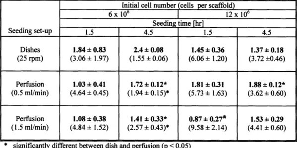
Table 3.1. Seeding and cultivation of cardiomyocytes in perfused cartridges ... 40
Table 3.2. The effects of seeding parameters ... 42
Table 3.3. Kinetics of change in live cell numbers ... 44
Table 3.4. Metabolic indices for 12 different seeding conditions ... 45
Table 4. 1. P values for the individual and interactive effects of culture time (T) and culture system (S) on cell viability and metabolism ... 57
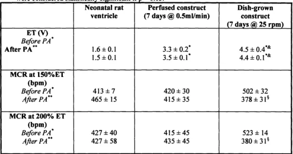
Table 4.2. Contractile properties of neonatal ventricles, 7-day perfused constructs and 7-day dish-grown constructs ... 64
Table 5.1. Glucose metabolism and biochemical properties of stimulated and non stimulated constructs ... 80
Table 6.1. Experimental design ... 92
Table 6.2. Metabolic properties of the constructs during pretreatment and culture ... 95
Table 7.1. Biomimetic approach to cardiac tissue engineering ... 107
Table 7.2. Medium gas composition and metabolic properties ... 113
Table 8.1 Boundary conditions ... 125
Table 8.2 Model parameters ... 130
Table 8.3 Important dimensionless numbers ... 131
Table 8.4 Channel Sherwood number...134
Table 8.5: Oxygen transport improvements in the presence of PFC emulsion ... 139
List of Figures
Figure 2.1. Tissue engineering based on cell cultivation on biomaterial scaffolds in bioreactors. 19
Figure 2.2. Representative bioreactors ... 20
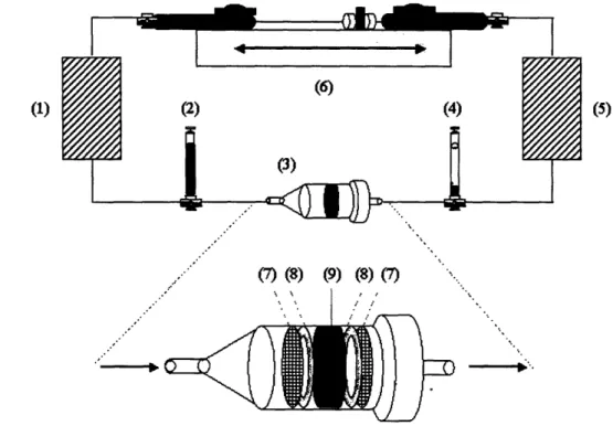
Figure 3.1 Seeding set-up for alternating flow perfusion ... 36
Figure 3.2. Morphology and contractile responses of cardiac myocyte constructs ... 41
Figure3 3. Seeding parameters ... 43
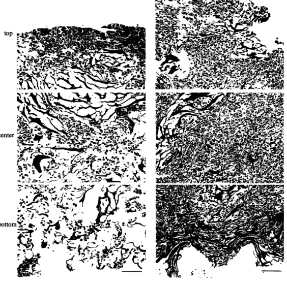
Figure 3.4. Cell distributions ... 47
Figure 4.1. Perfused cartridges with interstitial flow of culture medium ... 54
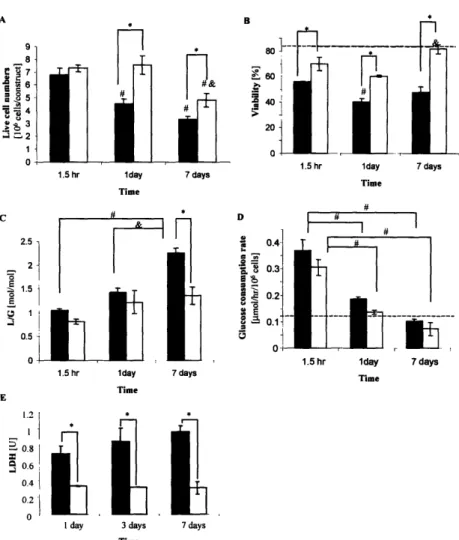
Figure 4 2. Cell viability and metabolism ... 58
Figure 4.3. Fractions of cells in the G0/G 1, S and G2/M phases of the cell cycle ... 60
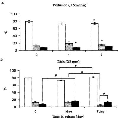
Figure 4.4. Cell distribution ... 61
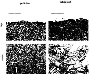
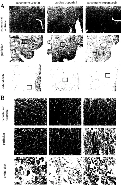
Figure 4.5.Tissue architecture and cell differentiation ... 62
Figure 5.1. Functional properties of engineered cardiac constructs ... 79
Figure 5.2. Tissue morphology and expression of contractile proteins ... 81
Figure 5.3. Ultrastructural features of stimulated and non-stimulated constructs at the end of cultivation (8-day) as compared to the neonatal rat ventricle ... 83
Figure 5.4. Temporal dependence of cell morphology on days of preculture and proposed mechanism ... 85
Figure 5.5. Expression of a number of cardiac proteins as assessed by RT-PCR ... 86
Figure 6.1. Contractile properties of constructs at the end of cultivation ... 96
Figure 6.2 Construct composition ... 97
Figure 6 3. Composition of initial cell suspension and constructs ... 98
Figure 6.4 Tissue morphology and expression of cardiac and fibroblast markers ... 99
Figure 6.5 Expression of cardiac Troponin I ... 100
Figure 7. .Schematics of the model system ... 109
Figure 7.2. Contractile properties of cardiac constructs ... 113
Figure 7.3. Protein and DNA content of cardiac constructs ... 114
Figure 7.4. Scanning electron micrographs of biorubber scaffold with a parallel channel array 115 Figure 7.5. Expression of cardiac markers: Troponin I and connexin-43 in cardiac constructs.. 117
Figure 8.1. Schematics of the construct with parallel channel array ... 126
Figure 8.2. Effect of PFC emulsion on the oxygen concentration ... 135
Figure 8.3. Model solution and validation ... 136
Figure 8.4. Comparison of predicted oxygen concentration profiles ... 137
Figure 8.5. Comparison of predicted oxygen concentration profiles (M) in a channel array supplemented with 0, 3.2, or 6.4% PFC emulsion at physiological cell density ... 138
1. INTRODUCTION
Cardiovascular disease is the leading cause of death in the Western world. It kills more people than next five leading causes of death including cancer. (AHA update 2004) Diseases related to myocardium, heart muscle, contribute to a significant portion of this death toll. Nearly 8 million people in the United States alone have suffered from myocardial infraction, with 800,000 new cases occurring each year [I].
Conventional treatment options, including drugs and assist devices are limited by inability of myocardium to regenerate after injury. Cell based therapies have been considered as a novel and potentially curative treatment option [2], either by utilization of cells alone [3-15] or by cultivation of cardiac construct in vitro that can be surgically attached to the myocardium [16-30]. While cell injection can be pursued as a treatment option for small scale injuries, improved cell
localization achieved by tissue engineering can potentially be utilized to repair large scale injuries and congenital malformations. In addition, engineered cardiac tissue can be utilized as a model system for study of normal and pathological cardiac function in vitro.
Most of the current tissue engineering approaches are limited by diffusional supply of oxygen from the surface of the construct to the interior resulting in a
non-uniform cell distribution with the shell of functional tissue on the outside and an empty interior. Construct cultivation in perfused cartridges markedly improved the uniformity of cell distribution but the overall cell density remained low, due to the oxygen diffusional limitations associated with cell seeding.
In standard culture systems (dishes, flasks, rotating vessels), cardiomyocytes did not align in parallel as in the native heart and they remained poorly differentiated due to the lack of orderly excitation-contraction coupling [17, 18]. Application of contraction alone via cyclic mechanical stretch significantly improved the level of cell differentiation and force of contraction, but some hallmarks of cardiac differentiation were still missing (M bands, IC discs) [29]. In addition, the distribution and frequency of gap junctions, critical for electrical signal propagation in the tissue, remained unclear. Notably, excitation-contraction coupling, which is crucial for the development and function of native myocardium [31], has not been established in any of these culture systems. The main objective of this thesis was to develop the tissue culture approach that would remedy
the problems associated with the conventional culture.
We hypothesized that functional, clinically sized (1-5 mm thick), compact cardiac constructs with physiologic cell densities can be engineered in vitro by mimicking cell microenvironment present in the native myocardium in vivo. The key parameters in the cell microenvironment
include: high cell density with multiple cell types, convective diffusive oxygen transport through a capillary network and orderly excitation contraction coupling (Tablel.1) During the course of this PhD thesis each of the factors present in the Table 1.1 was tested independently. The obtained results are summarized in six manuscripts, whose abstracts are included as Chapters
3-8.
Table 1.1 Factors governing cardiac tissue development in vitro and in vivo.
The organization of this thesis is as follows:
Chapter 2 described basicis of cardiac muscle structure and function and provides the overview of tissue culture methods up to date.
Chapter 3 address oxygen diffusional limitations during seeding by providing a method that enables uniform cell seeding at high initial densities (108cells/cm3) while maintaining viability. Seeding of cardiomyocytes at high initial densities is essential for cultivation of dense compact tissue since cardiomyocytes have limited ability to proliferate. The seeding method consists of two steps: rapid inoculation of gel/cell suspension onto collagen sponges followed by the application of alternating medium flow for convective oxygen supply.
Chapter 4 describes cultivation of cardiac constructs in the presence of convective-diffusive oxygen transport in perfusion bioreactors. Medium perfusion maintained aerobic cell metabolism, high viability and uniform distribution of cells expressing cardiac markers and superior contractile properties compared to the dish grown controls.
Chapter 5 describes establishment of orderly excitation-contraction coupling of engineered cardiac constructs during in vitro culture. Synchronous construct contractions were induced by suprathreshold electrical field stimulation using a commercial cardiac stimulator. Electrical stimulation enabled formation of tissue with elongated cells aligned in parallel and with organized ultrastructure remarkably similar to the one present in the native heart.
In vivo In vitro
Cells High density 5 108 cells/cm3 0.5 -1-108 cells/cm3
Multiple cell types (myocytes, Multiple cell types (myocytes, endothelial cells, fibroblasts) Fibroblasts)
Oxygen and nutrient supply Convection and diffusion Convection and diffusion in perfusion bioreactor Geometry Capillary network Parallel channel array in the
l____________ _ (diameter
l
0gm, spacing 20jim) scaffoldOxygen carrier Hemoglobin PFC Emulsion (Oxygent®)
Excitation-contraction Electrical signal propagation Electrical stimulation
Chapter 6 investigates the effect of multiple cell types on the properties of engineered cardiac tissue. Cardiac fibroblasts and cardiac myocytes were cultivated synchronously, separately or serially (pretreatment of scaffolds with fibroblasts followed by the addition of myocytes). Presence of fibroblasts remarkably improved contractile response of the engineered cardiac constructs with the superior biochemical and morphological properties in the pretreated group.
Chapter 7 describes co-culture of cardiac fibroblasts and myocytes on an elastic scaffold with capillary-like channel array perfused with culture medium supplemented with synthetic oxygen carrier (PFC emulsion). Presence of PFC emulsion resulted in significantly higher cell density and improved contractile properties compared to the constructs cultivated in the culture medium alone, presumably by increasing total oxygen content.
Chapter 8 describes a mathemactical model of oxygen distribution in the channeled cardiac construct perfused with oxygen carrier supplemented culture medium. The release of oxygen from PFC particles was estimated not to be rate limiting.
Chapter 9 summarizes the results of this thesis and identifies problems that need to be addressed in the future work.
REFERENCES:
1. Association, A.H., Heart Disease and Stroke Statistics-2004 Update. 2004.
2. Reinlib, L. and L. Field, Cell transplantation as future therapy for cardiovascular
disease?: A workshop of the National Heart, Lung, and Blood Institute. Circulation,
2000. 101: p. e82-e187.
3. Koh, G.Y., et al., Differentiation and long-term survival od C2C12 myoblast grafts in heart. Journal of Clinical Investigation, 1993. 92(3): p. 1115-1116.
4. Soonpaa, M.H., et al., Formation of nascent intercalated disks between grafted fetal cardiomyocytes and host myocardium. Science, 1994. 264: p. 98-101.
5. Li, R.K., et al., Cardiomyocyte transplantation improved heart function. Annals of Thoracic Surgery, 1996. 62: p. 654-660.
6. Taylor, D.A., et al., Regenerating functional myocardium: improved performance after skeletal myoblast transplantation. Nature Medicine, 1998. 4: p. 929-933.
7. Reinecke, H., et al., Survival, integration, and differentiation of cardiomyocyte grafts: a
study in normal and injured rat hearts. Circulation, 1999. 100(2): p. 193-202.
8. Sakai, T., et al., Fetal cell transplantation: a comparison of three cell types. Journal of Thoracic and Cardiovascular Surgery, 1999. 118(4): p. 715-724.
9. Condorelli, G., et al., Cardiomyocytes induce endothelial cells to trans-differentiate into
cardiac muscle: implications for myocardium regeneration. Proceedings of the National
Academy of Sciences USA, 2001. 98(19): p. 10733-10738.
10. Etzion, S., et al., Influence of embryonic cardiomyocyte transplantation on the
progression of heart failure in a rat model of extensive myocardial infarction. Journal of
11. Orlic, D., et al., Bone marrow cells regenerate infarcted myocardium. Nature, 2001. 410(5): p. 701-705.
12. Menasche, P., et al., Myoblast transplantation for heart failure. Lancet, 2001. 357(9252): p. 279-280.
13. Muller-Ehmsen, J., et al., Rebuilding a damaged heart: long-term survival of
transplanted neonatal rat cardiomyocytes after myocardial infarction and effect on
cardiacfunction. Circulation, 2002. 105(14): p. 1720-1726.
14. Muller-Ehmsen, J., et al., Survival and development of neonatal rat cardiomyocytes
transplanted into adult myocardium. Journal of Molecular Cellular Cardiology, 2002.
3 4(2): p. 107-116.
15. Roell, W., et al., Cellular cardiomyoplasty improves survival after myocardial injury. Circulation, 2002. 105(20): p. 2435-2441.
16. Akins, R.E., et al., Cardiac organogenesis in vitro: Reestablishment of three-dimensional
tissue architecture by dissociated neonatal rat ventricular cells. Tissue Engineering,
1999. 5(2): p. 103-118.
17. Bursac, N., et al., Cardiac muscle tissue engineering: toward an in vitro model for
electrophysiological studies. American Journal of Physiology: Heart and Circulatory
Physiology, 1999. 277(46): p. H433-H444.
18. Carrier, R.L., et al., Cardiac tissue engineering: cell seeding, cultivation parameters and tissue construct characterization. Biotechnology and Bioengineering, 1999. 64: p.
580-589.
19. Eschenhagen, T., et al., Three-dimensional reconstitution of embryonic cardiomyocytes in
a collagen matrix: a new heart model system. FASEB Journal, 1997. 11: p. 683-694.
20. Fink, C., et al., Chronic stretch of engineered heart tissue induces hypertrophy and functional improvement. FASEB Journal, 2000. 14: p. 669-679.
21. Leor, J., et al., Bioengineerred cardiac grafts: A new approach to repair the infarcted myocardium? Circulation, 2000. 102(suppl III): p. 11156-11161.
22. Kofidis, T., et al., In vitro engineering of heart muscle: Artificial myocardial tissue. Journal of Thoracic and Cardiovascular Surgery, 2002. 124(l): p. 63-69.
23. Li, R.-K., et al., Survival and function ofbioengineered cardiac grafts. Circulation, 1999. 100(Suppl II): p. 1163-1169.
24. Li, R.-K., et al., Construction of a bioengineered cardiac graft. Journal of Thoracic and Cardiovascular Surgery, 2000. 119: p. 368-375.
25. Papadaki, M., et al., Tissue engineering of functional cardiac muscle: molecular,
structural and electrophysiological studies. American Journal of Physiology: Heart and
Circulatory Physiology, 2001. 280(Heart Circ. Physiol. 44): p. H168-H178.
26. Radisic, M., et al., High density seeding of myocyte cells for tissue engineering Biotechnology and Bioengineering, 2003. 82(4): p. 403-414.
27. Radisic, M., et al., Medium perfusion enables engineering of compact and contractile
cardiac tissue. American Journal of Physiology: Heart and Circulatory Physiology, 2004.
286: p. H507-H516.
28. Shimizu, T., et al., Fabrication of pulsatile cardiac tissue grafts using a novel
3-dimensional cell sheet manipulation technique and temperature- responsive cell culture
surfaces. Circulation Research, 2002. 90(3): p. e40-e48.
29. Zimmermann, W.H., et al., Tissue engineering of a differentiated cardiac muscle construct. Circulation Research, 2002. 90(2): p. 223-230.
30. Zimmermann, W.H., et al., Cardiac grafting of engineered heart tissue in syngenic rats. Circulation, 2002. 106(12 Suppl 1): p. 1151-1157.
2. BACKGROUND'
Overview
A variety of exciting new strategies have emerged over the past decade to address the clinical problem of tissue failure. Tissue engineering is particularly significant because it can provide biological substitutes of compromised native tissues. As compared to the transplantation of cells alone, engineered tissues have the potential advantage of immediate functionality. As compared to transplantation of native tissues, engineered tissues can alleviate the scarcity of suitable tissue transplants, as well as donor-recipient compatibility and disease transmission (for allografts), and donor site morbidity (for autografts). Engineered tissues can also serve as physiologically relevant models for controlled studies of cells and tissues under normal and pathological conditions. Ideally, a lost or damaged tissue could be replaced by an engineered graft that can re-establish appropriate structure, composition, cell signalling and function of the native tissue. In light of this paradigm, the clinical utility of tissue engineering will likely depend on our ability to replicate the site-specific properties of the particular tissue across different size scales. In engineered constructs, the cells should conform to a specific differentiated phenotype, while the
composition and architectural organization of the extracellular matrix (ECM) should provide the necessary functional properties inherent to the tissue being replaced. Ideally, an engineered graft
should provide regeneration, rather than repair, and undergo remodelling in response to environmental factors.
In general, the tissue engineering requirements can be summarized as follows. To begin with, it is necessary to generate a graft of a desired size and shape to repair a specific defect. Next, the grafts should have the biochemical composition, histomorphology and ultrastructure mimicking those of the native tissue being replaced. Furthermore, it is often necessary for a graft to provide immediate functionality at some minimal level. For load-bearing tissues for example, mechanical competence of the engineered tissue can critically determine if the graft will survive implantation. An engineered graft should also have the capacity to fully integrate (structurally and functionally) with the adjacent host tissues. Additional requirements include specific structural and functional
I Significant part of this chapter has been described in Obradovic B, Radisic M, Vunjak-Novakovic G "Tissue Engineering of Cartilage and Myocardium "In: Cell Immobilization Biotechnology: Applications (V. Nedovic and R. Willaert, eds.) Kluwer Academic Publishers (in
properties such as compressive stiffness for cartilage, contractile function for myocardium, and vascularization for most tissues.
Cells, biomaterial scaffolds, biochemical and physical regulatory signals have been utilized in a variety of ways to engineer tissues, in vitro and in vivo. Tissue engineering generally involves the presence of reparative cells, a structural template, facilitated transport of nutrients and metabolites, and a provision of molecular and mechanical regulatory factors.
Tissue Engineering Model System
One envisioned scenario of clinically relevant tissue engineering involves the use of
autologous cells, a biodegradable scaffold (designed to serve as a structural and logistic template
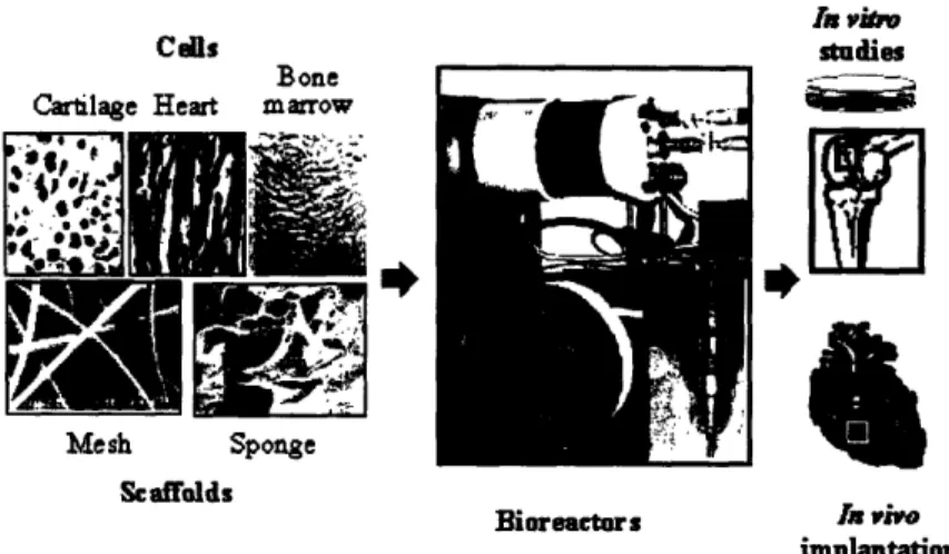
for tissue development), and a bioreactor (designed to enable environmental control and support cell differentiation and functional assembly into an engineered tissue) (Figure 2.1). Cells are generally isolated from a small tissue sample, expanded in culture under conditions selected to yield sufficient number for seeding a clinically sized scaffold and in some cases transfected to (over)express a gene of interest. Scaffolds should be made of biocompatible materials, preferentially those already approved for clinical use. Scaffold structure determines the transport of nutrients, metabolites and regulatory molecules to and from the cells, whereas the scaffold chemistry may have an important role in cell attachment and differentiation. The scaffold should biodegrade at the same rate as the rate of tissue assembly and without toxic or inhibitory products. Mechanical properties of the scaffold should ideally match those of the native tissue being replaced, and the mechanical integrity should be maintained as long as necessary for the new tissue to mature and integrate.
In this approach, a bioreactor should ideally provide all necessary conditions in in vitro environment for rapid and orderly tissue development by cells cultured on a scaffold. In general, a bioreactor is designed to perform one or more of the following functions: () establish a desired spatially uniform cell concentration within the scaffold during cell seeding, (ii) maintain controlled conditions in culture medium (e.g., temperature, pH, osmolality, levels of oxygen, nutrients, metabolites, regulatory molecules), (iii) facilitate mass transfer, and (iv) provide physiologically relevant physical signals (e.g., interstitial fluid flow, shear, pressure,
n Vib
Cells studies
Bone I - I
Cartilage Heart marow
*)
*E
Mesh Sponge
Scaffolds
Bioreactors In vvto
implantation
Figure 2.1. Tissue engineering based on cell cultivation on biomaterial scaffolds in bioreactors. Cells (e.g., from cartilage, heart or bone marrow) are cultured on a scaffold (e.g., highly porous, biodegradable mesh or a sponge) in a bioreactor (e.g., rotating bioreactor or perfused cartridge [1]). The resulting constructs are usedfor controlled in vitro studies or implanted in vivo (e.g., to repair an osteochondral defect [2] or injured myocardium [3]).
Three representative culture vessels that are frequently used for tissue engineering are compared in Figure 2.2. All culture vessels are operated in an incubator (to maintain the temperature and pH) with continuous gas exchange and periodic medium replacement.
Flasks contain constructs that are fixed in place by threading onto needles and cultured either statically or with magnetic stirring, with gas exchange through loosened side arm caps. Rotating vessels contain constructs that are freely suspended in culture medium between two concentric cylinders the inner of which serves as a gas exchange membrane. The vessel rotation rate is adjusted to maintain each construct settling at a stationary point within the vessel. This experimental set-up thus enables the evaluation of the effects of flow and mass transfer on engineered tissues and the selection of suitable culture environments to be further explored and optimised.
a)
b) Cultivation Static flask Mixed flask Rotating vessel
parameter
Vessel diameter 6.5 6.5 14.6/5.1
(cm)
Medium volume 120 120 110
(cm')
Tissue construct Fixed in place Fixed in place Freely settling or explantl)12 per vessel per n = 12 vessel n = 12 per vessel
Medium exchange
(3 cm3 per tissue Batch-wise Batch-wise Batch-wise per day)
Continuous
Continuous Continuous Continuous
Gas exchange C Cotnos via an internal
via surface aeration via surface aerational
membrane Stirring/rotation
Stirring/rotation 0 0.83 - 1.25 0.25 - 0.67
Flow conditions Static fluid Turbulent(2 ) Laminar3 )
Mixing None Magnetic stirring Setni
mechanism None Magnetic stirring rotational flow
Convection Convection
Mass transfer in Molecular
bulk medium diffusion (due to medium (due to tissue
stirring) settling)
Fluid shear at
tFluid shear at None Steady, turbulent Dynamic, laminar tissue surfaces
Reference 9-13 9-13 9-11, 14-19
1)5 mm diameter x 2 mm thick discs
(2) The smallest turbulent eddies had a diameter of 250 Jim and velocity of 0.4 cm/s [4]
3) Tissues were settling in a laminar tumble-slide regimen in a rotational field [5]
Figure 2.2. Representative bioreactors. a) Schematic presentation of tissue cultivation in static flasks, mixedflasks and rotating vessels b) Overview of the operating conditions for each vessel
type [based on 6, 7, 8].
Myocardium (cardiac muscle).
The myocardium (cardiac muscle) is a highly differentiated tissue composed of cardiac myocytes and fibroblasts with a dense supporting vasculature and collagen-based extracellular matrix. The myocytes form a three-dimensional syncytium that enables propagation of electrical signals across specialized intracellular junctions to produce coordinated mechanical contractions
K
V I J D I Ar /- I ,Woccupy 80-90% of the heart volume. The average cell density in the native rat myocardium is on the order of 5x10⁸ cells/cm3. Morphologically, intact cardiac myocytes have an elongated, rod shaped appearance. Contractile apparatus of cardiac myocytes consists of sarcomeres arranged in parallel myofibrils. High metabolic activity is supported by the high density of mitochondria and electrical signal propagation is provided by specialized intercellular connections, gap junctions [9, 10].
The control of heart contractions is almost entirely self-contained. Groups of specialized cardiac myocytes (pace makers), fastest of which are located in the sinoatrial node, drive periodic contractions of the heart. Majority of the cells in the myocardium are non-pace maker cells and they respond to the electrical stimuli generated by pace maker cells. Excitation of each cardiac myocyte is followed by the increase in the amount of cytoplasmic calcium which triggers mechanical contraction. The propagation of the electrical excitation through the tissue by ion currents in the extracellular and in the intercellular space results in synchronous contraction, that enables expulsion of the blood from the heart.
Clinical need
Cardiovascular disease is responsible for a preponderance of health problems in the developed countries, as well as in many developing countries. Heart disease and stroke, the principal components of cardiovascular disease, are the first and the third leading cause of death in the U.S., accounting for nearly 40% of all deaths. Congenital heart defects, which occur in nearly 14 of every 1000 newborn children [11] year of life [12]. Cardiovascular diseases result in substantial disability and loss of productivity, and largely contribute to the escalating costs of health care. About 61 million Americans (almost one-fourth of the population) live with cardiovascular diseases, such as coronary heart disease, congenital cardiovascular defects, and congestive heart failure, and 298.2 billion dollars were spent in 2001 to treat these diseases [13]. The economic impact of cardiovascular disease on the U.S. health care system is expected to grow further as the population ages.
Once damaged, the heart is unable to regenerate. Heart failure affects over five million Americans [14], and is the leading cause of morbidity and mortality in developed countries [15]. Currently, the only definitive treatment for end stage heart failure is cardiac transplantation. However, the limited availability of organs for transplantation has led to prolonged waiting periods that are often not survivable [16]. Repair of myocardial injuries has been attempted by injection of myogenic cells into scarred myocardium [17] and the replacement of scarred tissue with engineered grafts [18].
Tissue engineering of myocardium
Tissue engineering has emerged over the last decade as an interdisciplinary field with tremendous potential. Tissue engineering offers a possibility of creating tissue constructs to be used for repair of larger injuries or congenital malformations. In addition cardiac tissue constructs may be utilized for studies of normal and pathological tissue function in vitro. Substantial progress has been made in areas of biopolymers [19], cell-material interactions [20] and bio-mimetic culture devices [21]. In addition, functional tissues have been developed and implanted
in vivo, including (but not limited to) cartilage [22], bone [23], bladder [24] and blood vessels [25,Niklason, 1999 #343]. However, fundamental and all-encompassing problems remain. One of the most important ones is mass transfer into the tissues that are greater than 100-200 m in thickness, both during the in vitro cultivation and following implantation in vivo. This explains why tissue engineering has been most successful with tissues that are either thin (e.g., bladder) or have low oxygen requirements (e.g., cartilage). There are multiple challenges to be overcome to produce viable, functional cardiac tissue (Table 2.1 ).
Table 2.1 Key challenges of cardiac tissue engineering
In current approaches, fetal or neonatal rat cardiomyocytes are seeded onto scaffolds (collagen sponges, polyglycolic acid meshes) or cast in collagen gels, and cultivated immersed in
the culture medium in static or mixed dishes, spinner flasks or rotating vessels [26, 27]. The metabolism and viability of the resulting constructs are assessed during culture by monitoring
levels of glucose consumed, lactate produced and the release of lactate dehydrogenase from the samples of culture medium. Cell distribution, morphology and construct structure are assed by histology. Expression of cardiac specific markers is assessed by immunohistochemistry and confirmed by Western blots. Gene expression is assessed by RT-PCR. Cardiac specific ultrastructural features are detected by transmission electron microscopy. Functional assessment * Development of appropriate biodegradable and biocompatible scaffolds that
can provide adequate mass transfer, vascularization, and transduction of mechanical and electrical signals
* Development of bio-mimetic culture systems that promote differentiated function and excitation - contraction coupling of cardiac cells
* Development of functional vascular networks embedded into the cardiac muscle tissue to promote nutrient and oxygen transfer, angiogenesis and integration with the host vasculature.
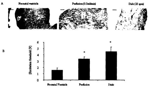
of engineered constructs has been based on electrophysiological studies [28, 29], monitoring of synchronous contractions in response to electrical stimuli [30, 31] and measurement of force of contraction in paced [32] or spontaneously contracting constructs [33].
Cells. Three dimensional cardiac tissue constructs were successfully cultivated in dishes using variety of scaffolds and cell sources. Fetal rat ventricular cardiac myocytes were expanded after isolation, inoculated into collagen sponges and cultivated in static dishes for up to 4 weeks [3]. The cells proliferated with time in culture and expressed multiple sarcomeres. Adult human ventricular cells were used in a similar system, although they exhibited no proliferation [34]. Fetal cardiac cells were also cultivated on alginate scaffolds in static 96-well plates. After 4 days in culture the cells formed spontaneously beating aggregates in the scaffold pores [35]. Cell seeding densities of the order of 108cells/cm3 were achieved in the alginate scaffolds using centrifugal forces during seeding [36]. Neonatal rat cardiac myocytes formed spontaneously contracting constructs when inoculated in collagen sponges within 36 hr [33] and maintained their activity for up to 12 weeks. The contractile force increased upon addition of Ca2+ and epinephrine.
Two-week constructs based on neonatal rat cardomyocytes exhibited spontaneously beating areas, whereas constructs based on embryonic chick myocytes exhibited no contractions and reduced in size by 60%. Immunohistochemistry, revealed presence of large number of non-myocytes in the constructs based on embryonic chick hear cells, while constructs based on neonatal rat cells consisted mostly of elongated cardiomyocytes [26].
Constructs based on the cardiomyocytes enriched by preplating exhibited lower excitation threshold (ET), higher conduction velocity, higher maximum capture rate (MCR), and higher maximum and average amplitude [37].
Scaffolds. The scaffolds utilized for cardiac tissue engineering include collagen fibers [38], collagen sponges [3, 33, 34] and polyglycolic acid meshes [26, 27, 37]. The main advantage of a synthetic scaffold such as PGA is that it provides mechanical stability, while scaffolds based on natural cell polymers such as collagen enable rapid cell attachment.
The scaffold free approaches include casting the cells in collagen gels followed by mechanical stimulation [39-41] and stacking of confluent cardiac cell monolayers [42]. The main advantage of scaffold free approaches is higher active force generated by such tissues. However, the main disadvantage remains tailoring the shape and dimensions of the scaffold free engineered tissues.
As an alternative, gels (Matrigel) were combined with scaffolds (collagen sponge) to achieve rapid cell inoculation and attachment along with the possibility of tailoring tissue shape and dimensions through the use of scaffolds [30].
Bioreactor hydrodynamics. The representative bioreactors utilized for tissue engineering of the myocardium include static or mixed dishes, static or mixed flasks and rotating vessels (Figure 2.1 and 2.2). These bioreactors offer three distinct flow conditions (static, turbulent, and laminar) and therefore differ significantly in the rate of oxygen supply to the surface of the tissue construct. Oxygen transport is a key factor for myocardial tissue engineering due to the high cell density, very limited cell proliferation and low tolerance of cardiac myocytes for hypoxia In all configurations oxygen is supplied only by diffusion from the surface to the interior of the tissue construct.
Static dishes remain the most widely used set-up for cardiac tissue engineering (Table 2.2). In
static dishes, oxygen and nutrients are supplied mainly by diffusion, which is capable of satisfying oxygen demand of only -100 gm thick surface layer of compact tissue, whereas the construct interior remained mostly acellular [43]. In contrast, cartilage has been successfully grown in static dishes to millimeter thicknesses. In orbitally mixed dishes, the rate of delivery of oxygen and nutrients to construct surfaces can be increased, but diffusion remains the main mechanism of mass transport within the tissue. Diffusional transport of oxygen to cardiac myocytes within dish-grown constructs resulted in prevalently anaerobic glucose metabolism [31]. Additional limitation of the culture in static or mixed dishes is that the bottom surface of the construct (the surface closest to the bottom of the dish) often lacks proper oxygenation and nutrients yielding asymmetric cell distribution with compact tissue mostly on the top surface.
To improve cell survival and assembly on all surfaces of the engineered tissue, cardiac constructs were cultivated suspended in the spinner flasks (Table 2.2). Cultivation in spinner flasks (at stirring rates of up to 90 rpm) improved construct properties [26], presumably due to enhanced mass transport at construct surfaces (Figure2.2). After 2 weeks of culture, constructs from mixed flasks had significantly higher cellularity index (-20ggDNA/construct) and metabolic activity (-150MTTunits/mg DNA) than those from static flasks (-.5ggDNA/construct and -50 MTT units/mg DNA). Mixing maintained medium gas and pH levels within the physiological range yielding a more aerobic glucose metabolism (L/G-1.5) in mixed flasks as compared to static flaks (L/G>2) [26]. Constructs contained a peripheral tissue-like region (50-70gm thick) in which cells stained positive for tropomyosin and organized in multiple layers in a
3-D configuration [37] Electrophysiological studies conducted using a linear array of extracellular electrodes showed that the peripheral layer of the constructs sustained macroscopically continuous impulse propagation on a centimeter-size scale [37]. However, construct interiors remained empty due to the diffusional limitations of the oxygen transport within the bulk tissue, and the density of viable myocytes was orders of magnitude lower than that in the neonatal rat ventricles [37]. Additional drawback of the cultivation in spinner flasks is that turbulent flow conditions may induce cell damage and dedifferentiation and result in the formation of a fibrous capsule at construct surfaces.
Table 2.2 Overview of cardiac tissue engineering studies
INITIAL SCAFFOLD CONSTRUC INTA
BIOREACTOR TYPE CELL SOURCE SCAFFOLD CONST RUC CELL REF.
TYPE T SIZE NMENUMBER
Static dish 6mm
(96 well plate) Fetal rat CM Alginate diameter 3- 105 [35] x mm thick
Static dish Bovine
Stam t m ) Fetal rat CM collagen 5 x 5 x I mm -104 [3]
(5 ml culture medium) (Gelfoam) _
(Gelfoam)
Static dish
Ne~~~~ovnaara
CMolg en
Bovine 20 x 15 x 2.52.106
106[33]
(4 ml culture medium) Neonatal rat CM collagen m(Tissue Fleece) Bovine
Static dish Neonatal rat CM collagen 5 x 5 x 3 mm 0.5. 106 [44
Collagen
~~~~0106
m4
Mixed dish Bovine 10 mm
(20rpm, 4ml culture Neonatal rat CM collagen diameter 6- 12- 106 [31] x 1.5 mm
medium) (Ultrafoam) thick
Spinner flask Embryonic chick PGA 5 mm
(0, 50, 90 rpm, CM diameter 1.3-8.106 [26]
120 ml culture medium) Neonatal rat CM x 2 mm thick
Spinner flask PGA PGA 5 mm
(50 rpm, Neonatal rat CM , s diameter 8- 106 [27,
120 ml culture medium) x 2 mm thick 7]
Rotating vessels PGA, sPGA,5 mm [26,
(II rpm, Neonatal rat CM IsPGA diameter 8 106 27,
100 ml culture medium) x 2 mm thick 37]
Rotating vessel Neonatal rat CM Bov 5 x 5 x 3 mm 0.5- 106 [44] collagen
11 mm
Perfused cartridge 1 m16 [3
Perfused cartridge Neonatal rat CM PGA diameter 24 1 453,
(0.2-3 mI/mi) _______
~mtik45]
x 2 mm thick
Cyclic stretch Embryonic chick [18,
(1.5-2 Hz, 1-20 % strain) CM Collagen gel 12.5-106 32,
Neonatal rat CM 401
Bovine 30 x 20x3
Cyclic stretch Human heart cells collagen mm 3-30- 106 [46
(1.3 Hz) (Gelfoam) 20 x 20x 30.0 [6
Laminar conditions of flow in rotating vessels (Table 2.2) enabled the maintenance of oxygen concentration in medium and pH within the physiological range, and resulted in mostly aerobic cell metabolism (L/G-1) [26]. The metabolic activity of cells within constructs increased into the range of values measured for neonatal rat ventricles (-250 units MTT/mg DNA) and was significantly higher than in mixed flasks (-150 units MTT/mg DNA). The index of cell hyperthrophy was also comparable to the neonatal tissue (18mg protein/mg DNA). However, the construct cellularity remained 2-6 times lower than in native heart ventricles. In the best experimental group (heart cells enriched for cardiac myocytes by pre-plating, laminin-coated PGA scaffolds, low serum concentration) the outer layer of viable tissue was up to 160 gm thick
[27]. Cells expressed cardiac-specific markers (e.g., tropomyosin, gap junction protein connexin, creatin kinase-MM, sarcomeric myosin heavy chain) at levels that were lower than in neonatal rat ventricles but higher than in constructs cultured in spinner flasks [27]. Electrophysiological properties were also improved, as evidenced by the prolonged action potential duration (APD, a measure of electrophysiological functionality of cell membrane), higher maximum capture rates (a measure of construct response to electrical pacing), and more physiological response to drugs. In particular, pharmacological studies done with 4-aminopyridine indicated that a decrease in transient outward potassium current may be responsible for the observed differences in APD and MCR [29]. Overall, dynamic laminar flow of rotating bioreactors improved properties of the peripheral tissue layer, but the limitations of the diffusional transport of oxygen to the construct interior were not overcome and constructs remained largely acellular.
Interstitial flow. In an attempt to enhance mass transport within cultured constructs, a perfusion bioreactors was developed that provides interstitial medium flow through the cultured construct at velocities similar to those found in native myocardium (-400-500 !xm/s, [47]). In such a system oxygen and nutrients are supplied to the construct interior by both diffusion and convection.
In early studies, constructs were prepared by seeding cardiac myocytes onto PGA scaffolds in mixed flasks, a method that has been successfully used to seed chondrocytes. After 3 days, constructs were transferred into a perfusion cartridge and pulsatile flow of medium through the construct was provided by a peristaltic pump (0.2 - 3 ml/min) (Table 2.2). Gas exchange between culture medium and incubator air occurred in an external coil of silicone tubing within the medium recirculation loop. Perfusion during construct cultivation improved cell distribution, viability and differentiation. However, the overall cell density remained low due to the limitations of oxygen transport to the cells inside constructs during scaffold seeding in mixed flasks [43, 45].
Mechanical stimulation. To provide appropriate mechanical stimulation neonatal rat cardiac myocytes were reconstituted in collagen gel and cultivated in the presence of cyclic stretch (Table 2.2). In one set-up neonatal rat ventricular myocytes were suspended in a gel consisting of collagen I and Matrigel [40]. For each piece of tissue, 0.7 ml of the cell/gel mix was poured into a well ( 1 x 17 x 4 mm) made of silicone rubber containing one set of Velcro coated silicone tubes (7 mm length, 3mm OD 2mm ID). The mixture was allowed to gel at 37°C for 60 min before culture medium was added. After 4 days in culture, tissues were transferred for an additional 6 days into a motorized stretching device that applied either unidirectional and cyclic stretch (1.5 Hz, strain rate of up to 20%). Mechanical stimulation enhanced the alignment of cardiac myocytes, and resulted in higher mitochondrial density and longer myofilaments. As compared to the non-stimulated controls, stimulation markedly increased the RNA/DNA ratio (by 100 %) and protein/cell ratio (by 50 %). The force of contraction was also higher in stretched constructs, both under basal conditions and after stimulation with isoprenaline [40].
In an improved set-up, neonatal rat cardiac cells were suspended in the collagen/Matrigel mix and cast into circular molds [32]. After 7 days in culture, the strips of cardiac tissue were placed around two rods each fixed to a stretching bar of a custom made mechanical stretcher and subjected to unidirectional and cyclic stretch at 10% strain rate and 2 Hz. Mechanical stimulation improved the formation of interconnected and aligned cardiac muscle bundles with morphological features resembling adult rather than immature native tissue. Fibroblasts and macrophages were found through the constructs, and the capillary structures positive for CD31 were also noted. Cardiomyocytes exhibited well developed ultrastructural features: sarcomeres arranged in myofibrils, with well developed Z, I, A H and M bands, gap and adherence junctions, T tubules, and well developed basement membrane. The constructs exhibited contractile properties similar to the native tissue with high ratio of twitch to resting tension and strong -adrenegenic response. Action potentials characteristic of rat ventricular myocytes were recorded.
Using another system for mechanical stimulation, cyclic mechanical stretch (1.33Hz) was applied to the constructs based on collagen scaffold and human heart cells (isolated from children undergoing repair of Tetralogy of Fallot) [46]. A rectangular piece of tissue was fixed at one end to the bottom of a square dish; the other end is attached to a steel rod the cyclic movement of which is induced by dynamically changing magnetic filed. Constructs subjected to chronic stretch had improved cell distribution and collagen matrix formation.
Summary
Tissue engineering can provide functional cell-based grafts to restore normal function of a compromised native tissue and to serve as physiologically relevant models for biological research. One approach to functional tissue engineering involves the in vitro cultivation of immature but functional tissue constructs by using: (i) cells isolated from a small tissue harvest and expanded in vitro, (ii) a biodegradable scaffold designed to serve as a structural and logistic template for tissue development, and (iii) a bioreactor designed to provide environmental conditions necessary for the cells to regenerate a functional tissue structure.
REFERENCES:
I. Freed, L.E. and G. Vunjak-Novakovic, Tissue engineering bioreactors, in Principles of Tissue Engineering, R.P. Lanza, R. Langer, and J. Vacanti, Editors. 2000, Academic Press: San Diego. p. 143-156.
2. Schaefer, D., et al., Tissue engineered composites for the repair of large osteochondral
defects. Transactions of the Orthopaedic Research Society, 2000. 25: p. 619.
3. Li, R.-K., et al., Survival and function of bioengineered cardiac grafts. Circulation, 1999. 100(Suppl II): p. II63-1169.
4. Vunjak-Novakovic, G., et al., Effects of mixing on the composition and morphology of
tissue-engineered cartilage. AIChE Journal, 1996. 42(3): p. 850-860.
5. Freed, L.E. and G. Vunjak-Novakovic, Tissue engineering of cartilage, in Biomedical Engineering Handbook, J.D. Bronzino, Editor. 1995, CRC Press: Boca Raton. p.
1788-1807.
6. Freed, L.E., . Martin, and G. Vunjak-Novakovic, Frontiers in tissue engineering: in vitro
modulation of chondrogenesis. Clinical Orthopaedics and Related Research, 1999. 367S:
p. S46-S58.
7. Freed, L.E. and G. Vunjak-Novakovic, Tissue engineering of cartilage, in The
Biomedical Engineering Handbook, J.D. Bronzino, Editor. 2000, CRC Press: Boca
Raton. p. 124-1-124-26.
8. Vunjak-Novakovic, G., Fundamentals of tissue engineering: scaffolds and bioreactors, in
Tissue Engineering of Cartilage and Bone, A.I. Caplan, Editor. 2003, John Wiley:
London. p. 34-51.
9. MacKenna, D.A., et al., Contribution of collagen matrix to passive left ventricular
mechanics in isolated rat heart. American Journal of Physiology, 1994. 266: p.
H1007-H1018.
10. Brilla, C.G., et al., Pharmacological modulation of cardiac fibroblast function. Herz, 1995.20: p. 127-135.
11. Gillum, R.F., Epidemiology of congenital heart disease in the United States. American Heart Journal, 1994. 127(4 Pt 1): p. 919-927.
12. Hoffman, J.I., Incidence of congenital heart disease: I. Postnatal incidence. Pediatric Cardiology, 1995. 16(3): p. 103-113.
13. Lysaght, M.J. and J. Reyes, The growth of tissue engineering Tissue Engineering, 2001. 7: p. 485-493.
14. Rich, M., Epidemiology, pathophysiology, and etiology of congestive heart failure in
older adults. Journal of the American Geriatrics Society, 1997. 45: p. 968-974.
15. Dominguez, L., et al., Trends of congestive heart failure: epidemiology contrast with
clinical trial results. Cardiologia, 1999. 44: p. 801-808.
16. Evans, R.W., Economic impact of mechanical cardiac assistance. Progress in Cardiovascular Diseases, 2000. 43: p. 81-94.
17. Soonpaa, M.H., et al., Formation of nascent intercalated disks between grafted fetal cardiomyocytes and host myocardium. Science, 1994. 264: p. 98-101.
18. Zinmmnermann, W.H., et al., Cardiac grafting of engineered heart tissue in syngenic rats. Circulation, 2002. 106(12 Suppl 1): p. 151-1157.
19. Wang, Y., et a., A tough biodegradable elastomer. Nature Biotechnology, 2002. 20: p. 602-606.
20. Hubbell, J.A., Bioactive biomaterials. Current Opinion in Biotechnology, 1999. 10: p. 123-129.
21. Niklason, L.E., et al., Functional arteries grown in vitro. Science, 1999. 284(5413): p. 489-493.
22. Freed, L.E., et al., Chondrogenesis in a cell-polymer-bioreactor system. Experimental
Cell Research, 1998. 240: p. 58-65.
23. Niklason, L.E., Engineering of bone grafts. Nature Biotechnology, 2000. 18: p. 929-930. 24. Oberpenning, F., et al., De novo reconstitution of a functional mammalian urinary
bladder by tissue engineering. Nature Biotechnology, 1999. 17: p. 149-155.
25. L'Heureux, N., et al., A completely biological tissue-engineered human blood vessel.
FASEB Journal, 1998. 12: p. 47-56.
26. Carrier, R.L., et al., Cardiac tissue engineering: cell seeding, cultivation parameters and
tissue construct characterization. Biotechnology and Bioengineering, 1999. 64: p.
580-589.
27. Papadaki, M., et al., Tissue engineering of functional cardiac muscle: molecular,
structural and electrophysiological studies. American Journal of Physiology: Heart and
Circulatory Physiology, 2001. 280(Heart Circ. Physiol. 44): p. H168-HI178.
28. Bursac, N., et al. Cardiac tissue engineering: an electrophysiological study. in Annual
Meeting of the BMES. 1998. Cleveland, OH.
29. Bursac, N., et al., Cultivation in rotating bioreactors promotes maintenance of cardiac
myocyte electrophysiology and molecular properties. Tissue Engineering, 2003. 9(6): p.
1243-1253.
30. Radisic, M., et al., High density seeding of myocyte cells for tissue engineering. Biotechnology and Bioengineering, 2003. 82(4): p. 403-414.
31. Radisic, M., et al., Medium perfusion enables engineering of compact and contractile
cardiac tissue. American Journal of Physiology: Heart and Circulatory Physiology, 2004.
286: p. H507-H516.
32. Zimmermann, W.H., et al., Tissue engineering of a differentiated cardiac muscle construct. Circulation Research, 2002. 90(2): p. 223-230.
33. Kofidis, T., et al., In vitro engineering of heart muscle: Artificial myocardial tissue. Journal of Thoracic and Cardiovascular Surgery, 2002. 124(1): p. 63-69.
34. Li, R.-K., et al., Construction of a bioengineered cardiac graft. Journal of Thoracic and Cardiovascular Surgery, 2000. 119: p. 368-375.
35. Leor, J., et al., Bioengineerred cardiac grafts: A new approach to repair the infarcted myocardium? Circulation, 2000. 102(suppl III): p. 11156-11161.
36. Dar, A., et al., Cardiac tissue engineering Optimization of cardiac cell seeding and
distribution in 3D porous alginate scaffolds. Biotechnology and Bioengineering, 2002.
37. Bursac, N., et al., Cardiac muscle tissue engineering toward an in vitro model for
electrophysiological studies. American Journal of Physiology: Heart and Circulatory
Physiology, 1999. 277(46): p. H433-H444.
38. Akins, R.E., et al., Cardiac organogenesis in vitro: Reestablishment of three-dimensional
tissue architecture by dissociated neonatal rat ventricular cells. Tissue Engineering,
1999. 5(2): p. 103-118.
39. Eschenhagen, T., et al., Three-dimensional reconstitution of embryonic cardiomyocytes in
a collagen matrix: a new heart model system. FASEB Journal, 1997. 11: p. 683-694.
40. Fink, C., et al., Chronic stretch of engineered heart tissue induces hypertrophy and functional improvement. FASEB Journal, 2000. 14: p. 669-679.
41. Zimmermann, W.H., et al., Three-dimensional engineered heart tissue from neonatal rat
cardiac myocytes. Biotechnology and Bioengineering, 2000. 68: p. 106-114.
42. Shimizu, T., et al., Fabrication of pulsatile cardiac tissue grafts using a novel
3-dimensional cell sheet manipulation technique and temperature- responsive cell culture
surfaces. Circulation Research, 2002. 90(3): p. e40-e48.
43. Carrier, R.L., et al., Perfusion improves tissue architecture of engineered cardiac muscle. Tissue Engineering, 2002. 8(2): p. 175-188.
44. van Luyn, M.J.A., et al., Cardiac tissue engineering: characteristics of in unison
contracting two- and three-dimensional neonatal rat ventricle cell (co)-cultures.
Biomaterials, 2002.23(24): p. 4793-4801.
45. Carrier, R.L., et al., Effects of oxygen on engineered cardiac muscle. Biotechnology and Bioengineering, 2002. 78: p. 617-625.
46. Akhyari, P., et al., Mechanical stretch regimen enhances the formation of bioengineered autologous cardiacmuscle grafts. Circulation, 2002. 106(12 Suppl 1): p. 1137-1142. 47. Fournier, R.L., Basic Transport Phenomena in Biomedical Engineering. 1998,
3. HIGH DENSITY SEEDING OF MYOCYTE CELLS FOR CARDIAC TISSUE
ENGINEERING2
INTRODUCTION
Due to the limited ability of cardiac myocytes to regenerate [ attempts have been made to repair myocardial injuries by injecting myogenic cells into the scarred myocardium [2-4], or in the case of large injuries to replace scarred tissue with engineered grafts [5, 6]. Three dimensional tissue constructs that express structural and physiological features characteristic of native cardiac muscle have been engineered using collagen gels [7-9], collagen fibers [10], collagen sponges [6, 11] and polyglycolic acid meshes [12-14], in conjunction with fetal or neonatal rat cardiac myocytes. In all cases, cells were seeded on scaffolds and cultivated immersed in culture medium in dishes [5, 6, 12, 14], spinner flasks [12-14] or rotating vessels [10, 12, 14]. Oxygen dissolved in medium was transported to the cells by molecular diffusion, which provided enough oxygen for an approximately 100 gm thick outer layer of functional tissue but not to the construct interior which remained relatively acellular [8, 12-14].
We previously developed a perfused bioreactor system in an attempt to enhance mass transport between culture medium and cells within cultured constructs [15]. Cells were seeded onto scaffolds in tissue culture dishes for 48 hours and subsequently cultured for 7 days with direct perfusion of culture medium. The transport of oxygen from the medium to the cells occurred via diffusion during cell seeding, and by a combination of diffusion and convection during cultivation. During cultivation, the flow of medium redistributed the cells evenly across the entire volume of the construct, but the cell density remained low due to the limitations in oxygen transport during cell seeding.
We hypothesized that rapid gel inoculation of hypoxia-sensitive cells in conjunction with direct medium perfusion through the seeded scaffold would result in high rate, yield, viability and uniformity of cell seeding. We report a new seeding strategy that enables the seeding of tissue engineering scaffolds at the initial cell densities comparable to those in adult rat myocardium (-108 cells/cm3, [16]), and was developed by combining the methods for rapid cell inoculation using a gel [17] and scaffold seeding with cell suspension in perfusion systems [18]. Medium perfusion through the cell-polymer construct is established immediately, in order to maintain the viability of inoculated cells during cell attachment to scaffolds and subsequent construct
2 Most of this chapter has been published in: Milica Radisic, Michelle Euloth, Liming Yang, Robert Langer, Lisa E. Freed and Gordana Vunjak-Novakovic:"High density seeding of myocytes cells for cardiac tissue engineering" Biotechnology and Bioengineering 82: 403-414, 2003
cultivation. The feasibility of the proposed seeding method was first evaluated in seeding and cultivation studies with neonatal cardiac myocytes. Subsequently, the effects of three system parameters (initial cell number, seeding time, seeding set-up) were systematically studied using C2C12 mouse muscle myoblast cell line. C2C12 cells were chosen as a model system due to their availability, ease of handling and evidence that they differentiate and express cardiac specific proteins [19-21] when grafted into the rat or mouse myocardium.
MATERIALS AND METHODS
Materials Dulbecco's modified Eagle Medium (DMEM), fetal bovine serum (FBS), N-2-Hydroxyethylpiperazine-N'-2-Ethane-Sulfonic Acid (HEPES), calcium and magnesium free phosphate buffered saline (PBS), lx trypsin-EDTA solution in PBS, Hank's Balanced Salt Solution (HBSS) and penicillin were all from Gibco (Grand Island, NY). Collagenase Type II was from Worthington (Freehold, NJ). Trypsin was from U.S. Biochemicals (Cleveland, OH). Matrigel® and dispase were from Becton-Dickinson (Two Oak Park, Bedford, MA). The C2C12 mouse muscle myoblast cell line was from the American Type Culture Collection (Manassas, VA). Tissue culture dishes and T175 flasks were from Costar (Cambridge, MA). 10% Formalin buffer was from Sigma Diagnostic (St. Louis, MO). Ethidium monoazide bromide (EMA) was from Molecular Probes (Eugene, OR). The flow cytometer (model FACScan) was from Becton-Dickinson (Bedford, MA). Orbital shaker was from Bellco, (type BTB, Vineland, NJ) and the Push/Pull PHD2000 syringe pump was from Harvard Apparatus (Holliston, MA). Polycarbonate perfusion cartridges were kindly donated by the Advanced Tissue Sciences (LaJolla, CA). Platinum cured silicone tubing and the multichannel peristaltic pump (L/Sm Masterflex) were from Cole-Parmer (Vernon Hills, IL). Reservoir bag was 32 ml gas permeable VueLife bag (American Fluoroseal Corporation, Gaithersburg, MD). Syringes were from Becton Dickinson (Two Oak Park, Bedford, MA) and three-way stop-cocks from Baxter Healthcare (Irvin, CA). Heating tape was from VWR (Bridgeport, Connecticut). The temperature controller was from Barnstead/Thermolyne (Dubuque, Iowa). The programmable cardiac stimulator was from Nihon Kohden (type SEC-3102). The electrodes (cm x 0.5cm) were custom made by coating 500 gm thick silicon wafers with 3000 A of silicon nitride, 100 A of Ti, and 1000 A of gold.
C2C12 cells, a cell line derived from murine myoblasts, were subcultured in T-75 flasks
(P4 to P9) in Dulbecco's Modified Eagle Medium (DMEM) containing 4.5 g/L glucose, supplemented with 10% fetal bovine serum (FBS), 10 mM N-2-hydroxyethylpiperazine N'-2-ethanesulfonic acid (HEPES), 2 mM L-glutamine and 100 units/ml penicillin. In order to prepare