HAL Id: hal-02619160
https://hal.inrae.fr/hal-02619160
Submitted on 25 May 2020
HAL is a multi-disciplinary open access
archive for the deposit and dissemination of
sci-entific research documents, whether they are
pub-lished or not. The documents may come from
teaching and research institutions in France or
abroad, or from public or private research centers.
L’archive ouverte pluridisciplinaire HAL, est
destinée au dépôt et à la diffusion de documents
scientifiques de niveau recherche, publiés ou non,
émanant des établissements d’enseignement et de
recherche français ou étrangers, des laboratoires
publics ou privés.
Tatiana Rochat, Erina Fujiwara-Nagata, Ségolène Calvez, Inger Dalsgaard,
Lone Madsen, Alexandra Calteau, Aurélie Lunazzi, Pierre Nicolas, Tom
Wiklund, Jean Francois Bernardet, et al.
To cite this version:
Tatiana Rochat, Erina Fujiwara-Nagata, Ségolène Calvez, Inger Dalsgaard, Lone Madsen, et
al..
Genomic Characterization of Flavobacterium psychrophilum Serotypes and Development of
a Multiplex PCR-Based Serotyping Scheme. Frontiers in Microbiology, Frontiers Media, 2017, 8,
�10.3389/fmicb.2017.01752�. �hal-02619160�
ORIGINAL RESEARCH published: 12 September 2017 doi: 10.3389/fmicb.2017.01752
Edited by: Shicheng Chen, Michigan State University, United States Reviewed by: Gregory D. Wiens, ARS-USDA, United States Mark L. Lawrence, Mississippi State University, United States *Correspondence: Eric Duchaud [email protected] Specialty section: This article was submitted to Microbial Physiology and Metabolism, a section of the journal Frontiers in Microbiology Received: 22 June 2017 Accepted: 28 August 2017 Published: 12 September 2017 Citation: Rochat T, Fujiwara-Nagata E, Calvez S, Dalsgaard I, Madsen L, Calteau A, Lunazzi A, Nicolas P, Wiklund T, Bernardet J-F and Duchaud E (2017) Genomic Characterization of Flavobacterium psychrophilum Serotypes and Development of a Multiplex PCR-Based Serotyping Scheme. Front. Microbiol. 8:1752. doi: 10.3389/fmicb.2017.01752
Genomic Characterization of
Flavobacterium psychrophilum
Serotypes and Development of a
Multiplex PCR-Based Serotyping
Scheme
Tatiana Rochat1, Erina Fujiwara-Nagata2, Ségolène Calvez3, Inger Dalsgaard4,
Lone Madsen4, Alexandra Calteau5, Aurélie Lunazzi1, Pierre Nicolas6, Tom Wiklund7,
Jean-François Bernardet1and Eric Duchaud1*
1Virologie et Immunologie Moléculaires, Institut National de la Recherche Agronomique, Université Paris-Saclay,
Jouy-en-Josas, France,2Department of Fisheries, Kindai University, Nara, Japan,3Biologie, Epidémiologie et Analyse du
Risque, Institut National de la Recherche Agronomique, Oniris, Nantes, France,4National Veterinary Institute, Technical
University of Denmark, Kongens Lyngby, Denmark,5CEA/Genoscope/LABGeM, CNRS-UMR8030, Université d’Evry –
Université Paris-Saclay, Evry, France,6Mathématiques et Informatique Appliquées du Génome à l’Environnement, Institut
National de la Recherche Agronomique, Université Paris-Saclay, Jouy-en-Josas, France,7Laboratory of Aquatic
Pathobiology, Environmental and Marine Biology, Faculty of Science and Engineering, Åbo Akademi University, Turku, Finland
Flavobacterium psychrophilum is a devastating bacterial pathogen of salmonids reared in freshwater worldwide. So far, serological diversity between isolates has been described but the underlying molecular factors remain unknown. By combining complete genome sequence analysis and the serotyping method proposed byLorenzen and Olesen (1997) for a set of 34 strains, we identified key molecular determinants of the serotypes. This knowledge allowed us to develop a robust multiplex PCR-based serotyping scheme, which was applied to 244 bacterial isolates. The results revealed a striking association between PCR-serotype and fish host species and illustrate the use of this approach as a simple and cost-effective method for the determination of F. psychrophilum serogroups. PCR-based serotyping could be a useful tool in a range of applications such as disease surveillance, selection of salmonids for bacterial coldwater disease resistance and future vaccine formulation.
Keywords: Flavobacterium psychrophilum, salmonid aquaculture, fish-pathogenic bacteria, genomics, serotype, mPCR
INTRODUCTION
Salmonids represent one of the most important fish groups in the aquaculture industry (FAO, 2016). However, the success and sustainability of salmonid aquaculture largely depend on disease control.
Flavobacterium psychrophilum, a bacterium belonging to the family Flavobacteriaceae, is considered one of the most important bacterial pathogens in freshwater salmonid aquaculture worldwide (Nematollahi et al., 2003). The two main clinical forms are rainbow trout fry syndrome (RTFS) and bacterial cold-water disease (BCWD) (Cipriano and Holt, 2005) that can both result in considerable economic losses (Antaya, 2008; Starliper, 2011). All salmonid fish, especially
coho salmon (Oncorhynchus kisutch) and rainbow trout (Oncorhynchus mykiss) are susceptible to RTFS and BCWD, as well as ayu (Plecoglossus altivelis), a fish related to salmonids. These infections are associated with skin ulcers, necrotic myositis, septicemia as well as exophthalmia (Nematollahi et al., 2003).F. psychrophilum has occasionally been detected in non-salmonid fish, samples of water, sediments and biofilms from rivers receiving outlet water from infected fish farms (Madetoja et al., 2002; Starliper, 2011). Despite extensive research on vaccine development (Plant et al., 2009; Gómez et al., 2014) and the recent marketing authorization of a vaccine in Chile, treatment still essentially relies on antibiotic administration (Schmidt et al., 2000). Diversity among F. psychrophilum isolates was reported at different levels, and a variety of typing schemes (e.g., biotypes, serotypes, and genotypes) was proposed accordingly. However, the correlation between these different schemes (Madsen and Dalsgaard, 2000; Madetoja et al., 2001; Ngo et al., 2017) and the impact of the different types on the onset and severity of the infection remain poorly understood.
Gram-negative bacteria have a complex set of surface polysaccharides that include the lipopolysaccharide (LPS) as well as capsular polysaccharides (CPS) or exopolysaccharides (EPS) (Raetz and Whitfield, 2002). These structures are thought to play important roles in the pathogenicity, immune escape, serum resistance, inflammation, adhesion and biofilm formation (Finlay and McFadden, 2006). Serotyping is based on the immunogenicity of various surface-exposed bacterial structures (e.g., LPS, capsular polysaccharides, flagella, peptidoglycan, etc.) biosynthesized by genes belonging to the accessory genome. This represents an advantage over many molecular typing methods (e.g., 16S rRNA sequencing, ribotyping, MLST) that rely on core genome genes, which are not phenotypically relevant determinants. LPS is one of the most pro-inflammatory compounds of Gram-negative bacteria typically consisting of three regions: a hydrophobic domain known as lipid A, a non-repeating “core” oligosaccharide, and a distal polysaccharide (orO-antigen). The O-antigen greatly varies between and within species, providing the main basis for many serotyping schemes. The structure of theO-polysaccharide of F. psychrophilum strain CSF259-93 has been solved and found to be an unbranched polymer of trisaccharide repeating units (MacLean et al., 2001). A 70 kb region (FP1299 to FP1234) encompassing genes predicted to be involved in the biosynthesis, export, modification and polymerization of polysaccharides was previously identified in the genome of F. psychrophilum JIP 02/86 (Duchaud et al., 2007). Several substitutions of genes enclosed in this region were noticed by comparing the genomes of strains CSF259-93 and JIP 02/86 (Wiens et al., 2014).
Serological differences amongF. psychrophilum isolates were noticed since early studies and different serotyping protocols were proposed, resulting in the description of a varying number of serotypes. Wakabayashi et al. (1994) described two distinct serotypes (O-1 and O-2) among a collection of Japanese isolates; a third serotype (O-3) was added in 1999 (Izumi and Wakabayashi, 1999) and a fourth one (O-4) in 2003 (Izumi
et al., 2003). In Denmark,Lorenzen and Olesen (1997)proposed a serotyping scheme based on three serogroups (Fp, Th and FpT). Using these schemes, some degree of association between serotypes and host fish species was observed. For example, strong relationship between host and O serotypes was reported, O-1, O-2, and O-3 corresponding to isolates infecting Coho salmon, ayu, and rainbow trout, respectively (Izumi and Wakabayashi, 1999). Moreover, the co-existence of genetically and serologically diverse isolates within individual farms was detected in Finland (Madetoja et al., 2001) and in the United Kingdom (Ngo et al., 2017). Finally,Mata et al. (2002) identified as much as seven different serovars (1–7) and also reported obvious links with host fish species. This serological diversity might have important consequences for the selection of appropriate candidate strain(s) for vaccine development (Gómez et al., 2014 and references therein), selective breeding of salmonids for increased disease resistance (Leeds et al., 2010), follow-up studies, epidemiological surveillance, disease control (Ngo et al., 2017), as well as for a better understanding of virulence and host resistance traits.
In the present study, we used the serotyping method proposed by Lorenzen and Olesen (1997) together with genome-wide association studies to identify key molecular determinants of serotypes. Accordingly, a multiplex PCR-based serotyping scheme (mPCR) was developed and successfully applied to a collection of 244 bacterial isolates.
MATERIALS AND METHODS
Genome Comparisons
The complete or draft genome sequences used in this study are listed in Supplementary Table S1. Genome comparisons were performed using the web interface MicroScope (Vallenet et al., 2017), which allows graphic visualization enhanced by a synchronized representation of synteny groups1. Comparison
of the gene content between strains was done by pairwise proteome similarity search using BlastP Bidirectional Best Hit and the MicroScope default parameters (i.e., >80% protein identity, >80% coverage). Analysis of gene organization was conducted using syntons (i.e., maximal set of orthologous gene pairs displaying a conserved organization) constructed with MicroScope default parameters (i.e., orthologous gene set having the same local organization in two strains were computed using BlastP Bidirectional Best Hit or having at least 30% identity on 80% of the shortest sequence and a genomic co-localization with allowed gap set to 5 genes). Evolutionary relationships between strains were analyzed using the DNA sequences of 1549 copy genes of the core genome identified by single-linkage clustering (E-value ≤ 1e−5 in blastp v2.2.26 pairwise proteome comparisons, alignment with amino-acid sequence identity ≥85% and length ≥70% of the shortest sequence). To mitigate the effect of recombination that introduces random and often large number of linked nucleotide differences per diversification event, a whole-genome MLST approach was
Rochat et al. Flavobacterium psychrophilum Multiplex PCR-Based Serotyping Scheme
TABLE 1 | Oligonucleotides used in this study.
Name Sequence Amplicon size ctrol_fw AGCAAATTTGGCTCTTTTGG 188 bp ctrol_rev TTGTAACAACGCCACCAGTT Type-1_fw ACCAACCTTCAAGATTATCGT 549 bp Type-1_rev GGGGAGTGGTTAGAACTGA Type-2_fw TTGAACGAAACTTATATGGATAGA 841 bp Type-2_rev TTACCAAAGAGCCCTTTAGTG Type-3_fw CGCCATGCAAGAAATTAGTT 361 bp Type-3_rev CCTGCGATCTCAACATATCA
adopted (Jolley and Maiden, 2010). Pairwise distances were computed as the fraction of genes where alleles differ between two strains and a tree was then obtained and drawn using the ‘nj’ and ‘plot.phylo’ functions of R package ‘ape’ (Popescu et al., 2012).
Bacterial Isolates and Serological Tests
The F. psychrophilum isolates used in the present study are listed in Supplementary Table S1. The antisera were raised in rabbits against the serotypes FpT (= NCIMB 1947T), Fd (=
DIFR 950106-1/1), and Th (= DK002 = 900406-1/3) (Dalsgaard and Madsen, 2000). The slide agglutination tests were performed using cross-absorbed antisera as previously described (Lorenzen and Olesen, 1997). Briefly, bacteria were grown on tryptone yeast extract salts broth (Holt et al., 1993) with 1.1% agar for 4 to 6 days. Bacterial colonies were suspended in sodium acetate buffer (0.05 M NaCO2CH3, 0.1 M NaC1, pH 7.5) and
heated at 55◦
C for 10 to 15 min, and 15 mM sodium azide was subsequently added. Absorbed anti-Th, anti-Fd and anti-FpT antisera were used undiluted. Ten microliter of antiserum was allowed to react with 10 to 15µL of the bacterial suspensions on a slide using a gentle rocking motion. The reaction was recorded macroscopically against a dark background after 1 to 2 min. Controls for autoagglutination were done in saline.
Multiplex PCR
DNA was extracted using a Wizard genomic DNA purification kit (Promega) according to the manufacturer’s instructions. Primers (Table 1) were designed using Primer3 web version 4.0.02
according to the complete and draft genome sequences available and conservation of their sequences was verified.FP0711, a core-genome gene highly conserved inF. psychrophilum was used as a positive control for DNA amplification. mPCR reactions were performed using DreamTaq DNA polymerase, 10× DreamTaq buffer (20 mM MgCl2) (Thermo fisher scientific) and 10µM
of each primer in a 50 µL final reaction volume. The mPCR amplification mix was heated at 95◦
C for 5 min, followed by 30 cycles of 95◦
C for 30 s, 52◦
C for 30 s, 72◦
C for 60 s, and a final extension at 72◦
C for 10 min. The amplified products were electrophoresed in 2.0% agarose gels run in 1× Tris–borate buffer with the GeneRuler DNA ladder mix (Thermo Fisher Scientific) as the molecular size standard.
2http://primer3.ut.ee
Statistical Analysis
The putative association between genotypes and host fish species was investigated using chi-squared tests. Post hoc tests were conducted by comparing chi-squared residual values with normal distribution (the corresponding p-values were adjusted for multiple testing using the Bonferroni correction procedure). These analyses were conducted with the R package.
RESULTS
Comparative Genomics Identify Key
Molecular Determinants of the
F. psychrophilum Serotypes
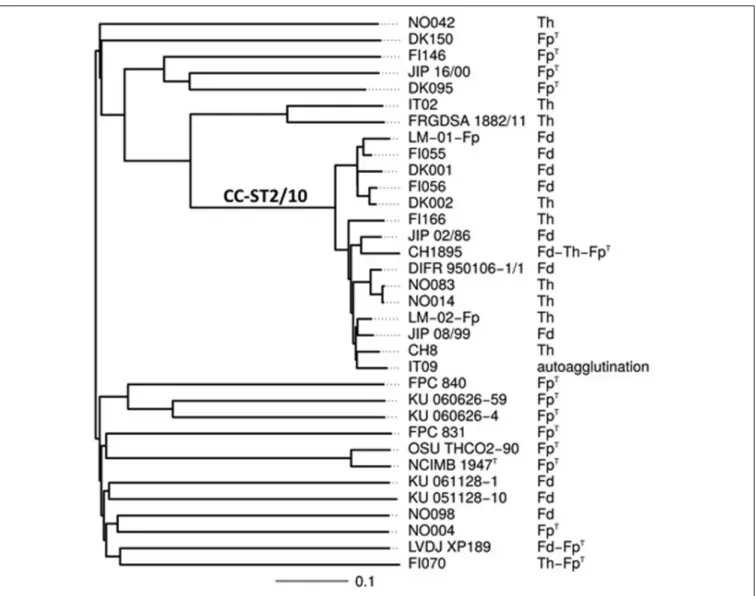
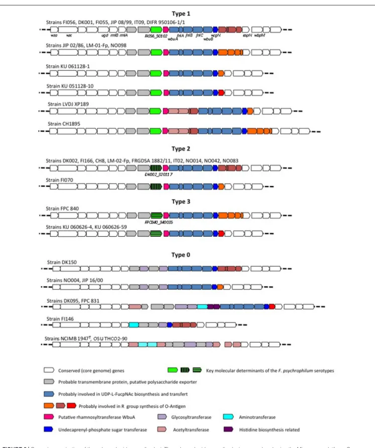
Using the slide agglutination test proposed by Lorenzen and Olesen (1997), serotyping was performed on the 34 strains which complete genomes were available in our laboratories. This serotyping scheme is able to discriminate 3 serotypes: FpT, Th, and Fd, with some strains reacting with multiple antisera. The results are shown in Table 2. Strikingly, all strains but one belonging to CC-ST2/10 display the serotypes Fd or Th (Figure 1). However, the distribution of strains with these serotypes does not strictly follow the tentative phylogeny based on whole-genome MLST. For instance, strains FI056 and DK002, though tightly clustered in the phylogenetic tree, belong to serotypes Fd and Th, respectively. Comparison of the gene content of these two strains revealed that they share 2243 genes (i.e., the core genome genes) but contain only four and two strain-specific genes, respectively. Two of them (FI056_50102 and DK002_320117) are co-localized, substituting each other, and lie in the predicted polysaccharide biosynthesis locus (Figure 2). FI056_50102 and DK002_320117 are orthologous genes ofFP1290 and FPSM_02202, both encoding hypothetical proteins in strains JIP02/86 and CSF259-93, respectively. These genes are adjacent to wbuA encoding a rhamnosyl transferase (Miyamoto et al., 2010), predicted to be involved in theO-antigen biosynthesis.
Extensive comparative genomic analysis of this locus revealed a globally well-conserved structural backbone. By grouping the strains harboring identical or nearly identical neighboring gene organization using FI056_50102/DK002_320117 as the seed, we detected another group of genomes containing a third gene, namedFPC840_340035 in strain FPC840, that substitutes FI056_50102/DK002_320117 at the same chromosomal location (Figure 2). Based on the presence/absence of these three genes, four main genomic organizations, hereafter designated “Types,” were identified. Strains FI056, DK002, and FPC840 are the typical examples of Type-1, Type-2, and Type-3, respectively. Type-0 encompasses a number of strains that do not belong to any of Types 1, 2 and 3 (i.e., not containing any genes related to FI056_50102, DK002_320117, or FPC840_340035) and displays a less-conserved genomic structure and a much broader genomic diversity. The type strain of F. psychrophilum, NCIMB 1947T, belongs to this type.
Comparing the serotypes of the 34F. psychrophilum strains to their Type revealed a remarkable correlation (Table 2). Strikingly,
TABLE 2 | Serotyping of F. psychrophilum isolates.
Strain Serotype mPCR Type
FI056 Fd 1 DIFR 950106-1/1∗ Fd 1 JIP 02/86 Fd 1 LM-01-Fp Fd 1 NO098 Fd 1 DK001 Fd 1 FI055 Fd 1 JIP 08/99 Fd 1 KU 061128-01 Fd 1 KU 051128-10 Fd 1 LVDJ XP189 Fd-FpT 1 CH1895 Fd-Th-FpT 1 IT9 auto-agglutination 1 DK002∗ ∗ Th 2 FI166 Th 2 CH8 Th 2 LM-02-Fp Th 2 FRGDSA 1882/11 Th 2 IT02 Th 2 NO042 Th 2 NO083 Th 2 NO014 Th 2 FI070 Th-FpT 2 NCIMB 1947T ∗ ∗ ∗ FpT 0 OSU THCO2-90 FpT 0 DK150 FpT 0 NO04 FpT 0 JIP16/00 FpT 0 DK095 FpT 0 FPC 831 FpT 0 FI146 FpT 0 FPC 840 FpT 3 KU 060626-04 FpT 3 KU 060626-59 FpT 3 ∗
Strain DIFR 950106-1/1 was used to produce the anti-Fd antiserum.
∗ ∗Strain DK002 = 900406-1/3 was used to produce the anti-Th antiserum. ∗ ∗ ∗
Strain NCIMB 1947Twas used to produce the anti-FpTantiserum.
all strains (n = 10) that only reacted with the anti-Fd antiserum belong to Type-1, all strains (n = 9) that only reacted with the anti-Th antiserum belong to Type-2, and all strains (n = 11) that only reacted with the anti-FpTantiserum belong to Type-0
(8 strains) or Type-3 (3 strains). A search for genes consistently present (>80% identity, >80% coverage) in all strains belonging exclusively to serotype Fd (i.e., FI056, DIFR 950106-1/1, JIP 02/86, LM-01-Fp, NO098, DK001, FI055, JIP 08/99, KU 061128-1, and KU 051128-10) and absent from all strains belonging exclusively to serotype Th (i.e., DK002, FI166, CH8, LM-02-Fp, FRGDSA 1882/11, IT2, NO042, NO083, and NO014) revealed a unique gene (FI056_50102 and its orthologs from the above-mentioned genomes, i.e.,IB65_06085, FP1290, LM01FP_150098, NO098_460117, DK001_50101, FI055_170072, JIP0899_1410015, KU06112801_120020, and KU05112810_320061, respectively). Conversely, a search for
genes consistently present (>80% identity, >80% coverage) in all strains belonging exclusively to serotype Th and absent in all strains belonging exclusively to serotype Fd revealed a unique gene (DK002_320117 and its orthologs from the above-mentioned genomes, i.e.,FI166_350102, CH008_450117, LM02FP_380101, FRGDSA1882_70059, IT2_340117, NO042_ 670087, NO083_60118, and NO014_350089, respectively).
The following particular cases were noticed: strain LVDJ XP189 reacted with the anti-Fd and anti-FpT antisera; strain CH1895 reacted with the anti-Fd, anti-Th and anti-FpTantisera and displayed technical variability between replicates; and strain IT9 was untypable due to auto-aggregation; these three strains belong to Type-1. Finally, strain FI070 reacted with the anti-Th and anti-FpTantisera and belongs to Type-2.
Development of a Multiplex PCR
GenesFI056_50102, DK002_320117 and FPC840_340035 do not display any primary sequence similarities. We therefore designed primers able to be used in a mPCR assay (Table 1). We included a positive control for PCR amplification that targets FP0711, a highly conserved gene ubiquitous in the F. psychrophilum genomes. To insure a straightforward discrimination of the types following gel electrophoresis, amplicons of 188, 361, 549 and 841 bp were selected for the positive control, Type-3, Type-1 and Type-2, respectively. Because of the diversity of Type-0 loci and the lack of a specific gene, no specific primers were designed for the latter and the amplification of only the control testified for Type-0 (Figure 3).
Screening of a F. psychrophilum Strain
Collection Using Multiplex PCR
The mPCR scheme was then used to screen a collection of 244 F. psychrophilum strains isolated from the fifties to 2015 from a wide variety of geographic areas and host fish species. All amplifications were successful and the results are show in Supplementary Table S2. This collection encompasses 28 strains that were included in the original paper ofLorenzen and Olesen (1997). A remarkable correlation between the previously reported serotypes obtained by these authors and the Types obtained using mPCR was observed for the 15 strains reacting with a unique antiserum using both agglutination and ELISA. Indeed: (i) the three strains (DIFR 911209-2, DIFR 930210-1, and DIFR 930413-1) that reacted exclusively with the anti-Fd antiserum belong to Type-1; (ii) the eleven strains (DIFR 900406-1, DIFR 910614-2, DIFR 910614-3, DIFR 910614-5, DIFR 911009-3, DIFR 911126-3, DIFR 930310-1, DIFR 930407-1, DIFR 930427-2, DIFR 930611-2, and Fi 147/93) that reacted exclusively with the anti-Th antiserum belong to Type-2; and (iii) the single strain (LFNW 16/90) that reacted exclusively with the anti-FpT antiserum
belongs to Type-0. The situation of four cross-reacting isolates is more complex but in perfect agreement with Lorenzen and Olesen’s observations: i.e., Fd-FpT cross-reacting isolates using agglutination assay should be considered more closely related to Fd whereas Th-FpT cross-reacting isolates more closely related
to Th. Using this interpretation, mPCR is consistent with the serotypes for four additional strains: (i) the three strains (LPAA
Rochat et al. Flavobacterium psychrophilum Multiplex PCR-Based Serotyping Scheme
FIGURE 1 | Serotypes and evolutionary relationships between the 34 F. psychrophilum strains. Serotypes are shown on a tree attempting to capture the
phylogenetic relationships between the 34 F. psychrophilum strains used in this study for which complete genomes were available. The tree was constructed based on whole-genome MLST profiles (see Materials and Methods for details). Distances are expressed in terms of the fraction of genes where alleles differ between strains. The branch that encompasses strains belonging to clonal complex 2/10 is indicated as CC-ST2/10.
11522, Fi 171/93, and Fi 206/93) that reacted with both anti-Fd and anti-FpTantisera in agglutination assay and exclusively with the anti-Fd antiserum in ELISA assay belong to Type-1 and (ii) the strain (DIFR 910516-1) that reacted with both anti-Th and anti-FpTantisera in agglutination assay and exclusively
with the anti-Th antiserum in ELISA assay belongs to Type-2. Finally, among the 28 isolates, the following particular cases (9 isolates) were noticed: (i) the two strains (LVDI 5/1 and Fi 196/93) that reacted exclusively with the anti-FpTantiserum in agglutination assay and that were untypable using ELISA belong to Type-1; (ii) the single strain (DIFR 910611-1) that reacted exclusively with the anti-FpT antiserum in both agglutination and ELISA belongs to Type-2; (iii) among the three strains that reacted with both anti-Fd and anti-Th antisera in agglutination but only with anti-Th in ELISA, DIFR 930324-1 belongs to Type-1 whereas Fi 88/93 and DIFR 9Type-106Type-19-Type-1 belong to Type-2; and
finally (iv), three strains (USC PT 4.1, JIP 28/86 and LPAA 11524) that reacted with the anti-Fd, anti-Th and anti-FpT antisera in agglutination assay but only with the anti-Th antiserum in ELISA belong to Type-1. In their publication, Lorenzen and Olesen (1997) concluded than the ELISA assay discriminate different serotypes most distinctly than agglutination assay. If we compare ELISA results reported by these authors with mPCR results, 91% of correlation was found (22 out of the 26 ELISA-typable strains).
Our data clearly show a significant association between the Types and the host fish species (Figure 4): 22 out of the 23 strains retrieved from Coho salmon belong to Type-0 (p-value = 0), 59 and 60 out of the 151 strains retrieved from rainbow trout belong to Type-1 and to Type-2, respectively (p-values = 1.8e−7 and 5.8e−7) and all 35 strains retrieved from ayu belong to Type-3 (p-value = 0).
FIGURE 2 | Genomic organization of the polysaccharide encoding loci. The polysaccharide encoding loci were analyzed using the Microscope platform. Gene conservation was predicted using both homology and synteny criteria. White arrows represent core genome genes (i.e., genes conserved in the whole genome dataset which deduced proteins display>80% identity over >80% coverage) and colored arrows represent genes belonging to the variable genome (i.e., genes not conserved in the whole genome dataset).
Rochat et al. Flavobacterium psychrophilum Multiplex PCR-Based Serotyping Scheme
FIGURE 3 | Multiplex PCR. Migration of the PCR products after Multiplex PCR amplification: (MM) GeneRulerTMmolecular marker; (1) strain NCIMB
1947T, revealing Type-0; (2) strain FI056 revealing Type-1; (3) strain DK002,
revealing Type-2; (4) strain FPC 840, revealing Type-3.
FIGURE 4 | Association between Types and host fish species.
DISCUSSION
Serological differences among F. psychrophilum isolates were reported since the pioneering studies in the nineties and different serotyping schemes were proposed (Wakabayashi et al., 1994;
Lorenzen and Olesen, 1997; Mata et al., 2002). However, the conventional protocols for the serotyping of F. psychrophilum isolates available so far suffer several drawbacks such as the choice of the appropriate scheme, the need to raise antisera from animals, and the bias of human interpretation of agglutination results. In addition, some strains display auto-agglutination, lack of reactivity, or cross-reaction with multiple antisera that make laborious reciprocal absorptions against heterologous strains necessary. Moreover, inconsistencies between the results of agglutination and ELISA have been reported (Lorenzen and Olesen, 1997). Hence, serotyping by conventional serological methods is costly, labor-intensive and requires significant technical expertise. Therefore, molecular serotyping using mPCR
is a relevant alternative to traditional serotyping, though identification of molecular markers that determine the serotypes is a prerequisite in order to target them with DNA probes. Indeed, mPCR serotyping assays have been developed for a number of bacterial pathogens such as Haemophilus parasuis, Pasteurella multocida, Salmonella sp., Streptococcus suis, S. pneumoniae and S. agalactiae (Kong et al., 2005;Liu et al., 2013;Harper et al., 2015;
Ma et al., 2016).
We took advantage of the availability of the whole-genome sequences of 34 F. psychrophilum isolates (Duchaud et al., in review) and of the previously published serotyping scheme of Lorenzen and Olesen (1997) to identify key molecular determinants of the serotypes. Focusing on strains FI056 and DK002 that are phylogenetically closely related but belong to two different serotypes (Fd and Th) (Figure 1), we identified the strain-specific genes FI056_50102 and DK002_320117 encompassed in the polysaccharide biosynthesis-encoding locus. Indeed, among the genes surrounding FI056_50102 or DK002_320117, some are predicted to be involved in the biosynthesis of the nucleotide-sugar precursors of O-antigen (rmlA and rmlB for the synthesis of dTDP-L-rhamnose; wbpM for the synthesis of UDP-N-Acetyl-D-quinovosamine and flnA, flnB and flnC for the synthesis of UDP-N-Acetyl-L-fucosamine) whereas others encode the sugar transferases required for the assembly of lipid-linked O-repeat units (wbuA, wbuB and wcgN, the latter encoding the initial undecaprenyl-phosphate sugar transferase) (Figure 2). Previous studies aiming at identifying F. psychrophilum antigens recognized by polyclonal rabbit antisera have been performed (Crump et al., 2001, 2003). LPS-containing O-polysaccharide and core-region lipo-oligosaccharide have reported to be immunological relevant structures targeted by these sera. It is therefore tempting to speculate that the O-polysaccharide, predicted to greatly vary within species, provides the antigenic specificity of the serotyping scheme used in this study. So far, theO-polysaccharide structure has been solved for only oneF. psychrophilum strain (CSF259-93), identified as belonging to Type 2 by mPCR (MacLean et al., 2001). Extensive biochemical analyses will be required for each F. psychrophilum serogroup to formally identify the nature of the carbohydrates targeted by each anti-serum.
GenesFI056_50102/DK002_320117 were therefore promising candidate markers for serotype affiliation. Following a genome-wide association study using the 34 F. psychrophilum isolates, four Types were defined and Types 1 and 2 were firmly associated to Fp or Th serotypes (Table 2). Type 3 was not associated to an exclusive serotype in the scheme defined by Lorenzen and Olesen as FpT serotype was shared between Type 0 and Type 3 isolates. However, its strong association with ayu (Figure 4 and Supplementary Table S2) indicates that Type 3 likely corresponds to the O2 serotype defined by
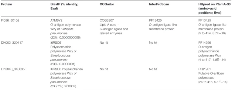
Izumi and Wakabayashi (1999). Remarkably, neither the three genes located at this position (FI056_50102, DK002_320117 and FPC840_340035) nor their encoded proteins display any primary sequence identity. Initially annotated as “hypothetical protein,” they are all predicted to be multipass transmembrane proteins, located in the inner membrane, with weak homologies to polysaccharide-biosynthesis proteins (e.g., O-antigen
TABLE 3 | In silico prediction of protein function. Protein BlastP (% identity;
Eval)
COGnitor InterProScan HHpred on PfamA-30 (amino-acid positions; Eval) FI056_50102 A7M6Y2 O-antigen polymerase Wzy of Klebsiella pneumoniae (22%; 0.0000000006) COG3307 Lipid A core – O-antigen ligase and related enzymes PF13425 O-antigen ligase-like membrane protein PF13425 O-antigen ligase-like membrane protein (5 to 414; 8.7E−16) DK002_320117 I6RSC6 Polysaccharide polymerase Wzy of Streptococcus pneumoniae (20%; 0.0000001) No hit No hit PF14296 O-antigen polysaccharide polymerase Wzy (4 to 417; 1.8E−14) FPC840_340035 I6RSC6 Polysaccharide polymerase Wzy of Streptococcus pneumoniae (23.27%; 0.00002) No hit No hit PF01901 Putative O-antigen polymerase (24 to 415; 9.1E−14)
polymerase,O-antigen ligase) (Table 3). They likely perform the same general function (i.e., an unknown step in polysaccharide biosynthesis) but their activity should lead to structural variations such as the nature of the glycosidic bounds, additional sugar ramification(s), chain length, etc. As no obvious orthologous genes have been identified in strains belonging to Type 0, one might hypothesize that other multipass transmembrane proteins encompassed in polysaccharide biosynthesis-encoding locus could be functional homologs in these strains. Because of the phylogenetic incongruence of these genes, one must conclude that gene exchange by recombination took place at this locus. Indeed, F. psychrophilum was found to have a very high homologous recombination rate that contributes to population diversification (Nicolas et al., 2008;Vos and Didelot, 2009).
Taking advantage of this sequence divergence, we developed a mPCR assay able to discriminate strains belonging to Types 1, 2, 3 and 0. Our mPCR assay proved to be successful as all 244 strains tested were typable using this scheme (Supplementary Table S2). Among these strains, 28 were analyzed in the original paper of Lorenzen and Olesen (1997)and an outstanding correlation between the previously reported ELISA serotypes and the mPCR results was observed. The situation is more complex using agglutination assay. As a matter of fact, one should keep in mind that the antisera consist of polyclonal antibodies that were processed by multiple rounds of reciprocal absorptions against different heterologous strains. Therefore, cross-reactivity observed using slide-agglutination could be considered as a technical drawback, often noticed using such assays. Other variations in the locus (e.g., gene substitution, polymorphism or differences observed in other genes of the locus for some strains) may also impact the serotype and could explain the puzzling results that were obtained with the remaining cross-reacting isolates, especially since as much as seven different serotypes were reported for F. psychrophilum using a broader scheme (Mata et al., 2002). Using the latter scheme, strain FPC840 belongs to serovar 7 and using Izumi’s scheme it belongs to
serovar O2. It is therefore tempting to speculate that Type-3 described in this paper actually corresponds to serovar 7 according to Mata and to serovar O2 according to Izumi. Another example is strain LVDI 5/1 that corresponds to serovar 5 using Mata’s scheme and to FpT usingLorenzen and Olesen’s (1997)
scheme. An attempt to correlate all the previously published serotyping schemes with mPCR is proposed in Supplementary Table S3. Because FpT encompasses many strains displaying a poorer conservation of the locus together with a wider genomic diversity, one might suspect that they could also belong to some other serovars observed byMata et al. (2002) or to other, yet undescribed serovars. One should conclude that the current serotyping schemes may not cover all existing serotypes of F. psychrophilum and that WGS is likely a promising and efficient approach to address this issue. Accordingly, our mPCR assay could be enriched by new targets allowing the detection of additional serogroups in the future.
Our results also revealed a striking association between mPCR-serotype and fish host species. All strains retrieved from coho salmon but one belong to Type-0. The only exception is the Chilean isolate MHC 1710K that belongs to Type 2. As previously proposed, Chilean fish farming practices have contributed to create overlapping host ranges as most fish farms often house mixed stocks of different salmonid species including coho salmon and rainbow trout (Avendaño-Herrera et al., 2009, 2014). The vast majority of strains retrieved from rainbow trout (119 among 151) belong to Types-1 and 2. However, 23 strains belong to Type-0 (20 from France, 2 from Switzerland, and one from Denmark) and 9 strains to Type-3 (8 from France and one from Israel). It is noteworthy that strains of serotypes Th and Fd were reported to be predominant in rainbow trout and associated to disease outbreaks (Lorenzen and Olesen, 1997; Dalsgaard and Madsen, 2000) and to tend to be more virulent than FpTisolates
in experimental infection model using rainbow trout (Madsen and Dalsgaard, 2000; Madetoja et al., 2002). Considering that Types-1 and -2 correspond to Fd and Th serotypes, our results
Rochat et al. Flavobacterium psychrophilum Multiplex PCR-Based Serotyping Scheme
showed that Th and Fd are equally predominant (40%) in our rainbow trout set of isolates. Previous studies reported quite similar results, with serotype Th observed more frequently (60%) in the United Kingdom (Ngo et al., 2017) and Fd occurring more frequently in Finland (Madetoja et al., 2002). Altogether, the mPCR typing method represents a promising epidemiological tool as it allows determining if an isolate belongs to one of the two serotypes associated to disease outbreaks in European fish farms. Finally, all strains retrieved from ayu or their eggs belong to Type-3. It is therefore very tempting to speculate that the nature of the Type provides a selective advantage according to the host that is infected. Nevertheless, these associations might also reflect the underlying genetic structure of the population without necessarily involving a selective advantage linked to the serotype. Indeed, MLST analysis ofF. psychrophilum population structure has also identified clear associations between particular clonal complexes and some fish host (Nicolas et al., 2008). Our identification of the genetic determinants of the serotypes provides a basis for future studies aiming at clarifying the links between serotypes, population structure, and virulence.
As previously observed for other bacterial pathogens, mPCR-based serotyping methods often perform better than traditional serotyping in terms of detection rate, accuracy, reproducibility and compatibility of the results between different laboratories (Harper et al., 2015; Ma et al., 2016). Therefore, our mPCR assay may be readily used in large-scale studies aiming at fish farming improvement. Indeed, this straightforward mPCR-based serotyping procedure might be very useful for a variety of non-academic purposes such as bacterial strain selection for vaccine development, selective breeding or epidemiological surveillance in fish farms.
AUTHOR CONTRIBUTIONS
TR: Substantial intellectual contribution throughout the study, data interpretation and manuscript preparation. EF-N, SC, and AL: Bacterial isolates management, DNA extraction and mPCR. ID and LM: Bacterial isolates management and serological
tests. AC and PN: Participation in genomic data analysis and handling. TW and J-FB: Participation in data interpretation. ED: Substantial intellectual contribution throughout the study, gene mining, interpretation of data, manuscript preparation and responsible for acquisition of funding. All authors read and approved the final manuscript.
FUNDING
The study was funded by the EU EMIDA ERA-NET project “ControlFlavobacteriaceae Infections in European Fish farms”. The France Génomique National infrastructure is funded as part of Investissement d’avenir program managed by Agence Nationale pour la Recherche (contract ANR-10-INBS-09).
ACKNOWLEDGMENTS
The authors are very grateful to Lisbeth Schade Hansen, Kári Karbech Mouritsen and Catherine Fournel for very skillful technical assistance; to Ellen Lorenzen, Ruben Avendaño-Herrera, Mitsuru Eguchi for providing bacterial strains and to Luc Jouneau and Sébastien Bridel for help with statistical analysis. The LABGeM (CEA/IG/Genoscope & CNRS UMR8030) and the France Génomique National infrastructure are acknowledged for support within the MicroScope annotation platform.
SUPPLEMENTARY MATERIAL
The Supplementary Material for this article can be found online at: http://journal.frontiersin.org/article/10.3389/fmicb. 2017.01752/full#supplementary-material
TABLE S1 | Genome sequences used in this study.
TABLE S2 | Screening of the Flavobacterium psychrophilum strain collection using mPCR.
TABLE S3 | Comparison of previously published serotyping schemes with mPCR.
REFERENCES
Antaya, C. (2008). Current eco-economical impacts of Flavobacterium
psychrophilum. MMG 445 Basic Biotechnol. eJ. 4, 16–21.
Aveñdano-Herrera, R., Houel, A., Irgang, R., Bernardet, J. F., Godoy, M., Nicolas, P., et al. (2014). Introduction, expansion and coexistence of epidemic Flavobacterium psychrophilum lineages in Chilean fish farms. Vet. Microbiol. 170, 298–306. doi: 10.1016/j.vetmic.2014.02.009
Avendaño-Herrera, R., Ilardi, P., and Fernández, J. (2009). “Significance of Flavobacterium diseases on salmonid farming in Chile,” in Proceedings of the 2nd Conference on Members of the Genus Flavobacterium, Paris.
Cipriano, R. C., and Holt, R. A. (2005).Flavobacterium psychrophilum, cause of
bacterial cold-water disease and rainbow trout fry syndrome.Fish Dis. Lealf. 86, 1–44.
Crump, E. M., Perry, M. B., Clouthier, S. C., and Kay, W. W. (2001). Antigenic
characterization of the fish pathogen Flavobacterium psychrophilum. Appl.
Environ. Microbiol. 67, 750–759. doi: 10.1128/AEM.67.2.750-759.2001 Crump, E. M., Perry, M. B., Gale, S., Crawford, E., and Kay, W. W. (2003).
Lipopolysaccharide O-antigen antibody-based detection of the fish pathogen
Flavobacterium psychrophilum. J. Mol. Microbiol. Biotechnol. 6, 182–190. doi: 10.1159/000077249
Dalsgaard, I., and Madsen, L. (2000). Bacterial pathogens in rainbow trout, Oncorhynchus mykiss (Walbaum), reared at Danish freshwater farms. J. Fish Dis. 23, 199–209. doi: 10.1046/j.1365-2761.2000.00242.x
Duchaud, E., Boussaha, M., Loux, V., Bernardet, J. F., Michel, C., Kerouault, B., et al. (2007). Complete genome sequence of the fish pathogen Flavobacterium psychrophilum. Nat. Biotechnol. 25, 763–769. doi: 10.1038/ nbt1313
FAO (2016).The State of World Fisheries and Aquaculture. Available at: http:
//www.fao.org/3/a-i5555e.pdf
Finlay, B. B., and McFadden, G. (2006). Anti-immunology: evasion of the host
immune system by bacterial and viral pathogens.Cell 124, 767–782. doi: 10.
1016/j.cell.2006.01.034
Gómez, E., Méndez, J., Cascales, D., and Guijarro, J. A. (2014).Flavobacterium
psychrophilum vaccine development: a difficult task. Microb. Biotechnol. 7, 414–423. doi: 10.1111/1751-7915.12099
Harper, M., John, M., Turni, C., Edmunds, M., St Michael, F., Adler, B., et al. (2015).
strains by use of the lipopolysaccharide outer core biosynthesis locus.J. Clin. Microbiol. 53, 477–485. doi: 10.1128/JCM.02824-14
Holt, R. A., Rohovec, J. S., and Fryer, J. L. (1993). “Bacterial cold-water disease,” in Bacterial Diseases of Fish, 1st Edn, eds V. Inglis, R. J. Roberts, and N. R. Bromage (New York, NY: Wiley-Blackwell), 3–22.
Izumi, S., Liu, H., Aranishi, F., and Wakabayashi, H. (2003). A novel serotype of Flavobacterium psychrophilum detected using antiserum against an isolate from amago,Oncorhynchus masou rhodurus Jordan & Gilbert, in Japan. J. Fish Dis. 26, 677–680. doi: 10.1046/j.1365-2761.2003.00502.x
Izumi, S., and Wakabayashi, H. (1999). Further study on serotyping of Flavobacterium psychrophilum. Fish Pathol. 34, 89–90. doi: 10.3147/jsfp.34.89 Jolley, K. A., and Maiden, M. C. J. (2010). BIGSdb: scalable analysis of bacterial
genome variation at the population level.BMC Bioinformatics 11:595. doi: 10. 1186/1471-2105-11-595
Kong, F., Ma, L., and Gilbert, G. L. (2005). Simultaneous detection and serotype
identification of Streptococcus agalactiae using multiplex PCR and reverse
line blot hybridization.J. Med. Microbiol. 54, 1133–1138. doi: 10.1099/jmm.0. 46244-0
Leeds, T. D., Silverstein, J. T., Weber, G. M., Vallejo, R. L., Palti, Y., Rexroad, C. E., et al. (2010). Response to selection for bacterial cold water disease resistance in rainbow trout.J. Anim. Sci. 88, 1936–1946. doi: 10.2527/jas.2009-2538 Liu, Z., Zheng, H., Gottschalk, M., Bai, X., Lan, R., Ji, S., et al. (2013).
Development of multiplex PCR assays for the identification of the 33 serotypes
ofStreptococcus suis. PLOS ONE 8:e72070. doi: 10.1371/journal.pone.0072070
Lorenzen, E., and Olesen, N. J. (1997). Characterization of isolates of Flavobacterium psychrophilum associated with coldwater disease or rainbow
trout fry syndrome II: serological studies.Dis. Aquat. Organ. 31, 209–220.
doi: 10.3354/dao031209
Ma, L., Wang, L., Chu, Y., Li, X., Cui, Y., Chen, S., et al. (2016). Characterization of
ChineseHaemophilus parasuis isolates by traditional serotyping and molecular
serotyping methods. PLOS ONE 11:e0168903. doi: 10.1371/journal.pone.
0168903
MacLean, L. L., Vinogradov, E., Crump, E. M., Perry, M. B., and Kay, W. W. (2001).
The structure of the lipopolysaccharide O-antigen produced byFlavobacterium
psychrophilum (259-93). Eur. J. Biochem. 268, 2710–2716. doi: 10.1046/j.1432-1327.2001.02163.x
Madetoja, J., Dalsgaard, I., and Wiklund, T. (2002). Occurrence ofFlavobacterium psychrophilum in fish-farming environments. Dis. Aquat. Organ. 52, 109–118. doi: 10.3354/dao052109
Madetoja, J., Hänninen, M. L., Hirvelä–Koski, V., Dalsgaard, I., and Wiklund, T.
(2001). Phenotypic and genotypic characterization of Flavobacterium
psychrophilum from Finnish fish farms. J. Fish Dis. 24, 469–479. doi: 10.1046/j.1365-2761.2001.00326.x
Madsen, L., and Dalsgaard, I. (2000). Comparative studies of Danish Flavobacterium psychrophilum isolates: ribotypes, plasmid profiles, serotypes and virulence.J. Fish Dis. 23, 211–218. doi: 10.1046/j.1365-2761.2000.00240.x Mata, M., Skarmeta, A., and Santos, Y. (2002). A proposed serotyping system for
Flavobacterium psychrophilum. Lett. Appl. Microbiol. 35, 166–170. doi: 10.1046/ j.1472-765X.2002.01157.x
Miyamoto, Y., Mukai, T., Naka, T., Fujiwara, N., Maeda, Y., Kai, M., et al. (2010). Novel rhamnosyltransferase involved in biosynthesis of serovar
4-specific glycopeptidolipid fromMycobacterium avium complex. J. Bacteriol.
192, 5700–5708. doi: 10.1128/JB.00554-10
Nematollahi, A., Decostere, A., Pasmans, F., and Haesebrouck, F. (2003). Flavobacterium psychrophilum infections in salmonid fish. J. Fish Dis. 26, 563–574. doi: 10.1046/j.1365-2761.2003.00488.x
Ngo, T. P., Bartie, K. L., Thompson, K. D., Verner-Jeffreys, D. W., Hoare, R.,
and Adams, A. (2017). Genetic and serological diversity ofFlavobacterium
psychrophilum isolates from salmonids in United Kingdom. Vet. Microbiol. 201, 216–224. doi: 10.1016/j.vetmic.2017.01.032
Nicolas, P., Mondot, S., Achaz, G., Bouchenot, C., Bernardet, J. F., and Duchaud, E.
(2008). Population structure of the fish-pathogenic bacteriumFlavobacterium
psychrophilum. Appl. Environ. Microbiol. 74, 3702–3709. doi: 10.1128/AEM. 00244-08
Plant, K. P., LaPatra, S. E., and Cain, K. D. (2009). Vaccination of rainbow
trout,Oncorhynchus mykiss (Walbaum), with recombinant and DNA vaccines
produced toFlavobacterium psychrophilum heat shock proteins 60 and 70. J.
Fish Dis. 32, 521–534. doi: 10.1111/j.1365-2761.2009.01008.x
Popescu, A.-A., Huber, K. T., and Paradis, E. (2012). ape 3.0: new tools for distance-based phylogenetics and evolutionary analysis in R.Bioinformatics 28, 1536–1537. doi: 10.1093/bioinformatics/bts184
Raetz, C. R., and Whitfield, C. (2002). Lipopolysaccharide endotoxins.Annu. Rev. Biochem. 71, 635–700. doi: 10.1146/annurev.biochem.71.110601.135414 Schmidt, A. S., Bruun, M. S., Dalsgaard, I., Pedersen, K., and Larsen, J. L. (2000).
Occurrence of antimicrobial resistance in fish-pathogenic and environmental
bacteria associated with four danish rainbow trout farms.Appl. Environ.
Microbiol. 66, 4908–4915. doi: 10.1128/AEM.66.11.4908-4915.2000
Starliper, C. E. (2011). Bacterial coldwater disease of fishes caused by Flavobacterium psychrophilum. J. Adv. Res. 2, 97–108. doi: 10.1016/j.jare.2010. 04.001
Vallenet, D., Calteau, A., Cruveiller, S., Gachet, M., Lajus, A., Josso, A., et al. (2017). MicroScope in 2017: an expanding and evolving integrated resource for
community expertise of microbial genomes.Nucleic Acids Res. 45, D517–D528.
doi: 10.1093/nar/gkw1101
Vos, M., and Didelot, X. (2009). A comparison of homologous recombination rates in bacteria and archaea.ISME J. 3, 199–208. doi: 10.1038/ismej.2008.93 Wakabayashi, H., Toyama, T., and Iida, T. (1994). A study on serotyping of
Cytophaga psychrophila isolated from fishes in Japan. Fish Pathol. 29, 101–104. doi: 10.3147/jsfp.29.101
Wiens, G. D., LaPatra, S. E., Welch, T. J., Rexroad, C. III, Call, D. R., Cain, K. D.,
et al. (2014). Complete genome sequence ofFlavobacterium psychrophilum
strain CSF259-93, used to select rainbow trout for increased genetic resistance
against bacterial cold water disease.Genome Announc. 2:e00889-14. doi: 10.
1128/genomeA.00889-14
Conflict of Interest Statement: The authors declare that the research was conducted in the absence of any commercial or financial relationships that could be construed as a potential conflict of interest.
Copyright © 2017 Rochat, Fujiwara-Nagata, Calvez, Dalsgaard, Madsen, Calteau, Lunazzi, Nicolas, Wiklund, Bernardet and Duchaud. This is an open-access article distributed under the terms of the Creative Commons Attribution License (CC BY). The use, distribution or reproduction in other forums is permitted, provided the original author(s) or licensor are credited and that the original publication in this journal is cited, in accordance with accepted academic practice. No use, distribution or reproduction is permitted which does not comply with these terms.