HAL Id: tel-01021258
https://tel.archives-ouvertes.fr/tel-01021258
Submitted on 9 Jul 2014HAL is a multi-disciplinary open access archive for the deposit and dissemination of sci-entific research documents, whether they are pub-lished or not. The documents may come from teaching and research institutions in France or abroad, or from public or private research centers.
L’archive ouverte pluridisciplinaire HAL, est destinée au dépôt et à la diffusion de documents scientifiques de niveau recherche, publiés ou non, émanant des établissements d’enseignement et de recherche français ou étrangers, des laboratoires publics ou privés.
Genes of innate immunity and their significance in
evolutionary ecology of free livings rodents
Alena Fornuskova
To cite this version:
Alena Fornuskova. Genes of innate immunity and their significance in evolutionary ecology of free livings rodents. Populations and Evolution [q-bio.PE]. Université Montpellier II - Sciences et Tech-niques du Languedoc; Masarykova univerzita (Brno, République tchèque), 2013. English. �NNT : 2013MON20103�. �tel-01021258�
UNIVERSITE•MONTPELLIER•II••
SCIENCES•ET•TECHNIQUES•DU•LANGUEDOC••
FACULTE•DES•SCIENCES•
• and• •MASARYK•UNIVERSITY,•BRNO•
FACULTY•OF•SCIENCE•
• THESIS•• • To•obtain•doctoral•degree• • Formation•doctorale:•Biologie•de•l'évolution•et•écologie•• Ecole•Doctorale:•Systèmes•Intégrés•en•Biologie,•Agronomie,•Géosciences,•Hydrosciences,• Environnement,•SIBAGHE• • • Presented•and•defended•publicly• • AUTHOR:•Alena•Fornuskova• • •GENES•OF•INNATE•IMMUNITY•AND•THEIR•SIGNIFICANCE•IN•
EVOLUTIONARY•ECOLOGY•OF•FREE•LIVING•RODENTS•
• Toll-like•receptor•polymorphisms•in•rodents•• • • Thesis•supervised•by•Dr.•Jean-François•Cosson/Dr.•Josef•Bryja• • Date•and•place•of•defence:•the•19•December•2013•in•CBGP• • • • • • • • • • • • • COMMITTEE:• • M.•Pierre•Boursot,•Director•of•research•CNRS,•Université•Montpellier•2,•Montpellier,•FR• •••••••••••••••••••••••••••Examiner•• M.•Petr•Ho ín,•Professor,•University•of•Veterinary•and•Pharmaceutical•Sciences,•Brno,•CZ•••••••••••••••••••••••••••••••••Reviewer• M.•Dirk•Werling,•Professor,•The•Royal•Veterinary•College,•University•of•London,•UK••• •••••••••••••••••••••••••••Reviewer• M.•Jan•Zima,•Professor,•Institute•of•Vertebrate•Biology•AS•CR,•Brno,•CZ• • • •••••••••••••••••••••••••••Examiner• M.•Josef•Bryja,•Assoc.•professor,•Institute•of•Vertebrate•Biology•AS•CR•and•Masaryk•University,•Brno,•CZ••••Supervisor•3
ABSTRACT•
Appropriate recognition of parasites is crucial for effective immune response, ensuring activation of adequate defence mechanisms. In vertebrates, it has frequently been demonstrated that genes encoding proteins involved in pathogen recognition by an adaptive immune system are often subject to intense selection pressures. On the contrary, much less information has been provided on the evolution of recognition mechanisms of innate immunity. The aim of this thesis is to describe the pattern of natural variation of innate immunity genes involved in pathogen recognition in rodents and to analyze the mechanisms of their evolution. We used murine rodents (subfamily Murinae) as a principal model group because they often live in our close proximity and thus are potential reservoirs of various pathogens dangerous to humans.
First, we studied the intraspecific variability of five bacterial sensing Toll-like receptors (TLR1, TLR2, TLR4, TLR5, and TLR6) in inbred strains derived from two subspecies of the house mouse (M. m. musculus, hereafter abbreviated as Mmm and Mus
musculus domesticus, Mmd). Wild-derived inbred strains are suitable tools for studying
variation of immunity genes because they provide information about alleles that occur in natural populations, and at the same time they occur at homozygous state. The most significant results include the findings of a stop codon in exon 2 of the Tlr5 gene in one Mmm strain and no variability in Tlr4 of Mmd. The results also provide the set of diagnostic SNPs for each gene allowing future studies of introgression of immunity genes across the house mouse hybrid zone and their possible role in the speciation process.
Following these results we decided to check whether the absence of Tlr4 polymorphism in Mmd reflects the pattern found in natural populations, or whether it is a consequence of insufficient sampling or subsequent breeding. We therefore sequenced Tlr4 in both subspecies across a large part of the Western Palaearctic region (in total 39 Mmm and 62 Mmd individuals), then we compared these results with variability on mitochondrial DNA (cytochrome b). The result confirmed our prediction that observed variability in Mmd is strongly reduced also in free-living populations (compared to Mmm), probably due to strong purifying selection by pathogens with which they met during the westward colonization. However, the influence of random evolutionary processes (e.g. drift during bottlenecks) cannot be excluded based on our data. At the intraspecific level, we could not find any sign of positive selection. Our results revealed also species specific variants of Tlr4 and an important role of recombination in Mmm during evolutionary history.
4
The last part of my dissertation is devoted to interspecific comparison of two receptors, TLR4 and TLR7. These two TLRs differ in the exposure and the ligands detection. TLR4 is an extracellular receptor detecting mainly bacterial ligands (especially lipopolysaccharides), while TLR7 is located inside the cell and detects ssRNA viruses. The aim of this part of the thesis was to describe variability of both receptors at the interspecific level and to reveal selection forces acting on TLRs in longer evolutionary time scale. In total we analyzed 23 rodent species of the subfamily Murinae in Europe, Asia and Africa. Our results suggest that purifying selection has been a dominant force in evolution of the Tlr4 and
Tlr7 genes, but we also demonstrated that episodic diversifying selection has shaped the
present species-specific variation in rodent Tlrs. Sites under positive selection were concentrated mainly in the extracellular domain of both receptors, which is responsible for ligand binding. The comparison between two TLRs lead us to the conclusion that the intracellular TLR7 is under much stronger negative selection pressure, presumably due to its interaction with viral nucleic acids, which are similar to those of the host and even small changes in TLR7 conformation could cause autoimmunity.
Key words: Toll-like receptors, receptors of innate immunity, Pattern Recognition Receptors,
selection, evolution, natural selection, genetic polymorphism, phylogeny, host-parasite coevolution, genetic diversity.
5
RÉSUMÉ
Une reconnaissance appropriée des parasites est essentielle pour une réponse immunitaire efficace, assurant l'activation adéquate des mécanismes de défense immunitaire. Chez les vertébrés, il a été fréquemment démontré que les gènes codant pour les récepteurs de l'immunité adaptative impliqués dans la reconnaissance des agents pathogènes sont souvent soumis à une intense pression sélective. En revanche, beaucoup moins d’études se sont intéressées à la sélection agissant sur les récepteurs de l'immunité innée. Le but de cette thèse est de décrire la variabilité naturelle des gènes de l'immunité innée impliqués dans la détection des agents pathogènes chez les rongeurs et d’analyser les mécanismes responsables de leur évolution. Ce travail s’est focalisé principalement sur les rongeurs de la sousfamille des Murinae de part leur présence fréquente à proximité des populations humaines et de leur rôle potentiel en tant que réservoirs d’agents pathogènes dangereux pour l’Homme.
Tout d´abord nous avons étudié la variabilité intraspécifique de cinq Toll-like récepteurs ciblant les bactéries (TLR1, TLR2, TLR4, TLR5 et TLR6) pour des lignées consanguines de souris domestiques issues d’une population sauvage de deux sous-espèces :
Mus musculus domesticus (Mmd) et Mus musculus musculus (Mmm). Les souches
consanguines constituent un outil adapté à l'étude de la variabilité des gènes immunitaires car elles confèrent une information sur les allèles présents dans les populations naturelles tout en bénéficiant de génotypes homozygotes. Les résultats les plus significatifs concernent la découverte d'un codon stop dans l'exon 2 du Tlr5 chez une lignée de Mmm et l’absence de variabilité du Tlr4 chez Mmd. Ces résultats ont également permis de constituer un jeu de SNPs diagnostics utilisable pour de futures études afin de mieux comprendre le rôle de l’introgression de ces gènes immunitaires dans les mécanismes de spéciation de la zone hybride de la souris domestique.
A la suite de ces résultats, nous avons décidé de vérifier si l‘absence de polymorphisme du Tlr4 chez Mmd reflète une absence de variabilité dans les populations naturelles, ou si il s’agit plutôt d’un effet de l'échantillonnage ou des croisements ultérieurs. Nous avons donc séquencé le gène Tlr4 pour les deux sous-espèces provenant de la région du Paléarctique Occidentale (au total 39 Mmm et 62 Mmd) puis nous avons comparé ces résultats avec la variabilité génétique d’un gène mitochondrial (cytochrome b). Nous avons confirmé notre prédiction : la variabilité de Tlr4 chez Mmd est fortement réduite par rapport à Mmm, probablement à cause d’agents pathogènes ayant exercé une sélection purifiante chez
6
Mmd durant la colonisation vers l’ouest. Cependant, l'influence de mécanismes évolutifs neutres, tel que la dérive consécutive à un goulot d’étranglement démographique, ne peut être exclue sur la base de nos données. De plus, nos résultats ont montrés que les deux sous-especes présentent des variants différents de Tlr4, et que la recombinaison a joué un rôle important dans le maintien de la variabilité chez Mmm.
La dernière partie de cette thèse a été consacrée à la comparaison interspécifique de deux récepteurs : TLR4 et TLR7. Ces deux TLRs se différencient à la fois par leur localisation et leur capacité de détection. TLR4 est un TLR extracellulaire reconnaissant principalement les ligands bactériens, essentiellement les lipopolysaccharides, tandis que TLR7 est localisé dans la cellule et détecte les virus à ARN simple brin. L‘objectif était de décrire la variabilité inter-spécifique de chaque récepteur et de révéler les mécanismes de sélection s’exerçant sur ces gènes au cours de leur évolution sur une échelle de temps plus importante. Nous avons analysé 23 espèces de Murinae provenant d’Europe, d’Asie et d’Afrique. Nos résultats suggèrent que la sélection purifiante est la force principale ayant agit sur l’évolution des gènes TLR4 et TLR7. Cependant, nous avons également mis en évidence des épisodes de sélection diversifiante qui ont pu être à l’origine des variations intra-spécifiques de TLRs observée aujourd’hui chez les rongeurs. Des sites sous sélection positive sont principalement concentrés dans les domaines extracellulaires des deux récepteurs, domaines responsables de la reconnaissance des agents pathogènes. Enfin, la comparaison entre ces deux TLRs montre que le TLR7 localisé dans le compartiment intracellulaire est soumis à une sélection négative plus forte. Cette sélection peut s’expliquer en raison des interactions du TLR7 avec les acides nucléiques viraux qui peuvent être similaires à ceux de l'hôte. Ainsi, un changement même faible dans la conformation du TLR7 pourrait provoquer des réactions auto-immunes chez l’hôte.
Mots clés: Toll-like receptors, récepteurs de l’immunité innée, Pattern Recognition
récepteurs, sélection, évolution, sélection naturelle, polymorphisme génétique,
7
ABSTRAKT
V•asné rozpoznání patogen• je zásadní pro efektivní imunitní odpov••, zajiš•ující aktivaci adekvátních obranných mechanism•. U obratlovc• je obecn• zdokumentováno, že molekuly adaptivní imunity, které se ú•astní rozpoznání patogen•, jsou •asto p!edm•tem intenzivních selek•ních tlak•. Naopak mnohem mén• údaj• je známo o selekci p•sobící na receptory vrozené imunity. Cílem této práce je popsat variabilitu gen• vrozené imunity, které se ú•astní detekce patogen• u voln• žijících hlodavc• a odhalit, které selek•ní síly na n• b•hem evoluce p•sobily. Práce je zam•!ena na pod•ele• Murinae, jelikož •asto žijí v bezprost!ední blízkosti lidí a jsou potencionálními nositeli r•zných pro •lov•ka nebezpe•ných patogen•. Nejprve jsme studovali vnitrodruhovou variabilitu p•ti anti-bakteriálních Toll-like receptor• (TLR1, TLR2, TLR4, TLR5 a TLR6) u inbredních linií myši domácí, které byly odvozeny z populací voln• žijících zví!at. Tyto kmeny jsou vhodným nástrojem pro studování variability imunitních gen•, jelikož nám poskytují informaci o alelách, které se v p!irozené populaci vyskytují, a zárove$ je možné vhodn• využít homozygotních genotyp• u inbredních jedinc•. Výsledkem této •ásti bylo popsání polymorfismu p•ti gen• TLR u dvou poddruh• myši domácí (Mus musculus musculus, Mmm a M. m. domesticus, Mmd). K nejzajímav•jším výsledk•m pat!ilo zjišt•ní stop kodonu v exonu 2 u Tlr5 a nulová variabilita Tlr4 u Mmd. Tato •ást práce zárove$ poskytla sadu diagnostických SNPs, které mohou být využity ke studiu introgrese Tlr gen• p!es hybridní zónu myši domácí a ke stanovení vlivu jejich polymorfismu na specia•ní procesy.
Na tento výsledek jsme navázali další studií, kdy jsme se rozhodli ov•!it, zda nízká variabilita Tlr4 u Mmd je reálným odrazem variability p!írodních populací, •i se jedná o nep!irozenou odchylku zp•sobenou nedostate•ným vzorkováním •i následným inbredním k!ížením. Osekvenovali jsme tedy Tlr4 u obou poddruh• z velké •ásti jejich výskytu v západním Palearktu (celkem 39 Mmm a 62 Mmd) a srovnali variabilitu Tlr4 se znakem na mitochondriální DNA. Hlavním výsledkem bylo potvrzení hypotézy, že variabilita u Mmd je oproti Mmm opravdu radikáln• snížena, pravd•podobn• vlivem silné selekce zp•sobené patogeny, se kterými se b•hem koloniza•ní cesty tento poddruh potkal. Vliv náhodných evolu•ních mechanism• (nap!. genetický drift p!i opakovaných sníženích efektivní velikosti populace) však nem•že být na základ• našich dat zcela vylou•en. Dále jsme zjistili, že oba poddruhy mají zcela odlišné Tlr4 varianty, a že u Mmm je variabilita Tlr4 udržována i mechanismem rekombinace.
8
Poslední •ást disertace je v•nována mezidruhovému srovnání dvou receptor• (TLR4 a TLR7). Ty se liší jednak lokalizací v bu•ce a jednak schopností detekce odlišných ligand•. TLR4 pat•í mezi extracelulární receptory detekující hlavn• bakteriální ligandy (p•edevším lipopolysacharidy), zatímco TLR7 je lokalizován na vnitrobun••ných membránách a detekuje ssRNA vir•. Cílem mezidruhového srovnání bylo popsat variabilitu obou zmín•ných receptor• a p•sobení selekce b•hem evoluce obou TLR. Celkem bylo pro analýzu použito 23 druh• hlodavc• z pod•eledi Murinae z Evropy, Asie a Afriky. P•estože se ob• molekuly vyvíjejí p•edevším pod vlivem purifikující selekce, která eliminuje negativní mutace a udržuje funk•nost receptor•, n•které jejich •ásti vykazovaly znaky pozitivní selekce. Místa pod pozitivní selekcí byla koncentrována p•edevším v extracelulární •ásti obou receptor•, která je zodpov•dná za rozpoznání patogen•. Následným srovnáním obou TLR jsme zjistili, že intracelulární TLR7 je pod mnohem v•tším negativním selek•ním tlakem, nebo! rozpoznává virové nukleové kyseliny, které jsou zna•n• podobné hostitelským molekulám, a jakákoliv i drobná zm•na by mohla vést k autoimunitním poruchám.
Klí•ová slova: Toll-like receptory, receptory vrozené imunity, Pattern-recognition receptory, selekce, evoluce, p•írodní selekce, polymorfismus, fylogeneze, koevoluce mezi hostitelem a patogenem, genetická variabilita.
9
The thesis was prepared in the laboratories of:
CENTRE DE BIOLOGIE ET GESTION DES POPULATIONS
(UMR INRA/IRD/CIRAD/MontpellierSupAgro) Campus International de Baillarguet
CS 30016
34988 Montfferrier sur Lez cedex France
&
INSTITUTE OF VERTEBRATE BIOLOGY
Research Facility Studenec
Academy of Sciences of the Czech Republic Studenec 122
675 02 Kon•šín Czech Republic
10
FUNDING
This thesis was supported by the French National Agency for Research projects CERoPath (grant number 00121 0505, 07 BDIV 012) http://www.ceropath.org/ and BioDivHealthSEA (grant number ANR 11 CPEL 002), and the Czech Science Foundation (grant number 206/08/0640). The thesis was partly funded by a three year French government fellowship and the fellowship from Masaryk University. My thesis was also supported by the project (MUNI/A/0937/2012) of Institute of Botany and Zoology of Masaryk University which provided me the generous financial support. Travelling expanses were partially funded by project of Institute of Vertebrate Biology, Academy of Sciences of the Czech Republic, Next-generation technologies in evolutionary genetics (CZ.1.07/2.3./20.0303) and by bilateral project BARRANDE (grant number MEB021130/24504WM).
11
ACKNOWLEDGEMENTS
Firstly, I would like to thank sincerely to my official thesis supervisors Josef Bryja and Jean-François Cosson, for their excellent mentoring, advices, and understanding over the past years. I appreciate the time they provide me and also their enormous patience. I would like to thank also to my “shadow” supervisors Jaroslav Piálek and Miloš Macholán for all long hours of discussion and big personal support during my whole stay in Studenec. I really admire their infinite enthusiasm (often infectious) and “active service” during weekends, holidays and late evenings in final phase of my thesis writing. Many thanks go also to Nathalie Charbonnel for all instrumental meetings and working coffee breaks. All these people gave me great advices when needed and I can easily say that once I would like to become such brilliant scientists as they are. I would like to thank also to other members of my thesis committee, Dr Pierrick Labbé and Dr Nicolas Bierne for their constructive advices and stimulant suggestions during the regular committee meetings. My special thanks belong to the French government, whose scholarship for Ph.D. students “en co-tutelle” has allowed my Ph.D. study in Montpellier and to Dr Jaroslav Piálek and his project (project 206-08-0640) funded by the Grant Agency of the Academy of Sciences of the Czech Republic from which I was partially paid. I thank also to Institute of Botany and Zoology of Masaryk University for the generous financial support from project MUNI/A/0937/2012. Because this thesis is the result of a great team work I tried to avoid using “I” in the text and replaced it by “we”.
Thanks to all my colleagues and dear friends from Studenec, especially to lab and mouse team A!a Bryjová, Michal Vinkler, Lucka Vl"ková, Zuzka Bainová, Iva Martincová, Dáša #ížková, Tania Aghová, Terka Králová, Ludovít $ureje, Hanka Konvi"ková, Jana Piálková, Wasimuddin, Start J. E. Baird and Joëlle Goüy de Bellocq, for making the research real, successful and often also more fun. Same thanks goes also to all my French colleagues and friends from CBGP, especially to girls and my very good friends Pascaline Dumas, Joséphine Piffaretti, Marie Pagès, Anne Laure Clamens, Caroline Tatard and Laure Sauné for their personal support and lot of funny moments. Than I would like to thank also to masculine part of CBGP, to phylogeny master Gael Kergoat, IT God Alex Dehne Garcia, fitness coach Laurent Soldati, IT genius Sylvain Piry, rat hunter Yannick Chaval and “brother in beer” Fabian Codamine. Special thanks go to my friend and „snowboard mate” Max Galan for his moral and technical support and precious friendship.
And last but not least I would like to thank to my family for all their support over the three years. I would like to especially thank to my mother, for her support and optimism. My
12
big thanks go also to my “big” brother who took care about whole family during my absence. My biggest thanks go to my boyfriend and great scientist Olda, who encouraged me every day and helped me to solve many problems and crazy hypothesis and again and again we spend our evenings in discussions about Tlrs and their evolution. He was my principal moral support when I lost my hope or motivation. He taught me how to be patient and tolerant and thanks to him I become better person and also better scientist I guess. I thank also to Mr Skype that gave us the possibility to be together and bridge the distance of 1600km and to my computer for smashing service during whole Ph.D and especially during last weeks.
Finally I would like to dedicate this work to my father who unfortunately died on 5th
December 2009. Thanks to him I am where I am and I am who I am. He was the person, who pushed me to study foreign languages especially French and English and who introduced me France as beautiful and magic country full of colours, fragrance and divine tastes. He opened me the world and awaked my passion for travelling and studing. Thanks to him I could become independent and strong person. He gave me wisdom advices but let me also do my own mistakes, because he knew that experiences are untransmittable. He supported me every time even when I choose science as my main life path and he used to say to me “fly low”!
13
PREFACE
Organisation•of•the•thesis•in•"co-tutelle"••
The thesis was from the beginning designed as the thesis under double co-supervision performed officially at the University of Montpellier 2 (Montpellier, France) and Masaryk University (Brno, Czech Republic). I have started my thesis at the Masaryk University in September 2009. I spent the first year by writing the project and application for the Grant of
French government and by preparing my first year in France.For next three years (from 2010
to 2013) I have spent seven months (from 1st March till the end of September) at the Research Facility Studenec of Institute of Vertebrate Biology of the Academy of Sciences of the Czech Republic ("IVB") and for the rest of the year (or better during winter) I have “migrated” southward to the Centre de Biology pour la Gestion des Populations, Montferriez-sur-Lez, France (CBGP). If you check the photos bellow you probably figure out my intention....
Photos: on the left, mouse breeding facility at IVB in Studenec in winter 2010, on the right, CBGP in winter
2010.
In CBGP, I had the opportunity to use the immense sample collections of rodents from Southeast Asia, which are also important vectors of emerging diseases (data collected mostly within the project CERoPath). At IVB, I was directly involved in projects using one of the best model systems for speciation studies, i.e. the European house mouse hybrid zone, which has been a subject of a long-term study at the institute. The material collected in the field during previous projects was complemented by a huge sample collection of both mouse subspecies from a large part of the Western Palaearctic area as well as by cca 20 inbred strains derived from wild populations. We have decided to focus on interspecific level of Tlr variation during my stays in CBGP and on intraspecific level during my work in Studenec.
14
LIST OF TABLES AND FIGURES
Chapter•1•and•2:•General•Introduction•and•Material•and•methods•
Fig. 1, Overview of localization of signalling PRRs and their MAMPs Fig. 2, VDJ recombination
Fig. 3, Function TCR and BCR Fig. 4, Frequency-dependent selection
Fig. 5, Balancing selection through negative frequency dependent selection. Fig. 6, Biochemical properties of amino acids
Fig. 7, Simplified description of the TLR4 structure
Fig. 8, Crystal structure of TLR ligand-binding domains with its ligands Fig. 9, Schematic of mammalian TLR signaling pathways
Fig. 10, Evolution of major vertebrate Fig. 11, Overview of composition
Fig. 12, Overview of localization and ligands of TLRs Fig. 13, Phylogeny of the subfamily Murinae
Fig. 14, Phylogeny of rats
Fig. 15, Phylogenetic relationships of the Indochinese Rattini
Fig. 16, Maximum likelihood tree reconstructed with a molecular clock hypothesis Fig. 17, Phylogenetic relationships among 10 mouse taxa
Fig. 18, Colonization routes of the house mouse subspecies starting from northern parts of Indian
subcontinent
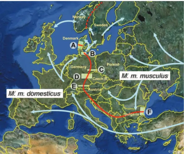
Fig. 19, The course of the M. m. musculus/M. m. domesticus hybrid zone in Europe
Fig. 20, Taxonomic structure of animals used for experiments by humans. by Daniel Engber
Chapter• 3.1:• Polymorphism• of• bacterial-sensing• TLRs• in• wild-derived• and• classical• laboratory•strains•of•Mus•musculus•
Fig. 1, Origin localities of wild-derived strains kept in Research Facility Studenec
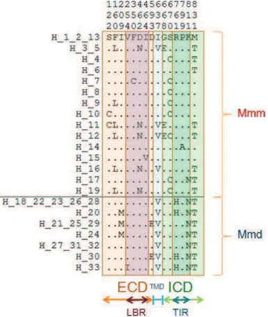
Fig. 2, Description of non-synonymous polymorphic sites (SNPs) and their position within TLRs Fig. 3, Haplotype network based on nucleic acids
Table 1, Summary of strains
Table 2, Overview of sequenced TLRs
Table 3, Overview of sites and regions important for ligand binding and dimerization Table 4, Genetic diversity of Tlrs in two subspecies of the house mouse
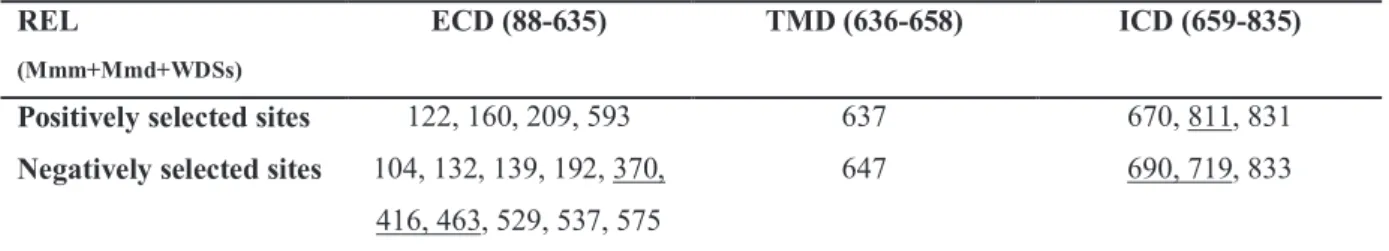
Table 5, Physicochemical properties of the amino acids involved in non-synonymous substitutions in
ligand binding regions
Chapter• 3.2:• Analysis• of• variability• at• intraspecific• level• in• wild• populations• of• Mus• musculus••
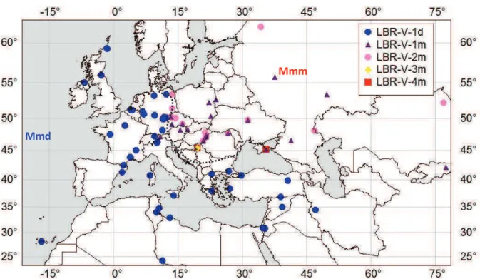
Fig. 1. Distribution of samples analyzed in this study
Fig. 2, Overview of Tlr4 non-synonymous substitutions in Mmd and Mmm Fig. 3, Ribbon diagram of the TLR4 ECD 3D structure
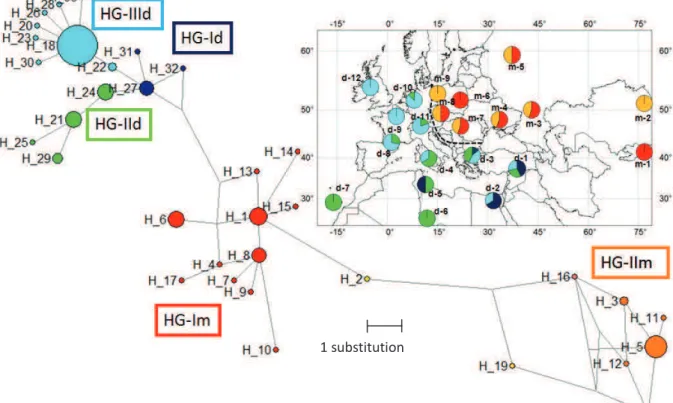
Fig. 4, Haplotype network and haplogroup distribution of Tlr4 (a) and mt-Cytb (b) Table 1, Genetic diversity of Tlr4 and mt-Cytb in two house mouse subspecies Table 2, Description of LBR variants
Table 3, Physicochemical properties of the amino acids involved in non-synonymous substitutions of
15
Table 4, Selection tested by REL in both subspecies together Supplementary materials
Fig. S1, Phylogeny based on Bayesian inference (MrBayes v.3.1) Tlr4 (a) and mt-Cytb (b) Fig. S2, Haplogroup definition Tlr4 (a) and mt-Cytb (b)
Fig. S3, Evidence of recombination between HG-Im and HG-IIm of Mmm
Table S1, Summary of sampled specimens, identification of haplotypes and NCBI GenBank accession
numbers.
Table S2, Binding sites between TLR4/LPS/MD-2
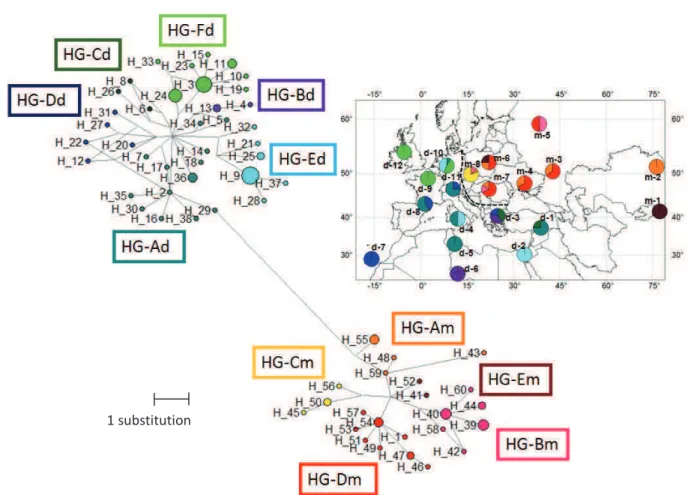
Chapter•3.3:•Analysis•of•variability•at•interspecific•level•of•wild•rodents• Fig. 1, Comparison of phylogenetic trees based on Tlrs and neutral markers Fig. 2, Distribution of sites under selection identified by SLAC and MEME Fig. 3, Sites under positive selection identified in evolutionary lineages by MEME
Fig. 4, Mapping of evolutionary conservation of amino acid positions in a protein molecule based on
the phylogenetic relations between homologous sequences
Table 1, Estimates of sequence diversity and average codon-based evolutionary divergence over all
sequence pairs for the exon 3 and particular domains of Tlr4 and Tlr7 genes
Table 2, Positively (MEME and SLAC-PS) and negatively (SLAC-NS) selected sites detected for the
exon 3 of Tlr4 and Tlr7 at p < 0.05
Supplementary materials
Fig. S1, Protein structure of TLR4 (a, c) and TLR7 (b, d) identified by SMART
Fig. S2, Phylogeny based on the exon 3 of Tlr4 (a) and Tlr7 (b) gene reconstructed by Bayesian
inference method in MrBayes
Fig. S3, Phylogeny based on the exon 3 of Tlr4 (a) and Tlr7 (b) gene reconstructed by maximum
likelihood method in RAxML
Fig. S4, Test of congruence between the presumably neutral and Tlr phylogenies (Tlr4 (a), Tlr7 (b))
Fig. S5, Superimposition of structures, tree clustering diagrams based on linkage distance, LBRTLR4 (a)
and LBRTLR7 (b)
Fig. S6, Analysis of LBR amino acid sequence charge at pH 7 (LRRFinder) for LBRTLR4 (a) and
LBRTLR7 (b)
Table S1, Summary of sampled specimens and identification of haplotypes Table S2, Primer description
Table S3, Residues binding to LPS in TLR4 based on knowledge of 3D-crystalography in human
predicted by Park et al. 2009
Table S4, Potential residues binding ssRNA predicted by Wei et al. 2009
Discussion•
Fig. 21, Hypothesis about the origin of reduced Tlr4 variability in Mmd.
Fig. 22, Hierarchical model outlining the evolutionary dynamics and biological relevance of the
16
LIST OF ABREVIATIONS
APC, anitgen presenting cells; BCRs, B cell receptors;
CARD, caspase activating and recruitment domain; CLRs, C-type lectin receptors;
CLS, classical laboratory strains;
CpG DNA, unmethylated cytosine-guanine dinucleotide sequences CpG, cytosine phosphate guanosine;
CRDs, carbohydrate recognition domains; CTD, C-terminal domain;
CTLD, C-type lectin-like domain; DCs, dendritic cells;
DC-SIGN, dendritic cell-specific intercellular adhesion molecule-3-grabbing nonintegrin; dN, number of non-synonymous substitutions per non-synonymous site;
dS, number of synonymous substitutions per synonymous site; dsRNA, double-stranded RNA;
ECD, N-terminal horseshoe-like extracellular domain, or extracellular domain; FDS, Frequency-dependent selection;
ICD, C-terminal intracellular domain; IFN, interferon;
IL, interleukin; IL-1•, Interleukin-1•; IL-6, Interleukin-6;
ILS, incomplete lineage sorting;
IRAK, interleukin-1-receptor-associated kinase; IRF, interferon-regulated factor;
LBR, ligand binding region;
LGP2, laboratory of genetics and physiology 2; LP, lipoproteins;
LPS, lipopolysaccharide; LRRs, leucine rich repeats; LS, laboratory strains;
MAL, MyD88 adaptor-like protein;
MAMPs, microbe associated molecular patterns; MBL, mannose-binding lectin;
MDA5, melanoma differentiation associated factor 5; MD-2, lymphocyte antigen 96;
MGL, Macrophage galactose-type C-type lectin; MHC, major histocompatibility complex;
Mmd, M. m. domesticus; Mmm, M. m. musculus; MR, mannose receptor;
MyD88, Myeloid differentiation factor 88; Myr, Million years ago;
NACHT, domain present in NAIP, CIITA, HET-E, TP-1; NALP, NACHT-, LRR-, and pyrin-domain containing proteins; NAP1, NAK-associated protein 1;
17 NF-•B, Nuclear factor •B;
NLRs, NOD-like receptors;
NOD, nucleotide-binding oligomerization domain; NPCs, neural progenitor cells;
nsSNPs, non-synonymous single nucleotide polymorphisms; ORs, opsonin receptors;
PAMPs, pathogen-associated molecular patterns; PG, peptidoglycan;
polyI:C, polyinosine-deoxycytidylic acid; PRRs, pattern recognition receptors; RIG-I, retionic acid-inducible gene I; RLRs, RIG-I like receptors;
SNPs, single nucleotide polymorphisms; SR , scavenger receptors;
ssRNA, single stranded viral RNA; TCRs, T cell receptors;
TIR, Toll/interleukin-1 receptor; TIRAP, TIR-associated protein; TLR, Toll-like receptor;
TMD, single transmembrane helix or transmembrane domain; TNF , tumour necrosis factor ;
TRAF, TNF-receptor-associated factor;
TRAF6, tumor necrosis factor receptor associated factor 6; TRAM, TRIF-related adaptor protein;
TRIF, TIR domain containing adapter-inducing!interferon!" WDS, wild derived strains
WT, wild type
TLR – used for proteins
18
TABLE OF CONTENTS
1 GENERAL INTRODUCTION ... 20
1.1 Immune system and recognition of antigens ... 20
1.1.1 Recognition receptors of innate immunity... 21
1.1.2 Antigen recognition in adaptive immunity ... 25
1.2 Evolutionary processes affecting evolution of immune receptors ... 27
1.2.1 Selection imposed by pathogens: adaptive evolution ... 29
1.2.2 Stochastic evolutionary processes ... 32
1.2.3 Polymorphism and the effect of non-synonymous substitutions on protein functions ... 33
1.2.4 Effect of non-synonymous substitutions on the function of pattern recognition receptors .... 35
1.3 Toll-like receptors – a general overview ... 36
1.3.1 A brief historical survey: from Toll to Toll-like receptors ... 36
1.3.2 Structure of TLRs... 36
1.3.3 Signalling of TLRs ... 39
1.3.4 Origin and function of the TLR family ... 40
1.3.5 Variability and polymorphism of TLRs... 44
1.3.6 Evolutionary forces acting on TLRs ... 45
1.4 Thesis aims ... 47
2 MATERIAL AND METHODS ... 48
2.1 Rodents and rodent-born infectious emergent diseases ... 48
2.1.1 Origin and radiation of the tribe Rattini in the Southeast Asia... 49
2.1.2 Tribe Murini and evolution of house mice (Mus musculus) ... 53
2.1.3 Rodent-borne diseases, emergence risk for humans and rodents as model species ... 57
2.2 Analysis of natural selection ... 60
2.2.1 Analysis of selection at the intraspecific level ... 60
2.2.2 Analysis of selection at the intraspecific or population level ... 61
3 RESULTS ... 62
3.1 Polymorphism of bacterial-sensing TLRs in wild-derived and classical laboratory strains of Mus musculus ... 63
3.1.1 Introduction ... 64
19
3.1.3 Laboratory techniques ... 65
3.1.4 Data analysis ... 66
3.1.5 Results ... 68
3.1.6 Discussion ... 71
3.2 Analysis of variability at intraspecific level in wild populations of Mus musculus ... 73
3.2.1 Introduction ... 75
3.2.2 Materials and methods ... 79
3.2.3 Results ... 82
3.2.4 Discussion ... 89
3.3 Analysis of variability at interspecific level of wild rodents ... 104
3.3.1 Introduction ... 106
3.3.2 Materials and methods ... 109
3.3.3 Results ... 115
3.3.4 Discussion ... 122
4 GENERAL DISCUSSION ... 142
4.1 Selection forces acting on TLRs in free living populations: intra- vs. inter-specific level ... 144
4.2 The role of recombination: instrument of stochastic processes or selection ... 147
4.3 Selection forces acting on TLRs: bacterial sensing vs. viral sensing ... 148
4.4 Selection forces acting on TLRs: ECD vs. ICD ... 151
4.5 TLRS IN SPECIATION RESEARCH - FUTURE PROSPECTS... 152
4.6 CONCLUSION ... 153 5 REFFERENCES ... 154 6 ANNEX ... 179 6.1 PRIMERS ... 179 6.2 PCR PROTOCOLS ... 180 6.3 CURRICULUM VITAE ... 181 6.4 ACCEPTED ARTICLE ... 185
20
1 GENERAL•INTRODUCTION••
1.1 Immune•system•and•recognition•of•antigens•
Ability of immune system to distinguish between self and non-self molecules is fundamental for fitness, i.e. for the survival of organisms and for their reproductive success. Evolution created a wide spectrum of defence mechanisms allowing organisms to deal with non-invited visitors (e.g. Danilova 2006). In the first line of self/non-self discrimination there are many receptors able to detect foreign molecules expressed by invading microbes considered as non-self (Danilova 2006). In this work, pathogens are referred to all infectious agents (e.g. diverse pathogenic viruses, unicellular or multicellular organisms), against which a host has to intervene.
In jawed vertebrates we can traditionally categorize recognition molecules into two major subdivisions corresponding to main branches of the immune system, i.e. innate and adaptive immunity (Medzhitov 2007). Yet, according to recent publications we have to take this division with caution because there are many cells (e.g. •• T cells, CD8•• T cells, B1 B cells, MZ B cells and natural killer T cells) whose classifications into a respective branch is not definite, because they evince patterns of both innate and adaptive immunity. Therefore they should be assumed rather as a bridge between both immunity branches and the immune system should be then considered as extremes of a continuum (Getz 2005; Borghesi and Milcarek 2007; Sun and Lanier 2009; Criscitiello and de Figueiredo 2013). However in this work I will stick to the traditional division into two branches mentioned above.
The function of innate and adaptive immune responses is closely related to creating a sophisticated network, although the two branches are fundamentally different. The first major difference is in the reaction time. Innate immunity receptors trigger an immediate answer to a pathogen invasion, while adaptive immunity needs more time to be manifested. The second main difference is their recognition specificity. Receptors of innate immunity provide non-specific responses, i.e. they detect similar molecules shared by groups of related pathogens that are essential for their survival and are different from molecules present in host cells. Recognition mechanisms of adaptive immunity are specific, what means that they are able to distinguish between individual forms or groups of microbes according to their antigens. Antigens are usually large molecules (proteins or polysaccharides) that can be found on the surface of microbes or they can be released through secretion into the extracellular fluid (e.g.
21
toxins) (Medzhitov and Janeway 1999). Finally, the third contrast is in their reaction for repeatedly encountered invaders (Schenten and Medzhitov 2011). The receptors of adaptive immune system exhibit an immunological memory (anamnestic response). They "remember" that they already met invading pathogens and during a subsequent exposure they improve their response and react more quickly and appropriately to the same pathogen. The innate immune system exhibits no memory response at all and repeated exposure to the same antigen does not lead to a qualitative or quantitative improvement of the following response. A receptor function can be temporarily upregulated as a result of exposure to pathogens, but the components of the innate immune system do not change permanently during individual’s lifetime. In the next part I shall present a short overview of main actors involving in antigen recognition of innate and adaptive immunity.
1.1.1 Recognition•receptors•of•innate•immunity••
Evolutionary older are receptors of innate immunity which probably arise 700 million years ago (mya) as a result of the first interactions with pathogens and before the separation of protostomes and deuterostomes (Kimbrell and Beutler 2001; Danilova 2006; Bosch 2013). In some forms they are present in all organisms from plants to vertebrates and provide an early and immediate host response (Akira et al. 2006). Altogether these germline-encoded receptors are called pattern recognition receptors (PRRs). PRRs behave as economical inspection, which detect essential and in general conserved microbial components called microbe associated molecular patterns (MAMPs) or similarly used equivalent pathogen associated molecular patterns (PAMPs) (Janeway 1989; Medzhitov and Janeway 2002). PAMPs are different from host structures (to fulfil the assumption about self and non-self discrimination), but in contrast they are often common for diverse microbes and therefore the number of PRRs
can be relatively low (Villaseñor-Cardoso and Ortega 2011). There are about 102 PRRs
known today which are able to recognize around 103 of PAMPs, however the number is
probably not definitive. PAMPs include, for example, lipopolysaccharides (LPSs) from cell wall of gram-negative bacteria, lipoproteins (LPs), phosphorylcholines, peptidoglycan (PG), lipotechoic acids from gram-positive cell wall, mannose (a terminal sugar common in microbial glycolipids and glycoproteins), bacterial and viral nucleic acids such as unmethylated cytosine-guanine dinucleotide sequences (CpG DNA), bacterial flagellin and pilin, the amino acid N-formylomethionin found in bacterial proteins, double-stranded
22
(dsRNA) and single stranded viral RNAs (ssRNA), glycolipids and zymosan from fungal cell walls. Injured, infected or transformed host cells are often also considered as PAMPs by PRRs (Gordon 2004). It is also not surprising that each microbe is composed of several different PAMPs and therefore it is detected by multiple PRRs. As a result, several PRRs detecting the same PAMPs can overlap in their function. PRRs are primarily present on the surface of dendritic, endothelial, and mucosal cells, lymphocytes and macrophages. Other types of PRRs react within phagolysosomes of phagocytes or in the cytosol. Therefore we can divide PRRs according to their function and localization into two groups: endocytic PRRs, mediating non-opsonic absorption of microbes, and signalling PRRs (Areschoug and Gordon 2008). Some of PRRs can be classed into both groups (e.g several delegates of C-type lectin receptors; CLRs).
Endocytic pattern recognition receptors
Endocytic pattern-recognition receptors are found on the surface of phagocytes and are responsible for the attachment of phagocytes to microbes and their subsequent destruction.
The first example of these receptors is the mannose receptor (MR). MRs belongs to a
subfamily of CLRs and bind mannose rich glycans and fucose groups on microbial
glycoproteins and glycolipids. MRs interact with gram positive and negative bacteria, fungal pathogens and envelope protein gp120 of human immunodeficiency virus (HIV) (Fraser et al. 1998; Lai et al. 2009). MRs participate also in the complement pathway (Medzhitov and Janeway 2002).
Other receptors present on the surface of phagocytes are scavenger receptors (SR). They bind components of bacterial cell walls such as LPSs, peptidoglycan or teichoic acids and dsRNA (Gough and Gordon 2000; Peiser et al. 2002). SRs are transmembrane receptors, which are composed of various domains (e.g. collagenous, cysteine-rich, C-type-lectin or
other domains) (Peiser et al. 2002). They mediate non-opsonic phagocytosis and cooperate
with Toll-like receptors (TLR), modulating the inflammatory response to TLR agonists (Areschoug and Gordon 2009).
The last type of main endocytic PRRs are opsonin receptors (ORs). These receptors bind microbes to phagocytes and include mainly mannose-binding lecitin, C-reactive protein (CRP) and complement pathway proteins, such as C3b and C4b etc.
23
Signalling pattern recognition receptors
Signalling pattern recognition receptors promotes secretion of intracellular regulatory molecules such as inflammatory cytokines and chemokines. These cytokines trigger innate inflammation, fever and phagocytosis. These signalling receptors can be divided into distinct families according to shared structural domains, ligand specificity, cellular distribution and downstream signalling pathways. Their common pattern is activation of adaptive immune response and they are often considered as a bridge between innate and adaptive immunity. Crosstalk between different families and redundancy of their function was described by several studies (Opitz et al. 2009; Loo and Gale Jr. 2011; Vasseur et al. 2011; Vasseur et al. 2012).
The first type of these receptors are NOD-like receptors (nucleotide-binding oligomerization domain) (NLRs). This large family contains more than 20 different NLRs (Carneiro et al. 2008; Vasseur et al. 2012) which share the cytostolic location and structural composition of three domains: ligand-sensing leucine rich repeats (LRRs), the NACHT domain (the NACHT domain has been named after NAIP, CIITA, HET-E and TP1), responsible for oligomerization and an effector domain (for example caspase recruitment domain, CARD) (Vasseur et al. 2012). NLRs are involved in intracellular recognition of peptidoglycans components, dsDNA and LPS, however, the full range of NOD PAMPs is still unknown. Among the best studied NLRs are NOD1 and NOD2 cytostolic proteins which bind muramyl dipeptides from bacterial cell walls and NALP (NACHT-, LRR-, and pyrin-domain containing proteins) subfamily sensing bacterial RNA and PG (Balamayooran et al. 2010).
Intracellular recognition of viral proteins, especially RNA helicases spread by viruses, is provided by RIG-I like receptors (RLRs). Up to date there are three known RLRs members: RIG-I (retionic acid-inducible gene I), MDA5 (melanoma differentiation associated factor 5) and LGP2 (laboratory of genetics and physiology 2). These receptors block viral replication via induction of interferons type I (IFN-• and IFN-!). Three principal domains were described as follows: CARDs involved in signalling, central DExD/H box RNA helicases domain with the capacity to hydrolyze ATP and interact with viral RNA and C-terminal domain (CTD) which is involved in autoregulation (Vasseur et al. 2011).
Extracellular receptors involved in antifungal immunity are C-type lectin receptors (CLRs) which have been already mentioned in the previous PRRs groups. CLRs are a large subfamily composed of 17 groups based on their phylogeny and domain organization
24
(Vazquez-Mendoza et al. 2013). Characteristic structures of CLRs are carbohydrate
recognition domains (CRDs) or C-type lectin-like domain (CTLD). Macrophage galactose-type C-galactose-type lectin (MGL), dendritic cell-specific intercellular adhesion molecule-3-grabbing nonintegrin (DC-SIGN), the mannose receptor (MR) including mannose-binding lectin (MBL) and Dectin-1(which can be classed as well in previous group, because of it ability to bind and phagocyte yeast and fungal-derived zymosan particles) are the most famous receptors of this group (Medzhitov 2007; Vazquez-Mendoza et al. 2013).
The most important and explored PRRs are Toll-like receptors (TLRs), a class of membrane proteins that play a fundamental role in pathogen recognition and activation of innate and adaptive immunity. Their fame came with finding that they are able to detect wide range of bacterial, fungal and viral PAMPs and they are in the front line of host defence against microbes (Akira et al. 2006; Medzhitov 2007; Beutler 2009). Common pattern of TLRs is their structural organization and downstream signalling through TIR domain. In humans, 10 different TLRs have been described. According to their localization and ligand detection we can divide them into two main groups: cellular bacterial-sensing on the cell surface and endosomal viral-sensing. Detailed description of TLRs and their signalling can be found in Chapter 1.3.
25
1.1.2 Antigen•recognition•in•adaptive•immunity••
The adaptive immune system evolved 500 mya in jawed vertebrates (Danilova 2006; Leulier and Lemaitre 2008; Flajnik and Kasahara 2010). The origin of adaptive immunity is still discussed, but the most probable seems the theory about transposons invasion (Travis 2009). There are two key players of antigen recognition, B lymphocytes (or B cells) and T lymphocytes (or T cells), which are highly specialized and adaptable. Each T and B lymphocyte contains protein molecules developed by somatic hypermutation, gene conversion or clonal gene rearrangements (assembled from gene segments: V-variable, D-diverse and J-joining) known as somatic or V(D)J recombination (Borghesi and Milcarek 2007; Medzhitov 2007). This mechanism enables a small number of genes to produce a huge number of different antigen receptors, which are then uniquely expressed on each individual lymphocyte.
More than 108 combinations of receptors are generated, although some are removed due to
self reactivity, while the others are improved in the host over time (Mogensen 2009). Structurally unique receptors allow pathogen-specific recognition of a vast number of
different antigens. The area of the antigen that binds to the antigen receptor is known as the
epitope. An antigen has usually multiple epitopes, which are specific for distinct receptors, to which they will bind, exclusively. Bellow I describe two major groups of antigen-specific receptors.
B cell receptors (BCRs) are membrane-bound immunoglobulin involved in humoral
immunity against extracellular pathogens and toxins. They are presented on the surface of B cells and recognize mostly bacterial components outside the cell (Schwensow et al. 2011). BCRs attack undigested antigens and break them into small peptides, which bind with major histocompatibility complex (MHC) molecules, and then with T cell receptors through the process known as immunological synapse.
T cell receptors (TCRs) are important in cell-mediated immunity. They can be found on the surface of T cells and are responsible for recognizing antigens bound to major histocompatibility complex (MHC) class I or class II molecules inside of infected cells. Appropriate TCRs reactivity to self MHC molecules is strictly controlled in the thymus and only correctly responding TCRs are released into the periphery (Jameson and Bevan 1998). Binding of an antigen activates signal transduction pathways which leads to cell proliferation,
differentiation and secretion of cytokines and growth factors (Choudhuri et al. 2005). MHC
26
must be able to present a wide range of peptides and are considered as the most polymorphic genes in vertebrate genomes. Mechanisms as recombination, codominant expression and polygenic locus are used to achieve such variability.
The activation of TCRs and BCRs is initiated and modulated by the signals from innate immune receptors. Both arms of the vertebrate immune system therefore create the complex and interconnected system of defence against microbe invasion.
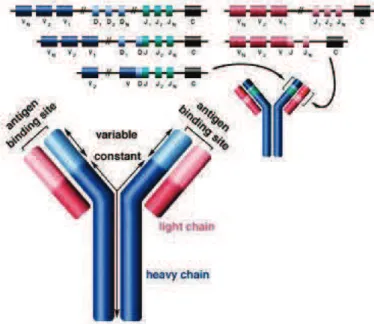
Fig. 2, VDJ recombination, an antibody is composed of two identical light and two identical heavy chains, and
the genes specifying them are found in the ‘V’ (Variable) region and the ‘C’ (Constant) region. In the heavy-chain ‘V’ region there are three segments; V, D and J, which recombine randomly, in a process called VDJ recombination, to produce a unique variable domain of each individual B cell. Similar rearrangements occur for light-chain ‘V’ region except there are only two segments involved; V and J.
Adopted from http://www.shaltech.com/about-lymphoma/
Fig. 3, Function TCR and BCR, the effector T Helper cells activate specific B cells through a phenomenon
known as an immunological synapse (BCR=B cell receptor, TCR =T cell receptor, IL = interleukin, and ILR = interleukin receptor). Activated B cells differentiate into plasma cells that subsequently produce antibodies which assist in clearing the host of the pathogen. CD4, CD40L are glycoproteins expressed on the surface of T helper cells, while CD40 is expressed on the surface of B cells and other anitgen presenting cells (APC).
27
1.2 Evolutionary•processes•affecting•evolution•of•immune•receptors•
Genetic diversity is important for survival and adaptability of species in changing environments. To decide which evolutionary forces shaped variation in specific genes is a difficult task because populations and species have to adapt to the abiotic environment (e.g. temperature, sunlight, pollution), to other species with which they interact (e.g. prey, predators, competitors, parasites) or more often to the combination of both. Moreover we should not underestimate neutral or non-adaptive evolutionary forces such as mutation, recombination and random genetic drift (Andrews 2010; Honnay 2013). Understanding processes which drive evolution of immune receptors is a fascinating research subject and big challenge.
According to the Red Queen hypothesis (the famous “running as fast as you can to stay in the same place”), firstly proposed by Leigh Van Valen (1973), organisms are running the arms race with other biological “partners” such as predators, source competitors at the intra- or interspecific levels or parasites. According to generally accepted theories the adaptation imposed by pathogens belongs among the most dynamic – continuous adaptive changes (Lederberg 1999; Zimmer 2001).
Parasites co-evolving with their hosts are viewed there as a key factor modulating different life traits of their hosts (e.g. population genetic structure, demographical changes, mating system, sexual dimorphism etc.) (Sheldon 1998). Generally microbes have an advantage during the arms race due to their shorter generation time and high mutation rates, which enhance genetic novelty and evolutionary potential, and so give better opportunity to update their invasion strategies tricking immune system of their hosts (Meyer 1991). Some bacteria generate surface proteins that bind to antibodies, rendering them ineffective; examples include Streptococcus pneumoniae (protein G), Staphylococcus aureus (protein A), and Peptostreptococcus magnus (protein L). Others are able to knock out or kill phagocytes. Some microbes can eventually mimic host cells and then block the interferon (IFN) production pathway or construct protective capsules (Mycobacterium) (Fortune et al. 2004). Another example is the ability of Mycobacterium leprae to suppress cell-mediated immunity or to play with immune system “hide-and-seek” inside host cells to avoid their detection (Maizels et al. 2004). Extracellular pathogens often alter their antigens (surface proteins), so we can imagine that it is like a thief who is escaping police by changing the coat and wig all the time and police is still one step behind (e.g. 84 known serotypes of Streptococcus
28
protein of Neisseria gonorrhoeae, or LPS from H. pylori and P. gingivalis) (Andersen-Nissen et al. 2005). The same strategy, i.e. antigenic variation and antigenic diversity, is used also by protozoan parasites (e.g. Plasmodium falciparum causing malaria) (Reeder and Brown 1996). Experts and prominent specialists in evasion strategies are viruses. Their short generation times and relatively high mutation rates give them huge advantage in red queen running with host defence mechanism and allows them quick adaptation to changing environment of their host. The best known example is influenza virus with constant replacement of its surface envelope proteins. Cytomegalovirus, another cunning invader, is able to evade the host defence system by expressing its MHC class I homologues thus pretending to be part of its body (Reyburn et al. 1997). Viruses can become also invisible for the immune system when they enter the latency state during which they are inactive (i.e. without replication). In this state, avirus particle does not cause a disease, but also does not produce any viral peptides which normally attract attention of different immune receptors. Viruses in the latent form can not be eliminated by the immune system and hence are sources of potential recurrent illnesses. Most famous for such a strategy are Herpes simplex viruses (Bowie and Unterholzner 2008). Another viral strategy is inhibition of immune response. Paramyxovirus for example can inhibit the type I IFN response which is included in the RIG-I signalling cascade. The most insolent viruses even use PRRs as entry ports (Yamada et al. 2005).
At first glance the only fair players seem to be parasitic worms, which evolve slower and therefore give a putative chance to the immune system to adapt. However, they are even bigger (literally) swindlers than their fast evolving cousins. During many millions of years of close coexistence with their hosts (parasites are often species specific) they have had time to evolve a sophisticated weapon arsenal designed to evade and modulate the host immune system (Wakelin 1996; Zaccone et al. 2006). Due to their generally bigger bodies it is very difficult for the immune system to eliminate them. There is a known pack of chameleons (e.g. coating with host proteins by schistosomes or filarial nematodes), the squad of chemical terrorists causing immunosuppression (e.g. hookworms producing a protein which binds the ß integrin CR3 and inhibits neutrophil extravasation or immunosuppression made by Burgia spp. or Nocardia brasiliensis) and also a group of nomads which avoids local inflammatory reactions by migrations through the host body (hookworms, Brugia spp., Wuchereria
bancrofti, or microfilariae of Onchocerca volvulus) (Pearce and Sher 1987; Wakelin 1996;
MacDonald et al. 2002). On the other hand long-term coevolution sometimes leads to a relatively stable relationship such as commensalism or mutualism. Moreover, according to the
29
hygiene hypothesis absolute elimination and subsequent absence of parasites can lead to autoimmune disorders (Zaccone et al. 2006).
As a consequence of co-evolution with microorganisms the immunity genes belong to the fastest evolving genes (Nielsen et al. 2005; Barreiro et al. 2009; Barreiro and Quintana-Murci 2010; Quach et al. 2013; Quintana-Quintana-Murci and Clark 2013). Since immune receptors are chief gatekeepers protecting against entrance of microbes, we suggest that antagonistic host-parasite interactions are the principal force shaping their evolution.
1.2.1 •Selection•imposed•by•pathogens:•adaptive•evolution•
The model which explains the evolution of immune receptors in the light of host-parasite co-evolution is called the matching alleles model (Frank 1993; Agrawal and Lively 2003). This model depicts the host-pathogen interaction as the process of reciprocal adaptive genetic change. In the context of microbe and pathogen it means receptors evolve to perfectly match the specific parasite structures (as the lock and key). In other words changes in gene frequencies resulting from selection acting on one population (species) create selective pressure for changes in gene frequencies in another population (species). This type of selection, called frequency-dependent selection, signifies that relative fitness of a genotype depends on its frequency (Carius 2001) (Fig. 4).
Frequency-dependent selection (FDS) can be positive or negative, Positive FDS favours the most numerous allele or genotype which thus increases its frequency and rapidly tends to fixation (Fig. 4c and d). Therefore, in essence the mode of this selection is directional and can be detected by important non-synonymous amino acid changes in different groups or lineages (Quintana-Murci and Clark 2013). Nevertheless positive FDS is less probable in the scope of immune genes.
In contrast, co-evolution determined by negative frequency dependent selection (NFDS) maintains high genetic diversity by favouring rare allelic variants (Takahata and Nei 1990; Stevens 2001) (Fig. 4a and b). In the context of the host-parasite interaction the mechanism of NFDS can be described as follows. The host immune system is adapted to tackle the most common parasite genotype and hence less common genotypes are favoured by natural selection. Rare gentoypes increase in frequency and subsequently become common therefore the cycle goes on (Fig. 5). NFDS is the type of balancing selection, which was already described in MHC genes (Takahata and Nei 1990; Bernatchez and Landry 2003;
30
Garrigan and Hedrick 2003; Aguilar et al. 2004; Bryja et al. 2006; Piertney and Oliver 2006; Smith et al. 2011). Balancing selection maintains genetic variation and leads to excess of polymorphism and excess of intermediate-frequency alleles. Besides NFDS, balancing selection can act as antagonistic and cyclic selection, selection in variable environment or through overdominance, i.e. heterozygote advantage where heterozygous genotypes confer higher fitness that homozygotes, for example, because they allow to recognize a wider variety of parasites (Doherty and Zinkernagel 1975).
Fig. 4, Frequency-dependent selection. (a) Negative frequency-dependent selection. The rarer a phenotype, the
higher its fitness. As a rare phenotype becomes more common, its fitness will decline, leading to a decrease in its frequency. (b) Thus, in negative frequency-dependent selection, the frequency of a phenotype will vary over time. (c) Positive frequency-dependent selection. The more common a phenotype is, the higher is its fitness. This means that over time, the more common phenotype is favored by selection and eventually becomes fixed in the population (d). Therefore, reduced variation is expected there. Schemes (a) and (c) represent simplification of relationships between fitness and genotype or fenotype, because these relationships are not neceserray linear. Adopted from Roy and Widmer (1999). The agreement for reuse of this picture in my disertation was provided by Elsevier using Copyright Clearance Center ("CCC"). Copyright © 2003, Elsevier
Except for various sorts of balancing selection typical Darwinian selection leads to loss of variation since harmful alleles are eliminated from the population by negative slection whereas beneficial alleles are fixed by positive selection. Both positive and negative selection can be detected in a genome on the basis of a decrease of polymorphism in the vicinity of a
31
selected locus. As the advantegous mutation is driven to fixation so are driven also tightly linked (“hitchhiking”) neutral or even slightly deleterious neighbouring DNA regions. Therefore, the presence of strong linkage disequilibrium can be used to identify sites which have recently been under selection. In addition, this process, called selective sweep, will result in overrepresentation of the region around the favoured locus in the population. In other words, besides apparent sequence homogeneity around the positively selected locus, a signature of a selective sweep is an excess of rare variants of this region in the population. A similar reduction of genetic variation can result from elimination of a harmful mutation by negative selection. The tighter the linkage with the counterselected locus, the greater the reduction in polymorphism. By contrast to the selective sweep, the effect of a harmful allele (called background selection) is not an excess of rare polymorphisms since such the allele causes one chromosome (or part of the chromosome) to merely drop out of the population (Hartl and Clark 1997). Genes affected by negative selection generally have very important functions. It was shown that this is a prevalent type of selection acting in the human genome (Quintana-Murci and Clark 2013). However, both positive and negative selections often act at the same time in different parts of the same genomic region, with varying strength and direction.
Fig. 5, Balancing selection through negative frequency dependent selection. Hosts resistant to parasite genotype
I are likely to be susceptible to parasite genotype II and vice versa. Since the parasite population evolution is closely associated with the evolution of its host’s population, the high frequency of parasite genotype I (left) will result in selection favouring hosts more resistant to this genotype (centre) yet susceptible to parasite genotype II. As a result, the latter genotype will prevail in the parasite population, which, in turn, will lead to selection in favour of the respective host resistance etc. Inspired by Freeman and Herron (2007).
We should keep in the mind that pathogen distribution and host specificity may vary in space and time (Hedrick 2002; Hedrick 2004). Local adaptation is a general phenomenon
32
found in most host-parasite relationships. That means that parasites in a particular area and time infect hosts from that area more efficiently than they infect hosts from another geographically distinct population. Local adaptation to pathogens (for example malaria) presented by different haplotype structure of TLR4 at distinct localities was shown for example in human populations from India, European-Americans and African-Americans (Mukherjee et al. 2009; Netea et al. 2012).
1.2.2 Stochastic•evolutionary•processes••
Although immunity genes are usually targeted by selection (Barreiro and Quintana-Murci 2010; Fumagalli et al. 2011; McTaggart et al. 2012), stochastic processes might also play an important role during their evolution (Grueber et al. 2013), however, their contribution is still difficult to assess. Selectively neutral evolutionary processes are brought about by random changes in a species´ gene pool that are neither advantageous nor disadvantageous for individual organisms nor they are connected to an increase or decrease of mean fitness of the respective population. Among principal stochastic mechanisms are neutral genetic drift (including bottlenecks and founder events) and neutral mutations (Futuyma 2005).
We can imagine genetic drift as a random process of gamete sampling. All genotypes have therefore the same chance to contribute to the next generation. The result of this random sampling is a shift in allele frequencies between subsequent generations. In most extreme situations one allele can either disappear from the gene pool or be fixed owing to genetic drift. Neutral mutations are mutations which do not affect the fitness of individuals. They are invisible for selection and therefore their fate depends on genetic drift. However we should note that the impact of genetic drift is not limited to neutral mutations. Due to genetic drift even advantageous mutations are eventually lost whereas some weakly deleterious mutations
may become fixed. This is often the case of small effective population sizes (Ne) where drift
can overcome the effect of selection (Nei and Tajima 1981). In such cases populations can pass through an adaptive valley towards higher adaptive peaks and thus follow a new evolutionary trajectory (Eyre-Walker and Keightley 2007). Such mechanism was described,
for example, in hominids which have Ne around 10,000 to 30,000 and about 30% of
non-synonymous mutations are effectively neutral while Drosophila which has Ne about 106, the