HAL Id: hal-01496179
https://hal.archives-ouvertes.fr/hal-01496179
Submitted on 7 May 2018
HAL is a multi-disciplinary open access
archive for the deposit and dissemination of
sci-entific research documents, whether they are
pub-lished or not. The documents may come from
teaching and research institutions in France or
abroad, or from public or private research centers.
L’archive ouverte pluridisciplinaire HAL, est
destinée au dépôt et à la diffusion de documents
scientifiques de niveau recherche, publiés ou non,
émanant des établissements d’enseignement et de
recherche français ou étrangers, des laboratoires
publics ou privés.
“Candidatus Bartonella rondoniensis” in Human Biting
Kissing Bugs (Reduviidae; Triatominae)
Maureen Laroche, Jean-Michel Berenger, Oleg Mediannikov, Didier Raoult,
Philippe Parola
To cite this version:
Maureen Laroche, Jean-Michel Berenger, Oleg Mediannikov, Didier Raoult, Philippe Parola.
Detec-tion of a Potential New Bartonella Species “Candidatus Bartonella rondoniensis” in Human Biting
Kissing Bugs (Reduviidae; Triatominae). PLoS Neglected Tropical Diseases, Public Library of Science,
2017, 11 (1), �10.1371/journal.pntd.0005297�. �hal-01496179�
RESEARCH ARTICLE
Detection of a Potential New Bartonella
Species “Candidatus Bartonella rondoniensis”
in Human Biting Kissing Bugs (Reduviidae;
Triatominae)
Maureen Laroche, Jean-Michel Berenger, Oleg Mediannikov, Didier Raoult,
Philippe Parola*
URMITE, Aix Marseille Universite´, UM63, CNRS 7278, IRD 198, INSERM 1095, IHU—Me´diterrane´e Infection, 19–21 Boulevard Jean Moulin, Marseille
*philippe.parola@univ-amu.fr
Abstract
Background
Among the Reduviidae family, triatomines are giant blood-sucking bugs. They are well
known in Central and South America where they transmit Trypanosoma cruzi to mammals,
including humans, through their feces. This parasitic protozoan is the causative agent of
Chagas disease, a major public health issue in endemic areas. Because of the medical and
economic impact of Chagas disease, the presence of other arthropod-borne pathogens in
triatomines was rarely investigated.
Methodology/Principal findings
In this study, seven triatomines species involved in the transmission of T. cruzi were
molecu-larly screened for the presence of known pathogens generally associated with arthropods,
such as Rickettsia, Bartonella, Anaplasmataceae, Borrelia species and Coxiella burnetii. Of
all included triatomine species, only Eratyrus mucronatus specimens tested positive for
Bar-tonella species for 56% of tested samples. A new genotype of BarBar-tonella spp. was detected
in 13/23 Eratyrus mucronatus specimens, an important vector of T. cruzi to humans. This
bacterium was further characterized by sequencing fragments of the ftsZ, gltA and rpoB
genes. Depending on the targeted gene, this agent shares 84% to 91% of identity with B.
bacilliformis, the agent of Carrion’s disease, a deadly sandfly-borne infectious disease
endemic in South America. It is also closely related to animal pathogens such as B. bovis
and B. chomelii.
Conclusions
As E. mucronatus is an invasive species that occasionally feeds on humans, the presence
of potentially pathogenic Bartonella-infected bugs could present another risk for human
health, along with the T. cruzi issue.
a1111111111
a1111111111
a1111111111
a1111111111
a1111111111
OPEN ACCESSCitation: Laroche M, Berenger J-M, Mediannikov
O, Raoult D, Parola P (2017) Detection of a Potential New Bartonella Species “Candidatus Bartonella rondoniensis” in Human Biting Kissing Bugs (Reduviidae; Triatominae). PLoS Negl Trop Dis 11(1): e0005297. doi:10.1371/journal. pntd.0005297
Editor: Ricardo E. Gu¨rtler, Universidad de Buenos
Aires, ARGENTINA
Received: September 8, 2016 Accepted: January 2, 2017 Published: January 17, 2017
Copyright:© 2017 Laroche et al. This is an open access article distributed under the terms of the
Creative Commons Attribution License, which permits unrestricted use, distribution, and reproduction in any medium, provided the original author and source are credited.
Data Availability Statement: All relevant data are
within the paper and its Supporting Information files.
Funding: The authors received no specific funding
for this work.
Competing Interests: The authors have declared
Author Summary
Triatomines are hematophagous insects including vectors of
T. cruzi, the agent of Chagas
disease, a huge public health issue, especially in South America. Whether these arthropods
carry other pathogenic microorganisms is currently unknown. We investigated the
pres-ence of different arthropod-borne pathogens, including
Bartonella spp., by quantitative
PCR.
Bartonella species were identified using ftsZ, gltA and rpoB gene sequencing and a
new genotype of
Bartonella spp. was detected in Eratyrus mucronatus specimens, an
important vector of
T. cruzi to humans. This agent is closely related to several human and
animal pathogens. Depending on the gene fragment used, this agent shares 84% to 91% of
identity with
B. bacilliformis, the agent of the deadly Carrion’s disease. The possibility of
transmission of potentially pathogenic bacteria could be an additional threat to human
health since
E. mucronatus bugs are more and more anthropophilic.
Introduction
Triatomine bugs (order Hemiptera, family Reduviidae, subfamily Triatominae) are
blood-sucking arthropods (“kissing bugs”), most of which can feed both on animals and humans. All
stages from first instar to male and female adults are strictly hematophagous and responsible
for a relatively large blood intake due to their large size. They are mainly sylvatic and feed on
small wild mammals but can also feed on birds and bats [
1
]. Triatomines occupy diverse
natu-ral ecotopes, such as mammal and bird nests, hollow trees, caves and rock fissures [
2
], but also
rural environments, as they can prosper in crevices in houses [
1
]. These arthropods are
distrib-uted world-wide but the vast majority of the 140 recognized species is found in the Americas
[
3
]. They are particularly well studied in South America, where they transmit an endemic
flag-ellate pathogen,
T. cruzi, the etiological agent of Chagas disease [
1
]. Also known as the
Ameri-can trypanosomiasis, Chagas disease is a neglected tropical disease, the first human parasitic
disease in the endemic areas.
T. cruzi is transmitted through the feces of infected kissing bugs,
causing heart failure 10 to 30 years post-infection for almost 30% of individuals [
4
].
Because of the public health impact of Chagas disease, studies on kissing bugs are mainly
focused on this theme. As a matter of fact, the presence of other human pathogens was never
described in the hundred years that it has been known that kissing bugs could transmit
patho-gens. Only the presence of
Wolbachia and Arsenophonus species was investigated based on the
fact that these obligate intracellular bacteria are known to be endosymbionts of many
arthro-pods [
5
,
6
]. The presence of zoopathogenic arthropod-borne viruses was also investigated. To
our knowledge, there is no report of pathogen detection in dejections or in triatomines
them-selves, except for
T. cruzi, although Arsenophonus nasoniae was once reported to be detected in
an eschar of a human [
7
]. Regarding viruses, two have been described in these bugs.
Triatoma
virus is reported as strictly entomopathogenic, particularly for its principal host, Triatoma
infestans [
8
], while
African swine fever virus was detected in Triatoma gerstaeckeri but not
transmitted to pigs [
9
].
French Guiana is an 84,000 km
2overseas department and region of France bordered by
Brazil and Suriname. Because of its many different ecosystems, particularly a dense rainforest,
French Guiana is a biodiversity hotspot and one of the 21 areas where Chagas disease is
endemic [
10
]. Among the 27 described species of triatomines in this area, many are invasive
species. That is to say that many of them temporarily leave their sylvatic or peridomestic
dwell-ings in order to invade houses. The main vector community of French Guiana comprises
highly anthropophilic bugs belonging to the
Panstrongylus, Rhodnius and Eratyrus genera [
10
].
They accidentally feed on humans [
11
] and also on potentially infected animals since they
eas-ily feed on domestic animals or wild mammals.
Aiming to add to knowledge regarding bacteria and triatomine association, we screened
seven species of triatomines bugs from French Guiana by molecular biology for the presence
of arthropod-borne bacteria such as
Rickettsia, Bartonella, Borrelia, Anaplasma, Wolbachia,
Ehrlichia species and Coxiella burnetii.
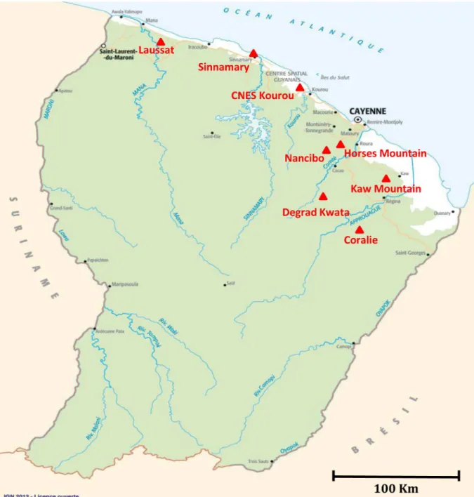
Fig 1. Distribution of sampling areas in French Guiana. Exact sampling sites are indicated by a triangle.
doi:10.1371/journal.pntd.0005297.g001
Methods
Triatomine collection, identification and selection
Triatomine specimens were collected in French Guiana from 1991 to 2013 using light traps
or interception traps by one of the authors (JMB) and by the Socie´te´ Entomologique
Antil-les-Guyane (SEAG) as part of an inventory of French Guiana’s insects. Triatomines were
caught in forests (Horses Mountains, Kaw Mountains), peridomiciliary areas (Degrad
Kwata, Kaw Mountains, Nancibo) or urban areas (Sinnamary, Kourou savannah) as
dis-played in
Fig 1
.
All triatomine specimens were morphological identified with the Be´renger et al.
morpho-logical key [
12
] and kept dried as insect collections. Seven
T.cruzi vectors were included in this
study:
Rhodnius prolixus (n = 10), Rh. pictipes (n = 10), Rh. robustus (n = 10), Triatoma
infes-tans (n = 10), Panstrongylus geniculatus (n = 10), P. rufotuberculatus (n = 4), Eratyrus
mucrona-tus (n = 23).
DNA extraction
Dried triatomines were rinsed in sterile water and air-dried on filter paper before cutting
lengthwise in two equal halves, using a sterile surgical blade for each specimen. One half and
the legs were stored at -20˚C as a backup sample and the other legless half was selected for
molecular analyses. Each half triatomine was crushed with a sterile pestle in 400
μL of a G2
buffer solution containing 40
μM of proteinase K (Qiagen) and incubated at 56˚C overnight.
After 1 minute of centrifugation at 7000 x g, 200
μL of the supernatant was then collected prior
to DNA extraction.
Triatominae genomic DNA was individually extracted using the EZ1 DNA
tissue extraction kit (Qiagen, Hilden, Germany) according to the manufacturer’s instructions.
Triatominae DNAs were then eluted in 100 μL of Tris EDTA (TE) buffer using the DNA
extracting EZ1 Advanced XL Robot (Qiagen) as previously described [
13
]. DNAs were either
immediately used or stored at -20˚C until used for molecular analysis. The DNA extracting
EZI Advanced XL Robot was disinfected after each batch of extraction as per the manufacturer
recommendations to avoid cross-contamination.
Table 1. Sequences of qPCR primers used to investigate the presence of pathogens’ DNA in the E. mucronatus samples. F: forward primer, R: reverse primer, P: qPCR probe.
Target organism Target gene Primer’s name Sequence (5’-3’)
Bartonella spp. Intergenic spacer IT2_F GGGGCCGTAGCTCAGCTG
ITS2_R TGAATATATCTTCTCTTCACAATTTC
ITS2_P 6FAM-CGATCCCGTCCGGCTCCACCA
Rickettsia spp. gltA gltA_F GTGAATGAAAGATTACACTATTTAT
gltA_R GTATCTTAGCAATCATTCTAATAGC
gltA_P 6FAM-CTATTATGCTTGCGGCTGTCGGTTC
Coxiella burnetii IS30A ITS30A_F CGCTGACCTACAGAAATATGTCC
ITS30A_R GGGGTAAGTAAATAATACCTTCTGG ITS30A_P 6FAM-CATGAAGCGATTTATCAATACGTGTATGC Borrelia spp. 16S 16S_F AGCCTTTAAAGCTTCGCTTGTAG 16S_R GCCTCCCGTAGGAGTCTGG 16S_P 6FAM- CCGGCCTGAGAGGGTGAACGG Anaplasmataceae 23S 23S_F TGACAGCGTACCTTTTGCAT 23S_R GTAACAGGTTCGGTCCTCCA 23S_P 6FAM- GGATTAGACCCGAAACCAAG doi:10.1371/journal.pntd.0005297.t001
Molecular analysis
DNA samples were individually tested by genus-specific PCR using primers and probes
target-ing specific sequences of
Bartonella spp., but also Rickettsia spp., Coxiella burnetii, Borrelia
spp., and all Anaplasmataceae species [
14
] as previously described [
15
] (
Table 1
). Real-time
quantitative PCR (qPCR) was carried out according to the manufacturer’s protocol using a
CFX Connect Real-Time PCR Detection System (Bio-rad, Hercules, CA, USA) with the
Euro-gentec Takyon qPCR kit (EuroEuro-gentec, Seraing, Belgium).
Bartonella elizabethae, Rickettsia montanensis, Coxiella burnetii, Anaplasma
phagocyto-philum and Borrelia crocidurae DNAs were used as positive qPCR controls for the primers
and probe targeting respectively all
Bartonella, Rickettsia, Coxiella burnetii and Borrelia
species.
DNAs were tested at different concentrations to avoid PCR inhibition. For each run, a PCR
mix without DNA was used as negative control. Standard PCR targeting a 710 bp region of the
invertebrate
cytochrome oxidase I (COI) gene was performed on PCR negative triatomines to
control DNA extraction.
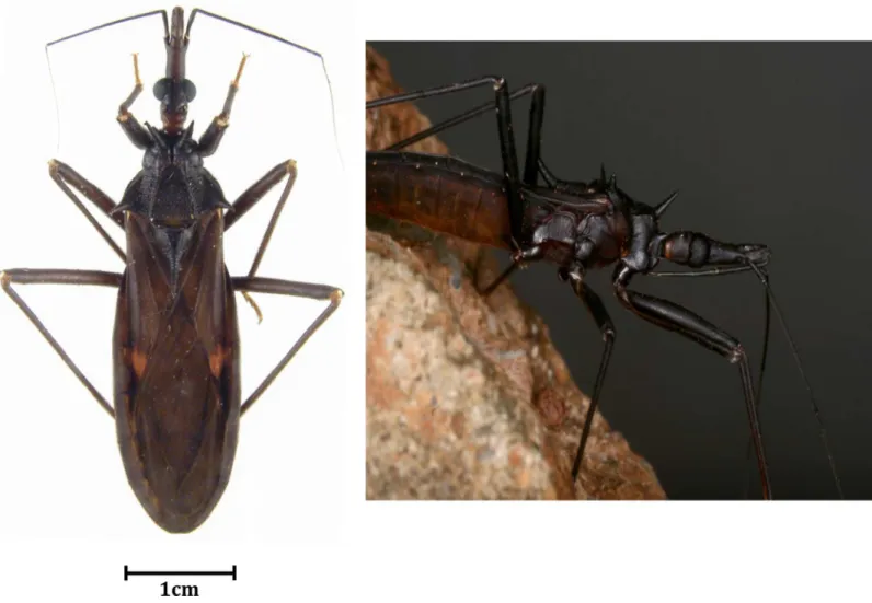
Fig 2. Pictures of alive and dead Eratyrus mucronatus in its environment and dried on paper.
doi:10.1371/journal.pntd.0005297.g002
Sequencing and GenBank accession numbers
DNA samples that were positive with
Bartonella-qPCR were submitted to conventional PCR
amplification using a Bio-Rad Thermocycler (Bio-Rad Laboratories, Hercules, CA) prior to
sequencing. For
Bartonella species identification, primers targeting Bartonella rpoB, gltA and ftsZ
genes fragments were used as previously described [
16
]. DNA from
Bartonella elizabethae served
as PCR positive control and mixture without DNA as negative control. The cycling protocol
con-sisted of 15 min at 95˚C followed by 35 cycles of denaturing at 95˚C for 30 s, annealing at 50˚C
for 30 s (58˚C for
rpoB gene), extension 1 min at 72˚C, followed by a final cycle of 1 min at 72˚C
and sampling held at 4˚C. Amplification products were separated by electrophoresis through a
1.5% agarose-tris-borate-EDTA gel containing ethidium bromide. PCR products were sequenced
using a Big Dye Terminator kit and an ABI PRISM 3130 Genetic Analyser (Applied BioSystems,
Courtabeauf, France). The sequences were analyzed using the ABI PRISM DNA Sequencing
Analysis software version 3.0 (Applied BioSystems) and compared to sequences available in the
GenBank database using the BLAST algorithm (
http://blast.ncbi.nlm.nih.gov/Blast.cgi
). The
par-tial sequences of
ftsZ and rpoB genes of Bartonella amplified from the sample EmG01 are available
in GenBank at #KX377404 and #KX377405.
Phylogenic analysis
Phylogeny of the detected
Bartonella with other members of the Bartonella genus was
estab-lished with TOPALi 2.5 software (Biomathematics and Statistics Scotland, Edinburgh, UK).
Table 2. Details of Eratyrus mucronatus collect from 1991 to 2013 in French Guiana.1,2,3,4,5,6,7: different areas of French Guiana. SEAG: Socie´te´ Ento-mologique Antilles-Guyane (Entomological Society for French West-Indies and Guiana). JMB: Jean-Michel Be´renger. BH: Bernard Hermier. CNES Kourou: Centre National d’Etudes Spatiales (National Center for Spatial Studies).
Eratyrus mucronatus specimens Sex Date of collection Location Area type Sampling person Ct values
EmG01 M 1998 Kaw Mountains Primary forest—peridomestic JMB 20.20 EmG02 M 1997 Kaw Montains1 Primary forest—peridomestic JMB 29.64
EmG03 M 1998 Kaw Mountains Primary forest—peridomestic JMB 28.56
EmG04 M 1995 Nancibo2 Rural- peridomestic BH Neg
EmG05 M 2010 Laussat3 SEAG 21.38
EmG06 M 2013 Horses Mountains4 Sylvatic SEAG Neg
EmG07 M 2013 Horses Mountains5 Sylvatic SEAG Neg
EmG08 M 2013 Horses Mountains5 Sylvatic SEAG 19.91
EmG09 M 2013 Horses Mountains5 Sylvatic SEAG 23.41
EmG10 M 2013 Horses Mountains5 Sylvatic SEAG Neg
EmG11 M 2013 Horses Mountains5 Sylvatic SEAG 24.78
EmG12 F 1993 Sinnamary6 Urban—peridomestic JMB Neg
EmG13 M 2013 Horses Mountains5 Sylvatic SEAG 13.98
EmG14 M 2013 Horses Mountains5 Sylvatic SEAG 13.23
EmG15 M 1995 Degrad Kwata7 Sylvatic–primary forest JMB 20.12
EmG16 M 1998 Kaw Mountains Primary forest—peridomestic JMB 19.69 EmG17 M 1998 Kaw Mountains Primary forest—peridomestic JMB 24.47 EmG18 M 1996 Kaw Montains1 Primary forest—peridomestic JMB Neg
EmG19 M 1996 Kaw Montains1 Primary forest—peridomestic JMB Neg
EmG20 M 2003 CNES Kourou Savannah JMB Neg
EmG21 M 2003 CNES Kourou Savannah JMB 25.91
EmG22 M 1991 Coralie8 Sylvatic–secondary forest JMB Neg
EmG23 M 1993 Kaw Montains1 Primary forest—peridomestic JMB Neg
Available sequences of
ftsZ, gltA and rpoB genes of validated Bartonella species were retrieved
from the National Center for Biotechnology Information (NCBI) based on the results of the
BLAST program. Multiple sequence alignment was performed with the ClustalW multiple
sequence alignment program, which is included in the BioEdit software.
Results
Triatominae collection
Triatomines were collected in eight different localities in French Guiana with no selection
regarding species and sex (convenient sampling). Among the triatomines collected,
Eratyrus
mucronatus (
Fig 2
) accounted for 20% of catches and 29.8% of the specimens analyzed. Details
related to collection, such as sampling area and triatomines’ sex, are indicated in
Table 2
. Of 23
E. mucronatus samples, 22 (95.6%) were male. Further details regarding other collected species
have been listed elsewhere [
12
].
Molecular detection
DNAs extracted from all the triatomines were included to assess the presence of
Bartonella
spe-cies. Of 23
Eratyrus mucronatus samples, 13 (56.5%) were positive by Bartonella spp.-specific
qPCR, with cycle threshold (Ct) values ranging from 13.23 to 25.91 (mean: 21.44) (
Table 2
).
These specimens were from six distinct regions and collected between 1993 and 2003. All
posi-tive specimens were male, and a majority of them were collected in sylvatic and peridomestic
areas: the Kaw Mountains (38.4%) and Horses Mountains (38.4%).
All samples tested negative for the presence of
Rickettsia spp., Borrelia spp. Anaplasma spp.,
Ehrlichia spp., Wolbachia spp. and Coxiella burnetii. Bartonella spp. was only detected in
Era-tyrus mucronatus specimens.
Sequencing
A 787 bp fragment of the
Bartonella spp. rpoB gene was amplified using conventional PCR
primers prior to sequencing. Only three
ITS2-qPCR positive samples were also positive for
rpoB by standard PCR. Sequencing failed for two of them. Comparison of the one rpoB
result-ing sequence against the NCBI database usresult-ing the BLASTN program indicated that it
pos-sessed 90% identity with the ATCC
Bartonella bacilliformis 35685D-5 strain (#CP014012.1)
and with the
B. bacilliformis KC583 strain (#CP000524.1). The next closest cultivated strains
were a
B. bovis strain [
17
] and a
B. chomelii strain [
18
], both with 89% identity. Our genotype
also possesses 87% identity with a
Bartonella ancashensis strain [
19
,
20
]. Available sequences of
Bartonella rpoB gene were retrieved from NCBI and compared to the Bartonella sequence
described hereby. This
Bartonella genotype formed a distinct clade, with a strain of Bartonella
bacilliformis as the closest clade based on rpoB gene analysis (
Fig 3
).
All samples allowed amplification of a single 333 bp fragment of the
ftsZ gene by standard
PCR. Blast analysis revealed 91% identity with the aforementioned 35685D-5 and KC583
B.
bacilliformis strains (
Fig 4
).
Fig 3. A consensus phylogenetic tree showing the relationships of the studied species of Bartonella species based on a portion of rpoB gene sequence comparison. GenBank accession numbers (or the only genome accession number) are indicated when the sequences initially originated
from Genbank. The sequences were aligned using ClustalW, and phylogenetic inferences were obtained using Bayesian phylogenetic analysis with TOPALi 2.5 software (Biomathematics and Statistics Scotland, Edinburgh, UK) within the integrated Maximum Likelihood application using the TrN + I +Г
model. Numbers at the nodes are percentages of bootstrap values obtained by repeating the analysis 100 times to generate a majority consensus tree. Bootstrap values below 80 were deleted from the final tree. The final set includes 756 base pairs. The new Bartonella sequence described in the present study is written in red.
A total of 12 out of 13 samples were successfully amplified by standard PCR targeting a
frag-ment of the
gltA gene. BLAST analysis showed 88% identity with uncultured Bartonella species
detected in bank voles [
21
], deer [
22
] and bats from Africa [
23
], but also with
B. bovis and B.
chomelii strains (
Fig 5
). Based on the
gltA gene, our genotype is 84% similar to B. bacilliformis
strains. Only a single
rpoB sequence was obtained but all ftsZ and gltA sequences obtained
were identical for all
E. mucronatus specimens.
BLAST analysis of the concatened sequence of the three genes revealed 99% of coverage
and 90% similarity with the two aforementioned
B. bacilliformis strains. Phylogenetic analysis
based on the concatened sequences revealed clustering of our
Bartonella strain with two B.
bacilliformis and B. ancashensis strains (
Fig 6
).
Discussion
Bartonella species are small fastidious gram-negative bacteria belonging to the
Alphaproteobac-teria class that are able to infect many mammals, including humans [
24
]. They are mostly
transmitted by arthropod vectors such as sandflies (
Lutzomyia verrucarum), human body lice
(
Pediculus humanus humanus), different fleas including cat fleas (Ctenocephalides felis), biting
flies and ticks [
25
]. Among the several
Bartonella species, some have been identified as human
pathogens, causing well-known vector-borne diseases such as Carrion’s disease (
B.
bacillifor-mis), trench fever (B. quintana), cat scratch disease (B. henselae) as well as endocarditis [
24
].
We hereby describe a novel
Bartonella genotype, phylogenetically related to several human
and animal pathogens, as shown by the phylogenetic analyses.
B. bacilliformis, a closely related
species, is the causative agent of the first and well-described human bartonellosis called Carrion’s
disease [
26
]. Transmitted through the bite of an infected phlebotomine sand fly,
L. verrucarum,
this South American endemic bacterium can induce a biphasic illness with two distinct
syn-dromes that can be concomitant or independent. An acute phase known as Oroya fever
mani-fests as a hemolytic fever linked to bacteremia that can range from 10 to 210 days and can be
fatal in 40–88% of individuals without treatment. The second syndrome called verruga peruana
manifests as blood-filled hemangiomas due to infection of the endothelium [
26
]. No human
cases of
B. bacilliformis infection have been reported in French Guiana to date [
27
]. Our
geno-type is also closely related to a strain of
B. ancashensis, a recently described Bartonella species
closely related to
B. bacilliformis that was isolated from the blood of two patients diagnosed with
a chronic stage of verruga peruana in Peru [
20
]. All data suggest that
B. ancashensis could be a
second agent. Our new agent is also closely related to
B. bovis strains. Isolated from cats, which
are only accidental hosts, this endocarditis [
28
].
E. mucronatus is a sylvatic triatominae bug involved in the transmission of T. cruzi [
11
]. It
is recognized now as an invasive species as it has been described around and inside houses
since 1959 [
29
] because of its attraction to artificial light sources [
30
]. They are known to feed
on bats, but also on small mammals such as xenarthrans and opossums [
11
]. Bats are widely
reported to be sources of many viral and bacterial pathogens [
31
], including
Bartonella spp.
worldwide, including in French Guiana [
32
], Nigeria [
33
], Guatemala [
34
] and Vietnam, for
Fig 4. A consensus phylogenetic tree showing the relationships of the Bartonella species studied based on a portion of ftsZ gene sequence comparison. GenBank accession numbers (or the only genome accession number) are indicated
when the sequences originated from Genbank at the beginning. The sequences were aligned using ClustalW, and phylogenetic inferences were obtained using Bayesian phylogenetic analysis with TOPALi 2.5 software (Biomathematics and Statistics Scotland, Edinburgh, UK) within the integrated Maximum Likelihood application using the ML SYM+I+Гmodel. Numbers at the nodes are percentages of bootstrap values obtained by repeating the analysis 100 times to generate a majority consensus tree. Bootstrap values below 80 were deleted from the final tree. The final set includes 292 base pairs. The new Bartonella sequence described in this study is written in red.
Fig 5. A consensus phylogenetic tree showing the relationships of the Bartonella species studied based on a portion of gltA gene sequence comparison. GenBank accession numbers (or the only genome accession number) are indicated when the sequences originated from
Genbank at the beginning. The sequences were aligned using ClustalW, and phylogenetic inferences were obtained using Bayesian phylogenetic analysis with TOPALi 2.5 software (Biomathematics and Statistics Scotland, Edinburgh, UK) within the integrated Maximum Likelihood application using the K81uf + I +Гmodel. Numbers at the nodes are percentages of bootstrap values obtained by repeating the analysis 100 times to generate a majority consensus tree. Bootstrap values below 80 were deleted from the final tree. The final set includes 200 base pairs. The new Bartonella sequence described in this study is written in red.
doi:10.1371/journal.pntd.0005297.g005
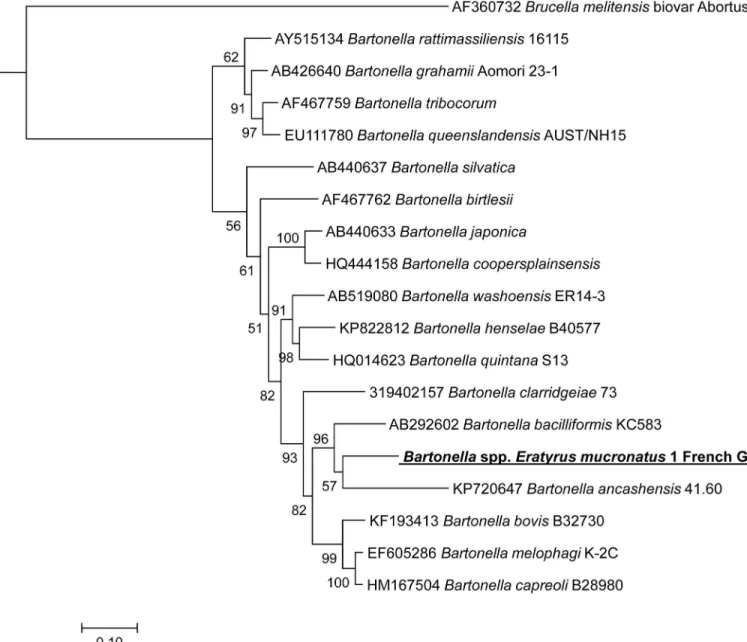
Fig 6. A consensus concatened phylogenetic tree showing the relationships of the Bartonella species studied based on a concatened sequence of Bartonella rpoB, ftsZ and gltA gene fragment. Concatenated rpoB, ftsZ and gltA sequences were aligned using CLUSTALW and
phylogenetic inferences obtained using Bayesian phylogenetic analysis [Ronquist F, Huelsenbeck JP. MrBayes 3: Bayesian phylogenetic inference under mixed models. Bio-informatics 2003; 19:1572–1574] with the TOPALi 2.5 software (Biomathematics and Statistics Scotland, Edinburgh, UK) with the integrated MrBayes application [ttp://mrbayes.csit.fsu.edu] with the HKY+I+Гsubstitution model. GenBank accession numbers are indicated at the beginning. Numbers at the nodes are bootstrap values obtained by repeating the analysis 100 times to generate a majority consensus tree. There were a total of 1245 positions in the final dataset. The scale bar indicates a 10% nucleotide sequence divergence.
example [
35
]. Therefore,
Bartonella spp. were frequently detected in hematophagous
arthro-pods feeding on bats such as bat flies (
Hippoboscidae, Streblidae, Nycteribiidae) [
36
] or
Cimex
adjunctus [
37
]. Triatomine vectors belonging to genera
Triatoma, Rhodnius, Panstrongylus
and
Eratyrus can be totally domiciliated or invasive, since they occasionally visit houses as
described in Bolivia [
38
], Brazil [
39
], Argentina [
40
] and Venezuela [
11
]. The presence of these
bugs around houses has long been known and has justified the establishment of chemical
con-trol campaigns, which after 5 years remain a failure in Bolivia [
38
]. The invasive behavior of
E.
mucronatus has not yet been described in French Guiana but data from Bolivia suggest that
eliminating it once it is settled is challenging [
38
]. Living in various ecotopes and not
host-spe-cific [
41
], these bugs can easily feed both on humans and animals [
38
], both of them potentially
bacteriemic, parasitemic or viremic at the blood meal time point.
Triatominae species are well-studied bugs, however, this work provides the first evidence to
our knowledge of infection with a bacterium that is not
a priori endosymbiotic. The specimens
we analyzed were dry, with no information regarding their engorgement status at the time of
collection. However, as they were collected using light traps or interception traps, we can
assume that they were seeking hosts and therefore probably non-engorged. Thus, we can
sup-pose that we did not detect DNA of a bacterium present in recently ingested blood but a
genu-ine infection. To support this hypothesis, the infection rate was considerable (56%) among
triatomines collected in very distant sampling periods, geographically and over time. Ct values
were also very low, increasing the possibilities that this bacterium multiplies within the bug’s
body. However, to evaluate the possibility of transmission of these
Bartonella spp., an
experi-mental model of infection, or at least information regarding the location of the bacteria in the
bug, would be necessary. Such information could not be obtained from our samples as they
were dry and old. Cultivation of any bacteria or any attempt to localize with
immunofluores-cence, for example, was not possible.
In continuing this work, it would be interesting to collect wild
E. mucronatus specimens in
order to isolate the bacterium and establish an experimental model of infection with this
arthropod/pathogen pair. This would reveal whether the bug is a simple carrier or an efficient
vector of this
Bartonella. The possible interaction between T. cruzi and this Bartonella spp. in
this insect is also unknown and could be investigated by monitoring the trypanosome’s cycle
and transmission in co-infected
E. mucronatus. Being phylogenetically closely related to two
severe human pathogens (
B. bacilliformis and B. ancashensis), it would also be important to
evaluate its pathogenicity. Because of the huge public health impact of Chagas disease in South
America, investigations on
Triatominae were limited to the study of their interactions with T.
cruzi. In fact, Triatominae bugs may host such bacteria as Bartonella species and, probably,
may be its vector.
Acknowledgments
We would like to thank the SEAG (Socie´te´ Entomologique Antilles Guyane) and Bernard
Her-mier for their contribution to the sample collection.
Author Contributions
Conceptualization: ML JMB PP.
Formal analysis: ML OM.
Investigation: ML JMB.
Methodology: ML JMB.
Resources: JMB PP.
Supervision: PP.
Validation: ML.
Writing – original draft: ML JMB.
Writing – review & editing: ML JMB PP OM DR.
References
1. Lazzari CR, Pereira MH, Lorenzo MG, Lazzari CR, Pereira MH, Lorenzo MG. Behavioural biology of Chagas disease vectors. Mem Inst Oswaldo Cruz. 2013; 108: 34–47. doi:10.1590/0074-0276130409 PMID:24473801
2. Monte GLS, Tadei WP, Farias TM. Ecoepidemiology and biology of Eratyrus mucronatus Stål, 1859 (Hemiptera: Reduviidae: Triatominae), a sylvatic vector of Chagas disease in the Brazilian Amazon. Rev Soc Bras Med Trop. 2014; 47: 723–727. doi:10.1590/0037-8682-0263-2014PMID:25626651
3. Schofield CJ, Galvão C. Classification, evolution, and species groups within the Triatominae. Acta Trop. 2009; 110: 88–100. PMID:19385053
4. Longo DL, Bern C. Chagas’ Disease. N Engl J Med. 2015; 373: 456–466. doi:10.1056/ NEJMra1410150PMID:26222561
5. Espino CI, Go´mez T, Gonza´lez G, do Santos MFB, Solano J, Sousa O, et al. Detection of Wolbachia bacteria in multiple organs and feces of the triatomine insect Rhodnius pallescens (Hemiptera, Reduvii-dae). Appl Environ Microbiol. 2009; 75: 547–550. doi:10.1128/AEM.01665-08PMID:19028913
6. Sorfova´ P, Skerı´kova´ A, Hypsa V. An effect of 16S rRNA intercistronic variability on coevolutionary anal-ysis in symbiotic bacteria: molecular phylogeny of Arsenophonus triatominarum. Syst Appl Microbiol. 2008; 31: 88–100. doi:10.1016/j.syapm.2008.02.004PMID:18485654
7. Edouard S, Subramanian G, Lefevre B, Santos Dos A, Pouedras P, Poinsignon Y, et al. Co-infection with Arsenophonus nasoniae and Orientia tsutsugamushi in a traveler. Vector Borne Zoonotic Dis. 2013; 13: 565–571. doi:10.1089/vbz.2012.1083PMID:23930974
8. Rozas-Dennis GS, Cazzaniga NJ. Effects of Triatoma virus (TrV) on the fecundity and moulting of Tria-toma infestans (Hemiptera: Reduviidae). Ann Trop Med Parasitol. 2000 Sep; 94(6):633–41. PMID: 11064765
9. Hess WR, Endris RG, Haslett TM, Monahan MJ, McCoy JP. Potential arthropod vectors of African swine fever virus in North America and the Caribbean basin. Vet Parasitol. 1987; 26: 145–155. PMID: 3326244
10. Pe´neau J, Nguyen A, Flores-Ferrer A, Blanchet D, Gourbière S. Amazonian triatomine biodiversity and the transmission of Chagas Disease in French Guiana: In Medio Stat Sanitas. PLoS Negl Trop Dis. 2016; 10: e0004427. doi:10.1371/journal.pntd.0004427PMID:26867025
11. Carrasco HJ, Segovia M, Londoño JC, Ortegoza J, Rodrı´guez M, Martı´nez CE. Panstrongylus genicula-tus and four other species of triatomine bug involved in the T. cruzi enzootic cycle: high risk factors for Chagas’ disease transmission in the Metropolitan District of Caracas, Venezuela. Parasit Vectors. 2014; 7: 602. doi:10.1186/s13071-014-0602-7PMID:25532708
12. Berenger J-M, Pluot-Sigwalt D, Page´s F, Blanchet D, Aznar C. The triatominae species of French Gui-ana (Heteroptera: Reduviidae). Mem Inst Oswaldo Cruz. 2009; 104: 1111–1116. PMID:20140371
13. Yssouf A, Almeras L, Terras J, Socolovschi C, Raoult D, Parola P. Detection of Rickettsia spp in ticks by MALDI-TOF MS. PLoS Negl Trop Dis. 2015; 9: e0003473. doi:10.1371/journal.pntd.0003473PMID: 25659152
14. Dahmani M, Davoust B, Benterki MS, Fenollar F, Raoult D, Mediannikov O. Development of a new PCR-based assay to detect Anaplasmataceae and the first report of Anaplasma phagocytophilum and Anaplasma platys in cattle from Algeria. Comp Immunol Microbiol Infect Dis. 2015; 39: 39–45. doi:10. 1016/j.cimid.2015.02.002PMID:25748051
15. Bessas A, Leulmi H, Bitam I, Zaidi S, Ait-Oudhia K, Raoult D, et al. Molecular evidence of vector-borne pathogens in dogs and cats and their ectoparasites in Algiers, Algeria. Comp Immunol Microbiol Infect Dis. 2016; 45: 23–28. doi:10.1016/j.cimid.2016.01.002PMID:27012917
16. Scola BL, Zeaiter Z, Khamis A, Raoult D. Gene-sequence-based criteria for species definition in bacteri-ology: the Bartonella paradigm. Trends in Microbiology. 2003; 11: 318–321. PMID:12875815
17. Bai Y, Malania L, Alvarez Castillo D, Moran D, Boonmar S, Chanlun A, et al. Global distribution of Barto-nella infections in domestic bovine and characterization of BartoBarto-nella bovis strains using multi-locus sequence typing. PLoS ONE. 2013; 8: e80894. doi:10.1371/journal.pone.0080894PMID:24278342
18. Antequera-Go´mez ML, Lozano-Almendral L, Barandika JF, Gonza´lez-Martı´n-Niño RM, Rodrı´guez-Moreno I, Garcı´a-Pe´rez AL, et al. Bartonella chomelii is the most frequent species infecting cattle graz-ing in communal mountain pastures in Spain. Appl Environ Microbiol. 2015; 81: 623–629. doi:10.1128/ AEM.03159-14PMID:25381240
19. Hang J, Mullins KE, Clifford RJ, Onmus-Leone F, Yang Y, Jiang J, et al. Complete Genome Sequence of Bartonella ancashensis Strain 20.00, isolated from the blood of a patient with Verruga Peruana. Genome Announc. 2015; 3: e01217–15. doi:10.1128/genomeA.01217-15PMID:26543106
20. Mullins KE, Hang J, Jiang J, Leguia M, Kasper MR, Ventosilla P, et al. Description of Bartonella anca-shensis sp. nov., isolated from the blood of two patients with verruga peruana. International Journal of Systematic and Evolutionary Microbiology. 2015; 65: 3339–3343. doi:10.1099/ijsem.0.000416PMID: 26296673
21. Buffet J-P, Marsot M, Vaumourin E, Gasqui P, Masse´glia S, Marcheteau E, et al. Co-infection of Borre-lia afzelii and Bartonella spp. in bank voles from a suburban forest. Comp Immunol Microbiol Infect Dis. 2012; 35: 583–589. doi:10.1016/j.cimid.2012.07.002PMID:22898354
22. Sato S, Kabeya H, Yamazaki M, Takeno S, Suzuki K, Kobayashi S, et al. Prevalence and genetic diver-sity of Bartonella species in sika deer (Cervus nippon) in Japan. Comp Immunol Microbiol Infect Dis. 2012; 35: 575–581. doi:10.1016/j.cimid.2012.07.001PMID:22832020
23. Dietrich M, Tjale MA, Weyer J, Kearney T, Seamark ECJ, Nel LH, et al. Diversity of Bartonella and Rick-ettsia spp. in bats and their blood-feeding ectoparasites from South Africa and Swaziland. PLoS ONE. 2016; 11: e0152077. doi:10.1371/journal.pone.0152077PMID:26999518
24. Regier Y, O’Rourke F, Kempf VAJ. Bartonella spp.—a chance to establish One Health concepts in vet-erinary and human medicine. Parasit Vectors. 2016; 9: 1.
25. Tsai Y-L, Chang C-C, Chuang S-T, Chomel BB. Bartonella species and their ectoparasites: selective host adaptation or strain selection between the vector and the mammalian host? Comp Immunol Micro-biol Infect Dis. 2011; 34: 299–314. doi:10.1016/j.cimid.2011.04.005PMID:21616536
26. Minnick MF, Anderson BE, Lima A, Battisti JM, Lawyer PG, Birtles RJ. Oroya fever and verruga per-uana: bartonelloses unique to South America. PLoS Negl Trop Dis. 2014; 8: e2919. doi:10.1371/ journal.pntd.0002919PMID:25032975
27. Sanchez Clemente N, Ugarte-Gil CA, Solo´rzano N, Maguiña C, Pachas P, Blazes D, et al. Bartonella bacilliformis: a systematic review of the literature to guide the research agenda for elimination. PLoS Negl Trop Dis. 2012; 6: e1819. doi:10.1371/journal.pntd.0001819PMID:23145188
28. Maillard R, Petit E, Chomel B, Lacroux C, Schelcher F, Vayssier-Taussat M, et al. Endocarditis in cattle caused by Bartonella bovis. Emerging Infect Dis. 2007; 13: 1383–1385. doi:10.3201/eid1309.070236 PMID:18252116
29. Vivas AS, Barazarte H, Molina-de-Ferna´ndez D. Primer registro de Eratyrus mucronatus. Stal; 1959.
30. Castro MCM, Barrett TV, Santos WS, Abad-Franch F, Rafael JA. Attraction of Chagas disease vectors (Triatominae) to artificial light sources in the canopy of primary Amazon rainforest. Mem Inst Oswaldo Cruz. 2010; 105: 1061–1064. PMID:21225207
31. Mu¨hldorfer K. Bats and Bacterial Pathogens: A Review. Zoonoses and Public Health. Blackwell Publish-ing Ltd; 2013; 60: 93–103. doi:10.1111/j.1863-2378.2012.01536.xPMID:22862791
32. Davoust B, Marie´ J-L, Dahmani M, Berenger J-M, Bompar J-M, Blanchet D, et al. Evidence of Bartonella spp. in blood and ticks (Ornithodoros hasei) of bats, in French Guiana. Vector Borne Zoonotic Dis. 2016; 16: 516–519. doi:10.1089/vbz.2015.1918PMID:27305604
33. Kamani J, Baneth G, Mitchell M, Mumcuoglu Y, Gutie´rrez R, Harrus S. Bartonella species in bats (Chir-optera) and bat flies (Nycteribiidae) from Nigeria, West Africa. Vector-Borne and Zoonotic Diseases. 2014; 14: 625–632. doi:10.1089/vbz.2013.1541PMID:25229701
34. Bai Y, Kosoy M, Recuenco S, Alvarez D, Moran D, Turmelle A, et al. Bartonella spp. in Bats, Guate-mala. Emerging Infect Dis. 2011; 17: 1269–1272. doi:10.3201/eid1707.101867PMID:21762584
35. Anh PH, Van Cuong N, Son NT, Tue NT, Kosoy M, Woolhouse MEJ, et al. Diversity of Bartonella spp. in Bats, Southern Vietnam. Emerging Infect Dis. 2015; 21: 1266–1267. doi:10.3201/eid2107.141760 PMID:26079810
36. Morse SF, Olival KJ, Kosoy M, Billeter S, Patterson BD, Dick CW, et al. Global distribution and genetic diversity of Bartonella in bat flies (Hippoboscoidea, Streblidae, Nycteribiidae). Infection, Genetics and Evolution. 2012; 12: 1717–1723. doi:10.1016/j.meegid.2012.06.009PMID:22771358
37. Reeves WK, Loftis AD, Gore JA, Dasch GA. Molecular evidence for novel Bartonella species in Tricho-bius major (Diptera: Streblidae) and Cimex adjunctus (Hemiptera: Cimicidae) from two southeastern bat caves, U.S.A. J Vector Ecol. 2005; 30: 339–341. PMID:16599175
38. Depickère S, Dura´n P, Lo´pez R, Martı´nez E, Cha´vez T. After five years of chemical control: colonies of the triatomine Eratyrus mucronatus are still present in Bolivia. Acta Trop. 2012; 123: 234–238. doi:10. 1016/j.actatropica.2012.05.005PMID:22634204
39. Ribeiro G, Gurgel-Gonc¸alves R, Reis RB, Santos CGSD, Amorim A, Andrade SG, et al. Frequent house invasion of T. cruzi-infected triatomines in a suburban area of Brazil. PLoS Negl Trop Dis. 2015; 9: e0003678. doi:10.1371/journal.pntd.0003678PMID:25909509
40. Cavallo MJ, Amelotti I, Gorla DE. Invasion of rural houses by wild Triatominae in the arid Chaco. J Vec-tor Ecol. 2016; 41: 97–102. doi:10.1111/jvec.12199PMID:27232130
41. Farfa´n-Garcı´a AE, Angulo-Silva VM. Triatoma dimidiata populations’ (Hemiptera: Reduviidae: Triatomi-nae) feeding behaviour in an endemic zone and related epidemiological implications. Revista de Salud Pu´blica. 2011; 13: 163–172. PMID:22030799