HAL Id: hal-02873658
https://hal.inrae.fr/hal-02873658
Submitted on 27 Nov 2020
HAL is a multi-disciplinary open access
archive for the deposit and dissemination of
sci-entific research documents, whether they are
pub-lished or not. The documents may come from
teaching and research institutions in France or
abroad, or from public or private research centers.
L’archive ouverte pluridisciplinaire HAL, est
destinée au dépôt et à la diffusion de documents
scientifiques de niveau recherche, publiés ou non,
émanant des établissements d’enseignement et de
recherche français ou étrangers, des laboratoires
publics ou privés.
Distributed under a Creative Commons Attribution - NonCommercial - NoDerivatives| 4.0
Dynamic measurement of cytosolic pH and [NO 3 – ]
uncovers the role of the vacuolar transporter AtCLCa in
cytosolic pH homeostasis
Elsa Demes, Laetitia Besse, Paloma Cubero-Font, Béatrice
Satiat-Jeunemaître, Sébastien Thomine, Alexis de Angeli
To cite this version:
Elsa Demes, Laetitia Besse, Paloma Cubero-Font, Béatrice Satiat-Jeunemaître, Sébastien Thomine,
et al.. Dynamic measurement of cytosolic pH and [NO 3 – ] uncovers the role of the vacuolar
transporter AtCLCa in cytosolic pH homeostasis. Proceedings of the National Academy of Sciences
of the United States of America , National Academy of Sciences, 2020, 117 (26), pp.15343-15353.
�10.1073/pnas.2007580117�. �hal-02873658�
Dynamic measurement of cytosolic pH and [NO
3
−
]
uncovers the role of the vacuolar transporter
AtCLCa in cytosolic pH homeostasis
Elsa Demesa, Laetitia Bessea, Paloma Cubero-Fontb, Béatrice Satiat-Jeunemaitrea, Sébastien Thominea,
and Alexis De Angelia,b,1
aInstitute for Integrative Biology of the Cell, Commissariat à l’Energie Atomique et aux Énergies Alternatives, CNRS, University Paris‐Sud, Université Paris-Saclay, 91198 Gif‐sur‐Yvette Cedex, France; andbBiochemistry and Plant Molecular Physiolgy, University Montpellier, CNRS, Institut National de Recherche pour l’Agriculture, l’Alimentation et l’Environnement (INRAE), Montpellier SupAgro, 34060 Montpellier Cedex 2, France
Edited by Julian I. Schroeder, Cell and Developmental Biology Section, Division of Biological Sciences, University of California San Diego, La Jolla, CA, and approved May 19, 2020 (received for review April 24, 2020)
Ion transporters are key players of cellular processes. The mech-anistic properties of ion transporters have been well elucidated by biophysical methods. Meanwhile, the understanding of their exact functions in cellular homeostasis is limited by the difficulty of monitoring their activity in vivo. The development of biosensors to track subtle changes in intracellular parameters provides invalu-able tools to tackle this challenging issue. AtCLCa (Arabidopsis thaliana Chloride Channel a) is a vacuolar NO3−/H+exchanger
reg-ulating stomata aperture in A. thaliana. Here, we used a geneti-cally encoded biosensor, ClopHensor, reporting the dynamics of cytosolic anion concentration and pH to monitor the activity of AtCLCa in vivo in Arabidopsis guard cells. We first found that ClopHensor is not only a Cl−but also, an NO3−sensor. We were
then able to quantify the variations of NO3−and pH in the cytosol.
Our data showed that AtCLCa activity modifies cytosolic pH and NO3−. In an AtCLCa loss of function mutant, the cytosolic
acidifi-cation triggered by extracellular NO3− and the recovery of pH
upon treatment with fusicoccin (a fungal toxin that activates the plasma membrane proton pump) are impaired, demonstrating that the transport activity of this vacuolar exchanger has a profound impact on cytosolic homeostasis. This opens a perspective on the function of intracellular transporters of the Chloride Channel (CLC) family in eukaryotes: not only controlling the intraorganelle lumen but also, actively modifying cytosolic conditions.
nitrate
|
stomata|
CLC|
Arabidopsis|
biosensorT
he fluxes of ions between cell compartments are driven by membrane proteins forming ion channels, exchangers, sym-porters, and pumps. Defects in the transport systems residing in intracellular membranes result in major physiological failures at the cellular and the whole-organism levels (1). The localization of transport systems in intracellular membranes prevents the use of in vivo electrophysiological approaches, considerably limiting our understanding of their cellular functions. Among the dif-ferent families of ion transporters identified, the CLC (Chloride Channel) family, which has been widely investigated in the last decades, constitutes a group of membrane proteins present in all organisms (2). The members of the CLC family function as anion channels or anion/H+exchangers sharing a similar structural fold(3, 4). In eukaryotes, all of the CLCs localized in intracellular membranes behave as anion/H+exchangers. In mammals,
muta-tions in intracellular CLCs lead to severe genetic diseases affecting bones, kidneys, and the brain (2). In plants, CLCs regulate nu-trient storage and photosynthesis and participate in drought and salt stress tolerance (5–11). In the last few decades, many studies addressed the biophysical properties of intracellular CLCs and provided a solid ground to understand the transport mechanisms of these exchangers (12–16). However, we still lack a molecular interpretation of the role of the CLC exchangers within cells,
preventing a full understanding of the defects observed in organ-isms carrying mutations inCLC genes (2).
Plant guard cells (GCs) constitute an appropriate experi-mental model to unravel CLC functions at the subcellular level. In plants, GCs are specialized cells gating the stomata pores at the leaf surface. Their biological function relies on the regulation of ion transport systems residing in the plasma membrane (PM) and vacuolar membrane (VM) (17–19). The VM delimits the largest intracellular compartment of GCs, the vacuole (17, 20). Stomata control gas exchanges between the photosynthetic tis-sues and the atmosphere, including water loss by transpiration. Two GCs delimit the stomata pore and regulate its aperture according to environmental conditions. The regulation of the stomata pore aperture is based on the capacity of GCs to change their turgor pressure and consequently, their shape. Increase and decrease of the turgor pressure in GCs open and close the sto-mata, respectively. Turgor changes in GCs depend on the accu-mulation/release of ions into/from the vacuole. Therefore, vacuolar ion transporters are key actors of stomata responses. The identification of a growing number of ion transporters and chan-nels that function in the VM of GCs highlighted the importance of intracellular transport systems selective for anions, such as NO3−,
Cl−, and malate2−, and for cations, such as potassium (7, 8, 21–25).
Significance
Intracellular transporters are key actors in cell biological pro-cesses. Their disruption causes major physiological defects. In-tracellular ion transporters are usually thought to control luminal conditions in organelles; meanwhile, their potential action on cytosolic ion homeostasis is still a black box. The case of a plant Chloride Channel (CLC) is used as a model to uncover the missing link between the regulation of conditions inside the vacuole and inside the cytosol. The development of an original live imaging workflow to simultaneously measure pH and anion dynamics in the cytosol reveals the importance of an Arabidopsis thaliana CLC, AtCLCa, in cytosolic pH homeostasis. Our data highlight an unsuspected function of endomembrane transporters in the regulation of cytosolic pH.
Author contributions: S.T. and A.D.A. designed research; E.D., L.B., P.C.-F., and A.D.A. performed research; E.D., L.B., P.C.-F., and A.D.A. analyzed data; and B.S.-J., S.T., and A.D.A. wrote the paper.
The authors declare no competing interest. This article is a PNAS Direct Submission.
This open access article is distributed underCreative Commons Attribution-NonCommercial-NoDerivatives License 4.0 (CC BY-NC-ND).
1To whom correspondence may be addressed. Email: alexis.deangeli@supagro.fr.
This article contains supporting information online athttps://www.pnas.org/lookup/suppl/ doi:10.1073/pnas.2007580117/-/DCSupplemental.
First published June 16, 2020.
PLANT
BIO
Anion channel and transporter families such as Slow Activating Anion Channels (SLAC/SLAH), Aluminum Activated Malate Transporter (ALMT), and CLC strongly influence GC function and stomata responses to environmental changes (7, 21–23, 26–29). However, the observed GC phenotypes and the bio-physical characteristics of these ion transport systems can be somehow difficult to reconcile (7, 8, 20, 21, 27, 30). The vacuolar CLC AtCLCa (Arabidopsis thaliana Chloride Channel a) is illus-trative of this difficulty. AtCLCa is known to act as a 2NO3−/1H+
exchanger driving the accumulation of NO3−into the vacuole (6,
31), suggesting a role in stomata opening. However, analysis of GC responses from AtCLCa knockout plants revealed that AtCLCa is not only involved in light-induced stomata opening but also, in abscisic acid (ABA)-induced stomata closure (7). This intriguing dual role questions the molecular interpretation of the subcellular role of AtCLCa.
Being anion/H+ exchangers, intracellular CLCs are expected to induce simultaneous modifications of [NO3−], [Cl−], and pH
in both the lumen of intracellular compartments and the cytosol. However, so far, only their role in regulating luminal-side con-ditions has been investigated in plants using isolated vacuoles (32) and in mammals in lysosomes and endosomes (14, 16, 33). In mammals, CLC-5 was shown to contribute to the acidification of endosomes (33), while CLC-7 activity was associated only with modest changes in lysosomal pH that could not be detected in all studies (16, 33). In both cases, the link between luminal acidifi-cation and the severe phenotypes observed in the corresponding knockout mice was not established (16, 33). In plants, no role of a CLC transporter in vacuolar pH regulation was so far dem-onstrated in vivo. Here, we hypothesized that AtCLCa activity affects cytosolic parameters in addition to its well-documented role in anion accumulation inside vacuoles. We therefore aimed to visualize whether the activity of an intracellular CLC like AtCLCa induces changes in the cytosolic pH and [NO3−, Cl−]
dynamics in living GCs.
In order to be able to detect simultaneously the subtle changes in cytosolic pH and anion concentration induced by the activity of an intracellular transporter, we introduced the genetically encoded biosensor ClopHensor into GCs as an experimental model. ClopHensor is a ratiometric biosensor originally de-veloped in mammalian cells with spectroscopic properties allowing us to measure [Cl−] and pH in parallel (34). Our results demonstrated that ClopHensor allows simultaneous measure-ments of the cytosolic pH, [Cl−] (34), and additionally, [NO3−],
which is an abundant anion in plant cells. We expressed Clo-pHensor in the cytosolic compartment (cyt) of Arabidopsis and conducted imaging experiments on GCs to visualize the sub-cellular effects of the activity of the NO3−/H+ exchanger
AtCLCa in vivo. We monitored by confocal laser scanning mi-croscope (CLSM) the changes in [Cl−]cytor [NO3−]cytin parallel
with pHcyt. We developed a specific image analysis workflow to
measure the fluorescence ratios of interest in GCs. A compar-ative study between GCs from wild-type and AtCLCa knockout mutant plants shows that the vacuolar exchanger AtCLCa not only controls the kinetics of [NO3−]cytchanges but also, actively
participates in the control of pHcyt. These results highlight an
unexpected role of AtCLCa in the regulation of pHcyt.
Fur-thermore, they open a perspective on the cellular functions of intracellular transporters in GCs that might provide an in-tegrated framework to understand the function of intracellular CLCs in other eukaryotic cells.
Results
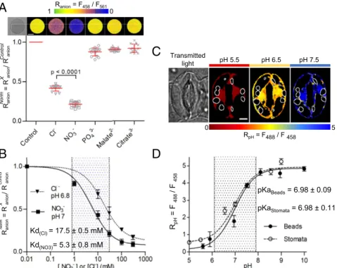
In Vitro Assays Reveal a Strong Affinity of ClopHensor for NO3−.In
contrast to mammalian cells, several anionic species are present in the millimolar range in plant cells (5, 35). Therefore, we in-vestigated the sensitivity of ClopHensor to Cl−, NO3−, PO43−,
malate2−, and citrate3−, the main anions present in the model
plant Arabidopsis (5). ClopHensor was previously shown to be insensitive to SO42−, which also accumulates to millimolar levels
in plant cells (34, 36). We used recombinant ClopHensor pro-teins bound to Sepharose beads and recorded the fluorescence upon exposure to a range of anions by CLSM after excitation at 458 nm (emission 500 to 550 nm), 488 nm (emission 500 to 550 nm), and 561 nm (emission 600 to 625 nm) (Fig. 1A). The ratio Ranion(F458/F561) was calculated from the ratio of the
fluores-cence intensity images after excitation at 458 nm (F458) and
561 nm (F561) to estimate the effect of anions on ClopHensor (SI
Appendixhas anRanioncalculation). No significant difference in
Ranionwas observed between the control (Ranionctrl= 1.14 ± 0.11)
and 30 mM PO43−(Ranionnorm= 0.88 ± 0.05), malate2−(Ranionnorm=
0.91± 0.02), and citrate3−(Ranionnorm= 0.92 ± 0.05) (Fig. 1A).
Mean-while, we found that ClopHensor was sensitive to Cl−(Ranionnorm=
0.42± 0.03) as previously reported (34) and remarkably, also to NO3−(Ranionnorm= 0.21 ± 0.03) (Fig. 1A). ClopHensor displayed
a higher affinity to NO3−(KdNO3= 5.3 ± 0.8 mM at pH 7) than to
Cl−(KdCl= 17.5 ± 0.5 mM at pH 6.8) (Fig. 1B). The sensitivity
range of ClopHensor was between 2 and 162 mM for Cl−(at pH 6.8) and between 0.6 and 48 mM for NO3−(at pH 7) (Fig. 1B).
Notably, in the physiological range of cytosolic pH (i.e., 6.8 to 8), theKdNO3of ClopHensor was between 5 and 25 mM (SI Appendix,
Fig. S1), which is in the range of the previously reported [NO3−]cyt
values of about 5 mM (35), therefore making it suitable to monitor the dynamics of this anion. Concerning chloride, KdCl of
Clo-pHensor was between 17.5 and 163 mM (SI Appendix, Fig. S1), values that are above the reported basal [Cl−]cytin plant cells of
about 10 mM (37). To test the pH sensitivity of ClopHensor in our in vitro assays, we calculated the ratioRpH(F488/F458) (SI Appendix
has anRpHcalculation). In agreement with a previous report (34),
we found a strong response ofRpHto pH variations with a steep
dynamic range of ninefold change between pH 6.1 and pH 7.9 and a pKa= 6.98 ± 0.09 (Fig. 1D). Neither the binding of NO3−nor that
of Cl−modified significantly the pH sensitivity of ClopHensor (SI
Appendix, Fig. S1), confirming its robustness as a dual anion and pH
biosensor.
ClopHensor Is a Robust and Sensitive Sensor of Cytosolic pH in A. thaliana GCs. We generated transgenicArabidopsis plants (eco-type Columbia 0 [Col-0]) expressing ClopHensor in the cytosol and nucleoplasmic compartments under the control of the Ubiquitin10 promoter (pUB10:ClopHensor). The expression of ClopHensor did not affect the development of the plants, in-dicating that its expression did not significantly interfere with the amount of anions available in the cytosol for cellular me-tabolism (SI Appendix, Fig. S2). To measure the pH sensitivity of ClopHensor in living GCs, stomata from pUBI10:Clo-pHensor were sequentially exposed to NH4-acetate–based
buffers to clamp the pHcytat defined values between 5 and 9
(Fig. 1C and D). We found that ClopHensor sensitivities to pH in vivo and in vitro were very similar. The mean <RpHcyt>,
calculated from each pixel in the stomata, showed that the pH titration curve of ClopHensor in GCs mirrored the in vitro assay (Fig. 1D). The pKa(6.98± 0.11) and the sensitivity range
of ClopHensor (between pH 6.1 and 7.9) measured in vivo matched the values measured in vitro (Fig. 1D). These findings demonstrate that 1) ClopHensor is a reliable reporter for in-tracellular pH changes in GCs, 2) the cytosolic environment does not affect ClopHensor properties with respect to pH, and 3) the ClopHensor sensitivity range is appropriate for mea-suring pHcytin GCs.
Settings and Design of the Experimental Workflow in GCs.The data we obtained open the possibility of measuring the variations of [NO3−]cyt, [Cl−]cyt, and pHcyt in vivo. This provides a unique
opportunity to disclose in living cells how ion fluxes across the PM and the VM of GCs affect cytosolic conditions. In order to
quantify [NO3−]cyt, [Cl−]cyt, and pHcytin GCs, we optimized the
fluorescence acquisition protocol in GCs expressing ClopHensor
(SI Appendix, Figs. S3 and S4) and determined the temporal
window to set up our experiments. First, to maximize the col-lected fluorescence and minimize photodamage by the laser, we selected stable transgenic lines expressing pUBI10:ClopHensor with high fluorescence in GCs after excitation at 458 nm (emission 500 to 550 nm), 488 nm (emission 500 to 550 nm), and 561 nm (emission 600 to 625 nm). Second, to quantify [NO3−]cyt,
[Cl−]cyt, and pHcyt, we excluded the fluorescent signals emitted
by chloroplasts (excitation 488 nm, emission 650 to 675 nm). Therefore, we developed an image processing workflow to ac-curately measure ClopHensor fluorescence in the cytosol of plant cells (SI Appendix, Fig. S4).
To derive the [NO3−]cyt, [Cl−]cyt, and pHcyt in GCs, we used
the calculation procedure described in Arosio et al. (34) (SI
Appendix). To obtain a quantitative estimation of the changes in
[NO3−]cyt and [Cl−]cyt induced by the applied treatments, we
determined in vivo theRanionratio in the absence of NO3−and
Cl−(i.e.,R0).R0is required to calculate the actual concentration
of Cl−and NO3−in the cytosol (SI Appendix). To this aim, we set
up experimental conditions where the initial endogenous [NO3−]cytand [Cl−]cytshould be below the sensitivity threshold of
ClopHensor. Selective microelectrode measurements have shown that, when plants are grown with less than 0.01 mM NO3−
supply, the cytosolic levels are below 0.5 mM (38). Therefore, we grewpUB10::ClopHensor plants in vitro in an NO3−-free medium
(0 mM NO3−medium) and determined the whole-plant [NO3−]
and [Cl−] at different days after germination (DAG) (SI
Ap-pendix, Table S1). We found that, in these conditions, the
whole-seedling endogenous content of NO3−and Cl− was decreasing
after germination. At DAG 14, Cl− was no longer detectable; meanwhile, [NO3−] was below the sensitivity threshold of ClopHensor
(i.e., 0.6 mM at pH 7). Subsequently, based on these data, we imaged the fluorescence in stomata frompUB10:ClopHensor plants grown in vitro for 14 d on an NO3−-free medium and
measured a mean ratioR0anionof 0.56± 0.07 (n = 29 stomata)
(SI Appendix, Fig. S3E).
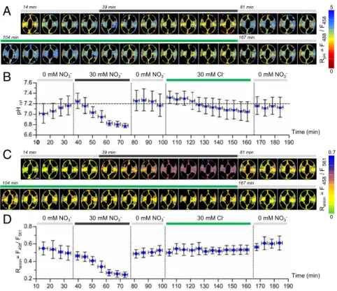
Dynamic Measurements of Cytosolic NO3−, Cl−, and pH in Arabidopsis
GCs.We challenged 14-d-old NO3−-starvedArabidopsis seedlings
expressing ClopHensor for the simultaneous detection in GCs of [NO3−]cyt, [Cl−]cyt, and pHcytchanges upon extracellular NO3−or
Cl−supply/removal (Fig. 2). The experimental design was based on the application of different extracellular conditions in a se-quence of five steps (Fig. 2). GCs were 1) perfused with NO3−-free medium to determine the ratioR0for each stomata;
2) exposed to 30 mM KNO3to observe [NO3−]cyt changes; 3)
washed out with NO3−-free medium; 4) exposed to 30 mM KCl
to observe [Cl−]cyt changes; and 5) washed out again with
NO3−-free medium. We applied 30 mM KNO3or KCl as these
concentrations are commonly used in stomata aperture assays (8, 39). To perform a full experiment, we imaged GCs for 190 min, and each stomata was imaged every 4 min with sequential exci-tation at 561, 488, and 458 nm. Fluorescence intensity recorded in NO3−-free medium was not altered after 190 min of
illumi-nation, indicating that ClopHensor was not significantly affected by photobleaching over the whole duration of the experiment (SI
Appendix, Figs. S5 and S6). Raw data suggested striking
varia-tions of the mean fluorescence intensity recorded after excitation
C
5 RpH= F488/ F 458 0 pH 6.5 pH 7.5 pH 5.5 Transmitted lightA
Ranion= F458/ F561 0 1 Kd(Cl) = 17.5 ± 0.5 mM Kd(NO3)= 5.3 ± 0.8 mMB
Ran ion = R an ion / R an ion Norm X Control Ran ion = R an ion / R an ion Norm X Cont rolD
pKaBeads= 6.98 ± 0.09 pKaStomata= 6.98 ± 0.11 RpH = F 488 / F 458Fig. 1. ClopHensor is sensitive to NO3−, Cl−, and pH. (A and B) In vitro ratio imaging of Sepharose beads decorated with ClopHensor in the presence of 30 mM Cl−, NO3−, PO43−, malate2−, and citrate3−. (A, Upper) False color images of representative beads displaying the fluorescence ratio Ranion(Ranion= F458/F561). (Scale bar: 50μm.) (A, Lower) normalized Rnorm
anion(mean value± SD; n ≥ 15 beads in each condition). The bracket indicates a statistically significant dif-ference. (B) In vitro dose–response analysis of ClopHensor showing Rnorm
anionin the presence of Cl−or NO3−from 0 to 300 mM at pH 6.8 and 7, respectively (mean value± SD; n = 15 beads in each condition). Data were normalized to control conditions and fitted withSI Appendix, Eq. S2. Dotted area, sensitivity range for NO3−(0.6 to 48 mM) of ClopHensor. (C) In vivo ratio imaging of Arabidopsis stomata expressing ClopHensor. False color images of a representative stomata showing the fluorescence ratio RpH(RpH= F488/F458) upon sequential exposure to NH4-acetate buffers at pH 5.5, 6.5, and 7.5. From left to right, transmitted light and false color images of RpHat pH 5.5, 6.5, and 7.5, respectively. White contours, localization of the chloroplasts subtracted during the analysis. (Scale bar: 5μm.) (D) Plot of RpHvs. pH showing that the pH dependence of ClopHensor in vivo (stomata; black circles; n≥ 10) and in vitro (Sepharose beads; white circles; n ≥ 15) is comparable (mean value± SD). Data were fitted withSI Appendix, Eq. S1. Dotted area, sensitivity range for pH (6.1 to 7.9) of ClopHensor.
PLANT
BIO
at 488 and 458 nm when NO3− was added to, or washed out
from, the extracellular medium; meanwhile, Cl− addition had less pronounced effects (SI Appendix, Fig. S5). Ratiometric im-ages forRpHandRanionwere established from the fluorescence
intensity images (Fig. 2A and C). The ratiometric maps for RpH
andRanionwere then used to compute the mean pHcyt(Fig. 2B)
and the meanRanionin the presence of extracellular NO3−and
Cl− for each cell (Fig. 2D). The results show that, differently from our observations with NO3−, Raniondoes not change
sig-nificantly upon addition of Cl−, suggesting that [Cl−]cytwas below
the range of sensitivity of ClopHensor. In addition, the com-parison of pHcytandRanionchanges in the presence of
extracel-lular NO3−during the experiment suggests a link between NO3−
transport and pH modification (Fig. 2B and D). Initially (step 1), in the NO3−-free medium, the pHcyt was 7.01 ± 0.19. Within
35 min, it increased and stabilized to 7.17± 0.18, while Ranionwas constant (Fig. 2D). Upon addition of 30 mM extracellular KNO3
(step 2), theRaniondecreased from a mean value in 0 mM NO3−
of 0.49± 0.08 to a value of 0.25 ± 0.03. The calculation of the [NO3−]cytshows that it increased from an initial value of 0.74±
0.25 to 4.91± 0.40 mM. In parallel, the pHcytdecreased to 6.78±
0.04. Both pHcyt and Ranion reached a plateau within 20 to
30 min, suggesting a coordination between the two parameters. At step 3, unexpectedly both pHcytandRaniondropped back to
their initial values in less than 4 min after removal of KNO3.
Finally (step 4), when the stomata were exposed to 30 mM KCl, a modest and not significant (P = 0.17, n = 6) decrease from pHcyt= 7.17 ± 0.20 to pHcyt= 7.05 ± 0.40 was observed, with a
rate of pH decrease lower than with 30 mM KNO3 (Fig. 2B).
Similar results were obtained when stomata were exposed to KCl only (SI Appendix, Fig. S7).
As a whole, these data demonstrate that ClopHensor enables us to simultaneously monitor in vivo the variations in [Cl−]cytor
[NO3−]cyt and pHcyt at a cellular resolution. In the conditions
tested, [Cl−]cytwas below the limit of detection of ClopHensor
for Cl−(i.e., 2 mM) (Fig. 2D). This suggests that in our experi-mental setting, ClopHensor was measuring essentially cytosolic NO3−variations. Notably, cytosolic NO3−and pH changes
ap-pear to be concerted, suggesting that they are governed by a common mechanism.
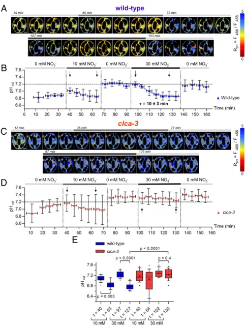
AtCLCa Accounts for Cytosolic Acidification in Response to NO3−.The
finding that ClopHensor can measure the dynamic changes of [NO3−]cytand pHcytin GCs opens the possibility to visualize the
activity of intracellular ion transport systems in living cells. We therefore used this sensor to address the role of the vacuolar 2NO3−/1H+exchanger AtCLCa in cytosolic NO3−and pH
ho-meostasis. AtCLCa is known to mediate the uptake of NO3−into
the vacuole driven by H+ extrusion into the cytosol (6, 40). Therefore, based on its biophysical properties, AtCLCa may be involved in the [NO3−]cytand pHcytresponses measured in Fig. 2.
To assess this possibility, we generatedclca-3 knockout mutant plants expressing ClopHensor by crossingclca-3 with a wild-type pUBI10:ClopHensor line. Patch-clamp experiments performed on vacuoles isolated from the wild type andclca-3 pUBI10::ClopHensor confirmed that clca-3 plants expressing pUBI10:ClopHensor were defective in vacuolar NO3− transport activity (SI Appendix,
Fig. S8). We then compared the dynamic changes of [NO3−]cyt
and pHcyt in stomata of 14-d-old nitrate-starved seedlings
from wild-type and clca-3 pUBI10:ClopHensor plants (Figs. 3 and 4). Since AtCLCa is highly selective for NO3− over Cl−,
we performed experiments applying extracellular KNO3only.
B
A
14min 39min 81min 104 min 167 min RpH = F 488 / F458 5 0 pH cy t 0 mM NO3- 30 mM NO3- 0 mM NO3- 30 mM Cl- 0 mM NO3 -Time (min)C
Rani on = F 458 / F 561 0 0.7 n i m 1 8 n i m 9 3 n i m 4 1 104 min 167 minD
Time (min) Rani on = F458 / F561 0 mM NO3- 30 mM NO3- 0 mM NO3- 30 mM Cl- 0 mM NO3 -0Fig. 2. ClopHensor reveals the dynamics of cytosolic pH, NO3−, and Cl−in Arabidopsis stomata. Epidermal peels from plants grown in vitro for 14 d in NO3−-free media were imaged (SI Appendix). (A and C) Representative false color ratio images of RpH(A) and Ranion(C) at different time points of a stomata sequentially exposed to NO3−-free medium (0 mM NO3−), 30 mM KNO3, and 30 mM KCl. Gray areas, localization of chloroplasts subtracted during the analysis. (Scale bars: 5μm.) (B) pHcytwas quantified at each time point from the corresponding RpHimages. (D) Quantification of the Ranionin the cytosol of GCs. (B and D) pHcyt(B) and Ranion(D), indicating [NO3−]cyt, change simultaneously upon extracellular application and removal of 30 mM KNO3. Horizontal error bars represent the time interval of 4 min for the sequential imaging of stomata. Data represent mean values± SD (n = 6).SI Appendix, Fig. S4shows the workflow for the calculation of pHcyt(B) and Ranion(D). Vertical dotted lines indicate changes of extracellular conditions. The horizontal dashed line (B) serves as a reference for pH 7.2.
Again, we designed experiments divided in five steps. GCs from the wild type andclca-3 were 1) perfused with NO3−-free
medium to establish the ratioR0of each stomata; 2) perfused with 10 mM KNO3; 3) washed out with NO3−-free medium; 4)
perfused with 30 mM KNO3; and 5) washed out with NO3−-free
medium.
Application of this five-step protocol to wild-type pUBI10:-ClopHensor GCs showed that [NO3−]cytvaries according to the
applied extracellular KNO3 concentration. We calculated the
[NO3−]cytto be 1.64± 0.32 and 4.74 ± 1.52 mM in 10 and 30 mM
KNO3, respectively (n = 8) (Fig. 3 A, B, and E). In the presence
of 10 mM KNO3, the [NO3−]cytreached a plateau in less than
4 min (Fig. 3B). However, in the presence of 30 mM KNO3in the
extracellular medium, the [NO3−]cyt rose progressively with a
time constant ofτ = 15 ± 3 min (Fig. 3B). Interestingly, GCs
maintained an [NO3−] gradient between the apoplast and the
cytosol of about sixfold when either 10 or 30 mM KNO3was
applied. In all cases, upon washout with NO3−-free medium, the
[NO3−]cytdropped back to concentrations close to the limit of
detection within 4 min. Inclca-3 pUBI10:ClopHensor GCs, the [NO3−]cytbehaved similarly to wild-type plants upon exposure to
10 mM KNO3, reaching 2.24± 1.47 mM (n = 15) (Fig. 3 C–E).
Further, similarly to the wild type, upon application of 30 mM KNO3, clca-3 pUBI10:ClopHensor GCs [NO3−]cyt increased to
6.34± 2.91 mM (n = 15) (Fig. 3D). However, in contrast with the wild type, [NO3−]cytincreased faster, reaching a plateau in less
than 4 min inclca-3 (τ < 3 min) compared with about 30 min in wild-typepUBI10:ClopHensor GCs (Fig. 3D). These data are in agreement with the involvement of the AtCLCa exchanger in buffering cytosolic NO3−. Furthermore, we found that the pHcyt
B
D
C
12min 36min 71min n i m 1 3 1 n i m 7 9 τ = 15 ± 3 minE
Wild-type τ = < 3 min clca-3 clca-3 wild-type clca-3 Time (min) Time (min) 0 mM NO3- 10 mM NO3- 0 mM NO3- 30 mM NO3- 0 mM NO3 -0 mM NO3- 10 mM NO3- 0 mM NO3- 30 mM NO3- 0 mM NO3-A
19 min 46min 78min n i m 3 5 1 n i m 7 0 1 wild-type Rani on = F 458 / F 561 0 0.7 Rani on = F 458 / F 561 0 0.7Fig. 3. The vacuolar NO3−/H+exchanger AtCLCa controls [NO3−]cytin Arabidopsis stomata. Epidermal peels from plants grown in vitro for 14 d in NO3−-free media were imaged (SI Appendix). (A and C) Representative false color ratio images of Ranionfrom wild-type (A) and clca-3 (C) stomata at different time points. Stomata were sequentially exposed to 0, 10, and 30 mM KNO3(horizontal bar in Upper). Gray areas, localization of chloroplasts subtracted during the analysis. (Scale bars: 5μm.) (B and D) [NO3−]cyt(mean± SD) at each time point in wild-type (B; n = 8) and clca-3 stomata (D; n = 15). Horizontal error bars represent the time interval of 4 min for the sequential imaging of stomata. Dotted areas, ClopHensor sensitivity threshold for NO3−. Vertical dotted lines indicate changes of extracellular conditions. Horizontal dashed lines indicate [NO3−]cyt= 6 mM. Black arrows, time points used for the box plot analysis in E. (E) Box plots of the [NO3−]cytat different time points (black arrows in B and D). Brackets indicate statistically significant differences. Blue boxes, the wild type (n= 17); red boxes, clca-3 (n = 15) stomata. Whiskers show the 10 to 90% percentiles. Crosses indicate the means.
PLANT
BIO
dynamics in the wild type and clca-3 were markedly different when extracellular KNO3was applied (Fig. 4A–D). In wild-type
GCs, the pHcytstabilized at 6.89± 0.05 (n = 8) at the beginning
of the experiments. Then, exposure to 10 mM KNO3induced an
initial slight pHcytincrease followed by a progressive and modest
acidification of the cytosol. Washing out with NO3−-free medium
provoked a fast increase of the pHcytto 7.21 ± 0.12 (n = 8).
Then, upon perfusion with 30 mM KNO3, a progressive and
marked acidification to pH 6.87 ± 0.13 (n = 8) with a time constant ofτ = 10 ± 3 min was observed. Finally, after washing out in NO3−-free medium, an alkalinization to 7.16± 0.16 (n =
8) was observed within 4 min. Inclca-3 pUBI10:ClopHensor GCs, a modest pHcyt acidification was observed upon exposure to
10 mM KNO3, as in the wild type. However, this pHcytdecrease
was not statistically significant in clca-3 plants (Fig. 4E). Re-markably, the perfusion of 30 mM KNO3, which induced a
marked acidification in wild-type GCs, did not induce any de-crease of pHcytinclca-3 GCs: pHcytremained stable at pH∼ 7.3
(n = 15) (Fig. 4 B and D). To exclude an effect of the sequence of KNO3application, we inverted step 2 and step 4 in the perfusion
protocol and obtained the same results (SI Appendix, Fig. S9). These findings show that the presence of the 2NO3−/1H+
ex-changer AtCLCa in the VM is associated with the pHcyt
modi-fication detected in wild-type GCs upon perfusion with 30 mM KNO3, suggesting a role of AtCLCa in the regulation of pHcyt.
We tested whether the application of KNO3has an effect on
stomata aperture at a whole-leaf level and performed leaf gas exchange measurements on detached leaves (SI Appendix, Fig. S10) (41, 42). In these experiments, we applied KNO3at the leaf
petiole, and we detected an increase of stomata conductance that was similar in the wild type andclca-3 (SI Appendix, Fig. S10 and
Table S4). The similar behavior of the wild type andclca-3 when
A
B
D
C
n i m 8 7 n i m 6 4 19 min n i m 3 5 1 n i m 7 0 1 n i m 1 7 n i m 6 3 n i m 2 1 n i m 1 3 1 n i m 7 9 clca-3 wild-typeE
pH cy t pH cy t τ = 10 ± 3 min Wild-type wild-type clca-3 Time (min) pH cy t clca-3 Time (min) 0 mM NO3- 10 mM NO3- 0 mM NO3- 30 mM NO3- 0 mM NO3 -0 mM NO3- 10 mM NO3- 0 mM NO3- 30 mM NO3- 0 mM NO3 -RpH = F 488 / F 458 5 0 RpH = F 488 / F 458 5 0Fig. 4. The vacuolar NO3−/H+exchanger AtCLCa regulates pHcytin Arabidopsis stomata. Epidermal peels from plants grown in vitro for 14 d in NO3−-free media were imaged (SI Appendix). (A and C) Representative false color ratio images of RpHfrom wild-type (A) and clca-3 (C) stomata at different time points. Stomata were sequentially exposed to 0, 10, and 30 mM KNO3(horizontal bar in Upper). Gray areas, localization of chloroplasts subtracted during the analysis. (Scale bars: 5μm.) (B and D) pHcyt(mean± SD) at each time point in wild-type (B; n = 8) and clca-3 stomata (D; n = 15). Horizontal error bars represent the time interval of 4 min for the sequential imaging of stomata. Vertical dotted lines, changes of extracellular conditions. Horizontal dashed lines indicate pH 7.2. Black arrows, time points used for the box plot analysis in E. (E) Box plots of the pHcytat different time points (black arrows in B and D). Brackets indicate statistically significant differences. Blue boxes, the wild type (n= 17); red boxes, clca-3 (n = 15). Whiskers show the 10 to 90% percentiles. Crosses indicate the means.
KNO3is applied converges with the finding that at a cellular
level AtCLCa does not determine the steady-state [NO3−]cyt
(Figs. 3 and 5). The subsequent application of 50μM ABA on detached leaves induced a similar decrease of the stomata con-ductance in both the wild type andclca-3 (SI Appendix, Fig. S10
and Table S4). These results with detached leaf gas exchange
measurements do not correlate with the observations made on stomata from isolated epidermis fromclca knockout (7). Such discrepancy between experimental methods and conditions has been reported as well for several well-known knockout mutants involved in ABA signaling and stomata regulation such as, for example,slac1, abi1, and abi2 (41).
AtCLCa Is Involved in pH Homeostasis upon Treatment of GCs with Fusicoccin.The results obtained upon treatment with extracellular KNO3 indicated that AtCLCa may be an important player in
pHcythomeostasis (Figs. 3 and 4). To test whether AtCLCa
in-fluences pHcyt regulation independently of the addition of its
anion substrates, NO3−and Cl−, we investigated its role in
re-sponse to the fungal toxin fusicoccin, which triggers stomata opening through a robust activation of the PM H+ pump (43, 44). In these experiments, we used stomata from plants grown in soil, and since we could not control the initial cellular [NO3−]
and [Cl−], we quantified the changes in pHcytand [NO3−]cytin
GCs asΔRpH=RpH,iandΔRanion=Ranion,i(Fig. 5 andSI Appendix).
Positive values of ΔRpH=RpH,i andΔRanion=Ranion,idenote
cyto-solic alkalinization and increase in [NO3−]cyt, respectively. To
correlate the changes in pHcytor [NO3−]cytwith the opening of
stomata, we started the experiments with closed stomata at the end of the dark period (Fig. 5). Thus, epidermal peels from the wild type andclca-3 were prepared 1 h before the onset of light. After incubation under the microscope for 20 min in a buffer containing 10 mM KNO3at pH 5.7, 10μM fusicoccin was
ap-plied for total time of 130 min. Stomata were imaged every 4 min, and we measured pore aperture,RpH, andRanionin each
stomata (Fig. 5). Fusicoccin induced a significantly lower open-ing inclca-3 (1.8 μm ± 0.1 at 152 min, n = 15) compared with wild-type (2.6μm ± 0.2 at 152 min, n = 14) stomata (Fig. 5 A and B), in agreement with previous results showing that light-induced sto-mata opening is reduced inclca knockout mutants (7). In wild-type stomata, fusicoccin induced a rapid increase of pHcytleading
to a ΔRpH=RpH,i= 0.12 ± 0.02 as early as 4 min after treatment
(n = 14) (Fig. 5C). Then, pHcytslowly recovered to almost reach
its initial value after 120 min (ΔRpH=RpH,i= 0.03 ± 0.02, n = 15)
(Fig. 5C). Notably, wild-type stomata not treated with fusicoccin did not open and did not exhibit a significant increase of the
Wild-type + fusicoccin Wild-type - fusicoccin Wild-type + fusicoccin clca-3 + fusicoccin pH cy t [anion] cy t 10 mM NO3 -+ Fusicoccin ΔR anion / R ani o n,i ΔR pH / R pH,i ΔR pH / R pH,i ΔR ani o n / R ani o n,i Aperture (μm) Aperture (μm) Time (min) ) n i m ( e m i T ) n i m ( e m i T
Time (min) Time (min)
Time (min)
B
A
C
E
D
F
10 mM NO3 -+ FusicoccinFig. 5. Fusicoccin-induced pHcytand [anion]cytdynamics during stomata opening. (A and B) Fusicoccin-induced stomata opening from wild-type (A) and clca-3 (B) plants expressing ClopHensor. Stomata from epidermal peels were prepared 1 h before light onset and equilibrated for 20 min in the presence of 10 mM KNO3before exposure to 10μM fusicoccin (vertical dotted line) for 120 min or without fusicoccin (dashed lines in A, C, and E). In clca-3, fusicoccin induced a significantly lower stomata opening compared with the wild type (dashed lines in B, D, and F; 50 to 80 min P< 0.01, 80 to 12 min P < 0.001). The stomata in A and B were imaged to monitor the changes of pHcyt(C and D) and [anion]cyt(E and F) during fusicoccin-induced stomata opening. (C and D) Time-resolved ΔRpH=RpH,iin wild-type (C) and clca-3 (D) stomata. In wild-type (C) and clca-3 (D) stomata, theΔRpH=RpH,iincreased, indicating higher pHcyt. Within 120 min, theΔRpH=RpH,isignificantly decreased (C; P< 0.01) in the wild type (D) but not in clca-3 (D; P = 0.45). (E and F) Time-resolved ΔRanion=Ranion,iin the wild type (E)
and clca-3 (F) during fusicoccin-induced stomata opening. In both the wild type + fusicoccin and− fusicoccin, ΔRanion=Ranion,iincreased over time, indicating an
increase in [NO3−]cyt. (F) In clca-3,ΔRanion=Ranion,iincreased significantly less than in the wild type (dashed line; P= 0.02 after 120 min). In all panels, n = 14 for
the wild type with fusicoccin, n= 15 for clca-3, and n = 5 for the wild type without fusicoccin. Data are shown as mean ± SEM. Brackets indicate statistically significant differences.
PLANT
BIO
ΔRpH=RpH,i (n = 5) (Fig. 5C). In clca-3 stomata, fusicoccin
in-duced a rapid increase of pHcytwith aΔRpH=RpH,i= 0.10 ± 0.01
after 4 min (n = 14) (Fig. 5D) as in the wild type. However, in contrast with the wild type, pHcytdid not recover its initial value in
clca-3 stomata, even after 120 min (ΔRpH=RpH,i= 0.09 ± 0.01, n =
15) (Fig. 5D). The rapid increase in pHcytobserved after fusicoccin
treatment (Fig. 5C and D) is likely due to the activation of the PM
H+pumps that are extruding H+in the apoplast (43, 44). In clca-3, the absence of an NO3−/H+antiporter pumping H+from the
vacuole into the cytosol accounts for the defect in pHcytrecovery
after fusicoccin-induced alkalinization (6). This result shows that the transport activity of AtCLCa in the VM contributes to the recovery after the cytosolic pH increase induced by fusicoccin. Interestingly, the quantification ofΔRanion=Ranion,iin the wild type
H+
clca-3
wild-type
vacuole cytosolCLCa
H+ NO3 -H+ H+ NO3 -H+ K+ NO3 -H+ H+ H+ K+ H+ H+ K+ [NO 3 -]cyt H+ NO3 -H+ NO3 -H+ H+ H+-ATPase H+ H+ATPaseB
clca-3
wild-type
vacuole cytosol H+ NO3 -H+ H+ K+ H+ H+ NO3 -K+ H+ H+ H+ K+ H+ H+ K+CLCa
NO3 -H+ H+ NO3 -H+ NO3 -H+ 8 6.8 pH cytpH
cytclca-3
wild-type
vacuole cytosol H+ NO3 -H+ K+ H+ H+ NO3 -K+ H+ H+ H+ K+ H+ H+ K+CLCa
NO3 -H+ H+ NO3 -H+ NO3 -H+ 8 6.8 pH cyt K+ 10 mM NO3 -pH 5.8 10 mM NO3 -pH 5.8 + Fusicoccin 10 mM NO3 -pH 5.8 + Fusicoccin H+ H+-ATPaseclca-3
wild-type
vacuole cytosolCLCa
H+ NO3 -H+ H+ K+ H+ H+ NO3 -H+ K+ NO3 -H+ H+ K+ H+ H+ K+ [NO 3 -]cyt 0 mM NO3 -pH 5.8 30 mM NO3 -pH 5.8 H+ NO3 -H+ NO3 -H+[NO
3-]
cytA
30 mM NO3 -pH 5.8D
C
H+ H+ATPase H+ H+-ATPase H+-ATPase t = 23 min t =152 min t = 97 min t =127 minFig. 6. A vacuolar exchanger modifies cytosolic homeostasis in Arabidopsis stomata. Illustration recapitulating the impact of the activity of AtCLCa on [NO3−]cytand pHcythomeostasis in GCs. (A) In the presence of 30 mM KNO3, NO3−enters the cell via NO3−transporters and channels residing in the PM. In the wild type (Left), the vacuolar AtCLCa exchanger (shown in red) pumps NO3−into the vacuole, slowing down [NO3−]cytincrease. In the absence of AtCLCa (Right ), [NO3−]cytstabilizes in less than 4 min. (B) In the presence of 30 mM KNO3, the transport activity of AtCLCa releases H+in the cytosol, inducing an acidification in wild-type GCs (Left). In the absence of AtCLCa, the cytosolic acidification does not occur (Right). (C and D) Fusicoccin triggers stomata opening, activating the PM H+-ATPase (shown in red). (C) During opening, a progressive increase of [NO
3−]cytreaches higher levels in the wild type (Left) than in clca-3 (Right). (D) Fusicoccin induces an increase of pHcytin both the wild type (Left) and clca-3 mutant (Right). Notably, in the wild type, within 130 min the pHcyt recovers to the initial value (Left). Differently, in clca-3, the pHcytdid not recover its initial value (Right).
showed an increase in [NO3−]cytover the time of the experiment
independently of fusicoccin application (Fig. 5E). Therefore, in-creased [NO3−]cytdoes not seem to determine stomata opening.
Intriguingly, the rate of [NO3−]cytincrease was significantly lower
inclca-3 than in the wild type (Fig. 5F). This is opposite to what one would expect, as AtCLCa removes NO3−from the cytosol to
store it in the vacuole. This surprising result suggests that a more complex regulation is involved, such as a feedback of the NO3−
transport capacity of the VM on PM NO3−uptake, as previously
observed at the whole-plant level (45).
Together, our results strengthen the hypothesis of the role of AtCLCa activity in regulating pHcyt in response not only to
fluctuations of extracellular [NO3−] but also, to other stimuli
such as stomata opening induced by the fungal toxin fusicoccin (Fig. 6). Indeed, in wild-type plants exposed to high [NO3−], the
dynamics of [NO3−]cyt and pHcyt were obviously correlated
(compare Fig. 3A and B with Fig. 4 A and B). In contrast, in clca-3 pUBI10:ClopHensor GCs, [NO3−]cytchanges were not mirrored
by pHcytchanges, showing that inclca-3 the two processes were
uncoupled (compare Fig. 3C and D with Fig. 4 C and D). In the case of fusicoccin treatment (Fig. 5), the H+import to the cytosol from the vacuole mediated by AtCLCa likely compensates for the increased H+ extrusion by the PM H+ pumps. However, the variations of pHcytand of [NO3−]cytdid not display the same
ki-netics. In contrast, pHcytrecovery seems important for sustained
stomata opening. The opening rate was not significantly different between the wild type andclca-3 during the initial pH increase triggered by fusicoccin, but opening slowed down inclca-3 com-pared with the wild type during the pH recovery phase.
Discussion
The involvement of CLCs in severe genetic diseases in humans and their major physiological functions in plants have attracted considerable attention to these anion transport systems. In-terestingly, the CLC family presents a dichotomy: the CLCs lo-calized in the PM are anion channels; those lolo-calized in intracellular membranes are anion/H+ exchangers (2). A
com-bination of electrophysiological, structural, and biochemical data provided a detailed understanding of the mechanisms allowing the anion/H+ exchange or the anion channel behavior at a
submolecular level in CLCs (2). However and despite intense research, the cellular function of intracellular CLCs has remained elusive (2). So far, the role of intracellular CLCs was exclusively considered from the point of view of the organelle lumen, while the impact on thecyt has been overlooked. Nev-ertheless, when a CLC exchanger pumps anions into an organ-elle, it simultaneously releases a stoichiometric amount of H+in the cytosol. Therefore, intracellular CLCs have the capacity to influence pHcyt and regulate anionic homeostasis. To test this
hypothesis in vivo, we usedArabidopsis GCs expressing the dual anion and pH biosensor ClopHensor to unravel the impact of a vacuolar CLC on the cytosol.
ClopHensor Is Able to Sense pH and NO3−in Plant Cells.ClopHensor
is a genetically encoded biosensor originally developed in mammalian cells. Its photophysical characteristics have been analyzed in depth (34, 46, 47). The advantageous properties of ClopHensor allow us to measure, simultaneously and in the same cell, two important intracellular parameters, pH and the con-centration of anions such as Cl−. Notably, changes in pH and [Cl−] can report the activity of different types of ion transporters in the VM and PM of plant cells. Before using ClopHensor in plant cells, we first checked its sensitivity toward other anions that, differently from animal cells, are present in the millimolar range in the cytosol (5) (Fig. 1). In vitro analysis demonstrated that ClopHensor is sensitive not only to Cl−but also, to NO3−,
while it is insensitive to PO3−, malate2−, and citrate3− at the
tested concentrations. Furthermore, ClopHensor sensitivity to
NO3−is even higher than that to Cl−(Fig. 1). The analysis of the
[NO3−]cyt, [Cl−]cyt, and pHcyt in living GCs demonstrated that
ClopHensor is able to report dynamic changes of these param-eters (Figs. 3–5). Interestingly, the cytosolic [NO3−]cyt we
esti-mated is in the same range as those previously reported in other cell types with selective microelectrodes (35, 48, 49). The agreement between our data and previous reports demonstrates the robustness of ClopHensor to measure [NO3−]cyt in
Arabi-dopsis GCs. Concerning pH, ClopHensor displays a steep dy-namic range fitting cytosolic conditions (Figs. 1, 2, and 4). The steepness of the pH sensitivity is particularly valuable to resolve subtle pH changes. The properties of ClopHensor for pH mea-surements match those of other pHluorin-derived pH sensors used previously to measure pHcytin plant cells (50, 51). Overall,
our results demonstrate that ClopHensor can be used to measure [NO3−] and pH in GCs. Other NO3−biosensors have been
de-veloped, such as NiTrak, which allows monitoring the activity of the nitrate transporter NRT1.1/NFP5.6 (52), and sNOOOpy, a nitrate/nitrite biosensor that has not been tested in plants yet (53). However, ClopHensor is the first biosensor able to report [NO3−] in the cytosol of plants in parallel with pH. Given the link
between anion and H+transport in plant cells, this dual capacity of ClopHensor is particularly relevant.
A Vacuolar CLC Is Involved in Cytosolic Ion Homeostasis.To reveal the impact of the activity of the vacuolar transporter AtCLCa, we challenged stomata of 14-d-old nitrate-starved seedlings with different extracellular media applied in a defined sequence (Figs. 2–4). Starting from an initial condition with no NO3−or Cl−in
the extracellular medium and within the GCs, we applied dif-ferent KNO3- and KCl-based media. In our conditions, [Cl−]cyt
was below the sensitivity range of ClopHensor. However, we obtained a remarkable result: [NO3−]cytin GCs can undergo rapid
variations (Figs. 2 and 3). To our knowledge, such variations of [NO3−]cythave not been described so far. Former reports available
from root epidermal cells or mesophyll protoplasts suggested that [NO3−]cyt was stable, at least in the short term (35, 49). These
studies were using invasive approaches without challenging cells with modification of the extracellular ion concentrations, possibly explaining why [NO3−]cytchanges were not observed. Interestingly,
our findings show that [NO3−]cytcan change rapidly, within minutes
(Figs. 2 and 3). This supports the hypothesis that [NO3−]cyt
varia-tions may act as an intracellular signal. A role of [NO3−]cytto adjust
cell responses to external nitrogen supply has been previously proposed (48, 54). A second remarkable observation we made is a progressive acidification of the cytosol in parallel with the [NO3−]cyt
increase. Conversely, [NO3−]cytdecrease is paralleled by a rapid
pHcytincrease (Figs. 2–4 and 6). These findings clearly show a link
between [NO3−]cytand pHcytchanges and suggest a common
mo-lecular mechanism underlying NO3−and pH variations.
The detected changes in pHcyt and [NO3−]cyt integrate the
transport reactions occurring at the PM and the VM, as well as metabolic reactions and cytosolic buffer capacity. Our data suggest that the observed changes may be due to H+-coupled
transport reactions. In Arabidopsis cells, AtCLCa is the major H+-coupled NO
3−transporter in the VM (6, 31). Therefore, to
test whether AtCLCa is responsible for the variations detected in the cytosol, we conducted comparative experiments between GCs from the wild type and from clca-3 knockout plants expressing ClopHensor (Figs. 3 and 4). We found that [NO3−]cyt
reaches a steady-state value faster in clca-3 GCs than in the wild type when exposed to extracellular KNO3 (Fig. 3). This
proves that in vivo the vacuolar transporter AtCLCa buffers the [NO3−]cyt, as expected from its function in accumulating
NO3− into the vacuole (6, 31). This finding may explain the
defect of stomata opening reported earlier on isolated epidermis and dehydration test on whole rosettes (7). Gas exchange mea-surements showed that, on detached leaves, the application of
PLANT
BIO
KNO3 induces an increase of the stomata conductance with a
similar trend in both the wild type andclca-3 (SI Appendix, Fig. S10). Further, in the same experiments both genotypes reacted similarly to the application of ABA. These results seem to be in contrast with the observations made at the level of stomata in isolated epidermis from clca knockout (7) (Fig. 5). Such dis-crepancy is not unique to clca mutants as it was reported for other well-known knockout mutants involved in stomata ABA signaling such as, for example, slac1, abi1, and abi2 (41). Nev-ertheless, all these mutants asclca display strong defects in tol-erance to drought stress at the whole-rosette or whole-plant level. Notably, high concentrations (i.e., 50μM) of ABA applied at the petiole of detached leaves are required to induce stomata closure inslac1 and abi mutants (41). In such conditions, other anion channels, like ALMT12/QUAC1, may bypass SLAC1 loss of function to allow stomata closure (26).
At the subcellular level, the most impressive consequence of knocking out AtCLCa was on the pHcyt(Fig. 4). Indeed, in sharp
contrast with wild-type GCs, no pH acidification could be de-tected in clca-3 GCs when [NO3−]cyt increased. These
un-expected findings reveal that AtCLCa solely accounts for the pH acidification detected in wild-type GCs. Moreover, we found that the absence of AtCLCa also perturbs pHcyt regulation during
stomata opening after treatment with fusicoccin (Fig. 5). The role of AtCLCa in the control of pHcytis therefore not limited to
sit-uations involving massive changes of the concentration of its an-ionic substrate. Together, the results highlight a previously overlooked role of AtCLCa in pHcythomeostasis. AtCLCa is not
the only H+-coupled transport system operating in the PM and VM of GCs (Fig. 6). However, our results indicate that under the conditions tested, the transport activity of AtCLCa is predominant and high enough to overcome the pH buffering capacity of the cytosol. Therefore, the use of a biosensor like ClopHensor allowed us to detect in vivo the activity of an intracellular transporter, AtCLCa, and its impact of the intracellular ion homeostasis.
The finding that a vacuolar transporter influences pHcyt
homeo-stasis opens a perspective on the cellular functions of intracellular ion transporters. A potential role of H+-coupled transporters in the regulation of pHcytwas proposed in the’80s (55–57) but was never
demonstrated. Instead, the role of intracellular ion transporters is nowadays commonly interpreted from the point of view of the or-ganelle, focusing on how these transporters regulate ion homeostasis in the lumen of the organelles. Our data provide strong experi-mental evidence supporting the hypothesis that proton-coupled in-tracellular transporters participate in the regulation of pHcyt. In the
plant cell, the VM is commonly considered as a“second layer” with respect to the PM, which is postulated to have a dominant action on intracellular conditions. Our findings show that VM transporters can actively modify the cytosolic conditions rather than“just buffering the cytosol” to maintain homeostatic values. AtCLCa is important in this process, but it might not be the only one (Fig. 6). It will be of interest to understand if and how other transporters like proton pumps or cation/H+exchangers (e.g., Na+/H+exchanger [NHX]) as well as ion channels affect cytosolic ion homeostasis.
Cytosolic pH Control, a Framework for CLC Functions.The results presented here relate to a specialized plant cell type, the GCs. The effect of AtCLCa on pHcytmay account for the unexpected
defect in stomata closure observed inclca knockout plants, while
its function in loading anions into the vacuole would rather lead to the prediction that it is solely involved in stomata opening (7). In this context, modification of pHcyt could be an important
component of AtCLCa function, as pHcyt is an important
pa-rameter in cell signaling (58). The results obtained with fusi-coccin argue in favor of this hypothesis. The treatment with fusicoccin was performed on GCs from mature plants, which allowed monitoring changes in stomatal aperture in parallel with pHcytand [anion]cytvariations. The misregulation of pHcytinclca
correlated with the defect in stomata opening. During the initial pHcytincrease that was not affected inclca, the rate of stomata
opening was similar in the wild type andclca. In the following phase, the defect in pHcyt recovery inclca mutant paralleled a
drop in the rate of stomata opening. Cytosolic pH modifications may modulate ion transport systems and enzymatic reactions to trigger stomata opening or closure. For example, the activity of vacuolar H+ATPase (V-ATPase) is modified by changes of the pHcyt (59). Our findings may also be relevant in the broader
context of other eukaryotic CLC exchangers. Indeed, the func-tion of intracellular CLCs has been interpreted assuming that their only role was to regulate the lysosomal, endosomal, or vacuolar lumen conditions (2). However, the cellular functions of the lysosomal CLC-7 and endosomal CLC-5 remain unclear in mammal cells. CLC-7 was proposed to acidify the lysosomal lu-men, but only modest and controversial effects were detected (14, 16). In the case of CLC-5, endosomes from knockout mice present impaired luminal acidification (33). Nonetheless, the con-nection between endosomal acidification and the severe defects caused by CLC-5 mutations in Dent’s disease is still unclear (2). Indeed, renal failure associated with some mutations in CLC-5 present impaired endocytosis in tubular cells, which is independent of endosomal acidification (33). Intriguingly, pHcytis known to
af-fect endocytosis (60, 61). The results we report here suggest that in eukaryotic cells, intracellular CLCs are part of the cytosolic pH balance machinery. These findings open a perspective on the function of these exchangers in eukaryotic cells and may provide a framework to understand the pathophysiological disorders caused by mutations in human CLC genes.
Methods
Wild-type Arabidopsis plants were Col-0 ecotype. The clca-3 knockout line corresponds to Gabi Kat GK-624E03-022319. Images were acquired with a Leica SP8 upright CLSM. Image analysis was performed with ImageJ. Detailed description of the methods is available inSI Appendix.
Data Availability. All data presented in the paper are described in the text and
SI Appendix. Biological materials are available from the corresponding
au-thor on request.
ACKNOWLEDGMENTS. This work was supported by LabEx Saclay Plant Sciences-SPS (ANR-10-LABX-0040-SPS) and by the ATIP-AVENIR-2018 program. P.C.-F. was supported by a postdoctoral grant from Fundacion Alfonso Martin Escudero. This work has benefited from the facilities and the expertise of Imagerie-Gif microscopy platform, which is supported by France-BioImaging (ANR-INBS-04 “Investments for the future”) and by Saclay Plant Science (ANR-11 IDEX-0003-02). We thank M. Dauzat (Laboratoire d’Ecophysiologie des Plantes sous Stress Environnementaux) and N. Sidibé (Institute for Inter-grative Biology of the Cell) for the help with experiments, Joni Frederick for reading the manuscript, D. Arosio (Consiglio Nazionale delle Ricerche) for pro-viding plasmid with ClopHensor and advice, R. Le Bars (Imagerie-Gif) for the help with microscopy, and M. Bianchi (Institute for Integrative Biology of the Cell) and S. Filleur (Institute for Integrative Biology of the Cell) for discussion.
1. P. B. Persson, A. Bondke Persson, Channels and channelopathies. Acta Physiol. (Oxf.) 218, 149–151 (2016).
2. T. J. Jentsch, M. Pusch, CLC chloride channels and transporters: Structure, function, physiology, and disease. Physiol. Rev. 98, 1493–1590 (2018).
3. E. Park, R. MacKinnon, Structure of the CLC-1 chloride channel from Homo sapiens. eLife 7, e36629 (2018).
4. R. Dutzler, E. B. Campbell, M. Cadene, B. T. Chait, R. MacKinnon, X-ray structure of a ClC chloride channel at 3.0 A reveals the molecular basis of anion selectivity. Nature 415, 287–294 (2002).
5. D. Geelen et al., Disruption of putative anion channel gene AtCLC-a in Arabidopsis suggests a role in the regulation of nitrate content. Plant J. 21, 259–267 (2000). 6. A. De Angeli et al., The nitrate/proton antiporter AtCLCa mediates nitrate
accumu-lation in plant vacuoles. Nature 442, 939–942 (2006).
7. S. Wege et al., Phosphorylation of the vacuolar anion exchanger AtCLCa is required for the stomatal response to abscisic acid. Sci. Signal. 7, ra65 (2014).
8. M. Jossier et al., The Arabidopsis vacuolar anion transporter, AtCLCc, is involved in the regulation of stomatal movements and contributes to salt tolerance. Plant J. 64, 563–576 (2010).
9. C. T. Nguyen et al., Characterization of the chloride channel-like, AtCLCg, involved in chloride tolerance in Arabidopsis thaliana. Plant Cell Physiol. 57, 764–775 (2016). 10. J. Böhm et al., Understanding the molecular basis of salt sequestration in epidermal
bladder cells of Chenopodium quinoa. Curr. Biol. 28, 3075–3085.e7 (2018). 11. A. Herdean et al., The Arabidopsis thylakoid chloride channel AtCLCe functions in
chloride homeostasis and regulation of photosynthetic electron transport. Front. Plant Sci. 7, 115 (2016).
12. A. Accardi, C. Miller, Secondary active transport mediated by a prokaryotic homo-logue of ClC Cl- channels. Nature 427, 803–807 (2004).
13. A. Picollo, M. Pusch, Chloride/proton antiporter activity of mammalian CLC proteins ClC-4 and ClC-5. Nature 436, 420–423 (2005).
14. A. R. Graves, P. K. Curran, C. L. Smith, J. A. Mindell, The Cl-/H+ antiporter ClC-7 is the primary chloride permeation pathway in lysosomes. Nature 453, 788–792 (2008). 15. A. A. Zdebik et al., Determinants of anion-proton coupling in mammalian endosomal
CLC proteins. J. Biol. Chem. 283, 4219–4227 (2008).
16. S. Weinert et al., Lysosomal pathology and osteopetrosis upon loss of H+-driven ly-sosomal Cl− accumulation. Science 328, 1401–1403 (2010).
17. M. R. G. Roelfsema, R. Hedrich, In the light of stomatal opening: New insights into “the Watergate.”. New Phytol. 167, 665–691 (2005).
18. T.-H. Kim, M. Böhmer, H. Hu, N. Nishimura, J. I. Schroeder, Guard cell signal trans-duction network: Advances in understanding abscisic acid, CO2, and Ca2+ signaling. Annu. Rev. Plant Biol. 61, 561–591 (2010).
19. E. A. G. MacRobbie, Signal transduction and ion channels in guard cells. Philos. Trans. R. Soc. Lond. B Biol. Sci. 353, 1475–1488 (1998).
20. Z. Andrés et al., Control of vacuolar dynamics and regulation of stomatal aperture by tonoplast potassium uptake. Proc. Natl. Acad. Sci. U.S.A. 111, E1806–E1814 (2014). 21. A. De Angeli, J. Zhang, S. Meyer, E. Martinoia, AtALMT9 is a malate-activated
vacu-olar chloride channel required for stomatal opening in Arabidopsis. Nat. Commun. 4, 1804 (2013).
22. T. Vahisalu et al., SLAC1 is required for plant guard cell S-type anion channel function in stomatal signalling. Nature 452, 487–491 (2008).
23. J. Negi et al., CO2 regulator SLAC1 and its homologues are essential for anion ho-meostasis in plant cells. Nature 452, 483–486 (2008).
24. H. Zhang et al., Two tonoplast MATE proteins function as turgor-regulating chloride channels in Arabidopsis. Proc. Natl. Acad. Sci. U.S.A. 114, E2036–E2045 (2017). 25. M. Jezek, M. R. Blatt, The membrane transport system of the guard cell and its
in-tegration for stomatal dynamics. Plant Physiol. 174, 487–519 (2017).
26. S. Meyer et al., AtALMT12 represents an R-type anion channel required for stomatal movement in Arabidopsis guard cells. Plant J. 63, 1054–1062 (2010).
27. C. Eisenach et al., ABA-induced stomatal closure involves ALMT4, a phosphorylation-dependent vacuolar anion channel of Arabidopsis. Plant Cell 29, 2552–2569 (2017). 28. J. Zhang et al., Identification of SLAC1 anion channel residues required for CO2
/bi-carbonate sensing and regulation of stomatal movements. Proc. Natl. Acad. Sci. U.S.A. 115, 11129–11137 (2018).
29. D. Geiger et al., Stomatal closure by fast abscisic acid signaling is mediated by the guard cell anion channel SLAH3 and the receptor RCAR1. Sci. Signal. 4, ra32 (2011). 30. V. Barragán et al., Ion exchangers NHX1 and NHX2 mediate active potassium uptake
into vacuoles to regulate cell turgor and stomatal function in Arabidopsis. Plant Cell 24, 1127–1142 (2012).
31. S. Wege et al., The proline 160 in the selectivity filter of the Arabidopsis NO(3)(−)/H(+) exchanger AtCLCa is essential for nitrate accumulation in planta. Plant J. 63, 861–869 (2010).
32. A. Carpaneto, A. Boccaccio, L. Lagostena, E. Di Zanni, J. Scholz-Starke, The signaling lipid phosphatidylinositol-3,5-bisphosphate targets plant CLC-a anion/H+exchange
activity. EMBO Rep. 18, 1100–1107 (2017).
33. G. Novarino et al., Endosomal chloride-proton exchange rather than chloride con-ductance is crucial for renal endocytosis. Science 328, 1398–1401 (2010).
34. D. Arosio et al., Simultaneous intracellular chloride and pH measurements using a GFP-based sensor. Nat. Methods 7, 516–518 (2010).
35. S. J. Cookson, L. E. Williams, A. J. Miller, Light-dark changes in cytosolic nitrate pools depend on nitrate reductase activity in Arabidopsis leaf cells. Plant Physiol. 138, 1097–1105 (2005).
36. J. M. Frachisse, S. Thomine, J. Colcombet, J. Guern, H. Barbier-Brygoo, Sulfate is both a substrate and an activator of the voltage-dependent anion channel of Arabidopsis hypocotyl cells. Plant Physiol. 121, 253–262 (1999).
37. H. H. Felle, The H+/Cl− symporter in root-hair cells of sinapis alba (An electrophysi-ological study using ion-selective microelectrodes). Plant Physiol. 106, 1131–1136 (1994).
38. A. J. Miller, X. Fan, M. Orsel, S. J. Smith, D. M. Wells, Nitrate transport and signalling. J. Exp. Bot. 58, 2297–2306 (2007).
39. N. Leonhardt et al., Microarray expression analyses of Arabidopsis guard cells and isolation of a recessive abscisic acid hypersensitive protein phosphatase 2C mutant. Plant Cell 16, 596–615 (2004).
40. E. Y. Bergsdorf, A. A. Zdebik, T. J. Jentsch, Residues important for nitrate/proton coupling in plant and mammalian CLC transporters. J. Biol. Chem. 284, 11184–11193 (2009).
41. F. Pantin et al., The dual effect of abscisic acid on stomata. New Phytol. 197, 65–72 (2013).
42. P. H. O. Ceciliato et al., Intact leaf gas exchange provides a robust method for mea-suring the kinetics of stomatal conductance responses to abscisic acid and other small molecules in Arabidopsis and grasses. Plant Methods 15, 38 (2019).
43. M. Marra et al., The fungal H(+)-ATPase from Neurospora crassa reconstituted with fusicoccin receptors senses fusicoccin signal. Proc. Natl. Acad. Sci. U.S.A. 92, 1599–1603 (1995).
44. F. Johansson, M. Sommarin, C. Larsson, Fusicoccin activates the plasma membrane H+-ATPase by a mechanism involving the C-terminal inhibitory domain. Plant Cell 5, 321–327 (1993).
45. D. Monachello et al., Two anion transporters AtClCa and AtClCe fulfil interconnecting but not redundant roles in nitrate assimilation pathways. New Phytol. 183, 88–94 (2009).
46. D. Arosio et al., Spectroscopic and structural study of proton and halide ion co-operative binding to GFP. Biophys. J. 93, 232–244 (2007).
47. R. Bizzarri et al., Development of a novel GFP-based ratiometric excitation and emission pH indicator for intracellular studies. Biophys. J. 90, 3300–3314 (2006). 48. A. J. Miller, S. J. Smith, Cytosolic nitrate ion homeostasis: Could it have a role in
sensing nitrogen status? Ann. Bot. 101, 485–489 (2008).
49. M. Van Der Leij, S. J. Smith, A. J. Miller, Remobilisation of vacuolar stored nitrate in barley root cells. Planta 205, 64–72 (1998).
50. A. Martinière, G. Desbrosses, H. Sentenac, N. Paris, Development and properties of genetically encoded pH sensors in plants. Front. Plant Sci. 4, 523 (2013).
51. A. Martinière et al., Uncovering pH at both sides of the root plasma membrane in-terface using noninvasive imaging. Proc. Natl. Acad. Sci. U.S.A. 115, 6488–6493 (2018). 52. C. H. Ho, W. B. Frommer, Fluorescent sensors for activity and regulation of the nitrate
transceptor CHL1/NRT1.1 and oligopeptide transporters. eLife 3, e01917 (2014). 53. M. Hidaka et al., Visualization of NO3−/NO2−dynamics in living cells by fluorescence
resonance energy transfer (FRET) imaging employing a rhizobial two-component regulatory system. J. Biol. Chem. 291, 2260–2269 (2016).
54. A. Krapp, Plant nitrogen assimilation and its regulation: A complex puzzle with missing pieces. Curr. Opin. Plant Biol. 25, 115–122 (2015).
55. F. A. Smith, J. A. Raven, Intracellular pH and its regulation. Annu. Rev. Plant Physiol. 30, 289–311 (1979).
56. A. Kurkdjian, J. Guern, Intracellular pH: Measurement and importance in cell activity. Annu. Rev. Plant Physiol. Plant Mol. Biol. 40, 271–303 (1989).
57. H. H. Felle, pH: Signal and messenger in plant cells. Plant Biol. 3, 577–591 (2001). 58. S. Behera et al., Cellular Ca2+ signals generate defined pH signatures in plants. Plant
Cell 30, 2704–2719 (2018).
59. F. Rienmüller et al., Luminal and cytosolic pH feedback on proton pump activity and ATP affinity of V-type ATPase from Arabidopsis. J. Biol. Chem. 287, 8986–8993 (2012). 60. K. Sandvig, S. Olsnes, O. W. Petersen, B. van Deurs, Inhibition of endocytosis from
coated pits by acidification of the cytosol. J. Cell. Biochem. 36, 73–81 (1988). 61. K. Sandvig, S. Olsnes, O. W. Petersen, B. van Deurs, Acidification of the cytosol inhibits
endocytosis from coated pits. J. Cell Biol. 105, 679–689 (1987).
PLANT
BIO
![Fig. 3. The vacuolar NO 3 − /H + exchanger AtCLCa controls [NO 3 − ] cyt in Arabidopsis stomata](https://thumb-eu.123doks.com/thumbv2/123doknet/13572907.421323/6.877.196.680.343.973/fig-vacuolar-exchanger-atclca-controls-cyt-arabidopsis-stomata.webp)
![Fig. 5. Fusicoccin-induced pH cyt and [anion] cyt dynamics during stomata opening. (A and B) Fusicoccin-induced stomata opening from wild-type (A) and clca-3 (B) plants expressing ClopHensor](https://thumb-eu.123doks.com/thumbv2/123doknet/13572907.421323/8.877.197.674.470.933/fusicoccin-induced-dynamics-stomata-opening-fusicoccin-expressing-clophensor.webp)
![Fig. 6. A vacuolar exchanger modifies cytosolic homeostasis in Arabidopsis stomata. Illustration recapitulating the impact of the activity of AtCLCa on [NO 3 − ] cyt and pH cyt homeostasis in GCs](https://thumb-eu.123doks.com/thumbv2/123doknet/13572907.421323/9.877.71.818.226.967/vacuolar-exchanger-cytosolic-homeostasis-arabidopsis-illustration-recapitulating-homeostasis.webp)
