HAL Id: hal-01246826
https://hal.archives-ouvertes.fr/hal-01246826
Submitted on 19 Dec 2015
HAL is a multi-disciplinary open access
archive for the deposit and dissemination of
sci-entific research documents, whether they are
pub-lished or not. The documents may come from
teaching and research institutions in France or
abroad, or from public or private research centers.
L’archive ouverte pluridisciplinaire HAL, est
destinée au dépôt et à la diffusion de documents
scientifiques de niveau recherche, publiés ou non,
émanant des établissements d’enseignement et de
recherche français ou étrangers, des laboratoires
publics ou privés.
Diverse spatio-temporal dynamical patterns of p53 and
cell fate decisions
Jean Clairambault, Jan Elias
To cite this version:
Jean Clairambault, Jan Elias. Diverse spatio-temporal dynamical patterns of p53 and cell fate
deci-sions. ICNAAM 2015 Session 70: ”Mathematical models and methods to investigate heterogeneity in
cell and cell population biology”, Jean Clairambault, Sep 2015, Rhodes, Greece. pp.4. �hal-01246826�
Diverse spatio-temporal dynamical patterns of p53 and cell
fate decisions
Jean Clairambault and Ján Eliaš
1Sorbonne Universités, UPMC Univ Paris 06, CNRS, UMR 7598, Laboratoire Jacques-Louis Lions, INRIA, Équipe MAMBA, 4, place Jussieu 75005, Paris, France
Abstract. The protein p53 as a tumour suppressor protein accumulates in cells in response to DNA damage and transactivates a large variety of genes involved in apoptosis, cell cycle regulation and numerous other processes. Recent biological observa-tions suggest that specific spatio-temporal dynamical patterns of p53 may be associated with specific cellular response, and thus the spatio-temporal heterogeneity of the p53 dynamics contributes to the overall complexity of p53 signalling. Reaction-diffusion equations taking into account spatial representation of the cell and motion of the species inside the cell can be used to model p53 protein network and could be thus of some help to biologists and pharmacologists in anticancer treatment.
Keywords: p53, Spatio-temporal dynamics, Cell fate decision, Reaction-diffusion equations PACS: 87.17.Aa, 82.20.Wt, 87.14.et
SPATIAL LOCALISATION OF P53 ENCODES INFORMATION ABOUT CELL FATE
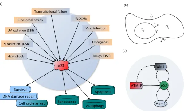
Whilst the p53 protein is present at very low level in normal cells mainly due to Mdm2-dependent degradation of p53 (p53 is very short-lived with the half-life 20 − 30 min [1]), it is extremely sensitive to even low levels of DNA damage thus either allowing for DNA repair or eliminating cells from becoming malignant [2], which are the properties attributing to tumour suppressing function of p53. Indeed, p53 can respond to several stresses such as DNA damage caused by γ-irradiation or drugs used in chemotherapy, oncogenes, heat and cold, which disrupt cellular integrity. p53 then may initiate processes leading either to a transient cell cycle arrest and DNA repair or to an irreversible cell fate including apoptosis, senescence or autophagy [3, 4], see Fig. 1(a).
The p53 protein, as a transcription factor, is largely nuclear when it is activated. In the nucleus, wild-type p53 promotes activation of several pro-survival, pro-apoptotic and other genes, likely with no preference for a particular group of genes [5]. In order for p53 to accumulate in the nucleus and become active, degradation-promoting effects of Mdm2, as the principal regulator of p53, must be suppressed [6]. Mdm2’s activity against p53 consists either in single ubiquitination of p53 followed by the nuclear export of p53 to the cytoplasm or Mdm2 promotes multiple ubiquitination with the subsequent export and degradation of p53 by the proteasome-degradation machinery in the cytoplasm [3, 6]. Persistent DNA damage may trigger another wave of nuclear accumulation of p53. Indeed, experiments in individual living breast cancer cells provided evidence for sustainedly oscillating p53 with pulses of fixed amplitudes and periods, which were visible even several days post-irradiation, [7, 8, 9].
Functional intracellular information may be encoded in the structural components of a cell (e.g., DNA or enzyme structures), however, information in cells can be also transmitted through the spatio-temporal dynamics of signalling proteins [13]. For instance,
• the aforementioned p53 oscillations triggered by γ-irradiation or drugs, maintained through the repeating nuclear
accumulation and removal of p53, are likely associated with transient cell cycle arrest and DNA repair, [9];
• damaged cells by γ-irradiation treated with nutlin, a small molecule inhibiting p53-Mdm2 interactions, showed sustained signalling of p53 of high level and were dominantly associated with senescence in [14] as well as in [15] where the cells were not treated with nutlin, however, they showed a delayed wave of p53 of high level several days post-irradiation, as the consequence of persistent genomic injury, associated with p21-dependent activation of senescence (p21 gene is another transcriptional target for p53);
• cells expressing p53 of high amplitude triggered apoptosis when exposed to UV radiation, [14].
(a) (b) Ω2 Ω1 Γ2 Γ1 n1 n2 (c)
FIGURE 1. (a) wt p53, “the guardian of the genome", is able to respond to a variety of stress stimuli and initiate cell cycle arrest, DNA damage repair, apoptosis or senescence; (b) cell scheme used in the spatio-temporal models for the p53 signalling in [10, 11, 12]. The cell consists of the nucleus Ω1, cytoplasm Ω2, nuclear membrane Γ1and cell membrane Γ2; n1and n2are the
unit normal vectors oriented outward from Ω1and Ω2. Organelles such as mitochondria and nucleoli can be easily involved in the
model; (c) scheme of the “minimal" network which is sufficient and necessary for oscillations of p53 in DNA damage response.
In addition, a certain fraction of p53 plays a part outside the nucleus [6], notably as a transcription-independent factor involved in the induction of apoptosis through its association with mitochondria [4, 16]. The cytoplasmic clearance of p53 may consequently lead to autophagy [16].
The spatio-temporal dynamical patterns of p53 become thus more intriguing because of the increased evidence that a chosen cell fate may be encoded in these patterns [13].
SPATIAL MODEL FOR P53
In [10, 11, 12] we have developed spatio-temporal models for activation and regulation of p53 which rely on reaction-diffusion equations simulating evolution of p53 and other proteins in 2D and 3D cells composed of the nucleus and the cytoplasm as on Fig. 1(b). In particular, these proteins are ATM as a sensor and transmitter of information about DNA damage and Mdm2 and Wip1 as negative regulators of p53 and ATM. Both Mdm2 and Wip1 are transcriptionally dependent on p53. These four proteins create a sufficient and necessary network for the oscillatory response of p53 to DNA double strand breaks (DSB) caused by γ-irradiation or drugs in vivo [9].
In brief, ATM in inactive state forms dimers that promptly dissociate into active monomers following occurrence of DSB. Such monomers then phosphorylate p53 on a residue, which is located very close to the Mdm2 binding domain, impeding Mdm2 to bind p53. Halted p53 ubiquitination results in p53 stabilisation in the nucleus where it acts transcriptionally towards many target genes, with the Mdm2 and Wip1 genes among them. The Wip1 protein dephosphorylates p53 and makes it a target for Mdm2 for degradation. Wip1 also dephosphorylates ATM molecules which create inactive dimers again, see Fig. 1(c), [10, 17] and references therein.
In the spatial setting as on Fig. 1(b), we can define a reaction diffusion system for an arbitrary protein network by du dt − div(D∇u) = f (u) in I× Ω1, dv dt − div(D∇v) = g(v) in I× Ω2, D∂ u ∂ n1 = P(v − u) = D∂ v ∂ n1 on I× Γ1, D ∂ v ∂ n2 = 0 on I× Γ2, (1)
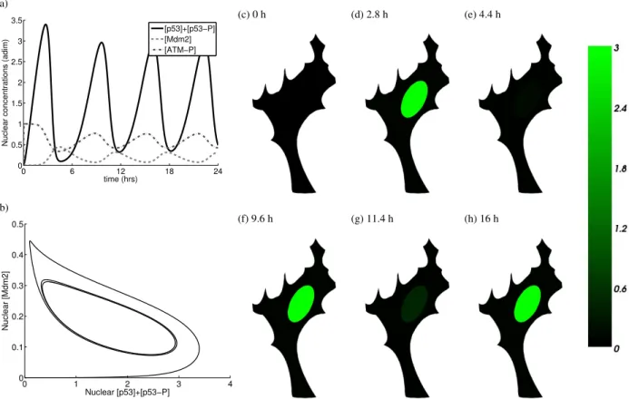
(a) 0 6 12 18 24 0 0.5 1 1.5 2 2.5 3 3.5 time (hrs)
Nuclear concentrations (adim)
[p53]+[p53−P] [Mdm2] [ATM−P] (b) 0 1 2 3 4 0 0.1 0.2 0.3 0.4 0.5 Nuclear [p53]+[p53−P] Nuclear [Mdm2] (c) 0 h (d) 2.8 h (e) 4.4 h (f) 9.6 h (g) 11.4 h (h) 16 h
FIGURE 2. (a) Nondimensionalised solution of the 2D p53 system: nuclear concentrations of p53 (p53 and p53-P), Mdm2 and ATM-P proteins. Since the dynamics of Mdm2 and Wip1 are similar their concentrations are almost identical curves. For this reason and for the clarity of image the concentration of Wip1 is not shown. The plotted concentrations are the total concentrations in the nucleus; (b) phase portrait of the nuclear concentration of p53 (p53 and p53-P) with respect to the nuclear concentration of Mdm2; (c)-(h) six 2D samples of the concentration of p53 (p53 and p53-P) captured at the time points when p53 and Mdm2 reach peaks in their concentrations.
where u = (u1, . . . , uN)T, ui= ui(t, x) : I × Ω1→ R and v = (v1, . . . , vN)T, vi= vi(t, x) : I × Ω2→ R, for i = 0, 1,. .., N −
1, denote nuclear and cytoplasmic concentrations of N species evolving in an open and bounded domain Ω = Ω1∪ Ω2⊂ Rd, d = 2, 3, and the time interval I = [0, T ], T > 0. Functions f : RN→ RN and g : RN → RN collect
protein-protein reaction terms and terms from gene regulatory networks. Avoiding any cross-diffusion, D is a diagonal N-by-N matrix with the diffusivities on the diagonal; P is a diagonal N-by-N with the permeabilities for the transport of the species through the nuclear membrane Γ1on the diagonal; div is the divergence operator. A 2D or 3D cell consists
of the two subdomains Ω1and Ω2representing the nucleus and the cytoplasm, respectively, Γ1is the nuclear membrane
and Γ2is the outer cellular membrane, Fig. 1(b). In our simplified framework, the cell can be as big as 10 − 100 µm,
it is able to proliferate and can have heterogeneous shape. However, we exclude cells of complicated structures and morphologies such as neurons or muscle cells. The nucleocytoplasmic transmission of species considered in (1) is driven by the difference of concentrations of the species in both compartments, that is by the so-called Kedem-Katchalsky boundary conditions. Zero-flux boundary conditions on the cellular membrane Γ2 and suitable initial
conditions are considered.
As it is demonstrated in [10, 11] and on Fig. 2, the model (1) applied to the p53 protein network with the chosen physiological parameters can reproduce observed oscillatory pattern of p53. Fig. 2(a) shows oscillations of the total nuclear concentrations of p53 (p53 and p53-P), Mdm2 and ATM-P (“-P" stands for phosphorylated) with the period 6.3 hours in the cell shown on Fig. 2(c)-(h), where the 2D solution of p53 and p53-P is plotted at six different times. Fig. 2(b) shows a phase portrait of the nuclear concentrations of p53 and Mdm2. More details about the models can be found in [10].
CONCLUSIONS
In the above spatio-temporal model, the oscillations were achieved not only by pure reactions between the species but also by the spatial localisation of cellular events either in the nucleus (e.g., gene transcription, ATM and p53 activation) or in the cytoplasm (e.g., mRNA translation) as well as by the migration of species in the compartments and between them [10, 11].
Other factors involved in different positive feedback loops regulating p53 may be important in different phases of the p53 signalling and may depend on spatial structure and other highly specific conditions [4]. As an example we can mention a p53-PTEN-PIP3-Akt feedback loop. In this loop Akt is phopshorylated at the cellular membrane by PIP3 and released back to the cytoplasm where it phosphorylates Mdm2. This is, according to some studies, essential modification of Mdm2 necessary for its translocation to the nucleus (although there are some discrepancies in the observations in other studies), [18] and citations therein. PTEN, as a substrate of p53, can inhibit the action of PIP3 towards Akt and thus it may enable p53 to reach high nuclear concentration. Another example is Mdm2 which primarily acts as a negative regulator of p53 establishing homeostasis in the DNA damage response (DDR). However, recently published studies [19, 20] show that following ATM-dependent phosphorylation of Mdm2, the phosphorylated Mdm2 may not target p53 for degradation but rather it enhances p53 synthesis. This is achieved by binding of Mdm2 to the nascent p53 mRNA which, passing likely through the nucleolus, changes Mdm2 to a positive regulator of p53.
In addition, the first p53-transcription independent wave of cells committing apoptosis in response to γ-irradiation is observed 30 minutes after DNA damage by rapid accumulation of p53 in the mitochondria [4]. The second delayed wave of apoptotic cells is determined by the concentrations of both pro-arrest and pro-apoptotic proteins [4, 5]. The p53 protein can activate both sorts of genes and the duration of expression of these genes and the concentration of their protein products are determined only by the concentration and duration of expression of p53 itself [5]. A cell with the damaged DNA evaluates the presence of both pro-arrest and pro-apoptotic proteins produced in a p53-dependent manner at any time during DDR. Apoptosis is initiated whenever the so-called “apoptotic ratio” reaches a certain threshold [5].
Spatial compartmental retention or clearance of p53 and localisation of other factors as well as more delicate spatial issues involved in precise timing of p53-dependent gene expression, post-translational modifications, promoter selectivity and dynamics determined by p53 mutational status, should not be excluded from further modelling of the cell fate decision and, for instance, intracellular actions of drugs used in chemotherapies.
REFERENCES
1. D. C. Olson, V. Marechal, J. Momand, J. Chen, C. Romocki, and A. Levine, Oncogene 8, 2353–2360 (1993). 2. K. H. Vousden, and D. P. Lane, Nature reviews Molecular cell biology 8, 275–283 (2007).
3. B. Vogelstein, D. Lane, and A. J. Levine, Nature 408, 307–310 (2000).
4. F. Murray-Zmijewski, E. A. Slee, and X. Lu, Nat Rev Mol Cell Biol 9, 702–712 (2008).
5. M. Kracikova, G. Akiri, A. George, R. Sachidanandam, and S. A. Aaronson, Cell Death Differ 20, 576–588 (2013). 6. D. Michael, and M. Oren, Seminars in Cancer Biology 13, 49–58 (2003).
7. G. Lahav, N. Rosenfeld, A. Sigal, N. Geva-Zatorsky, A. J. Levine, M. B. Elowitz, and U. Alon, Nat Genet 36, 147–150 (2004). 8. N. Geva-Zatorsky, N. Rosenfeld, S. Itzkovitz, R. Milo, A. Sigal, E. Dekel, T. Yarnitzky, Y. Liron, P. Polak, G. Lahav, and
U. Alon, Molecular Systems Biology 2, 1–13 (2006).
9. E. Batchelor, C. S. Mock, I. Bhan, A. Loewer, and G. Lahav, Molecular Cell 30, 277–289 (2008).
10. J. Eliaš, Mathematical model of the role and temporal dynamics of protein p53 after drug-induced DNA damage, Ph.D. thesis, Pierre and Marie Curie University (2015).
11. J. Eliaš, L. Dimitrio, J. Clairambault, and R. Natalini, Physical Biology 11, 045001 (2014).
12. J. Eliaš, and J. Clairambault, Computational and Structural Biotechnology Journal 10, 12–22 (2014). 13. J. E. Purvis, and G. Lahav, Cell 152, 945–956 (2013).
14. J. E. Purvis, K. W. Karhohs, C. Mock, E. Batchelor, A. Loewer, and G. Lahav, Science 336, 1440–1444 (2012). 15. R. Mirzayans, B. Andrais, A. Scott, and D. Murray, BioMed Research International 2012, 16 pp (2012). 16. D. R. Green, and G. Kroemer, Nature 458, 1127–1130 (2009).
17. J. Eliaš, L. Dimitrio, J. Clairambault, and R. Natalini, Biochimica et Biophysica Acta (BBA) - Proteins and Proteomics 1844, 232–247 (2014).
18. D. W. Meek, and U. Knippschild, Molecular Cancer Research 1, 1017–1026 (2003).
19. M. Gajjar, M. M. Candeias, L. Malbert-Colas, A. Mazars, J. Fujita, V. Olivares-Illana, and R. Fåhraeus, Cancer Cell 21, 25–35 (2012).
20. L. Malbert-Colas, A. Ponnuswamy, V. Olivares-Illana, A.-S. Tournillon, N. Naski, and R. Fåhraeus, Molecular Cell 54, 500–511 (2014).