HAL Id: hal-03064739
https://hal.archives-ouvertes.fr/hal-03064739
Submitted on 14 Dec 2020
HAL is a multi-disciplinary open access
archive for the deposit and dissemination of
sci-entific research documents, whether they are
pub-lished or not. The documents may come from
teaching and research institutions in France or
abroad, or from public or private research centers.
L’archive ouverte pluridisciplinaire HAL, est
destinée au dépôt et à la diffusion de documents
scientifiques de niveau recherche, publiés ou non,
émanant des établissements d’enseignement et de
recherche français ou étrangers, des laboratoires
publics ou privés.
Photonic jet lens
Sylvain Lecler, Stéphane Perrin, Audrey Leong-Hoi, Paul Montgomery
To cite this version:
Sylvain Lecler, Stéphane Perrin, Audrey Leong-Hoi, Paul Montgomery. Photonic jet lens. Scientific
Reports, Nature Publishing Group, 2019, 9 (1), �10.1038/s41598-019-41193-2�. �hal-03064739�
photonic jet lens
sylvain Lecler, stephane perrin , Audrey Leong-Hoi & paul Montgomery
Microsphere-assisted microscopy currently benefits from a considerable interest in the microscope-research community. Indeed, this new imaging technique enables the lateral resolution of optical microscopes to reach around λ/5 through a full-field and a far-field acquisition while being label-free. Despite the photonic jet clearly not being a relevant concept to justify the super-resolution phenomenon, we show here how it can be used to predict imaging formation and performance such as the image position and the microsphere magnification. This study allows a better understanding of the experimental measurements that have been observed over the last decade and that will be observed in coming years, through numerical simulations using different optical and geometrical parameters.
Supposedly known since 1908, the rigorous and complete Lorenz-Mie theory predicts the interaction of light with spherical particles1 and visible light scattered by dielectric microspheres2 in the far field. However, over the last
decades, the enhancement of computing power has made it possible to discover a focusing phenomenon in the near field of dielectric particles. Indeed, light is concentrated in a sub-diffraction-limit progressive beam known as photonic jet (PJ)3,4. This phenomenon is not specific to dielectric spherical-shape particles. As a matter of fact,
a square glass particle interacting with a plane incident wave is also able to generate a PJ (Fig. 1a)5. Moreover, at
this scale, propagation of light does not obey the classical laws of geometrical optics. The focusing effect is not only due to refraction of light by the particle but also to diffraction. The propagation of light inside the particle as well as around the particle must thus be considered in order to predict the generation of the PJ. Delayed inside the particle, the electromagnetic incident wave is also delayed continuously around the particle due to the continuity of the tangential electric field components, leading to a wavefront curvature as illustrated in Fig. 1b. In this work, only circular or cylindrical particles are considered.
The position and the width of the PJ irradiance peak depends on geometrical (e.g. the particle size) and opti-cal (e.g. the particle material and the wavelength of light) parameters. Since the ray-tracing is not relevant to study PJ performance, a rigorous electromagnetic model based on the Lorenz-Mie theory must be implemented. Figure 2 represents the evolution of PJ axial position P from the centre O of a microsphere as a function of its refractive index nm and for different radius ro. The wavelength of the plane wave incident on the microsphere is λ0 in air (ns = 1.00). The distance OP and the microsphere radius ro are expressed in wavelength λ0 due to the
invariant Maxwell equations by spatial scale modification. At a given radius ro, the higher the refractive index nm,
the smaller the distance OP, meaning the closer the PJ is to the center O. Nevertheless, at a given microsphere refractive index nm, an increase in the radius ro moves away the PJ. The black points indicate that the focus point P is on the microsphere interface, i.e. OP = ro. Through its asymptotic tendency (represented by the black dashed
curve), the PJ appears always inside the microsphere regardless of the radius ro when the refractive index is higher
than 1.83. It can be noted that using Snell’s law, the refractive index is higher and equals 2.0 for larger microsphere
sizes6.
In 2011, Wang et al. showed that dielectric microspheres do not only display the property of concentrating light in the near field, but can also perform a full-field super-resolved image in the far field7. In other words, the
microsphere is not merely a lens-like component focusing a near-field beam but it is also able to form an image. Amongst the numerous methods allowing microscopy to become nanoscopy8, microsphere-assisted
micros-copy is a new imaging technique which provides the advantage of being full field, label free, and easy to imple-ment. Miniature glass spheres have thus, once again, become “super-lenses” in microscopy, 350 years after van Leeuwenhoek used a single sphere of around 1 mm in size to visualize the first bacteria9. Although the
phenom-enon is currently still not explained, many papers studied performance of this imaging technique in 2D10–13 and
in 3D14–16. According to the Mie theory prediction, the PJ generation by a microsphere is not relevant to justify
this resolution improvement in the imaging process. Although the size of the focus spot overcome the diffraction limit, the full width at half maximum (FWHM) of the near-field beam waist is around a third of the wavelength17
which is higher than the lateral resolution obtained experimentally, i.e. around λ0/710 (or even λ0/1718 using
overestimated resolution criteria based on the gap measurement of features19). However, the PJ phenomenon can
explain the imaging formation in microsphere-assisted microscopy, i.e. the nature of the image, the position of the ICube, University of Strasbourg - CNRS (UMR 7357), 300 Bd Sébastien Brant, 67412, Illkirch, France. Correspondence and requests for materials should be addressed to S.P. (email: stephane.perrin@unistra.fr)
Received: 30 April 2018 Accepted: 15 February 2019 Published: xx xx xxxx
www.nature.com/scientificreports
www.nature.com/scientificreports/
image plane and the lateral magnification provided by the microsphere. Indeed, according to many studies on the PJ prediction3,20, the imaging process in microsphere-aided microscopy can be easily addressed by considering
the sphere as a photonic jet lens.
In this manuscript, the prediction of the image formation, i.e. the image nature, the image position and the lateral magnification, is descripted in microsphere-assisted microscopy by assimilating the microsphere as a clas-sical lens with geometrical optics. Indeed, due to the plane incident wave which can be assimilated to an infinitely distant object, the irradiance peak P of the PJ generated by the microsphere can be seen as the object focal point
F of a classical lens (Fig. 3a). The PJ generated by a soda lime glass microsphere has been numerically recon-structed using a plane incident wave in the TE mode. Assuming OP = OF and by means of Fig. 2, the nature of the image ′ ′A B of an object AB can then be retrieved (e.g., virtual imaging as schematically shown in Fig. 3b or real imaging as shown in Fig. 3c). Furthermore, a qualitative evolution of both the image position and magnification factor from the microsphere can be predicted as a function of the position of the object plane as well as the size and the refractive index of the microsphere.
Figure 1. Generation of a photonic jet through a square particle. (a) The normalised modulus of an electric
field interacting with the particle was retrieved using a finite element method in the TE mode. The size and the refractive index of the particle are 3 λ0 and 1.3 respectively, with λ0 being the wavelength in air. (b) A scheme
of wavefront distortion (red solid line) of the unitary plane wave is represented considering the continuity of the tangential component of the electric field in the particle and in the surrounding air medium, resulting in a photonic jet. Red arrows represent the wavefront vectors.
Figure 2. Axial position of the photonic jet irradiance peak. (a) The position of the photonic jet spot P is
estimated as a function of the microsphere refractive index nm for different radii ro. (b) In the Lorenz-Mie
model, the microsphere is illuminated by an incident plane wave with a wavelength of λ0 in air (ns = 1.0) in
order to retrieve the focusing phenomenon. Nine radius-dependent curves are represented, going from ro = 1 λ0
to ro = 9 λ0, with an increment of 1 λ0. The distance OP and ro are expressed in wavelength λ0. The black dots
show the position of the point P on the surface of the microsphere. For example, when nm = 1.52 and r0 = 4.0 λ0,
Results
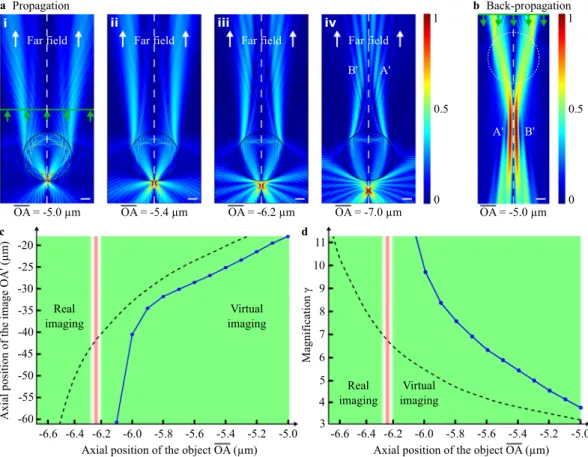
The influence of the object plane position on the image formation was firstly analysed. Therefore, the object has been displaced along the optical axis, and both the image plane position ′OA and magnification factor γ were
estimated. Furthermore, the comparison with the classical geometrical laws was studied. The results are shown in Fig. 4. The simulation was implemented in air (ns = 1.00) using here two emitting-point sources A and B as
men-tioned in Methods section (unlike the previous simulations where a plane wave illuminated the microsphere for finding the focusing spot). The object AB is placed at different axial position OA under a soda-lime-glass micro-sphere (nm = 1.52) having a radius ro of 5 μm. The wavelength λ0 of the emitting electric field being here of 500 nm, r0 equals thus 10 λ0 and the PJ spot is outside the microsphere at OP = −6.25 μm = −12.5 λ0 (Fig. 2a). When the
point sources are placed between the last dioptre of the microsphere and the PJ spot, i.e. OA = [−5.0 μm; −6.2 μm] (Fig. 4a.i and a.ii), the image is formed above the microsphere and is thus virtual (Fig. 4b). In this case, the further the object AB is from the microsphere, the further away is the position of the image ′ ′A B (Fig. 4c). Moreover, the magnification remains positive and increases steadily as shown in Fig. 4d. An asymptotic behaviour of the two curves appears when the object plane meets the focusing plane, i.e. when OA = OP. At this particular position, the image is located at infinity in the far field (Fig. 4a.iii). This phenomenon can also be found over a short distance (OA = [−6.2 μm;−6.3 μm]) which can be assimilated as the range of the Gouy phase shift21. In this range, the
magnification γ goes on to increase continuously until reaching infinity. This asymptotic tendency to infinity is similar in geometrical optics22 (black-dashed curve) when OA = ′OF :
γ = − ′ 1 1 (1) geo OA OF
Finally, beyond the P point position, the image ′ ′A B appears real and is formed above the microsphere as
shown in Fig. 4a.iv. The distance ′OA is then positive and γ becomes negative.
A modification of the microsphere refractive index nm provides also an influence on the imaging formation.
Indeed, an increase in nm results in the focusing point P being closer to the sphere interface. Furthermore, for nm
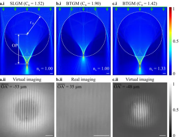
being higher than 1.8, we have shown that the focusing spot P is inside the sphere regardless to the microsphere size (Fig. 2a). In this situation, ′ ′A B should thus always be real. We validated this assumption using two
micro-spheres having different refractive index, i.e. a soda-lime-glass microsphere (nm = 1.52) and a
barium-titanate-glass microsphere (nm = 1.90), and a similar diameter (ro = 16 μm). A 600-nm-period positive
Ronchi grating (Aluminium features on glass wafer) was used as object and placed against the microsphere sur-face (OA = ro). When the PJ spot P is outside the microsphere (simulation in Fig. 5a.i), the latter generates a virtual
image (experiment in Fig. 5a.ii). Whereas, when P is inside the microsphere (simulation in Fig. 5b.i), the image appears to be real and less contrasted (experiment Fig. 5b.ii). It can be mentioned that, in geometrical optics, the effective focal length of a ball lens is smaller than the radius only when the refractive index is superior to 2.022.
This image process has already been experimentally presented in microsphere-assisted microscopy using both a 30 λ0-radius barium-titanate-glass microsphere and a 35 λ0-radius polystyrene microsphere (nm = 1.59)12. Maslov
and Astratov have also demonstrated the influence of the refractive index on the image process through simula-tions23. It has been shown that a microsphere having a radius of 3.2 λ
0 and a refractive index of 1.4, performs
Figure 3. Concept of photonic jet lens. (a) A glass microsphere forms a photonic jet where its irradiance peak
P can be seen as the object focal point F of the microsphere. In geometrical optics, this assumption leads to
the formation of (b) a virtual image when the object is placed between P and the microsphere, and (c) a real image when the object is placed further than P. The 2D FEM simulation was performed using an illumination wavelength λ0 of 500 nm in air. The refractive index of the 10 λ0-radius glass microsphere is 1.52, giving OP = 12.5 λ0.
www.nature.com/scientificreports
www.nature.com/scientificreports/
virtual imaging when the object is at 3.3 λ0 from the microsphere interface, i.e. above the focusing spot P position.
And, a 1.9-refractive-index microsphere results in real imaging. These results confirm our assumption which considers the microsphere as a photonic jet lens.
Placing now the 16 μm-radius barium-titanate-glass microsphere in water (ns∼1.33) provides a PJ which is
below the microsphere (simulation in Fig. 5c.i) and a virtual image occurs (experiment in Fig. 5c.ii). As a mat-ter of fact, according to Mie theory, a sphere with a refractive index nm, embedded in a surrounding medium
of refractive index ns and illuminated by a beam having a wavelength λ0 in air, is equivalent to a sphere having
a refractive index of nm/ns in air and being illuminated by an electric field wavelength of λ0/ns2. We can hence
deduce that a barium titanate microsphere immersed in water (ns∼1.33) is equivalent to a silica glass microsphere
(Cn = nm/ns∼1.90/1.33∼1.42) having an equivalent size parameter but illuminated with a wavelength λ0 divided
by ns. Numerical simulations and experiments in Fig. 5a,c confirm this concept. The clear interest of immersion
microsphere-assisted microscopy is thus to benefit from a smaller wavelength as in classical microscopy. It could be mentioned that considering the influence of the refractive index of the surrounding medium, new simulations are hence not required.
The influence of the probing wavelength on the magnification γ provided by the microsphere is also analysed. Here, the object AB is placed in contact with a 10 λ0-radius microsphere. The simulations are performed for
wavelengths λ0 in air within the visible range. As shown in Fig. 6a, the longer the wavelength λ0, the higher the
magnification γ following a linear evolution. Through the PJ evolution according to the wavelength (Fig. 2), the increase of γ can be understood. Indeed, for long wavelengths, the PJ appears closer to the microsphere and thus to the object AB, consequently making the magnification higher. In accordance, the image distance increases for
Virtual imaging Real imaging -6.2 -50
Axial position of the object OA (µm)
Axial positio no ft he imag eO A' (µm ) 0 . 6 - -5.8 -5.6 -5.2 -5.0 -45 -40 -30 -25 -20 c
Axial position of the object OA (µm)
Magnification γ 4 5 6 7 9 10 d OA = -5.0 µm a Far field OA = -5.4 µm Far field OA = -6.2 µm Far field Propagation 0 0.5 1 OA = -7.0 µm Far field B' A' -5.4 -35 -6.2 -6.0 -5.8 -5.6 -5.4 -5.2 -5.0 11 8 3 i ii iii iv b A' B' OA = -5.0 µm Back-propagation 0 0.5 1 6 . 6 - -6.4 -6.6 -6.4 Virtual imaging Real imaging -60 -55
Figure 4. Influence of the object plane position on the image formation. The electric field from two
out-of-phase point sources A and B, spaced 400 nm apart in air, is first propagated through the microsphere where (a) the normalised modulus of the electric field is represented for different axial positions OA. When, (a.i,a.ii) the object AB is above the PJ spot, (b) the back-propagation operation in the surrounding medium (from green line) is required to retrieve the potential virtual images A′ and B′. When, (a.iii) AB is in the intermediate axial range around the PJ, the image is formed at infinity. Finally, when (a.iv) the object AB is clearly below the PJ irradiance peak, the image becomes real. White-dotted lines represent the optical axis of the simulation and green arrows represent the direction of the time-reversed propagation. White scale bars represent 2 μm. (c) The image position and (d) the magnification are calculated as a function of OA. The simulations have been performed with a wavelength λ0 of 500 nm using a 1.52-refractive-index microsphere with a radius of 10 λ0. The
transverse distance AB is 400 nm. Black-dotted lines represent the evolution curves of the image plane position and the magnification using geometrical optics relations with a 5 μm-radius glass ball lens22. This assumption
shows a similar behaviour of the curves although the values are different at this scale (e.g. the focal length of the ball lens equals 7.30 μm and γ = n/(2 − n) = 3.17 when OA = −5.0 μm).
a.i SLGM (Cn= 1.52) b.i BTGM (Cn= 1.90) c.i BTGM (Cn= 1.42)
Virtual imaging Real imaging Virtual imaging
ns= 1.00 ns= 1.00 ns= 1.33
r0
OP
a.ii b.ii c.ii 0
0.5 1
0 0.5 1 OA' = -53 µm OA' = 35 µm OA' = -48 µm
Figure 5. Image formation depending on the refractive index contrast. Cn between the microsphere and the
surrounding medium. A soda lime glass microsphere SLGM (nm = 1.52), (a.i) generating a photonic jet outside
the microsphere (OP>ro) in air (ns = 1.00), thus (a.ii) a virtual image occurs. Whereas, using a
barium-titanate-glass microsphere BTGM (nm = 1.90) in air, (b.i) the focus spot P appears inside the microsphere, (b.ii)
providing a real image. Replacing the air immersion medium by water (ns = 1.33) allows (c.i) the photonic jet
position to be further than the microsphere interface (OP >ro) and (c.ii) the image to be once again virtual. In the three cases, the microspheres have a radius ro of 16 μm. The photonic jet beams are simulated using a plane
incident illumination with λ0 = 600 nm. The super-resolved images were experimentally measured using a
600-nm-period Ronchi grating placed against the microspheres (OA = ro). White scale bars represent 2 μm.
1,100 300 Wavelength λ0(nm) 500 700 900 3 4 5 6 7 8 Magnificatio n γ 2 Radius r0(µm) 3 4 5 6 7 8 Magnificatio n γ 2 2 3 4 5 6 7 8 a b
Figure 6. Magnification factor γ of the microsphere depending on optical and geometrical parameters. (a) γ is
expressed as a function of the emitting wavelength λ0. The object AB was spaced 400 nm apart and placed
against the microsphere having a refractive index of 1.52 (OA = r0 = 5 μm). For example, γ = 3.6 when λ0 = 500 nm. (b) γ is then calculated as a function of the microsphere radius r0 using Eq. 2 with λ0 = 500 nm.
Black-dashed line represents a curve fitting of γ using geometrical optics defined as γ = κ/f′ where f′ is the focal length of a ball lens and κ a constant value equalling 25.6 μm in this case22.
www.nature.com/scientificreports
www.nature.com/scientificreports/
high wavelengths. It should be noted that, here, the wavelength dependence of the microsphere refractive index24
is not considered, i.e. nm = 1.52 regardless the wavelength in the simulations. Thus, the generation of chromatic
aberrations does not come from the material dispersion, i.e. nm(λi), but from both the shape and the size of the
microsphere, as retrieved in optical fibre telecommunications.
Measuring the magnification γ as a function of the radius ro of the microsphere could provide wrong
interpre-tations of the results. Indeed, changing ro means that the distance OA is also modified. However, due to the scale
invariance from the Maxwell equations in the spatial domain, changing the sphere radius r1 for a constant source
wavelength in air λ1 is equivalent to changing the source wavelength in air λ2 with a constant sphere radius r2
with: λ = λ r r (2) 1 1 2 2
An increase in the wavelength is interpreted as a decrease in the radius of the microsphere. Thus, the magnifi-cation γ of the microsphere has been expressed as a function of its radius r0 in Fig. 6b from the previous results
(Fig. 6a). As in geometrical optics (black-dashed curve)22, the magnification γ clearly decreases when r
0 increases.
Indeed, a sphere radius increase leads to a longer distance between the PJ and the microsphere (as well as the object AB located at their default position in a plane tangent to the sphere) and hence, a reduction of γ. Reciprocally, when r0 decreases, the focus point P becomes closer to the object plane (Fig. 2), giving an
improve-ment of γ until it reaches an infinite value and an image located at infinity in the far field. By this means, the magnification should be considered to enable the imaging of the virtual image by the microscope objective. However, there is no interest in large magnification. Indeed, a too large magnification requires spheres with a small diameter, reducing the field of view and taking away the virtual image from the sphere.
Discussion
To date, the super-resolution phenomenon through microsphere-assisted microscopy has not been explained and is still in discussion. In this manuscript, it has been shown that the photonic jet does not form the basics of the lateral resolution enhancement. Indeed, the photonic jet effect (with a FWHM of around λ0/3 in air) is not
relevant to justify the observed resolution. The evanescent waves appear yet to be more pertinent to justify the lateral resolution enhancement and this approach is supported by several recent papers25–27. Moreover, the role
of the coherence should also be considered10. If the role of the evanescent waves is confirmed, we must
under-line that the photonic jet is a phenomenon based on propagative waves and can describe the imaging process. Nevertheless, it cannot be excluded that the imaging process could be slightly different through the evanescent wave approach, providing a shift in the focus point. Such a hypothesis could explain the observation of two image planes13,23. One of them would be the image of the propagative waves, i.e. small k wave vector, and the second
one, the image of the evanescent waves, i.e. large k wave vector. However, this is still only a hypothesis. The recent results concerning phase measurements and their exploitation for 3D reconstruction will be an opportunity for further investigation16.
Conclusion
This paper shows that photonic jet physics has to be considered in order to understand the imaging formation in microsphere-assisted microscopy. At this scale, the microsphere cannot be seen as a classical lens due to its small radius which makes the focus generation not only related to the refraction of light but also to the diffraction of light. However, considering the photonic jet irradiance peak as the focus point, we can deduce the nature of the image, i.e. real or virtual, as well as the tendency of the image position and the magnification from the micro-sphere. In this manuscript, simulations and experiments of performance of microsphere-assisted microscopy illustrate this phenomenon and allows the analogy with geometrical optics to be made. Indeed, it has been shown that placing the object plane before or after the photonic jet spot leads to a virtual or a real image, respectively. Furthermore, the magnification factor provides a behavior which is similar to geometrical optics. This similitude between geometrical optics and the photonic jet lens laws has been further highlighted through the study of the near-field focusing spot position by changing the refractive index of the microsphere and the surrounding medium. The use of high-refractive-index microspheres and immersion medium is also discussed, giving an obvious enhancement of performance due to the smaller equivalent wavelength in the surrounding medium. This effect can also be retrieved in immersion optical microscopy. Finally, the microsphere imaging process has been analysed as a function of the illumination wavelength and the size of the microsphere.
Method
To demonstrate the relevance of the photonic jet lens concept, the performance of microsphere-assisted micros-copy has been numerically computed using two rigorous numerical simulations in the TE mode. The propagation equations were solved using a 2D finite-element method (Comsol Multiphysics) with perfectly matched layers as radiation numerical boundary conditions. In this 2D model, the microsphere is reduced to a cylinder where the axis is along the y axis and is placed in a medium with a refractive index ns. The radius and the refractive index of
the cylinder are ro and nm, respectively. Two point sources A and B, serving as object, are placed symmetrically in
the horizontal plane below the microsphere at an axial distance OA. The dipoles A and B have a coherent phasor with a relative phase difference of pi in order to contribute to separate their images10,15. In the first simulation, the
complex electric field from A and B is transmitted through the microsphere and the far field is recorded (Fig. 7a). Second, if the image is virtual, this complex electromagnetic field is back-propagated (or time-reversal propa-gated) in the surrounding medium ns without the microsphere (Fig. 7b). It should be noted that, if the image is
recovered by finding the maxima values of the modulus of the electric field which is proportional to the irradiance of light. Finally, the axial position ′OA of the image and the magnification γ (defined as the ratio between
trans-versal distances ′ ′A B and AB) are determined. In the experiments, an objective lens above the microsphere
col-lects the far field from the object and relays it onto a camera sensor. Obviously, the performance of the objective lens must be significant to resolve the two point sources A′ and B′.
Moreover, experiments has been performed by introducing soda-lime-glass or barium-titanate-glass micro-spheres in a classical optical microscope, i.e. below the objective lens of the microscope (×20, NA = 0.4, Zeiss) and onto the object surface. The microspheres were illuminated by a light source having a central wavelength of 600 nm. The object consists of a Ronchi grating having a period of 600 nm with a groove of 300 nm and feature height of 100 nm. The images (real or virtual depending on both the sphere material and immersion conditions) were collected by matching the microscope object plane with the microsphere image plane. Finally, the images were recorded by a camera without implementing image processing algorithms. It should be mentioned that the microscope alone is able to resolve two details having a separation distance of at least 800 nm.
References
1. Mie, G. Beiträge zur Optik trüber Medien, speziell kolloidaler Metallösungen. J. Ann. Phys 25, 377–445, https://doi.org/10.1002/
andp.19083300302 (1908).
2. Van de Hulst, H. C. Light Scattering by Small Particles (John Wiley & Sons, New York, 1957).
3. Lecler, S., Takakura, Y. & Meyrueis, P. Properties of a three-dimensional photonic jet. Opt. Lett. 30, 2641–2643, https://doi.
org/10.1364/OL.30.002641 (2005).
4. Horiuchi, N. Light scattering: Photonic nanojets. Nat. Photonics 6, 138–139, https://doi.org/10.1038/nphoton.2012.43 (2012).
5. Pacheco-Pena, V., Beruete, M., Minin, I. V. & Minin, O. V. Multifrequency focusing and wide angular scanning of terajets. Opt. Lett. 40, 245–248, https://doi.org/10.1364/OL.40.000245 (2015).
6. Luk’yanchuk, B. S. et al. Refractive index less than two: photonic nanojets yesterday, today and tomorrow. Opt. Mater. Express 7, 1820–1847 (2017).
7. Wang, Z. et al. Optical virtual imaging at 50 nm lateral resolution with a white-light nanoscope. Nat. Commun. 2, 218, https://doi.
org/10.1038/ncomms1211 (2011).
8. Montgomery, P. C. & Leong-Hoi, A. Emerging optical nanoscopy techniques. Nanotechnol. Sci. Appl. 8, 31–44, https://doi.
org/10.2147/NSA.S50042 (2015).
9. Porter, J. R. A. van Leeuwenhoek: Tercentenary of his discovery of bacteria. Bacteriol. Rev. 40, 260–269 (1976).
10. Maslov, A. V. & Astratov, V. N. Imaging of sub-wavelength structures radiating coherently near microspheres. Appl. Phys. Lett. 108,
051104, https://doi.org/10.1063/1.4941030 (2016).
11. Darafsheh, A., Walsh, G. F., Negro, L. D. & Astratov, V. N. Optical super-resolution by high-index liquid-immersed microspheres. Appl. Phys. Lett. 101, 141128, https://doi.org/10.1063/1.4757600 (2012).
12. Lai, H. S. S. et al. Super-resolution real imaging in microsphere-assisted microscopy. PLoS ONE 11, e0165194, https://doi.
org/10.1371/journal.pone.0165194 (2016).
13. Ye, R. et al. Experimental imaging properties of immersion microscale spherical lenses. Sci. Rep. 4, 3769, https://doi.org/10.1038/
srep03769 (2014).
14. Wang, F. et al. Three-dimensional super-resolution morphology by near-field assisted white light interferometry. Sci. Rep. 6, 24703,
https://doi.org/10.1038/srep24703 (2016).
15. Kassamakov, I. et al. 3D Super-resolution optical profiling using microsphere enhanced Mirau interferometry. Sci. Rep. 7, 3683,
https://doi.org/10.1038/s41598-017-03830-6 (2017).
16. Perrin, S. et al. Microsphere-assisted phase-shifting profilometry. Appl. Opt. 56, 7249–7255, https://doi.org/10.1364/AO.56.007249
(2017).
17. Heifetz, A. et al. Photonic Nanojets. J. Comput. Theor. Nanosci. 6, 1979–1992, https://doi.org/10.1166/jctn.2009.1254 (2009).
Distance
z(
µm
)
b
Distance x (µm)
-
1
0
0
1
0
0
-10
10
-20
-30
0
0.5
1
A' B'
a
Distance
z(
µm
)
0
0.5
1
0
5
10
-10 -5
Distance x (µm)
0
-5
-10
10
5
A B
OA
Figure 7. Method of the numerical analysis of microsphere-assistedmicroscopy. To retrieve the image
position, the complex electric field from the out-of-phase emitting points A and B is first propagated through themicrosphere and maintained (green line on the top) to be then back-propagated in the surroundingmedium without the microsphere. (a) The real part of the electric field and (b) the normalized absolute value of the time-reversed electric field are represented. Here, A and B are placed between the microsphere and the irradiance peak of the photonic jet, leading to virtual images A0 and B0. The microsphere has a radius ro of 5 μm and a refractive index nm of 1.52. The wavelength λ0 is 500 nmand the surroundingmedium is air (ns = 1.0).
www.nature.com/scientificreports
www.nature.com/scientificreports/
18. Yan, Y. et al. Microsphere-coupled scanning Laser confocal nanoscope for sub-diffraction-limited imaging at 25 nm lateral
resolution in the visible spectrum. ACS Nano 8, 1809–1816, https://doi.org/10.1021/nn406201q (2014).
19. Sheppard, C. J. R. Resolution and super-resolution. Microsc. Res. Tech. 80, 590–598, https://doi.org/10.1002/jemt.22834 (2017).
20. Itagi, A. V. & Challener, W. A. Optics of photonic nanojets. J. Opt. Soc. Amer. A 22, 2847–2858, https://doi.org/10.1364/
JOSAA.22.002847 (2005).
21. Gouy, L. G. Sur une propriete nouvelle des ondes lumineuses. C. R. Acad. Sci. Paris 110, 1251 (1890). 22. Riedl, M.J. Optical Design Fundamentals for Infrared Systems, Second Edition (SPIE Press, Bellingham, 2001).
23. Maslov, A. V. & Astratov, V. N. Optical nanoscopy with contact Mie-particles: Resolution analysis. Appl. Phys. Lett. 110, 261107,
https://doi.org/10.1063/1.4989687 (2017).
24. Rubin, M. Optical properties of soda lime silica glasses. Sol. Energ. Mat. Sol. C. 12, 275–288,
https://doi.org/10.1016/0165-1633(85)90052-8 (1985).
25. Li, L., Guo, W., Yan, Y., Lee, S. & Wang, T. Label-free super-resolution imaging of adenoviruses by submerged microsphere optical
nanoscopy. Light Sci. Appl. 2, e104, https://doi.org/10.1038/lsa.2013.60 (2013).
26. Allen, K. W. et al. Super-resolution microscopy by movable thin-films with embedded microspheres. Ann. Phys. 527, 513–522,
https://doi.org/10.1002/andp.201500194 (2015).
27. Ben-Aryeh, Y. Increase of resolution by use of microspheres related to complex Snell’s law. J. Opt. Soc. Am. A 33, 2284–2288, https://
doi.org/10.1364/JOSAA.33.002284 (2016).
Acknowledgements
This work was supported by the University of Strasbourg and SATT Conectus Alsace. The authors thank Amandine Elchinger for her constructive input in writing the manuscript.
Author Contributions
All authors designed the project and the research. S.L. and S.P. implemented the computer program and performed the simulations. S.L. and S.P. processed the numerical data and analysed the results with A.L.-H.S.L. and S.P. wrote the manuscript. S.P. created the figures. P.M. supervised the whole work. All the authors reviewed the manuscript.
Additional Information
Competing Interests: The authors declare no competing interests.
Publisher’s note: Springer Nature remains neutral with regard to jurisdictional claims in published maps and
institutional affiliations.
Open Access This article is licensed under a Creative Commons Attribution 4.0 International
License, which permits use, sharing, adaptation, distribution and reproduction in any medium or format, as long as you give appropriate credit to the original author(s) and the source, provide a link to the Cre-ative Commons license, and indicate if changes were made. The images or other third party material in this article are included in the article’s Creative Commons license, unless indicated otherwise in a credit line to the material. If material is not included in the article’s Creative Commons license and your intended use is not per-mitted by statutory regulation or exceeds the perper-mitted use, you will need to obtain permission directly from the copyright holder. To view a copy of this license, visit http://creativecommons.org/licenses/by/4.0/.


