ORIGINAL ARTICLE
PTH improves titanium implant fixation more
than pamidronate or renutrition in osteopenic rats
chronically fed a low protein diet
R. Dayer&T. C. Brennan&R. Rizzoli&P. Ammann
Received: 9 February 2009 / Accepted: 27 July 2009 / Published online: 27 October 2009
# International Osteoporosis Foundation and National Osteoporosis Foundation 2009
Abstract
Summary We evaluated the effects of parathyroid hormone (PTH), pamidronate, or renutrition on osseointegration of titanium implants in the proximal tibia of rats subject to prolonged low-protein diets. PTH improved mechanical fixation, microarchitecture, and increased pull-out strength. Pamidronate or renutrition had lesser effects. PTH can thus improve implant osseointegration in protein-malnourished rats.
Introduction Protein malnutrition impairs implant osseoin-tegration in rats. PTH and pamidronate prevent deleterious effects of protein restriction introduced just prior to implantation. Whether these treatments improve osseointe-gration after chronic protein deprivation, i.e., in osteopenic bone at time of implantation, is unknown. We evaluated effects of PTH, pamidronate, or renutrition on resistance to pull-out of titanium rods implanted into the rat tibiae following isocaloric low-protein intake.
Methods Forty-one adult female rats received normal or isocaloric low-protein diets. Six weeks later, implants were surgically inserted into proximal tibiae. Following implantation, rats on low-protein diets were treated with
PTH (1-34), pamidronate, saline vehicle, or normal protein diets, for another 8 weeks. Tibiae were removed for micro-computerised tomographic morphometry and evaluation of pull-out strength.
Results Pull-out strength decreased in rats on isocaloric low-protein diets compared with normal protein group (−33.4%). PTH increased pull-out strength in low-protein group, even compared to controls from the normal protein group. PTH and pamidronate increased bone volume/tissue volume, bone-to-implant contact, and trabecular thickness, whilst trabecular separation was reduced, with a shift to more plate-like bone surrounding the implants.
Conclusions PTH reversed the deleterious effects of long-term protein undernutrition on mechanical fixation and bone microarchitecture and improved implant osseointegra-tion more than pamidronate or renutriosseointegra-tion, likely through changes to structure model index.
Keywords Implants . Nutrition . Pamidronate . Parathyroid hormone . Rodents
Introduction
As the population continues to age, undernutrition and protein deficiency persist amongst elderly patients living in nursing homes and in community-dwelling subjects [1–6]. This problem is widely acknowledged and has even driven the need for assessment tools to diagnose under- or malnutrition [5,7–9]. Decreases in dietary protein can be caused by age-related physiological changes, diseases of the gastrointestinal tract or of an inflammatory nature, physical impairment, and psychological as well as psycho-social issues. It is widely acknowledged that protein
Romain Dayer and Tara C. Brennan contributed equally to this paper. R. Dayer
:
T. C. Brennan:
R. Rizzoli:
P. Ammann (*)Division of Bone Diseases (WHO Collaborating Center for Osteoporosis Prevention), Department of Rehabilitation and Geriatrics, Geneva University Hospitals and Faculty of Medicine, 1211 Geneva 14, Switzerland e-mail: [email protected] R. Dayer e-mail: [email protected] T. C. Brennan e-mail: [email protected] R. Rizzoli e-mail: [email protected] DOI 10.1007/s00198-009-1031-x
undernutrition leads to prolonged illness periods [10], reduced immunity [11–13], and severely negative effects on the musculoskeletal system [14–16]. It is not surprising then that a high prevalence of undernutrition is observed in elderly patients undergoing total hip or total knee arthro-plasty [17–20] and that the prognosis for these patients is rather poor as it is difficult to stimulate food intake in these patients.
In a previous report, we demonstrated that 26 weeks of an isocaloric low protein diet induces a trabecular bone volume (BV) loss of around 75% in female rats and 70% in male rats. This was associated with a diminution of ultimate strength (or maximal load) in both females and males. Stiffness and energy were also negatively affected by the low protein diet in these animals. More recently, we reported that isocaloric low protein intake, introduced around the time of surgical implantation, impairs titanium implant osseointegration in adult female rats. This was associated with poor bone microarchitecture surrounding the implant [21].
Both bisphosphonates and parathyroid hormone (PTH) treatment have been reported to improve implant fixa-tion, allowing for more durable joint replacements [22– 32]. Indeed, in a recent study, we showed that both PTH and pamidronate were able to prevent the deleterious effects of an isocaloric protein restricted diet on bone microarchitecture close to the implant and on mechanical fixation in adult female rats [33]. In these experiments, rats were subject to low protein for only 2 weeks before the implants were inserted. Immediately following the implant surgery, PTH or pamidronate was administered for 8 weeks. Both PTH and pamidronate improved the mechanical fixation, even when compared to animals with normal protein intake.
It is more likely that a longer duration of protein malnourishment prior to implantation would more closely resemble what is often observed in the clinical setting, with many elderly patients having osteopenia caused by a long-term lack of sufficient dietary protein. No data has been reported on whether systemic treatment with bisphosphonates or PTH (1-34) improve the altered titanium implant osseointegration observed in prolonged low protein-induced osteopenic models, or modify the tridimensional trabecular microarchitecture in the vicinity of an implant. The effects of renutrition following implantation in such models have also never been tested. The aim of the current study was, therefore, to test the hypothesis that systemic treatments of PTH, bisphosph-onates, or renutrition are able to augment titanium implant osseointegration in a rat model of protein-malnourished-induced osteopenia, a model which closely resembles what is frequently observed in the clinical setting of orthopaedic wards.
Materials and methods Animals and diet
All experimental designs and procedures have received approval of the Animal Ethics Committee of the Geneva University Faculty of Medicine. Forty-one 10-month-old Sprague-Dawley female rats (Charles River Laboratories, L’Arbresle, France), housed individually at 25°C with a 12:12-h light–dark cycle, were employed. Rats were strictly pair-fed a laboratory diet provided by Provimi Kliba AG (Kaiseraugst, Switzerland) containing either 15% or 2.5% casein, 0.8% phosphorus, 1% calcium, 70– 80% carbohydrates, and 5% fat throughout the experi-mental period. Isocaloric intake by all rats was ensured by the addition of corn carbohydrate to the low protein diet, thus providing the same energy intake across all groups. Demineralised water was available ad libitum. Since the minimal protein intake in rats necessary to maintain normal bone homeostasis is 5% [34,35] and it is known that casein is not wholly utilisable by rats, a restriction to 2.5% casein corresponds to a reduction of more than 50% of the minimum recommended intake, and a reduction of 50% is often observed in patients who sustain a hip fracture [36,37].
Experimental design
One week after an initial adaptation period to a 15% casein-containing diet, rats were divided into five groups. One group received a diet containing 15% casein (n=8, 15% control group), and three groups were placed on a diet containing 2.5% casein until the end of the study. The final group received the 2.5% casein diet until the implantation, after which, they were commenced on 15% casein (n=8, renutrition group). Six weeks following the beginning of isocaloric protein restriction (i.e., at week7), a titanium implant was inserted in each tibia of the animals under general anaesthesia. We inserted implants into both tibias to avoid the possibility of disuse or overuse often observed when only one limb is operated on. Following implantation, rats from one of the groups receiving 2.5% casein were injected subcutaneously five times a week with human PTH (1-34; Eli Lilly and Company, Indianapolis, IN, USA) at a dose of 40μg/kg body weight per injection, until the end of the study (n=8, 2.5% PTH group). This dose has previously been demonstrated to effectively correct the altered bone mechanical properties associated with protein deprivation in rats [33, 38]. It is higher than that used in humans. Similarly, another of the groups receiving 2.5% casein-containing diet was treated five times a week during the first and the fifth weeks post-implantation surgery with subcutaneous injection of
3-amino-1hydroxypropylidene-1,1-bisphosphonate (Pamidronate, Novartis International AG, Basel, Switzerland) at a dose of 0.6 mg/kg (n=9, 2.5% pamidronate group). This dose has been shown to prevent the deleterious effect of isocaloric protein deprivation on bone strength [39]. Rats in the pamidronate group also received a subcutaneous injection of vehicle (sodium chloride, 0.9%, 0.5 ml/kg) five times a week in those weeks they did not receive pamidronate. All treatments and the vehicle were injected at the same volume per kilogramme body weight. The renutrition group was fed with a 15% casein-containing diet after the implantation as described above. Finally, the group fed a diet containing 15% casein (n=8, 15% control group), the renutrition group, and the remaining group on 2.5% casein diet (n=8, 2.5% control group) were injected with vehicle (sodium chloride, 0.9%, 0.5 ml/kg) five times a week during the 8 weeks following implantation. At 8 weeks post-implantation (i.e., at week15), blood was obtained from all rats by aortic puncture under general anaesthesia, and animals were killed by an overdose of ketamine hydrochloride. The tibias were removed for micro-computerised tomographic (μCT) morphometry and mechanical testing.
Implants design and preparation
The implants were made from commercially pure titanium (Institut Straumann AG, Waldenburg, Switzerland), were 1.0 mm in diameter and 4.1 mm long, and were sandblasted and acid etched on the non-threaded part. The threaded part was 1.25 mm long (type M1) and remained outside of the bone, permitting prehension for the pull-out test. This type of implant was selected as titanium is well tolerated by bone tissue. The rough surface, fashioned by sandblasting and acid-etching, ensures good osseointegration. The implants were cleaned in phosphate-free Deconex® (Borer Chemie AG, Zuchwil, Switzerland), rinsed in pure water in an ultrasonic bath, and sterilised with ethylene oxide. Implantation technique
Animals were anaesthetised by abdominal injections of ketamine (100 mg/kg) and xylazine (10 mg/kg). The skin of the bilateral tibial region was shaved and cleaned with 70% ethanol. We have previously provided a pictorial description of the implanted region [21]. Under aseptic conditions, a 10-mm incision was made anteriorly to gain access to the proximal medial section of the tibia metaphysis. The periosteum was reflected medially. The periosteum in adult rats is a relatively thin and fragile tissue, which cannot be replaced around the implant. It was thus left reclined. A 1.0-mm diameter hole was drilled through one of the cortices with a hand-held drill into which the implant was inserted. Rotatory speed did not exceed 2,000 rpm, and drilling was
accompanied by profuse saline irrigation to avoid thermal bone necrosis. The proximal limit of the entry point was delimited by a virtual line perpendicular to the long axis of the tibia and crossing the anterior edge of the growth plate centrally, which is curved in this central region anteriorly and inferiorly. The second anatomical landmark utilised was a virtual line passing from the inferior border of the tendinous insertion on the proximal anterior tibial crest to a medial tendinous insertion, which most likely corresponds to the pes anserinus in humans. The point of insertion was halfway between these two tendinous insertions. We have previously demonstrated by X-ray examination that employing such landmarks leads to the implant being in contact with the trabecular bone of the proximal tibial metaphysis [21], at a distance of 2 mm distal to the growth plate along the great axis of the tibia. The implant was inserted perpendicularly to the surface of the cortical bone on the medial part of the proximal tibia. The depth of the inserted implant section into the bone corresponded to the length of the non-threaded section of the implant (2.85 mm). Only the threaded part remained outside the bone. The implants were specifically designed taking into account the X-ray determined dimen-sions of the proximal tibia in adult female Sprague-Dawley rats. After insertion of the implant, the skin was sutured using a 3-0 resorbable polyglactic suture (Vycril®; Ethicon, Spreintenbach, Switzerland). Postoperative analgesia was maintained by subcutaneous injection of buprenorphine (0.06 mg/kg) twice a day for 3 days.
Micro-computerised tomographic (μCT) morphometry The tibias were carefully excised immediately after death and frozen at−20°C in plastic bags. The night before μCT analysis, the bones were thawed slowly at 7° and maintained at room temperature. During the whole analysis, they were immersed in saline solution. Parameters of volume, architecture, and percentage of implant surface in direct contact with trabecular bone (percentage of attach-ment, bone-to-implant contact) were studied using μCT morphometry (μCT 40; Scanco Medical AG, Bassersdorf, Switzerland). The voxel was 20μm in all spatial directions. For penetrating radio-opaque titanium and improving signal-to-noise ratio, the system was set to 70 kEV with a 350-ms integration time. The number of tomographic projections was 1,000 projections per 360°. Images were filtered with a 3-D Gauss philtre with Sigma 1.5 voxels and then segmented with threshold 1.4 cm−1for bone (25% of maximal grey scale value, visually chosen) and threshold 5.8 cm−1 for titanium (attenuation coefficient). Trabecular bone was analysed on a circular band of 0.5 mm around the implant (Fig. 2). Relative BV/tissue volume (TV), trabec-ular number, trabectrabec-ular thickness, and trabectrabec-ular separation were calculated by directly measuring the 3-D distances in
the trabecular network. Connectivity density based on Euler number (Conn.D) and the structure model index (SMI) were calculated. When a bone voxel was in the vicinity (60 µm boundary) of a titanium voxel, this titanium voxel was classified as“attached surface”. With a distance of 3 voxels (60µm) from the detected titanium surface, the presence or absence of bone can be accurately determined (IPL, image processing language software, available with the µCT 40, Scanco Medical AG, Bassersdorf, Switzerland). The arte-facts of the titanium in the CT image prevented us from analysing the presence of bone at a range closer to the implant [40, 41]. The number of attached surface voxels divided by the total number of titanium surface voxels yielded the percentage bone-to-implant contact. The tridi-mensional reconstructions obtained were always verified against the original grey scale images.
Pull-out test
After μCT analysis, implanted tibias were subjected to a pull-out test. The tibias were held in place on a metal device by two flat supports 5.6 mm apart. Another metal piece was screwed onto the threaded, i.e., external part of the implant and was used to grip the implant during the test. Pull-out strength was determined as the peak force, i.e., the maximum failure force, applied to completely loosen the implant from the bone, and was measured with a servo-controlled electromechanical system (Instron 1114; Instron Corp., High Wycombe, UK) with the actuator displacement speed set at 2 mm/min. Reproducibility was 13.4% (3.3– 22), as evaluated by the mean of the absolute differences between paired-sample measurements (left/right). A pre-liminary study showed that freezing the tibias did not alter pull-out strength values. The mean of the absolute differ-ences between values obtained before or after freezing procedure was 12.5% (0.9–35.5). Regression between values obtained before or after freezing procedure was characterised by an r2coefficient of 0.96 [21].
Biochemical assays
Plasma insulin-like growth factor I (IGF-I) was measured by immunoenzymometric assay, with a kit from Nichols Institute (San Juan Capistrano, CA, USA) following the manufacturer’s instructions, and osteocalcin employing a kit from Biomedical Technologies Inc. (Stoughton, MA, USA).
Statistical analysis
All results are expressed as mean ±SEM. Significance of difference was evaluated by analysis of variance (ANOVA) and post-analysis by Tukey’s honestly significant difference
test (Statistical Package for the Social Sciences (SPSS) 14.0; SPSS Inc., Chicago, IL, USA).
Results
Effect of PTH, pamidronate, or renutrition on pull-out strength
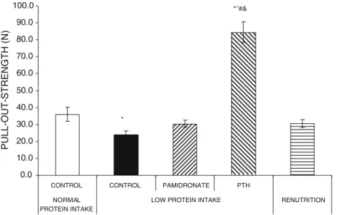
As previously shown [21, 33], mean pull-out strength values were decreased in rats fed an isocaloric low protein diet (−33.4%) compared with animals in the normal protein group (Fig. 1). Neither pamidronate nor renutrition were able to increase mechanical fixation above the low protein control group. PTH treatment, on the other hand, markedly increased the pull-out strength compared to the control animals receiving the same diet (2.5%). Pull-out strength values in the 2.5% PTH group were also higher than in animals never subject to a low protein diet, i.e., the 15% control group, as well as compared to the renutrition and pamidronate-treated groups.
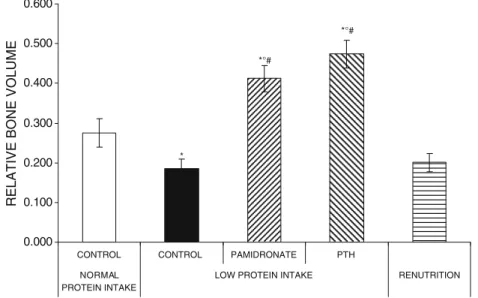
Effect of PTH, pamidronate, or renutrition onμCT morphometry of the trabecular bone around the implant To evaluate the effects of PTH, pamidronate, and renu-trition on pull-out strength, we analysed the trabecular bone microarchitecture around the implant and bone-to-implant contact. BV/TV values and bone-to-implant contact were lower in the low protein group compared to the normal protein group (Figs. 2, 3, and 4). Both PTH and pamidronate treatments increased BV/TV values and bone-to-implant contact compared with the low-protein intake control group and the renutrition group. Systemic administration of PTH or pamidronate under protein restriction increased BV/TV and bone-to-implant contact to values higher than those observed in control animals with normal protein intake. Finally, renutrition was unable to increase BV/TV or bone-to-implant contact compared to the low-protein control group.
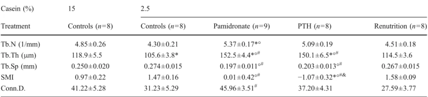
Protein restriction, PTH, and pamidronate affected other parameters of the trabecular bone architecture in the vicinity of the implant (Table1). A significant increase of trabecular number was noted in the 2.5% pamidronate group compared with the 15% and the 2.5% controls. Isocaloric protein restriction was associated with lower values of trabecular thickness when compared to the normal protein intake control group. PTH and pamidronate treatments increased trabecular thickness when compared to the low-protein control animals, the normal protein control animals, or the animals in the renutrition group. Accordingly, similar changes were noted for trabecular separation when compared to 2.5% and 15% control groups. This was associated with
alterations of the trabecular structure, as illustrated by changes in the SMI. Protein restriction induced a shift toward more rod-like bone microstructures, although more plate-shaped bone was observed in the animals treated with PTH or pamidronate compared with 2.5% controls and the renutrition group. This effect was stronger under PTH
treatment, with a decrease of SMI even when compared to the values obtained with pamidronate or in the normal protein control group. A non-significant decrease of connec-tivity density was noted in the low-protein-fed animals. This value was higher with pamidronate treatment, when com-pared to animals in the renutrition group.
0.0 10.0 20.0 30.0 40.0 50.0 60.0 70.0 80.0 90.0 100.0
CONTROL CONTROL PAMIDRONATE PTH
NORMAL PROTEIN INTAKE
LOW PROTEIN INTAKE RENUTRITION
PULL-OUT-STRENGTH (N)
*
*°#&
Fig. 1 Effects of systemic pamidronate or parathyroid hormone (PTH) treatment under isocaloric low protein intake or renutrition on pull-out strength of titanium implant inserted in the proximal tibial metaphysis of adult female rats 8 weeks after surgery, i.e., after 14 weeks of protein restriction. Treatments with pamidronate or PTH and renutrition were started immediately after implantation. Values are
means±SEM, asterisk corresponds to p<0.05 vs the control normal protein intake group, degrees corresponds to p<0.05% vs the control low protein intake group, number sign corresponds to p<0.05% vs the renutrition group, and ampersand corresponds to p<0.05% vs the pamidronate low protein group, as evaluated by analysis of variance and post-analysis by Tukey’s honestly significant difference test
Normal protein Control Low protein Control
Low protein Pamidronate Low protein PTH Renutrition BV/TV: 0.279 BIC: 0.663 BV/TV: 0.363 BIC: 0.928 BV/TV: 0.513 BIC: 0.838 BV/TV: 0.230 BIC: 0.567 BV/TV: 0.171 BIC: 0.506 1.0 mm 1.0 mm 1.0 mm 1.0 mm 1.0 mm Fig. 2 Microtomographic morphometry by microcom-puted tomography. Microtomo-graphic tridimensional reconstruction of the trabecular bone around titanium implants at 8 weeks after implantation. The two upper panels are from samples in the control groups (15% control and 2.5% control), and the three lower ones are from samples in the pamidronate and parathyroid hormone (PTH) low protein intake groups (2.5% pamidronate and 2.5% PTH) and renutrition group. Relative bone volume (BV/TV) around titanium implants and bone-to-implant contact (BIC) of the corresponding samples are indicated
Effect of isocaloric protein restriction and renutrition on IGF-I, osteocalcin, and on body weight
Isocaloric low protein intake decreased plasma IGF-I levels (Table2) consistent with previously shown data [21,34,35,
42]. Protein restriction was associated with lower body weight, despite strict pair-feeding with isocaloric diets. Renutrition increased plasma IGF-I levels and body weight
when compared to the three other groups subjected to protein restriction throughout the entire study.
Isocaloric protein restriction was not associated with significant changes of osteocalcin levels, when compared to the 15% controls and the renutrition groups (Table 2). Treatment with pamidronate was associated with a reduc-tion of osteocalcin levels when compared with the 2.5% control group and the renutrition group. PTH induced
0.000 0.100 0.200 0.300 0.400 0.500 0.600
RELATIVE BONE VOLUME
*
*°# *°#
CONTROL CONTROL PAMIDRONATE PTH
NORMAL PROTEIN INTAKE
LOW PROTEIN INTAKE RENUTRITION
Fig. 3 Effects of systemic pamidronate or parathyroid hormone treatment under isocaloric low protein intake, or renutrition on relative bone volume (BV/TV) around titanium implants in the proximal tibial metaphysis of adult female rats 8 weeks after surgery. Values are means±SEM, asterisks corresponds to p<0.05 vs the control normal
protein intake group, degrees corresponds to p<0.05% vs the control low protein intake group, and number sign corresponds to p<0.05% vs the renutrition group, as evaluated by analysis of variance and post-analysis by Tukey’s honestly significant difference test
0.000 0.100 0.200 0.300 0.400 0.500 0.600 0.700 0.800 0.900 1.000
%OF BONE IMPLANT CONTACT
*°# *°#
*
CONTROL CONTROL PAMIDRONATE PTH
NORMAL PROTEIN INTAKE
LOW PROTEIN INTAKE RENUTRITION
Fig. 4 Effects of systemic pamidronate or parathyroid hormone treatment under isocaloric low protein intake or renutrition on bone-to-implant contact in the proximal tibial metaphysis of adult female rats 8 weeks after surgery. Values are means ± SEM, asterisk corresponds to p<0.05 vs the control normal protein intake group,
degrees corresponds to p<0.05% vs the control low protein intake group, and number sign corresponds to p<0.05% vs the renutrition group, as evaluated by analysis of variance and post-analysis by Tukey’s honestly significant difference test
significant higher levels of osteocalcin when compared to the animals treated with pamidronate.
Discussion
We have previously shown that an isocaloric low protein diet induces increased bone resorption and depressed bone formation, resulting in bone loss in rats [34]. The results of the current study indicate that a prolonged isocaloric low protein intake also results in deleterious effects on titanium implant osseointegration. Systemic treatment with PTH (1-34) markedly increased the mechanical fixation of the implant under protein
restric-tion. Interestingly, PTH treatment in addition to a low protein diet was able to increase the pull-out strength needed to completely loosen the implants, even compared to control animals with normal protein intake. We also observed that PTH treatment significantly improved bone-to-implant contact and induced a higher relative BV in the vicinity of the implants. PTH also induced an increase in trabecular thickness, a decrease in trabecular separation, and a shift to more plate-like bone structures, which have all been shown to improve some of the biomechanical properties that aid in the prevention of bone fragility and decrease fracture risk [43]. It is also likely, therefore, that such improvements in these properties contribute to the osseointegration of titanium implants. It should be noted
Table 1 Effects of pamidronate or parathyroid hormone under isocaloric dietary protein restriction or renutrition on bone architecture around the titanium implant
Casein (%) 15 2.5
Treatment Controls (n=8) Controls (n=8) Pamidronate (n=9) PTH (n=8) Renutrition (n=8) Tb.N (1/mm) 4.85±0.26 4.30±0.21 5.37±0.17*° 5.09±0.19 4.51±0.18 Tb.Th (μm) 118.9±5.5 105.6±3.8* 152.5±4.4*°# 150.1±6.5*°# 114.5±3.6 Tb.Sp (mm) 0.250±0.020 0.274±0.015 0.197±0.011°# 0.203±0.013°# 0.267±0.015 SMI 0.97±0.22 1.47±0.16 0.01±0.42°# −1.07±0.32*°#& 1.58±0.09 Conn.D. 41.22±5.28 31.23±5.29 45.96±3.51# 37.20±4.31 27.59±3.77 Values are means±SEM. Results are trabecular number (Tb.N), trabecular thickness (Tb.Th), trabecular separation (Tb.Sp), structure model index (SMI), and connectivity density based on Euler number (Conn.D.). SMI is an estimate of the rod characteristics of trabecular bone (0 value = purely plate-shaped bone, 3 value=purely rod-like bone, negative values indicate a solid mass with only few holes in it). Isocaloric protein restriction was started 6 weeks before bilateral tibial insertion of titanium implants. After surgery, rats were treated with daily subcutaneous injections of a saline vehicle (control), pamidronate, or human parathyroid hormone (PTH), as specified in the“Materials and methods”. Another
group received a diet containing 2.5% casein until the implantation; after the surgery, animals in this group received a diet containing 15% casein and were treated with saline vehicle (renutrition group). Animals were sacrificed at 8 weeks after implantation
asterisk corresponds to p<0.05 vs the control normal protein intake group; degrees corresponds to p<0.05% vs the control low protein intake group; number sign corresponds to p<0.05% vs the renutrition group; ampersand corresponds to p<0.05% vs the pamidronate low protein group (as evaluated by ANOVA and post-analysis by Tukey’s HSD test)
Table 2 Effects of isocaloric protein restriction on insulin-like growth factor I, osteocalcin plasma levels, and change in body weight Casein (%) 15 2.5
Treatment Controls (n=8) Controls (n=8) Pamidronate (n=9) PTH (n=8) Renutrition (n=8) IGF-I (ng/mL) 687.1±35.8 480.7±20.2*# 427.9±17.7*# 444.3±26.3*# 678.3±29.8 Osteocalcin (nmol/L) 10.8±0.5 11.9±0.9 6.7±0.2*°# 12.6±0.7& 10.3±0.5 Δ Weight (g) −13.8±8.6 −70.1±8.2*# −66.4±6.0*# −71.1±7.4*# −22.6±5.5
Values are means±SEM. Isocaloric protein restriction was started 6 weeks before bilateral tibial insertion of a titanium implant. After surgery, rats were treated with daily subcutaneous injections of a saline vehicle (control), pamidronate, or human parathyroid hormone (PTH), as specified in “Materials and methods”. Another group received a diet containing 2.5% casein until the implantation; after the surgery, animals in this group
received a diet containing 15% casein (renutrition group) and were treated with saline vehicle. Determinations were performed 8 weeks after implantation, when rats were killed. Values are mean insulin-like growth factor I plasma levels (IGF-I) and mean difference of weight from the beginning of the experiment (Δ weight)
asterisk corresponds to p<0.05 vs the control normal protein intake group; degrees corresponds to p<0.05% vs the control low protein intake group; number sign corresponds to p<0.05% vs the renutrition group; ampersand corresponds to p<0.05% vs the low protein group treated with pamidronate (as evaluated by ANOVA and post-analysis by Tukey’s HSD test)
that PTH treatment may have a greater anabolic effect on bone in rats than in humans, as an increased mean wall thickness has been reported in rats given intermit-tent PTH treatment [44], whereas this parameter is unchanged in humans [45]. Treatment with pamidronate, an inhibitor of bone resorption, was also able to effectively prevent the negative impact of isocaloric protein restriction on bone architecture surrounding the titanium implants. Pamidronate induced an increase in relative BV, bone-to-implant contact, trabecular number, and trabecular thickness, even when compared to control animals with normal protein intake. Improved trabecular separation and a shift to more plate-like trabecular structures were also noted when compared to control animals on an isocaloric low protein diet. However, these positive morphometric effects of pamidronate were not associated with significant increases in the pull-out strength. Finally, renutrition implemented immediately following implantation was unable to improve mechanical fixation, relative BV, bone-to-implant contact, or any of the other measured parameters of trabecular bone micro-architecture around the titanium implant, despite com-pletely correcting the low IGF-I levels induced by the low protein diet. It should also be noted that in other studies performed in our group, it has been reported that dietary protein intake in patients who sustained hip fractures averaged between 0.6 and 0.7 g/kg of body weight [46,
47], whereas the recommended daily allowance for elderly adults is 1–1.2 g/kg of body weight [48], with anything below 0.8 g/kg of body weight being considered to represent protein undernutrition [14]. Furthermore, attempts to improve protein intake have been performed with insufficient success [36, 49]. It is very difficult to change the habits of both the patients and the caretakers. In addition, following dental implant procedures, mastic-ular function is often decreased, and maintaining food intake, let alone increasing it in order to obtain higher dietary protein, is rarely possible. In our opinion, there is no doubt that the best case scenario would be to improve protein intake post-surgery; however, it remains to be the situation, even within care-facilities, and such intervention is therefore not common practise. As such, post-surgical protein malnutrition remains a problem. This, taken with the results of the current study, highlights the need to look to treatments to improve osseointegration following implantation. It should be noted that one limitation of the current study is that while casein is a very common protein source for rats, it is not wholly digestible or utilisable by rats [50], and therefore, the rats receiving the low protein diet may actually be receiving less than 50% of the minimal requirement for protein.
The changes in bone remodelling previously observed with protein deprivation [34] were also associated with
decreased IGF-I concentrations and a resistance to IGF-I at the level of the target organs. In the present study, decreased plasma IGF-I was found in all animals exposed to the isocaloric low protein diet; however, IGF-I levels returned to normal in the renutrition group only. This confirms the low IGF-I levels under a low protein diet. Such malnutrition may also interfere with the process of osseointegration, in which both bone resorption and formation are implicated [51,52].
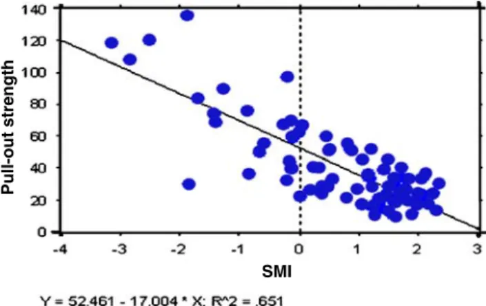
A recent study in animals indicated that treatments with PTH or pamidronate immediately following implantation fully prevented the deleterious effects associated with an isocaloric low protein diet commenced 2 weeks prior to implantation. Both treatments significantly increased pull-out strength in animals fed an isocaloric low protein diet, and PTH even induced a higher mechanical fixation when compared to animals with normal protein intake [33]. The current results establish that PTH treatment provides beneficial effects following long-term protein deprivation, a condition which more closely mimics the nutritional status of many elderly patients in orthopaedic wards undergoing total hip or knee arthroplasty [17–20]. The increases seen in bone-to-implant contact and relative BV following PTH treatment contribute to the positive effects of PTH on implant fixation after long-term low protein diet. We have previously shown that bone-to-implant contact is a strong predictor of pull-out strength [21]. It should be noted, however, that relative BV and bone-to-implant contact were not significantly different between PTH and pamidronate treatments, yet the pull-out strength values with PTH were higher than those obtained with pamidro-nate. Linear regression analysis indicates that there is a relationship between SMI and pull-out strength, character-ised by an r2value of 65% (Fig.5). SMI characterises the form of the trabecular structure, describing whether this
Pull-out strength
SMI
Fig. 5 Regression analysis between structure model index (SMI) and pull-out strength. Regression analysis revealed that SMI is a good predictor of pull-out strength, with an r2value of 0.651: Y=2.249− 0.038×X; R2=0.651
bone takes on rod-like shapes or more plate-like structures and was the only microstructural parameter which predicted pull-out strength. This is the first time a relationship between SMI and implant pull-out strength has been reported and may indeed underlie the interesting differences in pull-out strength observed between the PTH-treated and alendronate-treated rats. These results do not rule out the involvement of other factors, including enhanced material level properties and/or an increase in bone ingrowth into the microscopic pits of the textured titanium surface with PTH treatment. It is possible that this would generate a stronger bone interlacing and mechanical coupling at the interface. The nanometric pits on the titanium surface are largely below the resolution power of the µCT. The only technique available for such a tridimensional analysis would be a synchrotron [53], which was not available for this study.
Another possible explanation for the less potent effects of pamidronate under long-term protein deprivation is linked to the observation that isocaloric protein restriction is initially able to transiently decrease both bone resorption and bone formation [35]. Decreased bone formation may be the dominant feature of the changes in bone remodelling seen during the first weeks of protein undernutrition. This is also consistent with the strong positive effects of PTH treatment under such a diet, promoting bone formation into the microscopic pits on the titanium surface and thus inducing stronger mechanical fixation of the implant. With bone formation already depressed by protein deprivation, pamidronate, whose effects are primarily to inhibit bone resorption, is unable to reverse this deleterious effect [34]. However, our results regarding osteocalcin levels do not support this hypothesis. Osseointegration represents a complex interaction in which local bone biology plays a key role. This could contribute to the difficulty of drawing explanations based on biochemical markers reflecting general bone metabolism.
Finally, renutrition was unable to correct the deleterious effects of protein restriction on implant fixation, bone-to-implant contact, and the other tested parameters of trabecular bone microarchitecture, even when applied over a longer period (8 weeks) than protein deprivation (6 weeks). It should be noted, however, that plasma IGF-I was increased by protein renutrition, mimicking observa-tions seen in clinical studies [54,55]. This lack of effect of renutrition on osseointegration, even with increased IGF-I plasma levels (when compared to the other low protein groups), was unexpected. Indeed, in a previous study, essential amino acid supplements, at doses, which effec-tively increased plasma IGF-I, corrected bone strength via an increase in cortical thickness [42]. Taken together, these results combined with the current data support the hypothesis that essential amino acids may be more efficacious than protein repletion alone. Another possibility
is that IGF-I production stimulated by protein intake affects trabecular bone less than it does cortical bone.
In conclusion, in an experimental model of prolonged isocaloric dietary protein restriction, we have shown, for the first time, that systemic treatment with PTH is able to completely reverse the negative effects of long-term protein undernutrition on titanium implant osseointegration. In animals fed a low protein diet, mechanical fixation was significantly increased by PTH treatment, even when compared with the animals fed a normal protein diet. These positive effects were associated with increased bone-to-implant contact and with beneficial changes in the trabecular structure around the implant, including increased relative BV, thicker trabeculae, diminished trabecular separation, and a more plate-like trabecular network. Pamidronate treatment also significantly increased bone-to-implant contact and relative BV, even when compared with animals in the normal protein group. Positive changes in other parameters of trabecular bone microarchitecture around the implant were also noted; however, a significant corresponding increase in the mechanical fixation of the implant was not observed, while renutrition was unable to improve bone microarchitecture or pull-out strength values.
Acknowledgements The authors thank S. Clément for expert animal care, diet preparations, and blood collection; Dr. A. Laib for technical help in using µCT; Dr. M. Cattani, I. Badoud and V. Chatelain for biomechanical testing; and M. Perez for secretarial assistance. This research project was supported by Swiss National Research Foundation (Grant 3200BO-100714).
Conflicts of interest None.
References
1. Smoliner C, Norman K, Scheufele R, Hartig W, Pirlich M, Lochs H (2008) Effects of food fortification on nutritional and functional status in frail elderly nursing home residents at risk of malnutri-tion. Nutrition 24:1139–1144
2. Preben Ulrich P (2005) Nutritional care: the effectiveness of actively involving older patients. J Clin Nurs 14:247–255 3. Guigoz YY, Lauque SS, Vellas BJBJ (2002) Identifying the
elderly at risk for malnutrition. The mini nutritional assessment. Clin Geriatr Med 18:737–757
4. Van Nes M-C, Herrmann FR, Gold G, Michel J-P, Rizzoli R (2001) Does the mini nutritional assessment predict hospitaliza-tion outcomes in older people? Age Ageing 30:221–226 5. Feldblum I, German L, Castel H, Harman-Boehm I, Bilenko N,
Eisinger M, Fraser D, Shahar D (2007) Characteristics of undernourished older medical patients and the identification of predictors for undernutrition status. J Nutr 6:37
6. Rizzoli R, Ammann P, Bourrin S, Chevalley T, Bonjour JP (2001) Protein intake and bone homeostasis. In: Burckhardt P, Dawson-Hughes B, Heaney RP (eds) Nutritional aspects of osteoporisis. Academic, San Diego, pp 219–235
7. Omran ML, Morley JE (2000) Assessment of protein energy malnutrition in older persons, part I: history, examination, body composition, and screening tools. Nutrition 16:50–63
8. Omran ML, Morley JE (2000) Assessment of protein energy malnutrition in older persons, part II: laboratory evaluation. Nutrition 16:131–140
9. Soini H, Routasalo P, Lagstrom H (2004) Characteristics of the mini-nutritional assessment in elderly home-care patients. Eur J Clin Nutr 58:64–70
10. Edington J, Boorman J, Durrant ER, Perkins A, Giffin CV, James R, Thomson JM, Oldroyd JC, Smith JC, Torrance AD, Blackshaw V, Green S, Hill CJ, Berry C, McKenzie C, Vicca N, Ward JE, Coles SJ (2000) Prevalence of malnutrition on admission to four hospitals in England. Clin Nutr 19:191–195
11. Chandra RK (1997) Nutrition and the immune system: an introduction. Am J Clin Nutr 66:460S–463S
12. Paillaud E, Herbaud S, Caillet P, Lejonc J-L, Campillo B, Bories P-N (2005) Relations between undernutrition and nosocomial infections in elderly patients. Age Ageing 34:619–625
13. Wintergerst ES, Maggini S, Hornig DH (2007) Contribution of selected vitamins and trace elements to immune function. Ann Nutr Metab 51:301–323
14. Rizzoli R (2008) Nutrition: its role in bone health. Best Pract Res Clin Endocrinol Metab 22:813–829
15. Bruyere O, Brandi ML, Burlet N, Harvey N, Lyritis G, Minne H, Boonen S, Reginster JY, Rizzoli R, Akesson K (2008) Post-fracture management of patients with hip Post-fracture: a perspective. Curr Med Res Opin 24:2841–2851
16. Roubenoff RR (2000) Sarcopenia and its implications for the elderly. Eur J Clin Nutr 54(Suppl 3):S40–S47
17. Gherini S, Vaughn BK, Lombardi AV Jr, Mallory TH (1993) Delayed wound healing and nutritional deficiencies after total hip arthroplasty. Clin Orthop 293:188–195
18. Greene KA, Wilde AH, Stulberg BN (1991) Preoperative nutritional status of total joint patients. Relationship to postoper-ative wound complications. J Arthroplast 6:321–325
19. Marin LA, Salido JA, Lopez A, Silva A (2002) Preoperative nutritional evaluation as a prognostic tool for wound healing. Acta Orthop Scand 73:2–5
20. Rai J, Gill SS, Kumar BR (2002) The influence of preoperative nutritional status in wound healing after replacement arthroplasty. Orthopedics 25:417–421
21. Dayer R, Rizzoli R, Kaelin A, Ammann P (2006) Low protein intake is associated with impaired titanium implant osseointegra-tion. J Bone Miner Res 21:258–264
22. Aleksyniene R, Hvid I (2004) Parathyroid hormone—possible future drug for orthopedic surgery. Medicina (Kaunas) 40:842– 849
23. Skripitz R, Aspenberg P (2004) Parathyroid hormone—a drug for orthopedic surgery? Acta Orthop Scand 75:654–662
24. Shanbhag AS (2006) Use of bisphosphonates to improve the durability of total joint replacements. J Am Acad Orthop Surg 14:215–225
25. Tokugawa Y, Shirota T, Ohno K, Yamaguchi A (2003) Effects of bisphosphonate on bone reaction after placement of titanium implants in tibiae of ovariectomized rats. Int J Oral Maxillofac Implants 18:66–74
26. Narai S, Nagahata S (2003) Effects of alendronate on the removal torque of implants in rats with induced osteoporosis. Int J Oral Maxillofac Implants 18:218–223
27. Skoglund B, Holmertz J, Aspenberg P (2004) Systemic and local ibandronate enhance screw fixation. J Orthop Res 22:1108–1113
28. Astrand J, Aspenberg P (2004) Topical, single dose bisphospho-nate treatment reduced bone resorption in a rat model for prosthetic loosening. J Orthop Res 22:244–249
29. Soininvaara TA, Jurvelin JS, Miettinen HJ, Suomalainen OT, Alhava EM, Kroger PJ (2002) Effect of alendronate on peripros-thetic bone loss after total knee arthroplasty: a one-year,
randomized, controlled trial of 19 patients. Calcif Tissue Int 71:472–477
30. Wilkinson JM, Stockley I, Peel NF, Hamer AJ, Elson RA, Barrington NA, Eastell R (2001) Effect of pamidronate in preventing local bone loss after total hip arthroplasty: a randomized, double-blind, controlled trial. J Bone Miner Res 16:556–564
31. Skripitz R, Aspenberg P (2001) Early effect of parathyroid hormone (1–34) on implant fixation. Clin Orthop Relat Res 392:427–432
32. Shirota T, Tashiro M, Ohno K, Yamaguchi A (2003) Effect of intermittent parathyroid hormone (1-34) treatment on the bone response after placement of titanium implants into the tibia of ovariectomized rats. J Oral Maxillofac Surg 61:471–480 33. Dayer R, Badoud I, Rizzoli R, Ammann P (2007) Defective
implant osseointegration under protein undernutrition: prevention by PTH or pamidronate. J Bone Miner Res 22(10):1526–1533 34. Ammann P, Bourrin S, Bonjour JP, Meyer JM, Rizzoli R (2000)
Protein undernutrition-induced bone loss is associated with decreased IGF-I levels and estrogen deficiency. J Bone Miner Res 15:683–690
35. Bourrin S, Toromanoff A, Ammann P, Bonjour JP, Rizzoli R (2000) Dietary protein deficiency induces osteoporosis in aged male rats. J Bone Miner Res 15:1555–1563
36. Delmi M, Rapin CH, Bengoa JM, Delmas PD, Vasey H, Bonjour JP (1990) Dietary supplementation in elderly patients with fractured neck of the femur. Lancet 335:1013–1016
37. Rizzoli R, Ammann P, Chevalley T, Bonjour JP (2001) Protein intake and bone disorders in the elderly. Jt Bone Spine 68:383– 392
38. Ammann P, Gasser JA, Rizzoli R (2003) Protein intake determines net bone strength response to PTh or GH, two anabolic agents increasing bone turnover. J Bone Miner Res 2:S386
39. Mekraldi S, Toromanoff A, Rizzoli R, Ammann P (2005) Pamidronate prevents bone loss and decreased bone strength in adult female and male rats fed an isocaloric low-protein diet. J Bone Miner Res 20:1365–1371
40. Butz F, Ogawa T, Chang TL, Nishimura I (2006) Three-dimensional bone-implant integration profiling using micro-computed tomogra-phy. Int J Oral Maxillofac Implants 21:687–695
41. Rebaudi A, Koller B, Laib A, Trisi P (2004) Microcomputed tomographic analysis of the peri-implant bone. Int J Periodontics Restor Dent 24:316–325
42. Ammann P, Laib A, Bonjour JP, Meyer JM, Ruegsegger P, Rizzoli R (2002) Dietary essential amino acid supplements increase bone strength by influencing bone mass and bone microarchitecture in ovariectomized adult rats fed an isocaloric low-protein diet. J Bone Miner Res 17:1264–1272
43. Ito M, Nishida A, Koga A, Ikeda S, Shiraishi A, Uetani M, Hayashi K, Nakamura T (2002) Contribution of trabecular and cortical components to the mechanical properties of bone and their regulating parameters. Bone 31:351–358
44. Wronski TJ, Pun S, Liang H (1999) Effects of age, estrogen depletion, and parathyroid hormone treatment on vertebral cancellous wall width in female rats. Bone 25:465–468
45. Jiang Y, Zhao JJ, Mitlak BH, Wang O, Genant HK, Eriksen EF (2003) Recombinant human parathyroid hormone [1–34] [ter-iparatide] improves both cortical and cancellous bone structure. J Bone Miner Res 18:1932–1941
46. Schurch M-A, Rizzoli R, Slosman D, Vadas L, Vergnaud P, Bonjour J-P (1998) Protein supplements increase serum insulin-like growth factor-i levels and attenuate proximal femur bone loss in patients with recent hip fracture: a randomized, double-blind, placebo-controlled trial. Ann Intern Med 128:801–809
47. Geinoz G, Rapin CH, Rizzoli R, Kraemer R, Buchs B, Slosman D, Michel JP, Bonjour JP (1993) Relationship between bone
mineral density and dietary intakes in the elderly. Osteoporos Int 3:242–248
48. Gaillard C, Alix E, Boirie Y, Berrut G, Ritz P (2008) Are elderly hospitalized patients getting enough protein? J Am Geriatr Soc 56:1045–1049
49. Wong S, Lau EM, Lau WW, Lynn HS (2004) Is dietary counselling effective in increasing dietary calcium, protein and energy intake in patients with osteoporotic fractures? A random-ized controlled clinical trial. J Hum Nutr Diet 17:359–364 50. Kasaoka S, Oh-hashi A, Morita T, Kiriyama S (1999) Nutritional
characterization of millet protein concentrates produced by a heat-stable [alpha]-amylase digestion. Nutr Res 19:899–910
51. Carinci F, Volinia S, Pezzetti F, Francioso F, Tosi L, Piattelli A (2003) Titanium-cell interaction: analysis of gene expression profiling. J Biomed Mater Res B Appl Biomater 66:341–346
52. Taylor JC, Cuff SE, Leger JP, Morra A, Anderson GI (2002) In vitro osteoclast resorption of bone substitute biomaterials used for implant site augmentation: a pilot study. Int J Oral Maxillofac Implants 17:321–330
53. Weiss P, Obadia L, Magne D, Bourges X, Rau C, Weitkamp T, Khairoun I, Bouler JM, Chappard D, Gauthier O, Daculsi G (2003) Synchrotron X-ray microtomography (on a micron scale) provides three-dimensional imaging representation of bone ingrowth in calcium phosphate biomaterials. Biomaterials 24:4591–4601 54. Musey VC, Goldstein S, Farmer PK, Moore PB, Phillips LS
(1993) Differential regulation of IGF-1 and IGF-binding protein-1 by dietary composition in humans. Am J Med Sci 305:131–138 55. Jensen JE, Jensen TG, Smith TK, Johnston DA, Dudrick SJ
(1982) Nutrition in orthopaedic surgery. J Bone Joint Surg Am 64:1263–1272