HAL Id: tel-01083693
https://tel.archives-ouvertes.fr/tel-01083693
Submitted on 17 Nov 2014
HAL is a multi-disciplinary open access
archive for the deposit and dissemination of sci-entific research documents, whether they are pub-lished or not. The documents may come from teaching and research institutions in France or abroad, or from public or private research centers.
L’archive ouverte pluridisciplinaire HAL, est destinée au dépôt et à la diffusion de documents scientifiques de niveau recherche, publiés ou non, émanant des établissements d’enseignement et de recherche français ou étrangers, des laboratoires publics ou privés.
Neuroblastoma and gastrointestinal stromal tumor as a
target for natural killer lymphocytes : the role of
ncr3/nkp30
Michaela Semeraro
To cite this version:
Michaela Semeraro. Neuroblastoma and gastrointestinal stromal tumor as a target for natural killer lymphocytes : the role of ncr3/nkp30. Cancer. Université Paris Sud - Paris XI, 2013. English. �NNT : 2013PA11T045�. �tel-01083693�
UNIVERSITE PARIS XI
FACULTE DE MEDECINE PARIS-SUD
THESE
pour obtenir le grade de
DOCTEUR DE L’UNIVERSITE PARIS XI
Champ disciplinaire : ImmunologieEcole doctorale de rattachement : Cancérologie, Biologie, Médecine, Santé Présentée et soutenue publiquement par
Michaela SEMERARO
Le 05 Septembre 2013
NEUROBLASTOMA AND
GASTROINTESTINAL STROMAL TUMOR AS
A TARGET FOR NATURAL KILLER
LYMPHOCYTES: THE ROLE OF NCR3/NKp30
Directeur de thèse : Pr. Laurence ZITVOGEL Composition du jury :
Président Prof Christian Auclair
Rapporteur Prof Eric Tartour
Rapporteur Dr Christophe Caux
“Homo sum,
humani nihil a me alienum puto”
REMERCIEMENTS
C’est un honneur et un plaisir pouvoir remercier les personnes qui m’ont suivie tout au long de ce travail de thèse.
Tout d’abord, je tiens à remercier mon Directeur de thèse, le Professeur Laurence Zitvogel pour m’avoir accueillie dans son laboratoire, pour m’avoir dirigée et soutenue dans tout le déroulement de cette thèse. Sa perspicacité scientifique, son dynamisme et les efforts à fin d’établir un lien solide entre recherche fondamentale et application clinique, sont un exemple pour moi.
Je tiens à remercier les membres du jury :
Le Professeur Christian Auclair pour avoir accepté de présider le jury de ma thèse,
Merci au Professeur Eric Tartour et au Docteur Christophe Caux d’avoir accepté de juger ce travail et de m’avoir donné des conseils précieux,
Merci au Professeur Olivier Delattre d’avoir accepté de contribuer avec son expertise à l’évaluation de ce travail.
Je tiens à remercier le Docteur Dominique Valteau-‐Couanet et le Docteur Véronique Minard pour m’avoir encouragé à entreprendre un travail de recherche dans l’immunologie du Neuroblastome et pour m’avoir soutenue dans le déroulement de cette thèse.
Je suis particulièrement reconnaissante au Docteur Sylvie Rusakiewicz qui m’a encadré pendant mon master et ma thèse, ses conseils et sa disponibilité m’ont permis de progresser tout en améliorant ma démarche scientifique. Je lui suis reconnaissante non seulement pour son soutien mais également pour sa grande richesse humaine et pour l’appui sincère à tous les instants.
Merci au Docteur Nathalie Chaput pour ses conseils judicieux, pour la disponibilité à la discussion scientifique et pour ses pertinentes remarques.
Merci au Docteur Nicolas Delahaye qui m’a accueillie dans l’Unité 1015 et m’encadré pour mes premiers travaux. Sa gentillesse, son écoute, le partage de ses connaissances scientifiques et son amitié ont étés d’un grand soutien.
Un grand merci à tous mes collègues de l’Unité 1015 : l’aide, le soutien, les conseils et le partage scientifique m’ont été si précieux tout au long de cette thèse !
Je remercie ma famille qui me soutient depuis toujours, en particulier je tiens à remercier mon père qui m’a transmis la «nécessité de la Connaissance». Merci à mon mari Stéphane, qui, en m’accompagnant pendant ces années de thèse, m’a encore démontré d’être la personne plus courageuse que je connais…
Merci aux patients qui acceptent de fournir leur matériel biologique : leurs efforts de tous les jours sont notre source d’inspiration.
Table of contents
ABSTRACT pg 6
RESUME 8
LIST OF ABBREVIATIONS 10
1. INTRODUCTION 12
1.1 THE TUMOR MICROENVIRONMENT: THE ROLE OF THE IMMUNE SYSTEM 12
a. The complexity of cancer development 12
b. The tumor-‐host interactions: the immune microenvironment 13
1.2 NATURAL KILLER LYMPHOCYTES (NK) 18
a. Characteristics and physical parameters of activation 18
b. NK cells development 18
c. NK functions and subsets 19
d. NK cell Receptors 21
e. The NKp30 (CD337) receptor 26
f. Natural Killer cells and cancer 30
1.3 GASTROINTESTINAL STROMAL TUMOR (GIST) 33
a. Characteristics of the disease 33
b. GIST staging system 34
c. Mutational analysis 35
d. Treatment: the Imatinib Mesylate revolution 37
e. Role of the immune system in GIST 38
1.4 NEUROBLASTOMA 42
a. Characteristics of disease 42
b. Histopathological features 44
c. Biological features 45
d. Staging 47
e. Treatment 49
f. New drugs 51
g. Neuroblastoma and Immune system 53
h. Immunotherapy of Neuroblastoma 56
2. RESEARCH OBJECTIVES 59
2.1 Paper 1 61
2.2 Paper 2 62
3. DISCUSSION AND FUTURE PERSPECTIVES 64
Curriculum Vitae 68
Supplemental papers 83
Supplemental Paper 1
Supplemental Paper 2
ABSTRACT
Since Burnet and Thomas formulated in 1957 the cancer immunosurveillance theory, the scientific world has made tremendous progress to identify the immune cells involved in this process. Natural Killer (NK) cells have emerged as a major component of the innate immunosurveillance of several hematological and solid malignancies. The activity of NK-‐cells is mainly mediated through their wide variety of receptors with activating and inhibitory functions. Among the versatile receptors present on NK cells, the activating receptor NCR3/NKp30 is a major receptor involved in both direct killing of target cells and mutual NK and dendritic cell activation.
Gastrointestinal stromal tumors (GIST) and Neuroblastoma (NB) are known to be tumors sensitive to NK immunosurveillance. In a recent study we showed that alternative splicing of NCR3/NKp30 gene can affect NK cell function and GIST patient’s outcome. In order to better characterize the GIST tumor-‐infiltrating lymphocytes, we analyzed the CD3+, T regulatory (Treg) and NK lymphocytes infiltration within primary localized GIST tumors and we determined their prognostic value. We described that, before treatment, NK cells are mainly localized in fibrous trabeculae while T lymphocytes are in the tumor nests in HLA-‐I positive tumor cells contact. Moreover infiltrating NK cells displayed a secreting CD56bright phenotype, and accumulate in tumor nests after Imatinib (IM) treatment. Importantly CD3+ and NK lymphocytes independently predicted progression free survival (PFS). These results highlight the importance of the immune infiltrate in re-‐ define the GIST risk stratification and allow enhancing the immune response in the therapeutic decisions.
We next investigated the proportions of NK cells in blood and bone marrow (BM) in a cohort of localized and metastatic NB; a high proportion of CD56bright NK cells was associated with metastatic NB and with poor response to induction treatment within the metastatic NB. Moreover, infiltrated BM presented NKp30 down regulation. The expression of the NKp30 ligand, B7-‐H6, was found on BM neuroblasts, while the soluble protein, sB7-‐H6 correlated with resistance to treatment. Furthermore the transcriptional status of NKp30/NCR3 dictated the event-‐free survival rates of HR-‐NBs
with minimal residual disease post-‐induction chemotherapy: in particular patients presenting a high proportion of the immunosuppressive isoform (NKp30c) compared to the pro-‐inflammatory isoform (NKp30b), presented a worse outcome. We further demonstrated the significant role of monocytes to amplify the NKp30 activation response.
These researches in GIST and NB, two different but at the meantime NK-‐sensitive diseases support the effort to define new immunological therapeutic approaches and to determine their optimal use.
RESUMÈ
Depuis la formulation de la théorie de l’immuno-‐surveillance en 1957 par Burnet et Thomas, le monde scientifique s’est efforcé d’identifier les cellules immunitaires impliquées dans ce processus. Les lymphocytes Natural Killer (NK) constituent une composant majeure de l’immuno-‐surveillance innée dans plusieurs cancers hématologiques et solides. L’activité des lymphocytes NK passe principalement par une grande variété de récepteurs avec un rôle activateur ou inhibiteur. Parmi les récepteurs activateurs présents à la surface des lymphocytes NK, le récepteur NCR3/NKp30 a un rôle majeur dans la toxicité directe contre la cellule cible et dans l’activation des cellules dendritiques.
Les tumeurs stromales gastrointestinales (GIST) et le Neuroblastome (NB) sont deux tumeurs sensibles à l’immuno-‐surveillance par les lymphocytes NK. Dans une étude récente notre équipe a démontré que l’épissage alternatif du gène NCR3/NKp30 peut être déterminant dans la fonction NK et dans la survie des patients atteints de GIST. Afin de caractériser les lymphocytes infiltrant le GIST, nous avons effectué une recherche visant à analyser l’infiltrat des lymphocytes CD3+, des lymphocytes T régulateurs (Treg) et des lymphocytes NK dans des tumeurs GIST localisés, et corréler ces résultats à la survie des patients. Nous avons mis en évidence que, avant traitement, les lymphocytes NK sont surtout localisés au niveau des fibres trabéculaires qui entourent la tumeur, alors que les lymphocytes T sont localisé à l’intérieur de la tumeur en contact avec les cellules tumorales qui expriment HLA-‐I.
Nous avons aussi observé que les cellules NK ont un phénotype plutôt CD56bright et migrent à l’intérieur de la tumeur après traitement par Imatinib. L’analyse de survie a mis en évidence que les lymphocytes NK et T peuvent prédire la survie sans progression (PFS). Ces résultats mettent en évidence l’importance de l’infiltrat immunitaire dans la prédiction du risque de rechute dans le GIST et surlignent l’importance de viser une réponse immunitaire dans les protocoles thérapeutiques.
Nous avons ensuite déterminé la proportion de lymphocytes NK dans le sang périphérique et dans la moelle dans une cohorte de Neuroblastome (NB) localisé et
métastatique : une infiltration plus important par les NK CD56bright a été observé chez les patients présentant une maladie métastatique et chez les patients avec une réponse mineure au traitement d’induction. De plus, les NK présents dans les échantillons de moelle osseuse infiltrés par les neuroblastes, présentaient une expression plus basse du récepteur NKp30. L’expression du ligand de NKp30, B7-‐H6, a été mise en évidence sur les neuroblastes infiltrant la moelle osseuse, et sa forme soluble, sB7-‐H6, a été retrouvée être positivement corrélée à l’extension de maladie et inversement à la réponse au traitement d’induction.
L’analyse de l’épissage alternatif du gène NCR3/NKp30 a permis de mettre en évidence l’impact des isoformes NKp30 sur la survie sans progression chez les patients atteints de NB de haut risque en maladie minimale résiduelle après chimiothérapie d’induction. En particulier, les patients présentant un taux élevé de l’isoforme pro-‐inflammatoire (NKp30b) par rapport à l’isoforme immunosuppressive (NKp30c), présentent une meilleure survie sans évènement. Nous avons aussi démontré le rôle des monocytes dans l’amplification de la réponse NKp30 dépendant.
Les résultats de notre recherche dans le GIST et dans le NB, deux maladies différentes mais toutes les deux sensibles aux lymphocytes NK, surlignent l’importance d’intégrer de nouvelles options thérapeutiques aptes à cibler le système immunitaire.
LIST OF ABBREVIATIONS
ADCC antibody-‐dependent cellular cytotoxicity BAT3: HLA–B-‐associated transcript 3
BM : bone marrow
CD cluster of differentiation CDNA : complementar DNA
c-‐kit : Stem Cell Factor Receptor (or CD117) DC dendritic cell
dNTP : desoxyriboNucleotide Tri-‐Phosphate DNA : deoxyribose nucleic acid
DNAM-‐1: DNAX Accessory Molecule-‐1 EBV : Epstein-‐Barr-‐virus
FACS : fluorescence-‐activated cell sorting FasL: Fas ligand
FcR: Fc immunoglobulin receptor Fig : figure
GIST : gastrointestinal stromal tumor
GM-‐CSF: Granulocyte and Macrophage -‐ Colony Stimulating Factor G-‐CSF: granulocyte colony stimulating factor
H : hour
HIV : human immunodefiency virus HLA : human leukocyte antigen HR-‐NB : High risk neuroblastoma IFNg: interferon-‐ g
Ig : immunoglobulin IL : interleukin IM : imatinib
ITAM : immunoreceptor tyrosine-‐based activation motif ITIM : immunoreceptor tyrosine-‐based inhibition motif lncRNA: long non-‐coding RNA
KIR : killer cell Ig-‐like receptor
MFI: Mean Fluorescence Intensity
MHC I: major histocompatibility complex class I MICA (ou B): MHC-‐class I-‐related chain A (ou B) mRNA : messanger RNA
miRNA : microARN NB : neuroblastoma
NCR : natural cytotoxicity receptor NK : cell natural killer cell
NKp30L : NKp30 ligand
PBMC: Peripheral Blood Mononuclear Cells PCR: polymerase chain réaction
PI3K: phosphatidylinositol-‐3 kinase PTK: protein tyrosine kinase
qPCR: quantitative PCR RNA Ribonucleic acid rs: reference SNP RT reverse transcription sB7-‐H6 : soluble protein B7-‐H6 SCH : stem cell harvest
SHP SH2 domain containing phosphatase SNP: Single-‐Nucleotide Polymorphism TCR : T Cell Receptor
TGFb : transforming growth factor beta TLR : Toll-‐like Receptor
TILS : Tumor Infiltrated Lymphocytes TNF-‐α tumor necrosis factor α
TRAIL : TNF-‐related apoptosis-‐inducing ligand ULBP UL16 binding protein
1. INTRODUCTION
1.1 THE TUMOR MICROENVIRONMENT: THE ROLE OF THE IMMUNE SYSTEM
a. The complexity of cancer development
Tumor development and progression depend on several factors according to the type of cancer. However current models try to incorporate heterogeneous biological factors in order to establish basic processes involved in cancer development.
Cancer is due to a multi-‐step process with successions of genetic transformations of a normal cell with consequent cell growth out of control.
The tumor, formed of these abnormal cells, may remain within the tissue in which it originated (in situ cancer), or it may begin to invade nearby tissues (invasive cancer) and spread into the blood or lymph to colonize new organs (metastases). [1]. This model is required to have more complex rules: in fact not only genomic aberrations but also an alteration in homeostasis, proliferation and microenvironment interaction are needed.
Hallmarks, as enunciated by Hanahan and colleagues [2], acquired in the neoplastic state are:
- Cancer cells stimulate their own growth by different mechanisms like producing their own growth factors, increasing tumor-‐associated stroma, expressing constitutively activated growth cell receptors.
- Cancer evades inhibitory signals by genomic mutations with consequent
constitutive activation of signaling circuits usually triggered by activated growth factor receptors.
- Cancer invades local tissue and spreads to distant sites: different classes of metastasis genes mediate this multi-‐steps process.
- Cancer achieves immortality by loss of control on the replication system - Cancer resists cell death by evading the programmed cell death mechanisms
(apoptosis)
- Cancer stimulates the growth of blood vessels to supply nutrients to tumors (auto sustained new angiogenesis)
In the recent vision of tumor development, new emerging recognized hallmarks shared by almost all tumors are[3]:
- Deregulation of cellular energetics: capability to modify, or reprogram, cellular metabolism in order to most effectively support neoplastic proliferation.
- Evasion of immunological control: evading immune surveillance.
- Genome instability and mutation: cancer cells present severe chromosomal abnormalities.
- Inflammation with the evidence that chronic inflammation is linked to the development of many types of cancers.
Cancer, as a multistage process, occurs in the context of different organs with different features, and then researches have to take into account the organ microenvironment and the host (=patient) biological background.
b. The tumor-‐host interactions: the immune microenvironment
The tumor microenvironment shows a structured organization composed of extracellular matrix, secreted soluble factors (growth factors, proteases) and non-‐ neoplastic host cells, including abnormal fibroblasts, vascular endothelial cells and immune cells. The composition of the tumor microenvironment is highly variable; several differences are seen between patients and often in different areas of the same tumor. Moreover, tumor microenvironment changes its features as the disease progresses [4].
The tumor microenvironment presents distinctive survival conditions, compared to those found in normal tissue, like hypoxia, acidic conditions and low glucose levels. Thus, only the cells with genetic mutations that permit them to survive in severe conditions will continue to grow and contribute to the tumor spreading.
These conditions of growth impact also the normal cells surrounding the tumor that, consequently, exhibit altered characteristics compared to corresponding cells in normal tissue.
Experiments have illustrated the importance of the stroma in tumor development [5]. In direct contact with stroma, immune cells are present to varying degrees. Tumor–host interactions are mediated indirectly through extracellular matrix
molecules and soluble bioactive molecules released from host or neoplastic cells or both, and directly mediated through cell-‐surface molecules.
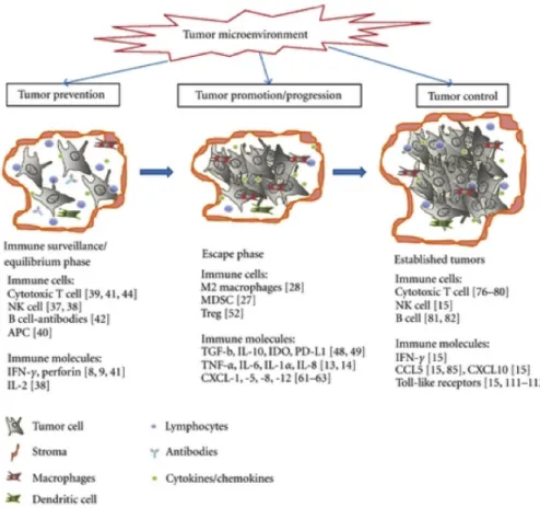
The observation of infiltrating inflammatory cells in tumors is an old observation since Virchow in 1863 postulated that cancer is due to a chronic inflammation [6]. This inflammation mainly consists of innate immune system cells (neutrophils, macrophages, eosinophils, dendritic cells, natural killer lymphocytes) but also of adaptative immune response cells (lymphocytes B and T) all of which are capable of producing an assorted array of cytokines and cytotoxic mediators (Fig 1).
Tumor-‐infiltrating immune cells play dual roles with potential to either eliminate or promote malignancy [7]. Despite exerting a key role in host protection, tumor surveillance by the immune system may eventually fail. As postulated in the theory of cancer immunoediting, tumor cells are initially eliminated by the immune system before becoming clinically detectable. This is then followed by an equilibrium phase, where selection processes for less immunogenic tumor variants take place until the tumors finally “escape” the immune surveillance [8]. On the other hand, the persistent inflammation associated with chronic infections may also encourage new tumor formation.
In the latest years many clinical studies have underlined the link between immunological tumor infiltrations and progression or response to treatment in different types of cancer like breast, ovarian, colorectal, gastric, skin and hepatocellular cancers and many others [9, 10].
In various solid tumors the presence of tumor-‐infiltrating immune cells correlates with better overall survival but the role of some immune cells like T regulatory lymphocytes and/or macrophages can be controversial [11].
To summarize, the actors of the microenvironment immune infiltrate are:
- Neutrophils (PMN): make up a significant portion of the inflammatory cell infiltrate found in a wide variety of human cancers [12, 13]. PNN are a component of the innate immune systems aiding in tumor immune evasion by matrix degradation, immunosculpting, tumor cell proliferation, increased
metastasis, and enhanced angiogenesis. Recent studies demonstrate that PNN can also protect from metastasis progression by matrix metalloproteinase-‐8 production, which plays a protective role in cancer through its ability to regulate the inflammatory response [14].
- Macrophage: Macrophages are a primary source of secreted pro-‐inflammatory cytokines. These cells can be generally categorized as type 1 (M1) or type 2 (M2). M1 macrophages secrete cytokines such as interleukin 12 (IL-‐12) and can assist in the generation of T-‐helper 1 (Th1) adaptive immunity and communicate a direct cytotoxic effect to tumor cells. M2 macrophages secrete immunosuppressive cytokines and promote tumor cell growth and neoangiogenesis [15]. Tumor-‐associated macrophages (TAMs) are, in general, of the M2 phenotype, and infiltration by these cells has been shown to be an independent predictor of poor prognosis in multivariate analysis in many malignancies [16].
- Monocyte/Myeloid-‐derived suppressor cells (MDSCs): MDSCs are a component of the innate immune cells. MDSCs promote cancer growth by blocking the adaptive immune response via the direct secretion of substances that affect T-‐ cell function as well as the induction of adaptive T regulatory (Treg) cells [17]
- T Lymphocytes: several studies have demonstrated the role of T lymphocytes in controlling tumor growth. CTL (cytotoxic T lymphocytes), able to produce IFNg and to eliminate cancer cells, are mainly CD8+ T cells while CD4+ lymphocytes (T-‐ helper) participate in the tumor micro-‐environment, both in enhancing tumor growth (immunosuppression), and in enhancing CTL activity [18].
Treg (regulatory T cells) are CD4+CD25+ lymphocytes expressing the transcriptor factor FoxP3 [19] with a suppressive capacity on regular T lymphocytes function. Many studies have correlated increased Treg presence with disease progression [20].
In the family of CD4+ lymphocytes, Th17 lymphocytes display a great degree of context-‐dependent plasticity. Th17’s role in cancer immunity is ambiguous [21]
but experiments involving adoptive cell transfer therapies have demonstrated their an antitumor activity [22].
Vα24-‐invariant NKT cells are an evolutionarily conserved sub-‐lineage of T cells characterized by reactivity to self-‐ and microbial-‐derived glycolipids presented by an HLA class-‐I–like molecule, CD1d. Several studies have revealed strong positive associations between the numbers of tumor infiltrating or circulating NKTs with improved disease outcome in patients even with diverse types of CD1d-‐negative solid tumors [23].
- B Lymphocytes: are essential for establishing chronic inflammatory states that are associated with pre-‐malignant lesions. Their role in tumor control is controversial because B cells can enhance T cell responses by producing Abs, stimulatory cytokines and serving as APC [24] while, on the other hand, B-‐cells can promote tumor growth [25] by a regulatory immunosuppressive pattern.
- Dendritic cells (DC): Although DCs are notoriously known to be the most potent T cell activating cell type, their function as antigen-‐presenting cells (APC) is markedly compromised in the cancer microenvironment. DC might be also polarized into immunosuppressive/tolerogenic regulatory DC, which limits the activity of effector T cells and supports tumor growth and progression [26] .
- Natural Killer cells are lymphocytes at the border between innate and adaptative immunity; they play an important role in the innate immune response against cancer. Several studies highlight their role in the elimination of tumor metastases and small tumor[27]. Natural killer lymphocytes characteristics and functions will be detailed in the next chapter.
1.2 NATURAL KILLER LYMPHOCYTES (NK)
a. Characteristics and physical parameters of activation
NK are a component of the innate immune system defined “killers” due to their capacity to kill tumoral, infected or stressed cells without the requirement of priming by antigen presenting cell (APC) in contrast to cytolytic T cells [28, 29]. NK cells are large granular lymphocytes, with cytoplasm enriched of lytic granules. They are highly conserved in the phylogenesis because cytolitic cells have been part of the immune defense system approximately 500 million years ago[30]. NK, which are relatively short lived cells renewing each 2 weeks [31] are present in the blood, where they represent 5-‐15% of total lymphocyte population; in secondary lymphoid organs like lymph nodes, spleen and tonsils and in some organs like lung, liver and placenta.
Human NK cells are defined within the lymphocyte gate on the flow cytometric analyzer as CD3 negative and CD56 positive (which is a neuronal cell adhesion protein) cells.
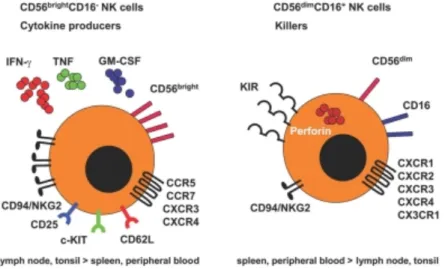
Different functions are known for CD3-‐CD56bright cells and CD3-‐CD56dim cells: while the bright ones are able to produce large amount of cytokines but not to kill tumor target, NK CD56dim can kill some tumor target but are less able to produce cytokines and chemokines after activation. In a healthy adult, circulating NK cells are mostly with a CD56dim phenotype. The majority of human NK cells are CD56 dim and express high levels of the FcγRIIIA, low affinity receptor for the Fc portion of immunoglobulin G (CD16).
b. NK cells development
NK cells are derived from a CD34+ hematopoietic stem cell and they do not need an intra-‐thymus development like T lymphocytes. The exact development of NK cells is still not really known but interaction with some soluble factors and receptors in bone marrow (like c-‐kit ligand, Flt3 and IL-‐15) are needed. The development occurs not only in the bone marrow but also in secondary lymphoid organs where CD56bright NK (immature) could be found [32]. During the maturation process, NK cells
differentiate into highly proliferative CD56bright cells. Terminal differentiation involves a down-‐regulation of CD56, changes in the receptor profile and acquisition of cytotoxic function [33]. In the meantime, NK cells are “educated” by recognition of MHC class I molecules in developing inhibitory receptors, which are found in CD56dim NK. Cytokines such as IL-‐15, IL-‐2, and IL-‐21, are involved in NK cell development and acquisition of effector functions, in particular, IL-‐15 is required for the maturation and survival of NK cells [31]. The high affinity receptor for IL-‐2 is mostly expressed on the CD56bright subset.
c. NK functions and subsets
NK cells are cytolytic effector lymphocytes, which, unlike cytotoxic T cells, can directly induce the death of tumor cells and virus-‐infected cells in the absence of specific immunization via perforin/granzyme production or death receptor (Fas ligand, TNFa, TRAIL pathway), and moreover the NK CD56bright cells are a major producer of IFNg, which is the key to activate naïve T lymphocytes in the lymph nodes [34]. But NK cells also produce many other cytokines with both pro inflammatory and immunosuppressive functions, such as tumor necrosis factor–α (TNF-‐α) and interleukin 10 (IL–10), respectively, and growth factors such as GM-‐CSF (granulocyte macrophage colony-‐stimulating factor), and IL-‐3, in this way they can positively [35, 36] or negatively [37] influence host T and B immunity.
The CD56bright NK subset generally requires 2 signals to produce IFN-‐g: the IL-‐12 released by monocytes, macrophages and DCs and the cytokines IL-‐1, IL-‐2, IL-‐15 or IL-‐18, or the engagement of an NK activating receptor [38]. The CD56dim NK lymphocytes are the one able to directly kill a target cells through a mechanism called antibody-‐dependent cellular cytotoxicity (ADCC). This subset mainly expresses CD16 (Fcg receptor IIIA), which can bind to the constant (Fc) region of immunoglobulin when they are immobilized on the cell surface of a target cell. This receptor-‐ligand binding is followed by a CD16-‐mediated activation signal that results in NK-‐cell degranulation and perforin dependent target cell lysis.
CD56bright and CD56dim completely differ for their chemokine receptors and adhesion molecules repertoire (Fig. 2): while the bright subset express CCR7, CXCR3, CD2,
CD11c, CD44, CD49e, CD54 and CD62L, the CD56dim NK express CXCR1, CX3CR1 and CD11a. This different repertoire induces different migratory properties: the CD56bright preferentially migrates to secondary lymphoid organs whereas the CD56dim cells migrate to the acute inflammatory sites. Moreover, CD56bright NK cells express high levels of the inhibitory CD94/NKG2A complex recognizing HLA-‐E, but lack KIRs receptors which are, in contrast expressed by CD56dim NK cells.
As mentioned before, NK cells may also play immuno-‐regulatory functions mainly by the IL-‐10, IL-‐21 and HLA-‐G production. Experimental in vitro produced “regulatory” NK cells [39] present a CD56+, CD16+, NKp30+, NKp44+, NKp46+, CD94+, CD69+, CCR7+ and are able to down-‐regulate the immune response. Interestingly, the CD56bright NK cells produce high amounts of the immunosuppressive cytokine IL-‐10. In mouse model, tumor derived IL-‐18 is able to expand a subset of NK cells that express the c-‐Kit receptor [40]. These NK cells regulate innate NK cell functions. Up to now, this NK subset has not been yet identified in human.
In the sites of peripheral inflammation, CD56bright also could be found (40%) with an activated phenotype (CD69+). Upon stimulation by IL-‐12, IL-‐15 and IL-‐18, CD56bright NK cells produce IFNγ and increase the production of TNFα from monocytes.
Figure 2. Characteristics of NK subsets (CD56bright and CD56dim), (From Lunemann et al 2009)
d. NK cell Receptors
The main characteristic of NK cell regulation is the protection of self-‐cells (normal) and to destroy the non-‐self (transformed) cells. The machinery to effectuate this regulation includes: a) recognition of “missing self” by inhibitory receptors when the inhibitory ligands (self proteins-‐MHC I) are down-‐regulated like in infected or transformed cells [41] b) recognition of ligands expressed on “stressed” or transformed cells by NK activating receptors[42]. These NK receptors are shown in Fig 3.
Fig 3. Natural Killer Receptors (from Vivier E et al Science 2011)
Inhibitory Receptors
Normal cells are characterized by the presence of self-‐antigens like MHC-‐class I molecules and the recognition of MHC class I molecules by NK inhibitory receptors dominates over the activation signals and blocks the effector functions of NK cells.
The receptors responsible for this inhibitory function include the MHC class I specific killer immunoglobulin-‐like receptors (KIRs) and CD94/NKG2A.
KIR receptors recognize the polymorphic MHC class I molecules. The inhibitory signal results from the presence of the immunoreceptor tyrosine-‐based inhibition motifs (ITIM) in the cytoplasmic domain of the inhibitory receptors. The phosphorylation of ITIMs upon MHC I-‐ligand engagement of inhibitory receptors results in the recruitment and specifically binding of Src-‐Homology-‐2 (SH-‐2) domain containing protein phosphatases. Activated phosphatases such as SHP-‐1 and -‐2 are able to dephosphorylate multiple targets in the activating pathway, thereby mediating its negative signaling. As a result, the activating receptor signaling is directly inhibited by the de-‐phosphorylation of ITAM-‐recruited protein-‐tyrosine kinases[43].
The different inhibitory KIR receptors with the correspondent ligands are depicted in Fig 4.
Other inhibitory receptors are:
• CD94/NKG2A, which recognizes the non-‐classical class I molecule HLA-‐E, with a consequent inhibitory ITIM signal
• CD161 (KLRB1)
• ILT2 (Immunoglobulin like transcript 2), which recognizes classical HLA-‐I and HLA-‐G but also CMV MHC-‐I homologue.
• LAIR-‐1 (CD 305) which recognizes Ep-‐CAM • P75 and IRp60
Activating NK Receptors
NK activating receptors are necessary for the initial activation of NK cell effector functions. These are non-‐covalent associated with transmembrane-‐anchored signaling adaptor proteins like CD3ζ, FcεRIγ, DAP10 or DAP12. The engagement of activating receptors activates “first line” protein tyrosine kinases (PTKs) of the Src-‐family, which phosphorylate immunoreceptor tyrosine-‐based activation motifs (ITAM) in the cytoplasmic tail of the adaptor proteins. Recruitment and activation of “second line” PTKs of the Syk-‐family like Syk and ZAP70 then results in the initiation of the downstream signaling cascade.
NK activating receptors are non-‐MHC specific receptors and include: low affinity Fc receptor FcγRIII (CD16), antibody activating receptor; NKG2D, a triggering receptor; the Natural Cytotoxicity Receptors (NCRs: NKp46/NCR1, NKp44/NCR2, and NKp30/NCR3) and the activating homologs of inhibitory MHC class I receptors (members of the Ly49, KIR and NKG2 families) [44].
CD16: NK cells mediate antibody-‐dependent cellular cytotoxicity (ADCC) by
expressing a low affinity Fc receptor FcγRIII (CD16). This receptor possesses ITAM motifs and, upon ligation, activates src-‐family tyrosine kinases (eg. Lck) and phosphorylates tyrosine residues contained within the ITAM motifs [45, 46]. This signal activates NK cells which results in secretion of cytokines, and antibody-‐
dependent cellular cytotoxicity (ADCC) leading to apoptosis as a consequence of Fas-‐ligand induced cell death [47-‐50].
NKG2D: NKG2D is a type II disulphide-‐linked dimer with a lectin like extracellular
domain which requires the association with the adaptor subunits DAP10 (DAP10 or DAP12 in mice) that mediate signaling, since the intracellular domain of NKG2D has no signaling motifs. NKG2D is expressed by CD8+ αβ-‐T cells (but also CD4+ in some tumors, eg melanoma), by almost all human γδ-‐T cells and human and murine NK cells. [51]
In human NK cells the triggering of NKG2D induces cytotoxicity but no cytokine release. Importantly, since NKG2D has a downstream signaling pathway that is distinct from the activating KIR and C-‐type lectin receptors, the triggering via NKG2D is less susceptible to blocking by KIR-‐ or NKG2A generated inhibitory signals. As a consequence, the signaling through human NKG2D was postulated to override inhibition signals generated by MHC class I engagement, and thus NKG2D functions as a primary cytotoxicity receptor rather than a co-‐receptor [52, 53].
The ligands for human NKG2D are the MHC class I chain related proteins A and B (MICA/B) and the UL16 binding proteins ULBP-‐1, -‐2, -‐3 and -‐4. In normal tissue low levels of MICA/B are found mainly on epithelial cells and fibroblasts. MIC molecules are highly polymorphic since at least 50 different MICA and more than 15 MICB alleles are currently known [54]. MICA/B are glycoproteins that contain MHC-‐like α1-‐, α2-‐ and α3-‐domains but, in contrast to MHC class I molecules, do not require β2-‐microglobulin or peptides for stable surface expression. The family of the UL16-‐binding proteins (ULBPs) is NKG2D ligands that are glycophosphatidyl inositol (GPI)-‐linked surface molecules, which initially were identified by their ability to bind to the human CMV-‐derived membrane glycoprotein UL16 [53].
Induction or up-‐regulation of NKG2D ligands may occur with pathogen related cellular stress, viral infection or tumor cell transformation. High MICA and MICB expression was found on epithelial tumors and on CMV infected epithelial tissues
or fibroblasts. Indeed, the induced expression of NKG2D ligands were shown to markedly enhance the sensitivity of tumors to NK cells in vitro and in vivo. In Neuroblastoma and other cancers, a soluble form of MICA (sMICA) was identified in patient sera derived from metalloprotease-‐mediated proteolytic shedding from the tumor cell surface: sMICA impairs NKG2D-‐mediated immune surveillance of tumors by triggering internalization of surface NKG2D [51].
Natural Cytotoxicity Receptors (NCRs): these are three Ig-‐like molecules termed
NKp46 (CD335), NKp44 (CD336) and NKp30 (CD337). NKp46 and NKp30 are present both in resting and activated NK cells, whereas NKp44 is acquired only on activation. NCRs are characterized by two Ig-‐C2 (NKp46) [55] or one Ig-‐V (NKp30 and NKp44) [56, 57] domains in the extracellular portion. Their short cytoplasmic tail lacks the typical tyrosine-‐based activating motifs but instead the trans-‐membrane regions contain positively charged amino acids that allow association of ITAM-‐ bearing polypeptides, CD3ζ and FcεRIγ for NKp46 and NKp30, while NKp44 associates with DAP12[58, 59]. Interestingly, NCR, instead of representing individual receptors, appear to form a molecular complex because both their expression and functions are coordinated [60]. NCR ‘s ligands are not completely known: virale hemoagglutinin (HA) and vimentin expressed on Mycobacterium tuberculosis-‐infected human monocytes are known to be NKp46 ligands, NKp46 could recognizes different type of tumoral cells and neutrophils; NKp44 cans also recognize HA and Mycobacterium but also Pseudomonas Aeruginosa; the known ligands for NKp30 are B7-‐H6 and BAT-‐3 molecules which are present mostly in stressed or tumoral cells [61](see next paragraph) (Fig 5).
Co-‐receptors
The function of NCR is supported and enhanced by the simultaneous engagement of different co-‐receptors (Fig.4). These include non-‐restricted surface molecules such as 2B4 (CD144), NTBA, NKp80 and CD59.
2B4 and NTBA are members of the CD2 sub-‐family of the immunoglobulin superfamily. Whereas in normal NK cell engagement of both coreceptors results in triggering cytotoxicity, they transduce inhibitory signals in NK cells derived
from X-‐linked lymphoproliferative disease (EBV infected cells). NKp80 participates in NK-‐myeloid cells cross talk; its ligand is AICL (activation induced C-‐ type lectin). Another frontier-‐line co-‐receptor is DNAM-‐1 (CD226), which is an activating molecule that recognizes poliovirus receptor and Nectin -‐2 (highly expressed by different tumors)[44].
e. The NKp30 (CD337) receptor
At the end of 1990s Pende and colleagues identified NKp30 as a novel 30-‐kDa triggering receptor expressed by all resting and activated human NK cells [57]. It is a type I trans-‐membrane protein characterized by a single V-‐type immunoglobulin (Ig) extracellular domain. The intracellular tail of NKp30 has no
signaling motif but its trans-‐membrane domain associates via a charged amino acid with immune-‐receptor based activation motif (ITAM)-‐bearing adapters (CD3ζ and FcRγ). Activation of NKp30 receptor by its ligand induces activation of the NFkB canonical pathway [62]. A recent study [63] identified the flexible stalk region of NKp30 as an important module for ligand recognition and related signaling of the corresponding full-‐length receptor proteins. In the same study region of glycosilation of the ectodomain of NKp30 is responsible of differential binding affinities and signaling capacities.
NKp30 encoding gene (NCR3) is located in the class III region of the human MHC on chromosome 6 (locus 6p21.3), where several other genes with immune function are found (TNFa eg). Phylogenic analysis of NKp30 in different mammalian species shows that gene sequences are very conserved except in exon 4 that encodes the intracellular domain[64].
NCR3 encodes six alternative spliced transcripts, 4 of these encode for a protein. NKp30a, NKp30b and NKp30c produce a V-‐type Ig extracellular domain and NKp30d, Nkp30e and NKp30f produce a C-‐type one. The NKp30 a, b and c are the most represented in tissues. Due to the NCR3-‐exon 4 alternative splicing, these extracellular domains are coupled with three different intracellular domains (Fig 6).
Figure 6 Structure of the NCR3 / NKp30 gene
We demonstrated [65] (see supplemental article 1) that the three different isoforms Nkp30a, NKp30b and NKp30c, issues from the exon 4 alternative splicing in the
intracellular domain, transmit distinct signals and thus mediate different cell functions: NKp30a and NKp30b are immunostimulatory isoforms that trigger TH1 cytokine production while NKp30c promotes IL-‐10 secretion, relaying an immunosuppressive signal (Fig 7).
Figure 7. NKp30 isoforms significant differ their functions in vivo
Ligation of NKp30 triggers the canonical pathway of nuclear factor-‐κB (NF-‐κB) activation. NKP30C fails to activate NF-‐kB pathway while induce a rapid
phosphorylation of p38 MAP kinase.
NKp30 is the only receptor involved both in tumor cell lyses and lyses of normal cells [57, 66, 67]. It is mostly restricted to NK cells but it could be expressed also on umbilical cord blood T cells after IL-‐15 stimulation and on endometrial epithelium after progesterone exposure.[68] IL-‐2 can enhance NKp30 surface protein expression while TGFβ can reduce the expression.
A striking feature of NKp30 is its involvement in the crosstalk between NK cells and DCs. NK cells are able to lyse immature DCs through engagement of NKp30 despite MHC I expression, but mature DCs are protected from lyses [66]. Additionally, NK cells that have been stimulated through NKp30 can produce tumor necrosis factor α (TNF-‐α), which then induces maturation of DCs [67]. DCs in return produce cytokines like IL-‐12 or IL-‐18 that can stimulate NK cells [42]. The mutual control and regulation
of DCs and NK cells again is an interesting example of how cells of the immune system communicate and work together in numerous ways; NK cells and DCs through their communication among each other are crucial in bridging the innate and the adaptive immune response.
In contrast to NKp44 and NKp46, NKp30 has not been linked to the binding of viral hemagglutinins. However, NKp30 has been implicated in the recognition of several pathogens:
• Cytomegalovirus: the CMV tegument protein pp65 bind NKp30 but this engagement leads to an inhibition of NK ability to kill. [69]
• Trypanosoma brucei infected erythrocytes [70]
• Filovirus infected DCs : NKp30 is able to recognize virus like particles of the filovirus type and activate NK cells to produce cytokines [71].
Others known ligands for NKp30 are BAT3 and B7-‐H6.
BAT3 or HLA-‐B associated transcript-‐3 is found among the genes of the MHC cluster,
specifically in the MHC III region but it does not show any homology to the common MHC proteins. The intracellular BAT3 protein was found to bind and activate NKp30 [72]. This protein could be exposed on surface or secreted or expressed on exosomes from mature DCs [73] after stress or heat shock. In this way BAT3 regulates the NK cell mediated cytotoxicity against immature DCs.
B7-‐H6 is a novel member of the B7 protein family (co-‐stimulatoty and co-‐inhibitory
transmembrane proteins that contribute to the immune response regulation) expressed by certain tumor cell lines and a subset of primary tumor cells (melanoma and carcinoma) or hematological tumors, (lymphoma and leukemia) but not by healthy cells [74]. The protein structure is similar to other members of the B7 family and, particularly, B7-‐H6 is a PD-‐L1/B7-‐H3 homologue. Membrane-‐bound B7-‐H6 activates NK cells, leading to degranulation and IFNg secretion. B7-‐H6 has two extra-‐cellular Ig domains encoded by exons with adjacent phase 1 introns and an intracytoplasmic domain, which contains signaling motifs like ITIMs. This suggests that, upon engagement, B7-‐H6 could induce a response in the NK cell target through interaction with intracellular signaling proteins. The absence of B7-‐H6 transcripts in normal tissues and presence in tumor cell define B7-‐ H6 as a novel example of stress induced self-‐molecule. Little is known about expression modulation by tumor editing or by microenviroment. Matta et al recently demonstrated