HAL Id: tel-02918168
https://tel.archives-ouvertes.fr/tel-02918168
Submitted on 20 Aug 2020HAL is a multi-disciplinary open access archive for the deposit and dissemination of sci-entific research documents, whether they are pub-lished or not. The documents may come from teaching and research institutions in France or abroad, or from public or private research centers.
L’archive ouverte pluridisciplinaire HAL, est destinée au dépôt et à la diffusion de documents scientifiques de niveau recherche, publiés ou non, émanant des établissements d’enseignement et de recherche français ou étrangers, des laboratoires publics ou privés.
chemins avec la transcription
Elena Cerutti
To cite this version:
Elena Cerutti. La réparation par excision de nucléotides à la croisée des chemins avec la transcription. Molecular biology. Université de Lyon, 2019. English. �NNT : 2019LYSE1057�. �tel-02918168�
N° d’ordre NNT : 2019LYSE1057
THESE de DOCTORAT DE L’UNIVERSITE DE LYON
opérée au sein de
l’ Université Claude Bernard Lyon 1
École Doctorale
N° 340Biologie Moléculaire Intégrative et Cellulaire
Spécialité de doctorat
: Réparation de l’ADNSoutenue publiquement le 10/05/2019, par :
Elena CERUTTI
Nucleotide Excision Repair at the
crossroad with transcription
Devant le jury composé de :
Orioli, Donata – PA – IGM, Università di Pavia Rapporteure Coin, Frédéric – DR – IGBMC, Université de Strasbourg Rapporteur Schaeffer, Laurent – PU – INMG, Université Lyon 1 Examinateur Giglia-Mari, Giuseppina – DR – INMG, Université Lyon 1 Directrice de thèse Mari, Pierre-Olivier – CR – INMG, Université Lyon 1 Co-directeur de thèse Diaz, Jean-Jacques – DR – CRCL, Université Lyon 1 Invité
UNIVERSITE CLAUDE BERNARD - LYON 1
Président de l’UniversitéPrésident du Conseil Académique
Vice-président du Conseil d’Administration
Vice-président du Conseil Formation et Vie Universitaire Vice-président de la Commission Recherche
Directrice Générale des Services
M. le Professeur Frédéric FLEURY M. le Professeur Hamda BEN HADID M. le Professeur Didier REVEL M. le Professeur Philippe CHEVALIER M. Fabrice VALLÉE
Mme Dominique MARCHAND
COMPOSANTES SANTE
Faculté de Médecine Lyon Est – Claude Bernard Faculté de Médecine et de Maïeutique Lyon Sud – Charles Mérieux
Faculté d’Odontologie
Institut des Sciences Pharmaceutiques et Biologiques Institut des Sciences et Techniques de la Réadaptation Département de formation et Centre de Recherche en Biologie Humaine
Directeur : M. le Professeur G. RODE Directeur : Mme la Professeure C. BURILLON Directeur : M. le Professeur D. BOURGEOIS Directeur : Mme la Professeure C. VINCIGUERRA Directeur : M. X. PERROT
Directeur : Mme la Professeure A-M. SCHOTT
COMPOSANTES ET DEPARTEMENTS DE SCIENCES ET TECHNOLOGIE
Faculté des Sciences et Technologies Département Biologie
Département Chimie Biochimie Département GEP
Département Informatique Département Mathématiques Département Mécanique Département Physique
UFR Sciences et Techniques des Activités Physiques et Sportives
Observatoire des Sciences de l’Université de Lyon Polytech Lyon
École Supérieure de Chimie Physique Électronique Institut Universitaire de Technologie de Lyon 1 École Supérieure du Professorat et de l’Éducation Institut de Science Financière et d'Assurances
Directeur : M. F. DE MARCHI
Directeur : M. le Professeur F. THEVENARD Directeur : Mme C. FELIX
Directeur : M. Hassan HAMMOURI Directeur : M. le Professeur S. AKKOUCHE Directeur : M. le Professeur G. TOMANOV Directeur : M. le Professeur H. BEN HADID Directeur : M. le Professeur J-C PLENET Directeur : M. Y. VANPOULLE
Directeur : M. B. GUIDERDONI Directeur : M. le Professeur E. PERRIN Directeur : M. G. PIGNAULT
Directeur : M. le Professeur C. VITON
Directeur : M. le Professeur A. MOUGNIOTTE Directeur : M. N. LEBOISNE
« Dans la vie, rien n'est à craindre, tout est à comprendre. »
ACKOWLEDGEMENTS/REMERCIEMENTS/RINGRAZIAMENTI
It has been almost 4 years since I started my PhD and I would like to thank all the people that, in one way or another, helped me to get here.
First, I would like to thank Laurent Schaeffer, Donata Orioli and Frederic Coin for accepted to be part of my defence jury and to read and correct my manuscript. Thank you for the kind remarks about my work and for the comments that helped me improve my thesis.
I would like to thank my thesis director, Ambra. I have started my first internship in your group in 2013, when I didn’t even know what a pipette was, and, since then, you have never stopped to teach me, science or not, something that have certainly enriched me as a person and a scientist. I also would like to thank my thesis co-director, Pierre-Olivier. I really thank you for your patience explaining me over and over again all the microscopy stuffs, even when I was looking at you with by best “fish face”. I am pretty sure that your teaching skills, and your persistence, have borne its fruits; thank you a lot.
Je voudrais aussi remercier tous les membres de l’équipe, qui se sont succédés pendent ces presque 4 ans. En particulier, je voudrais remercier Laurianne, qui m’a introduit en première au sujet du nucléole et de l’ARN Pol I et qui a partager avec moi ses connaissances au début de ma thèse. Merci beaucoup pour ton aide et pour avoir beaucoup discuter avec moi, de science et non, toujours en essayant de comprendre mon française « bizarre ». Ensuite, je voudrais ainsi remercier Anna, qui a débarquée en France au même temps que moi. T’as toujours été très gentille avec moi et ça été très enrichissant pour moi de faire ta connaissance. Parmi les membres présentes de l’équipe, un merci très spécial va surement à Lise-Marie. Merci pour m’avoir aidé, expliqué, supporté, réexpliqué, soutenu, réexpliqué, appris, réexpliqué, montré, écouté, compris… et je ne connais pas d’autres mots pour t’exprimer toute ma reconnaissance. Ça été un très grand plaisir de travailler avec toi et je suis sûre que tu seras une excellente chercheuse. Ensuite, je voudrais dire un grand merci à Damien. Ça été très drôle d’avoir fait ta connaissance et je te remercie vivement pour m’avoir fait rigoler autant. Surtout dans les moments plus difficiles, tu m’as toujours remonté le moral avec tes blagues, et ça c’était tous ce qu’il fallait, donc merci beaucoup. Je veux remercier aussi Charlène, qui est une super technicienne. Ça ne fait pas longtemps que t’es là et probablement t’as fait ma connaissance dans une période pas très facile pour moi, mais t’as toujours été très compréhensive avec moi et ton aide a été très précieux. Je te remercie et j’espère qu’on aura encore l’occasion de passer du temps ensemble et mieux se connaitre. Finalement, je voudrais remercier tous les stagiaires que j’ai pu rencontrer pendent ces presque 4 ans. Ceux que j’ai encadré ou ceux que j’ai juste aidé, vous m’avez tous appris à être
aussi patient que je voudrais que les autres soient avec moi et à vous transmettre mes connaissances comme je voudrais qu’elles me soient transmises. Je ne sais pas si j’ai fait un bon boulot avec vous, mais surement vous l’avez fait avec moi.
Vorrei ringraziare ora la mia famiglia. Quando ho deciso di passare quasi 4 anni in un altro Paese, che per quanto vicino resta comunque un altro mondo, non sapevo cosa sarebbe successo. Ora posso dirvi che l’ancora di sicurezza che voi rappresentate mi è mancata molto. Ciononostante, vi ringrazio per essere sempre stati dalla mia parte e avermi sempre incoraggiato in questa decisione. Vi ringrazio perché, senza il vostro aiuto, molto probabilmente non avrei mai scritto questa tesi e non mi sarei mai resa conto di ciò che voglio veramente fare. Quindi vi ringrazio per questo. Ringrazio anche le mie amiche, Valentina, Veronica e Cristina, che nonostante la lontananza, hanno sempre fatto in modo che non mi perdessi niente delle loro vite, né loro della mia. Sarà un piacere immenso potervi vedere molto più spesso ed essere presente per tutto il bello che verrà. Infine vorrei ringraziare Filippo, che durante questi quasi 4 anni è sempre stato al mio fianco in ogni momento. Ti vorrei ringraziare per ogni piccola e grande cosa che hai fatto per me, ma sarebbe una lista infinita. Quindi ti dico che tutto quel che ho fatto e tutto quel che sono ora è perché ho te al mio fianco, e tutto ciò che farò e tutto quel che sarò, se avrò te al mio fianco, sarà sicuramente meraviglioso.
Merci à tous! Elena
SUMMARY
Introduction
Cells are the units of organic life and store in their nuclei what constitutes the instruction manual for proper cellular functioning, in the form of the DNA molecule. There are different types of DNA, one of them is ribosomal DNA (rDNA), which is transcribed by RNA polymerase 1 (RNAP1) and is the first step of an important cellular function: ribosomal biogenesis. RNAP1 is responsible for more than 60 % of the total transcriptional activity of the cell and all transcription performed by RNAP1 takes place in a specific subcellular compartment: the nucleolus.
Unfortunately, the integrity of DNA is continuously challenged by a variety of endogenous and exogenous agents (e.g. ultraviolet light, cigarette smoke, environmental pollution, oxidative damage, etc.) that cause DNA lesions, which interfere with proper cellular functions resulting in aging or premature aging of the tissue and eventually, the organism as a whole.
To prevent the deleterious consequences of persisting DNA lesions, all organisms are equipped with DNA repair mechanisms. One of these systems is the Nucleotide Excision Repair (NER). NER removes helix-distorting DNA adducts such as UV-induced lesions (Cyclo-Pyrimidine Dimers and 6-4 Photoproducts, CPD and 6-4PP). NER exists in two distinct sub-pathways depending where DNA lesions are located within the genome. Global Genome Repair (GG-NER or GGR) will repair DNA lesion located on non-transcribed DNA. While, the second sub-pathway is directly coupled to transcription elongation and repairs DNA lesions located on the transcribed strand of active genes. This second sub-pathway is designated as Transcription-Coupled Repair (TC-NER or TCR).
The NER system has been linked to rare human diseases classically grouped into three distinct NER-related syndromes. These include the highly cancer prone disorders Xeroderma Pigmentosum (XP), Cockayne syndrome (CS), and trichothiodystrophy (TTD).
The objective of my thesis was to clarify some aspects of the repair mechanism at the crossroad with transcription. More particularly, my scientific objectives were:
- To concretely identify the players involved in the observed relocation of RNAP1 that occurs after UV irradiation. We identified two proteins involved in this relocation, which are specifically involved in the return of the RNAP1 within the nucleolus after completion of the DNA repair reaction.
- To clearly decipher the molecular mechanism of XAB2 function during DNA repair and to understand whether the function in transcription might be mechanistically similar to the function during DNA Repair.
Results
RNAP1
We have published in the PNAS journal, the article “Mechanistic insights in transcription-coupled nucleotide excision repair of ribosomal DNA”, of which I am co-first author. Within this study, we have demonstrated the importance of a fully functional NER mechanism in order to repair UVC lesion on ribosomal genes and that during DNA repair of UV lesions, the RNAP1 is displaced at the periphery of the nucleolus and returns within the nucleolus after DNA repair completion. Furthermore, we have observed that, in a GGR deficient cell line, the RNAP1 transcription restart after repair was not coupled to the re-entry of RNAP1 within the nucleolus.
On these bases, we investigated possible proteins responsible for this re-entry of RNAP1 and that do not take part in the repair process.
We studied two candidate proteins because of their function in RNAP1 transcription and in the cell migration and organelle movement.
- Nuclear Actin: Actin was first described in the cytoplasm where it serves the functions of cellular shape maintenance, cell motility and muscle contraction. However, several studies identified its presence in the nucleus and, more importantly, its interaction with RNAP1.
- Nuclear Myosin I: In the cytoplasm, actin and myosin are part of the motor protein superfamily. In the nucleus, similarly to actin, Nuclear Myosin I has been described to correlate with RNAP1, but it interacts with the polymerase in an indirect way.
In the absence of these two proteins, we have demonstrated that 40h after UVC exposure transcription of ribosomal DNA restarts, but neither RNAP1 nor the rDNA do not relocate within the nucleolus, as it would be expected. In light of these results, we investigated the possibility of these two candidates playing a role in the repair mechanism of NER and we found that there is no evidence of their implication in the repair process, showing that these proteins control the re-entry of RNAP1 and rDNA within the nucleolus without interfering with the DNA repair reaction. In light of these results, we wanted to investigate if the involvement of Nuclear Actin and Nuclear Myosin I in RNAP1 relocation after repair were transcription-dependent. To do so, we depleted these proteins in cells containing a LacO/LacR-GFP system that allow us to visualize rDNA and we performed RNA fish and RNAP1 Immunofluorescence experiments after Cordycepin
exposure. Cordycepin is an adenosine analogue that inhibits 47S rRNA synthesis in eukaryotic cells in a reversible way. The results we have obtained demonstrate that, in contrast with what happens for UV-irradiation, in the absence of Nuclear Actin and Nuclear Myosin I, 40 hours of recovery after a 2h of Cordycepin treatment, rDNA transcription restarts and the rDNA/RNAP1 relocate within the nucleolus. This result confirms that the rDNA/RNAP1 re-entry within the nucleolus after DNA repair is transcription-independent and that Nuclear Actin and Nuclear Myosin I are responsible for this UV-specific re-entry.
We have then investigated the possible interaction between Nuclear Actin and Nuclear Myosin I, known to be both implicated in different cellular events such as cell migration or muscle contraction, and RNAP1 by Co-Immunoprecipitation experiment on chromatin extracts. Furthermore, we explored the binding profile of Nuclear Actin and Nuclear Myosin I on the ribosomal DNA before and after UV-C exposure. The results we obtained suggest that Nuclear Actin and Nuclear Myosin I could work synergistically to bind rDNA and that both are needed in the same process of repositioning rDNA/RNAP1 within the nucleolus once repair is completed.
XAB2
The XPA-binding protein 2 was identified because of its ability to interact with XPA, a central factor to both GGR and TCR pathways. XAB2 is an 855 amino acids protein consisting of 15 tetratricopeptide repeats. IP experiments demonstrated that a fraction of XAB2 is able to interact with CSA and CSB as well as RNAP2. Since the XAB2 role in the TCR pathway has been poorly investigated so far, our aim was to decipher its function and its mechanism in both DNA repair and RNAP2 transcription.
XAB2 during DNA repair
Normally, 24h after UVC exposure, lesions on the active genes transcribed by RNAP2 are repaired and transcription restarts. In the absence of XAB2, transcription does not restart, suggesting that this protein plays a role in this process. However, it was not known whether XAB2 was implicated directly in the DNA repair reaction or whether it was playing a role in transcription restart after completion of DNA repair. Thanks to a technique developed in our research group, we could demonstrate that XAB2 plays a role in the repair reaction and this role induces a long lasting block of transcription after UV irradiation.
In addition, we investigated the dynamic behavior of XAB2 during repair. Surprisingly, we could show that XAB2 is released from the damaged area, while all the other NER proteins studied so far strongly accumulate on the local lesion.
Without any damage, XAB2 has certain mobility within the nucleus. This mobility is the result of XAB2 diffusion but also of the interactions with the chromatin. Interestingly, when we measured the mobility of XAB2 within CSA and CSB deficient cell lines and without any damage, we observed an increased immobile fraction in both CSA and CSB deficient cell lines, suggesting that CSA and CSB are responsible for the release of XAB2 from a more immobile substrate.
We then investigated, by Co-IP on nuclear extracts, the interaction between XAB2 and RNAP2 before and after UV-C exposure in WT cells and in CSA and CSB deficient cell lines, in order to decipher the mechanistic behavior of XAB2 during repair. The results we obtained demonstrated the presence of XAB2 in a pre-mRNA splicing complex before and after UV-C exposure and that a portion of XAB2 interacts with RNAP2 during the first steps of NER in WT cells, suggesting a role of XAB2 in the damage recognition by the repair machinery.
XAB2 during transcription
It has been demonstrated that XAB2 interacts with the RNAP2. In order to clearly understand the role of XAB2 in RNAP2 transcription we studied the mobility of RNAP2 in presence and absence of XAB2. For this purpose, we used a stable cell line produced in our lab expressing a RNAP2 tagged at the N-terminal with the Green Fluorescent Protein. Using the FRAP technique, we measured and compared the dynamic behavior of RNAP2 in the presence and in the absence of XAB2. The results of this analysis show that in absence of XAB2, RNAP2 bound fraction largely decreased, suggesting that XAB2 plays a role in keeping RNAP2 on the chromatin during the pause of transcription.
RESUMÉ
Introduction
Les cellules sont les unités de la vie organique et stockent dans leurs noyaux ce qui constitue le manuel d’instruction pour le bon fonctionnement cellulaire, sous la forme de la molécule d’ADN. Il existe différents types d’ADN, dont l’ADN ribosomique (ADNr), qui est transcrit par l’ARN Polymérase 1 (ARNP1) et est la première étape d’une fonction cellulaire importante : la biogenèse ribosomique. L’ARNP1 est responsable de plus de 60 % de l’activité transcriptionnelle totale de la cellule et toute transcription effectuée par l’ARNP1 a lieu dans un compartiment subcellulaire spécifique : le nucléole.
Malheureusement, l’intégrité de l’ADN est continuellement remise en question par une variété d’agents endogènes et exogènes (p. ex., la lumière ultraviolette, la fumée de cigarette, la pollution de l’environnement, les dommages oxydatifs, etc.) qui causent des lésions de l’ADN qui interfèrent avec les correctes fonctions cellulaires, entraînant le vieillissement ou le vieillissement prématuré du tissu et, finalement, de l’organisme dans son ensemble.
Afin de prévenir les conséquences néfastes des lésions persistantes de l’ADN, tous les organismes sont équipés de mécanismes de réparation de l’ADN. Un de ces systèmes est la réparation par excision de nucléotides (NER). NER supprime les adduits d’ADN déformant l’hélice tels que les lésions induites par les UV (dimères de cyclo-pyrimidine et 6-4 photo produits, CPD et 6-4PP). NER existe dans deux sous voies distinctes selon l’endroit où les lésions de l’ADN se trouvent dans le génome. Global Genome Repair (GG-NER ou GGR) réparera la lésion d’ADN située sur l’ADN non transcrit. Inversement, la deuxième sous voie est directement couplée à l’allongement de la transcription et répare les lésions de l’ADN situées sur le brin transcrit des gènes actifs. Cette deuxième sous voie est désignée Transcription-Coupled Repair (TC-NER ou TCR).
Le système NER a été associé à des maladies humaines rares classées généralement en trois syndromes distincts liés aux NER. Il s’agit notamment des troubles hautement cancéreux xeroderma pigmentosum (XP), du syndrome de Cockayne (CS) et de la trichothiodystrophie (TTD). L’objectif de ma thèse était de clarifier certains aspects du mécanisme de réparation à la croisée des chemins avec la transcription. Plus particulièrement, mes objectifs scientifiques étaient les suivants :
- Identifier concrètement les acteurs impliqués dans la relocalisation observée de l’ARNP1 après l’irradiation UV. Nous avons identifié deux protéines impliquées dans cette relocalisation,
qui sont spécifiquement impliquées dans le retour de l’ARNP1 e du ADNr dans le nucléole après l’achèvement de la réaction de réparation de l’ADN.
- Décrypter clairement le mécanisme moléculaire de la fonction de la protéine XAB2 pendant la réparation de l’ADN et comprendre si sa fonction en transcription pourrait avoir un mécanisme similaire à la fonction pendant la réparation de l’ADN.
Résultats
ARNP1
Nous avons publié dans le journal PNAS un article intitulé « Mechanistic insights in transcription-coupled nucleotide excision repair of ribosomal DNA », dont je suis co-premier auteur. Dans le cadre de cette étude, nous avons démontré l’importance d’un mécanisme de NER entièrement fonctionnel afin de réparer les lésions UVC sur les gènes ribosomiques et que lors de la réparation des lésions UV à l’ADNr, l’ARNP1 est déplacé à la périphérie du nucléole et retourne dans le nucléole après la réparation de l’ADN. De plus, nous avons observé que, dans une lignée cellulaire déficiente en GGR, le redémarrage de la transcription par l’ARNP1 après réparation n’était pas couplée à la rentrée du RNAP1 dans le nucléole.
Sur ces bases, nous avons recherché les protéines possibles responsables de cette rentrée de l’ARNP1 et qui ne participent pas au processus de réparation.
Nous avons étudié deux protéines candidates en raison de leur fonction dans la transcription par l’ARNP1 et dans la migration cellulaire et le mouvement des organites.
- Actine nucléaire : l’actine a été décrite pour la première fois dans le cytoplasme où elle sert les fonctions de maintien de forme cellulaire, de motilité cellulaire et de contraction musculaire. Toutefois, plusieurs études ont identifié sa présence dans le noyau et, plus important encore, son interaction avec l’ARNP1.
- Myosine nucléaire I : Dans le cytoplasme, l’actine et la myosine font partie de la superfamille des protéines motrices. Dans le noyau, de la même manière que l’actine, la myosine nucléaire I a été décrite comme étant en corrélation avec l’ARNP1, mais elle interagit avec la polymérase de façon indirecte.
En l’absence de ces deux protéines, nous avons démontré que 40h après l’exposition aux UVC, la transcription de l’ADN ribosomique redémarre, mais ni l’ARNP1 ni l’ADNr ne se relocalise à l’intérieur du nucléole, comme on pouvait s’y attendre. À la lumière de ces résultats, nous avons examiné la possibilité que ces deux candidats jouent un rôle dans le mécanisme de réparation du NER et nous avons constaté que, dans nos conditions, il n’y a aucune preuve de leur implication
dans le mécanisme de réparation, montrant que ces protéines contrôlent la rentrée de l’ARNP1 et de l’ADNr dans le nucléole sans interférer avec la réaction de réparation de l’ADN.
À la lumière de ces résultats, nous voulions déterminer si l’implication de l’actine nucléaire et de la myosine nucléaire I dans la relocalisation de l’ARNP1 après réparation dépendait de la transcription. Pour ce faire, nous avons dépléter ces protéines dans des cellules contenant un système Laco/LacR-GFP qui nous permettent de visualiser l’ADNr et nous avons réalisé des expériences d’ARNfish et d’immunofluorescence contre l’ARNP1 après exposition à la cordycépine. La cordycépine est un analogue de l’adénosine qui inhibe la synthèse de l’ARNr 47S dans les cellules eucaryotes de manière réversible. Les résultats que nous avons obtenus démontrent que, contrairement à ce qui se passe pour l’irradiation aux rayons UV, en l’absence d’actine nucléaire et de myosine nucléaire I, 40 heures de récupération après 2h de traitement par cordycépine, la transcription de l’ADNr redémarre et l’ADNr et l’ARNP1 se relocalise à l’intérieur du nucléole. Ce résultat confirme que la rentrée de ADNr/ARNP1 dans le nucléole après la réparation de l’ADN est indépendante de la transcription et que l’actine nucléaire et la myosine nucléaire I sont responsables de cette rentrée spécifique aux UV.
Nous avons ensuite étudié l’interaction possible entre l’actine nucléaire et la myosine nucléaire I, connue pour être à la fois impliquée dans différents événements cellulaires tels que la migration cellulaire ou la contraction musculaire, et l’ARNP1 par une expérience de co-immunoprécipitation sur des extraits de chromatine. De plus, nous avons examiné le profil de liaison de l’actine nucléaire et de la myosine nucléaire I sur l’ADN ribosomique avant et après l’exposition aux UV-C. Les résultats que nous avons obtenus suggèrent que l’actine nucléaire et la myosine nucléaire I pourraient travailler de manière synergique pour lier l’ADNr et que les deux sont nécessaires dans le même processus de repositionnement de ADNr/ARNP1 à l’intérieur du nucléole une fois la réparation terminée.
XAB2
La protéine XPA-binding protein 2 a été identifiée en raison de sa capacité à interagir avec XPA, un facteur central des voies GGR et TCR. XAB2 est une protéine de 855 acides aminés composée de 15 répétitions de tétratricopeptides. Les expériences d’immunoprécipitation ont démontré qu’une fraction de XAB2 est capable d’interagir avec CSA et CSB ainsi qu’avec l’ARNP2. Depuis que le rôle de XAB2 dans la voie TCR a été peu étudié jusqu’à présent, notre but était de déchiffrer sa fonction et son mécanisme dans la réparation de l’ADN et la transcription par l’ARNP2.
Normalement, 24h après l’exposition aux UVC, les lésions sur les gènes actifs transcrits par l’ARNP2 sont réparées et la transcription redémarre. En absence de XAB2, la transcription ne redémarre pas, ce qui suggère que cette protéine joue un rôle dans ce processus. Toutefois, on ne savait pas si XAB2 était directement impliqué dans la réaction de réparation de l’ADN ou s’il jouait un rôle dans le redémarrage de la transcription après la réparation de l’ADN. Grâce à une technique développée dans notre groupe de recherche, nous avons pu démontrer que XAB2 joue un rôle dans la réaction de réparation et ce rôle induit un bloc permanent de la transcription après irradiation UV.
De plus, nous avons étudié le comportement dynamique de XAB2 pendant la réparation. Étonnamment, nous avons pu montrer que XAB2 est libéré de la zone endommagée, tandis que toutes les autres protéines du NER étudiées jusqu’à présent s’accumulent fortement sur la lésion. Sans aucun dommage, XAB2 a une certaine mobilité dans le noyau. Cette mobilité est le résultat de la diffusion de XAB2 mais aussi des interactions avec la chromatine. Il est intéressant de noter que lorsque nous avons mesuré la mobilité de XAB2 dans des lignées cellulaires déficientes par CSA et CSB et sans aucun dommage, nous avons observé une fraction immobile plus importante dans les lignées cellulaires déficientes par CSA et CSB, ce qui laisse entendre que CSA et CSB sont responsables du rejet de XAB2 à partir d’un substrat plus immobile.
Nous avons ensuite étudié, par Co-IP sur des extraits nucléaires, l’interaction entre XAB2 et l’ARNP2 avant et après l’exposition aux UV-C dans des cellules WT et dans des lignées cellulaire déficientes par CSA et CSB, afin de déchiffrer le comportement mécanistique de XAB2 pendant la réparation. Les résultats que nous avons obtenus ont démontré la présence de XAB2 dans un complexe d’épissage de pré-ARNm avant et après l’exposition aux UV-C et qu’une partie de XAB2 interagit avec l’ARNP2 pendant les premières étapes de la réparation de l’ADN dans les cellules WT, suggérant un rôle de XAB2 dans la reconnaissance des dommages par la machinerie de réparation.
XAB2 pendant la transcription
Il a été démontré que XAB2 interagit avec l’ARNP2. Afin de bien comprendre le rôle de XAB2 dans la transcription par l’ARNP2, nous avons étudié la mobilité de l’ARNP2 en présence et en absence de XAB2. À cette fin, nous avons utilisé une lignée cellulaire stable produite dans notre laboratoire exprimant une ARNP2 étiqueté au terminal N avec la GFP. En utilisant la technique de FRAP, nous avons mesuré et comparé le comportement dynamique de l’ARNP2 en présence et en absence de XAB2. Les résultats de cette analyse montrent qu’en absence de XAB2, la fraction
immobile de l’ARNP2 a largement diminué, ce qui suggère que XAB2 joue un rôle dans le maintien de l’ARNP2 sur la chromatine pendant la pause de transcription.
ABBREVIATIONS
5’ETS 5’ External Transcribed Spacer 6-4PP 6-4 photoproducts
alt-EJ alternative End Joining AP site Abasic site
APE1 AP-endonuclease 1 BER Base Excision Repair BPS Branch-Point Sequence BRCA2 Breast Cancer 2
BRE TFIIB Recognition Element CAF-1 Chromatin Assembly Factor-1 CAK CDK-Activating Kinase
Cdk7 Cyclin-dependent kinase 7 Cdk9 Cyclin-dependent kinase 9
CHD1 Chromodomain-Helicase-DNA-binding protein 1 CK2 serine/threonine kinase
CPD Cyclobutane Pyrimidine Dimers
CPSF Cleavage and Polyadenylation Specificity Factor CS Cockayne Syndrome
CSA Cockayne syndrome A CSB Cockayne syndrome B CstF Cleavage stimulatory Factor CTD C-Terminal Domain
CtIP C-terminal binding protein-Interacting Protein DDB1 DNA Damage-Binding protein 1
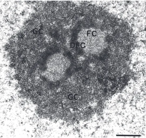
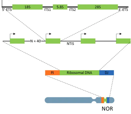
DDB2 DNA Damage-Binding protein 2 DFC Dense Fibrillar Component DJ Distal Junction
Dna2 DNA2-like helicase
DNA-PK DNA-dependant Protein Kinase DNMT1 DNA Methyltransferase 1 DNMT3 DNA Methyltransferase 3 DNTT DNA nucleotidyltransferase
DOT1L Disruptor Of Telomeric silencing 1-Like DPE Downstream Promoter Element DSB Double Strand Break
DSIF DRB Sensitivity Inducing Factor EF Elongating Factors
ELL Eleven-nineteen Lysine-rich Leukemia protein ERCC1 Excision Repair Cross-Complementation group 1 EXO1 Exonuclease 1
FACT Facilitates Chromatin Transcription FC Fibrillar Center
GC Granular Component GG-NER Global-Genomic Repair HMG High Mobility Group HP1 Heterochromatin Protein 1 HR Homologous Recombination Inr Initiator element
LIG1/LIG3 DNA Ligase 1/3 MLH1 MutL homolog 1 MMR Mismatch Repair
MSH2 MutS protein homolog 2 MSH6 MutS protein homolog 6 Nbs1 Nibrin
NELF Negative Elongation Factor NER Nucleotide Excision Repair NFR Nucleosomal-Free Region NHEJ Non-Homologous End-Joining NOR Nucleolar Organizer Region NoRC Nucleolar Remodelling Complex NTC Nineteen Complex
NTIS Non-Transcribed Intergenic Spacer regions NTP Nucleosides Triphosphate
NTR Nineteen-Related
PAF1C Pol II-Associated Factor 1 Complex PAH Polycyclic Aromatic Hydrocarbons PARP-1 Poly [ADP-ribose] Polymerase 1 PCNA Proliferating Cell Nuclear Antigen PH Pleckstrin Homology
PIC Pre-Initiation Complex PJ Proximal Junction
PMS2 Mismatch repair endonuclease PMS2 PNBs PeriNucleolar Bodies
Polβ DNA polymerase β
P-TEFb positive-Transcription Elongation Factor PTM Posttranslational Modification
PTRF Pol 1 and Transcript Release Factor rDNA ribosomal DNA
RES Retention and Splicing complex RFC Replication factor C
RNAP1 RNA Polymerase 1 RNAP2 RNA Polymerase 2 RNAP3 RNA Polymerase 3 ROS Reactive Oxygen Species RPA Replication Protein A
RRN3 RNAP1-specific transcription initiation factor rRNA ribosomal RNA
SL1 Selectivity Factor 1 snRNA small nuclear RNA snRNA small nuclear RNA
snRNP small nuclear ribonucleoprotein SSA Single-Strand Annealing
SSB Single Strand Brake ssDNA single strand DNA TAFs TBP-Associated Factors TBP TATA-Binding Protein
TC-NER Transcription-Coupled Repair TFIIA Transcription Factor II A TFIIB Transcription Factor II B
TFIID Transcription Factor II D TFIIE Transcription Factor II E TFIIF Transcription Factor II F TFIIH Transcription Factor II H TFIIS Transcription Factor II S TSS Transcription Start Site TTD Trichothiodistrophy
TTF-1 Transcription Termination Factor UBF Upstream Binding Factor
UCE Upstream Control Element USP7 Ubiquitin Specific Peptidase 7 UVSS UV-sensitive Syndrome
UVSSA UV Stimulated Scaffold protein A XAB2 XPA-Binding protein 2
XLF XRCC4-like factor
XP Xeroderma Pigmentosum
XPA Xeroderma Pigmentosum, Complementation group A XPB Xeroderma Pigmentosum, Complementation group B XPC Xeroderma Pigmentosum, Complementation group C XPD Xeroderma Pigmentosum, Complementation group D XPF Xeroderma Pigmentosum, Complementation group F XRCC1 X-ray repair cross-complementing protein 1
XRCC4 X-ray repair cross-complementing protein 4 XRN2 5'-3' Exoribonuclease 2
TABLES AND FIGURES
Table 1. Rate of DNA damage per human cell per day 4
Figure 1. Base Excision Repair mechanism 5
Figure 2. Mismatch Repair mechanism 6
Figure 3. Homologous Recombination mechanism 7
Figure 4. Non-Homologous End-Joining mechanism 8
Figure 5. GGR recognition of UV lesions 10
Figure 6. TCR recognition of UV lesions 11
Figure 7. NER common pathway 12
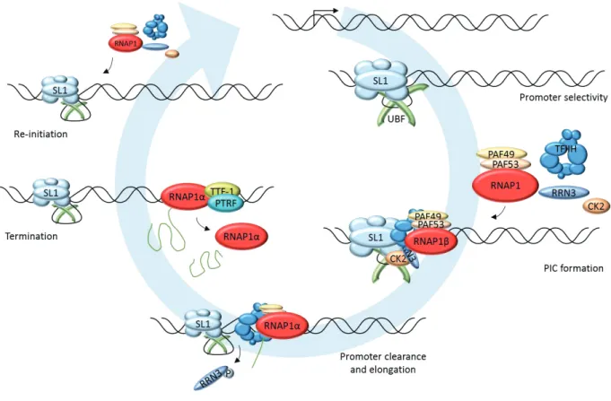
Figure 8. RNA Polymerase 2 transcription cycle 19 Figure 9. Organization of the nucleolus in human cells 22 Figure 10. Transcription unit of ribosomal DNA 24 Figure 11. RNA Polymerase 1 transcription cycle 27
TABLE OF CONTENTS
INTRODUCTION 1
1. DNA Damage and Repair 2
1.1. Types of DNA lesions 2
1.2. Repair mechanisms 5
1.2.1. Base Excision Repair 5
1.2.2. Mismatch Repair 6
1.2.3. Homologous Recombination 6
1.2.4. Non-Homologous End-Joining 7
1.3. Nucleotide Excision Repair 9
1.3.1. Lesions recognition 9
1.3.1.1. GGR 9
1.3.1.2. TCR 10
1.3.2. Opening, Excision and Synthesis 11
1.3.3. Transcription restart 13
1.3.4. Associated diseases 13
1.3.4.1. Xeroderma Pigmentosum (XP) 13
1.3.4.2. Cockayne Syndrome (CS) 14
1.3.4.3. Trichothiodistrophy (TTD) 14
1.3.4.4. UV-sensitive Syndrome (UVSS) 14
1.3.5. Chromatin context 14
2. RNA Polymerase 2 transcription 16
2.1. Initiation 16
2.2. Proximal pausing and Elongation 17
2.3. Termination 18
2.4. Splicing 19
2.5. Chromatin context 20
3. RNA Polymerase 1 transcription 22
3.1. The Nucleolus 22
3.2. Ribosomal DNA 23
3.3. Transcription cycle 24
3.3.1. Initiation 25
3.3.2. Promoter escape and Elongation 26
3.3.3. Termination 26
3.4. Chromatin context 27
RESULTS 29
Mechanistic Insights in Transcription-Coupled Nucleotide Excision Repair of Ribosomal DNA 30
Supplementary Information 40
Abstract 50
Results and Discussion 51
Bibliography 56
Figures 57
Extended data 61
Methods 68
Unravelling the molecular role of XAB2 during transcription and DNA repair 75
Introduction 76
Results 78
XAB2 involvement in DNA repair 78
XAB2 dynamic during TC-NER 79
XAB2 pre-mRNA splicing complex during repair 80
RNAP2 dynamic in absence of XAB2 82
Material and Methods 83
Discussion 89
Figures 91
Supplementary Information 95
Bibliography 98
CONCLUSIONS AND PERSPECTIVES 101
1. DNA Damage and Repair
The DNA macromolecule enclosed in the nucleus of our cells represents the foundation of life for all organisms. It contains all the genetic information, which characterizes our phenotype, e.g. the colour of our eyes, as well as all the instructions that are required for the proper functioning of our physiological processes.
Unfortunately, the DNA structure is continuously challenged by an extended number of damaging agents, which are responsible for the production of a variety of DNA lesions. These DNA-damaging factors originate from three main sources (1):
a. Environmental agents such as UV light, ionizing radiation, air polluting agents and other genotoxic agents, e.g. cigarette smoke
b. Products of cellular metabolism such as reactive oxygen species (ROS) generated by oxidative respiration and products of lipid peroxidation
c. Spontaneous disintegration of DNA chemical bonds, e.g. hydrolysis of nucleotide residues or deamination of cytosine, adenine and guanine
The consequences of a defective DNA damage repair can be deleterious for the organism survival. In actively dividing cells, the damaged DNA strand can be replicated in a permanent mutated form, increasing the risk of cancer development (2). On the other hand, lesions may also block the transcription process, causing cell death or senescence and contributing to aging (3). In order to protect the DNA integrity and to prevent formation of deleterious mutations, cells developed several repair mechanisms able to eliminate DNA persisting lesions.
1.1. Types of DNA lesions
The most succinct, and at the same time complete, definition of DNA damage is:
“Damage to DNA consists of any change that introduces a deviation from the usual double-helical structure”(4)
The various types of damages that our DNA can be subject to are listed below and their frequency rate is represented in Table 1.
Deamination: is a very common type of hydrolytic lesion and leads to the conversion of one base to another, resulting in a mismatch. For example, deamination of cytosine results in uracil, which preferentially pairs with adenine rather than guanosine, thus creating a mismatch. Then, when the cell undergoes replication, this will result in the substitution of a C:G pair with an A:T pair.
Depurination: is another very common lesion caused by hydrolytic loss of nitrogenous bases. Depurination is strongly affected by temperature and, among the total loss of nitrogenous bases, it occurs at a much higher rate compared to pyrimidine loss (5). In general, loss of nitrogenous bases leads to abasic sites, which may promote DNA strand breakage or base mismatch.
Alkylation: the most common alkylation reaction involves methylation of nitrogenous bases. It usually affects a nitrogen or an oxygen atom, thus resulting in a variety of modified bases, which have different pairing properties. Alkylated bases may also be subject to further modification, like for the 5-methylcytosine that is then deaminated to thymine, resulting in DNA mismatch. Furthermore, methylation has also a role in the alteration of chromatin architecture and deregulation of gene expression, through methylation of cytosine residues in CpG islands.
Oxidation: can affect the purine bases, as well as the pyrimidines. Oxidation of purines leads to 8-oxo-G or 8-oxo-A, while pyrimidine oxidation lead to glycol or -hydroxy compounds formation. Base oxidation is often coupled to strand breaks, both caused by ROS. Accumulation of oxidative damages is related to ageing (6).
Strand breaks: are the result of the disruption of the phosphodiester bond between two adjacent deoxyribose residues on one strand (Single-Strand Breaks, SSB) or on both strands (Double-Strand Breaks, DSB) of the DNA helix. Strand breaks frequently occur during normal DNA manipulations, such as transcription or replication, or genotoxic stresses caused by cell metabolism, such as ROS accumulation. DSB are less common in cells, since their presence is lethal; however, they are physiologically necessary for specific phases of cell cycle and only for some type of cells in order to allow homologous recombination of genetic information, e.g. they occur in germ cells during meiosis or in developing lymphocytes during V(D)J recombination. DSB can also be induced by exogenous agents like high-energy electromagnetic radiations (mostly ionizing radiations).
Dimerization: Dimers normally do not exist in DNA and their formation is most often caused by high-energy and short-wavelength UV light ranging between 280 and 350 nm (UV-B and UV-A). Dimerization can occur between two bases on the same strand or between bases from different DNA strands. There are two types of dimers in DNA: 6-4 photoproducts (6-4PP) that can involve T/C, C/C or T/T or Cyclobutane Pyrimidine Dimers (CPD) involving only T/T.
Bulky adducts and Inter/Intra-strand crosslinks: These lesions, as well as dimerizations, create physical impediments for processes such as transcription or replication of the DNA
double helix. Bulky lesions are formed by aromatic compounds, such as Polycyclic Aromatic Hydrocarbons (PAH), or by products of benzopyrene metabolism in vivo (a composite of cigarette smoke). Inter and Intra-strand crosslinks can be caused by intercalating agents, such as psoralen that is activated in presence of UV-A light, antibiotics as Mitomycin C, or even classic anticancer agents such as cisplatin derivatives (7).
Table 1. Rate of DNA damage per human cell per day
Information of different damages rates have been taken from (2, 8-11)
Rate of DNA lesions per human cell per day
Single strand break 55.000
Depurination 10.000
Deamination 400
Oxidation 1000
Alkylated bases 5000
Intra-strand crosslink 10
Double strand break 10-50
Dimerization
1.2. Repair mechanisms
All the repair mechanisms described below include a series of factors that participate in diverse and tightly regulated steps and some proteins are shared by different repair pathways. All these mechanisms are coupled to a signaling machinery communicating with the cell cycle in order to inhibit, when it is necessary, cell cycle progression and to give the cell the time for repair (12). Therefore, it has to be highlighted the intrinsic complexity of these repair mechanisms and the importance of their regulation.
1.2.1. Base Excision Repair
Base Excision Repair is responsible for the detection and repair of the most common type of DNA damages. Indeed, BER recognizes and removes small helix distorting lesions such as base oxidation by Reactive Oxygen Species (ROS), deamination and alkylation. ROS are generated, among others, as a product of the normal mitochondrial activity. Without BER, these lesions would potentially block DNA replication and increase the prevalence of disease causing mutations within
the genome.
In the initiation step of BER, a damage specific DNA glycosylase identifies and removes the damaged base through cleavage of the bond between the target base and deoxyribose, leaving an intact abasic site (AP site). At least 11 different mammalian DNA glycosylases are known, which are sorted into four super families based on their structural characteristics. The intact AP site is then processed by a human AP-endonuclease 1 (APE1), which leads to the formation of a single strand break. A DNA polymerase (polβ) performs the removal of the sugar residue and the insertion of the new nucleotide. Finally, a Ligase (LIG1/LIG3) and the scaffolding protein XRCC1 seal the nick (13, 14).
1.2.2. Mismatch Repair
The Mismatch Repair (MMR) pathway is involved in the correction of errors that escape polymerase proofreading during replication. MMR is responsible for the detection and repair of insertion/deletion loops and base-base mismatches; defects in this pathway result in a mutator phenotype leading to a strong cancer predisposition.
The mismatch recognition process starts with the binding to the lesion of the MSH2/MSH6 heterodimer, named MutSα. MutSα undergoes an ATP-driven conformational change and recruits the MLH1/PMS2 complex, MutLα. This ternary complex can translocate in both directions along the DNA. When it encounters a strand discontinuity, it recruits the Proliferating Cell Nuclear Antigen complex (PCNA) and the EXO1 exonuclease to initiate degradation of the nicked strand. The resulting single strand is stabilized by Replication Protein A (RPA), then the gap is filled by DNA polymerase δ and the remaining nick is filled by DNA ligase 1 (LIG1) (15, 16).
1.2.3. Homologous Recombination
Homologous Recombination (HR) is one of two repair mechanisms for Double Strand Breaks (DSB). DSBs can arise from ionizing radiation, anticancer treatments, DNA replication errors leading to replication fork collapse, and even ROS. Even if the rate of DSBs is estimated at 10 per cell, per day, they are considered one of the most harmful lesions for the cell since even one unrepaired DSB can lead to deleterious mitosis or chromosome instability.
HR is a highly conserved, error-free mechanism that only occurs during the S or G2 phases of the cell cycle, when a homologous template via the sister chromatid is available. The first step of HR is defined by DSB end resection involving the MRN complex, formed by Mre11, Rad50 and Nbs1, and the C-terminal binding protein-Interacting Protein (CtIP), which activates the MRN complex to initiate the resection. More extensive resection, performed by the exonuclease 1 (EXO1) together with the Dna2/BLM complex, contributes to the formation of a 3’ single-strand DNA which is protected by the recruitment of RPA proteins. RPA competes with Rad51, such that Figure 2. Mismatch Repair mechanism
and promote Rad51 binding. Actually, Rad51 drives the defining step of HR represented by the invasion of the 3’ ssDNA into a homologous duplex. After strand invasion of the sister chromatid, a DNA polymerase can synthetize the new DNA on the damaged strand (17-19).
Another pathway responsible for DSBs processing is termed Single-Strand Annealing (SSA). SSA concerns DSBs occurring on genes containing sequence repeats, such as ribosomal DNA, and it requires a homology of at least 20 bp. SSA is a non-conservative mechanism since it requires deletion of several nucleotides. Similar to HR, the process starts with DNA end resection, but Rad52 aligns the two DNA repeated sequences and the generated ssDNA tails are removed by the ERCC1/XPF nuclease (20).
1.2.4. Non-Homologous End-Joining
DSB can also be repaired via an additional pathway termed Non-Homologous End-Joining (NHEJ). NHEJ mediates the direct ligation of the broken DNA molecule and, since it does not require a homologous template for repair, it is not confined to a certain phase of the cell cycle. Therefore, it is the major and faster mechanism that repairs DSB, but it is also potentially error-prone, due to the absence of a template.
The initial steps of NHEJ is the recognition and binding of the Ku heterodimer (Ku70/Ku80) to the DSB in a ring-shaped structure, which can accommodate the double strand DNA helix. The Ku heterodimer’s function is to recruit the DNA-dependant protein kinase catalytic subunit (DNA-PKcs) and to stimulate its autophosphorylation. Upon DNA-PKcs phosphorylation, multiple DNA end-processing factors are also recruited to the break site to prepare the DNA ends for ligation, such as the endonuclease Artemis and the DNA nucleotidylexotransferase (DNTT) which catalyses the addition of nucleotides in the 3ʹ terminus without a DNA template. These DNA end-processing steps make DNA ends compatible for ligation where the final phase is mediated by the complex DNA Ligase4/XRCC4/XLF (18, 21, 22).
Similar to NHEJ, DSBs can be processed by an alternative form of NHEJ, termed alt-EJ. This additional mechanism normally acts when the classical-NHEJ is compromised and it requires a Figure 3. Homologous Recombination
homology between the double strands DNA of less than 20 bp. One of the first proteins implicated in alt-EJ is the Poly [ADP-ribose] Polymerase 1 (PARP-1) that binds the DSB in a competitive way with the Ku heterodimer, thus determining the choice between the c-NHEJ and alt-EJ. DNA ends are then processed by the MRN complex, stimulated by the phosphorylated CtIP. The DNA polymerase θ binds directly to resected DNA and synthetizes the new strand. DNA Ligase 1 or 3, with the recruitment or not of XRCC1, perform the end-ligation (18, 22).
Figure 4. Non-Homologous End-Joining mechanism
1.3. Nucleotide Excision Repair
The Nucleotide Excision Repair mechanism (NER) was described for the first time in the early 1960s thanks to the work of Setlow, Howard-Flanders and Hanawalt (23-25). NER is the repair pathway responsible for detection and repair of a broad variety of helix-distorting DNA lesions, such as UV-induced Cyclobutane Pyrimidine Dimers (CPDs) or 6-4 Pyrimidine-pyrimidone Photoproducts (6-4PPs), but also of oxidative damage, bulky lesions and intrastrand crosslink formed by cancer chemotherapeutic drugs such as cisplatin (3, 26, 27).
The NER process starts with the recognition of the DNA lesion after which, the damaged strand is incised on both sides of the ≈ 30 nucleotide long stretch before it can be removed. A new DNA patch is synthetized using the undamaged complementary strand and is ligated with the contiguous filament. (28) The NER mechanism will be described in more details below.
1.3.1. Lesions recognition
The lesion detection step is a specific process that divides the NER mechanism into two sub-pathways: Global-Genome Repair (GG-NER), operating throughout the genome, and Transcription-Coupled Repair (TC-NER), which detects lesions on the transcribed DNA strand of transcriptionally active genes (29).
1.3.1.1. GGR
The UV light induced 4PPs and CPDs cause different distortions to the DNA double helix. 6-44PPs create a DNA alteration sufficient to be directly detected by the XPC/hRAD23b/Centrin2 complex, while CPDs cause a minor distortion of the double helix that is not detectable by the XPC complex but will be first recognized by the DNA Damage-Binding protein 2 complex (DDB2). It is for this reason that detection and repair of CPDs is slower than that of 6-4PPs (30, 31).
The DDB2 protein (encoded by the xpe gene) forms a heterodimeric complex with the DDB1 protein (also termed XPE binding-factors) and together are part of the CUL4-ROC1 ubiquitin ligase complex that ubiquitinates DDB2, XPC and histones upon DNA damage. DDB2 binds to the CPD lesion thanks to a hydrophobic pocket that accommodates the lesion and creates a protrusion that facilitates recognition of the damage by the XPC complex (31-33).
The XPC protein has been shown to bind to the strand opposite the lesion. Studies conducted on Rad4, the yeast orthologue of XPC, has demonstrated that a β-hairpin domain encircles two nucleotides opposite the damage, displaying increased ssDNA character due to the thermodynamic destabilization caused by the lesion (34). The hRAD23b protein stabilizes XPC by
protecting it from degradation by the ubiquitin–proteasome system and facilitates XPC binding to the damaged site, but it rapidly dissociates from XPC upon binding to damaged DNA strand (35, 36). The centrin2 protein is a calcium-binding protein of the calmodulin family and its interaction with XPC via its C-terminal domain has been shown to stimulate NER activity (37, 38). The XPC/hRAD23b/Centrin2 complex melts the DNA around the lesion and recruits the multiprotein complex TFIIH. Okuda et al. have recently demonstrated that XPC recruits TFIIH by binding to the pleckstrin homology (PH) domain of TFIIH subunit p62 (39).
1.3.1.2. TCR
The arrest of an elongating RNAPII by the lesion present on the transcribed strand is the damage recognition step in TCR. The fate of RNAPII and the way NER system has access to the lesion have not been clarified yet, but several scenarios have been proposed (40). Sarker et al. suggested that elements of the NER machinery might have access to the lesions upon polymerase remodelling (41). Alternatively, a translesional mechanism has been described, in order to clear the way for GGR to find and repair the lesion post-transcriptionally, but this could result in transcriptional mutagenesis (42, 43). Another hypothesis that is presented is that, since stalled RNAP2 can lead to cell death, RNAP2 may undergo degradation with transcript abortion (44). An additional scenario is represented by a backtracking mechanism of the RNAP2. Backtracking, or reverse translocation, can occur to allow space for the repair complex to operate and it could be
TFIIS-mediated. Indeed TFIIS has been found to facilitate the transcript cleavage activity of RNAP2, which is necessary for resumption of elongation (45-47). Furthermore, Chiou at al. demonstrated that RNAP2 dissociates from the template during transcription-coupled repair (48).
After RNAP2 arrests, it recruits the CSB homodimer, a transcription elongation factor that translocates along the DNA template with RNAP2. CSB tightly binds the stalled RNAP2 and alters the RNAP2/DNA interface by wrapping the DNA around itself (49). CSB is a SWI/SNF-like DNA-dependent ATPase; its C-terminal region is required for RNAP2 interaction and CSA translocation to the nucleus and undergoes SUMOylation (50, 51). CSB colocalizes with other types of DNA lesions, such as oxidative damage, double-strand breaks and interstrand crosslink (52).
Subsequently, CSB, together with the stalled RNAP2, recruits the CSA protein. CSA is a WD40 motif-containing protein that interacts with Cullin4A (Cul4A) and ROC1/Rbx1 ubiquitin E3 ligase and is initially inhibited after UV irradiation via its association with the COP9 signalosome and later becomes activated to ubiquitinate and degrade CSB (53). Actually, CSA-dependent degradation of CSB is required for recovery of RNA synthesis after UV damage (54). CSA is also responsible for the recruitment of the UVSSA/USP7 complex (55).
The UVSSA protein and its partner USP7 are associated with elongating RNAP2. After the block of transcription, UVSSA and USP7 strongly bind to RNAP2. These factors facilitate CSA and CSB-dependent ubiquitination of the phosphorylated form of RNAP2, which deubiquitylates CSB to stabilize it and facilitate RNAP2 recycling for transcription restart after repair (56-58). Similar to TFIIH recruitment by XPC in GGR, UVSSA binds to the p62 PH domain and recruits TFIIH to the site of damage (59).
1.3.2. Opening, Excision and Synthesis
After the lesion recognition step, both GGR and TCR converge into the same pathway with the recruitment of the TFIIH complex in 5’ to the damage. TFIIH is a basal transcription/repair complex of 10 subunits divided between the core and the CAK. Two of the core proteins, the Figure 6. TCR recognition of UV
ATPase/helicases XPB and XPD, are responsible for unwinding the DNA to create a 30 nucleotide repair bubble. Concerning the XPB protein, only its ATPase activity is essential for NER, while XPD must be active as both ATPase and helicase. Another TFIIH subunit, the small p8 protein (TTD-A) has a NER specific role by acting as a stabilizer of TFIIH and facilitating the formation of the DNA repair bubble (60, 61).
Once the pre-excision complexes are assembled, XPA, RPA and XPG are sequentially recruited. The XPA protein binds the DNA close to the 5’ side of the bubble and is a central NER factor: it promotes the dissociation of the CAK complex from the TFIIH core; it interacts with TFIIH, RPA and PCNA and it recruits the XPF/ERCC1 endonuclease (62). RPA is a three subunit protein that binds the ssDNA opposite the lesion in order to protect it from other ssDNA binding proteins and from degradation (63). Interestingly, a novel protein has been found interacting with XPA, named XPA-Binding protein 2 (XAB2) (64), and its role in NER will be described in more details in the Results section.
The dual incision event is accomplished by the two endonuclease XPF/ERCC1 and XPG. XPG is the first recruited by TFIIH to the 3’ side of the bubble, but this incision is only triggered later, following the 5’ incision by XPF/ERCC1, which is recruited by XPA. There is a defined order for the incision step and, since XPF/ERCC1 generates a free 3’ hydroxyl group, the replication machinery can initiate the synthesis of the new strand even before the second incision has taken place (65, 66).
The DNA replication machinery necessary for the synthesis of the new DNA strand is recruited to the repair bubble by the sliding clamp Proliferating Cell Nuclear Antigen (PCNA) and the Replication factor C (RFC) (67). The DNA polymerase ε is responsible for DNA synthesis in replicative cells, while DNA polymerase δ and κ synthetize the new strand in non-dividing cells (68). The proliferative status of the cell also defines the DNA ligase used for the sealing of the new strand with the contiguous filament. In proliferating and non-proliferating cells the nick is sealed by LIG3/XRCC1 and LIG1 respectively (63).
Figure 7. NER common pathway
1.3.3. Transcription restart
After that repair of UV lesions on actively transcribed genes is completed, transcription by the RNAP2 and RNA synthesis need to restart. Multiple factors have been shown to be required for transcription resumption. One of these factors is the RNA polymerase 2 elongation factor Eleven-Nineteen Lysine-Rich Leukemia protein (ELL) that interacts with TFIIH via the Cyclin dependent kinase 7 (Cdk7) and has been shown to serve as a docking site and to promote resumption of transcription (69). It has also been shown that knockdown of TFIIS results in high levels of hyperphosphorylated RNAP2 and impaired transcription recovery, suggesting a post-repair function (70). Furthermore, some chromatin remodelling factors, such as DOT1L (71), HIRA and FACT, have been proposed to cooperatively work together to promote recovery of RNA synthesis (72).
1.3.4. Associated diseases
Mutations in different NER proteins can lead to several rare recessive photosensitive syndromes such as: Xeroderma Pigmentosum, Cockayne syndrome, Trichothiodystrophy and UV-sensitive syndrome. Despite these diseases have been extensively studied, there still are clinical outcomes that are not totally understood, such as the sun-sensitive skin associated with skin cancer predisposition occurring only in the case of XP patients.
1.3.4.1. Xeroderma Pigmentosum (XP)
Xeroderma pigmentosum is a rare and inherited disease caused by mutations in one of the seven complementation groups encoding for proteins XP-A to XP-G. An additional complementation group, XPV, is NER proficient and the disease is due to mutation in DNA polymerase η, a translesional polymerase responsible for replication of DNA containing unrepaired UV-lesions.
XP patients present parchment skin and freckles on skin regions exposed to the sun and they have marked skin sun sensitivity with a >1000-fold increased risk to develop skin cancers. The mean age of onset of these symptoms is 2 years and the mean age of onset of skin cancer is 8 years old. In addition to skin abnormalities, a fraction of XP patients (≈ 30%) displays progressive neurologic degeneration. Although there is no treatment for the disease at the time, some precautionary measures can be adopted, as special UV-filters which are applied on windows or to avoid skin exposure to the sun without a sunscreen (73, 74).
1.3.4.2. Cockayne Syndrome (CS)
Cockayne Syndrome is caused by mutations on the CSA and CSB complementation group proteins, so CS patients have a defective TCR repair pathway. The symptoms of the disease are mostly neurological, such as neurodemyelinnation, mental retardation, hearing loss, growth failure and microcephaly. Unlike the other NER diseases, CS also include immature sexual development. Surprisingly, CS patients are apparently not predisposed to develop skin cancer. Many of the clinical symptoms of CS patients are difficult to explain just in light of a partial defective NER, but they could be due to the CSA and CSB transcriptional engagement. Approximately 10% of CS cases present rare mutations in XPB, XPD, XPG or ERCC1 leading to a XP/CS combined phenotype. This includes XP skin phenotype, including cancer predisposition, in addition to neurological abnormality of CS (54, 73).
1.3.4.3. Trichothiodistrophy (TTD)
Trichothiodistrophy is caused by certain mutations on the XPB and XPD genes and on the TTDA gene. The hallmark of TTD is the presence of sulphur-deficient brittle hair. Depending on the severity of the case, other symptoms can include ichthyosis, microcephaly, neurological defects, premature aging feature and intellectual disability. Some TTD patients may also present photosensitivity, but without skin cancer predisposition (73).
1.3.4.4. UV-sensitive Syndrome (UVSS)
UV-sensitive syndrome is a rare autosomal recessive disease that can be caused by mutations on CSA, CSB or UVSSA genes. Unlike other NER syndromes, it presents a relatively mild phenotype with sun sensitivity, freckles, skin dryness and abnormal pigmentation. UVSS patients develop
normally and they do not have skin cancer predisposition (40).
1.3.5. Chromatin context
The damaged DNA present in the nucleus exists in the form of chromatin, coiled and compacted in nucleosome, and is therefore not always easily accessible to be repaired. Therefore, repair systems also have to deal with the chromatin environment. Indeed, it has been proposed a model of chromatin rearrangement for the NER mechanism, called “Access, Repair, Restore”(75, 76), which has been recently improved by the “Access/Prime and Repair/Restore” model in which chromatin and chromatin-associated proteins are not only impeding access to repair machineries but are also actively promoting repair (77). In order to access the lesion for repair, histone proteins have to be mobilized. Several chromatin-associated factors have been proposed for this step, such
as BRG1 and INO80 (78, 79). A recent study demonstrated that also factors involved in chromatin compaction are recruited to UV-damaged chromatin, such as the Heterochromatin Protein 1 (HP1), underlying the concept that chromatin organization has not to be simply considered as a barrier, but that chromatin-associated proteins can play an active role in repair (80). At the Repair/Restore step of chromatin rearrangement around a UV lesion, factors responsible for de
novo incorporation of histones have been found: new H3.1 histones are deposited by CAF-1 and
One of the fundamental mechanisms for the life of all organisms consists in the translation of the genetic information, contained in the DNA, into the proteins or to various classes of functional RNAs, in order to accomplish specific cellular roles. This mechanism is said transcription and is performed by multiple enzymatic complexes, the DNA-dependent RNA Polymerases.
There are three RNA polymerases operating in mammalian cells: RNA polymerase 1 transcribes the pre-ribosomal rRNA 47S; RNA polymerase 2 synthetizes precursors of mRNAs and most snRNAs and microRNAs; RNA polymerase 3 is in charge of tRNAs, rRNA 5S and other small RNAs transcription. In order to provide a background for the results presented, in this section and in the next one, RNAP2 and RNAP1 transcription cycles will be detailed.
2. RNA Polymerase 2 transcription
RNA polymerase 2’s complete structure has been identified thanks to X-ray crystallography in 2003 and it has allowed for a better understanding of its organization and function (83, 84). We can distinguish a core composed of 10 subunits (RPB1, RPB2, RPB3, RPB5, RPB6, RPB8, RPB9, RPB10, RPB11 and RPB12) and a stalk formed by the heterodimer RPB4 and RPB7, which is required for initiation of transcription but not for the elongation step. Within the core, 5 subunits are commonly shared between RNAP1, RNAP2 and RNAP3. The two main RNAP2 subunits RPB1 and RPB2 form a deep positively charged cleft in which the open ssDNA can enter and be transcribed by the active site. The other core subunits are necessary for maintain this structure.
An important functional characteristic of the RPB1 subunit is the presence of a C-Terminal Domain (CTD). The CTD, which is specific to RNAP2, contains tandem repeats of a heptad sequence: Tyr-Ser-Pro-Thr-Ser-Pro-Ser. Modifications of phosphorylated and dephosphorylated states of Ser 2, 5 and 7 regulate all the steps of the transcription cycle, and even mRNA processing (85, 86).
2.1. Initiation
RNAP2 transcription cycle is depicted in Figure 8. The first step of RNAP2 transcription is the recognition of the promoter by the transcription machinery. Promoters of RNAP2 transcribed genes present a great variability and there are not universal core promoter (87). Sequences found in core promoters include the TATA box, the initiator element (Inr), the TFIIB recognition element (BRE) and the Downstream Promoter Element (DPE) (88). The core promoter elements represent binding site for subunits of the transcription machinery and serve to orient the pre-initiation complex (PIC) at the transcription start site (TSS) (89).
The PIC assembly starts with the binding of the TFIID subunit TBP to the TATA element; TAF1/2 components of TFIID have been implicated in Inr recognition, in order to strengthen interaction with the promoter. Then TFIIA and TFIIB are recruited to stabilise promoter bound-TFIID. Next, RNAP2, in association with TFIIF, adds to the growing PIC. This drives the association of TFIIE and lastly of TFIIH (90). An alternative PIC assembly pathway proposes a preassembled complex of RNAP2, the Mediator complex and the General Transcription Factors (except TFIID and TFIIA) binding directly to the promoter (91).
After PIC formation, the DNA double helix has to be separated around the TSS in order to form an open complex suitable for transcription. This opening occurs thanks to the ATP-dependent helicase activity of the TFIIH subunit XPB. RNAP2 is the only polymerase that requires this kind of activity for DNA melting (92, 93). The DNA template is then placed in the active site of RNAP2 and, after a series of abortive transcripts, a RNA product of more than 10 nucleotides is produced and this allows RNAP2 to leave the promoter (94). The required step for this switch to a stable elongation form is the phosphorylation of the CTD Ser5 by the TFIIH subunit Cdk7 (95). This CTD phosphorylation is also important for the process of RNA transcript because it is recognized by the capping enzymes that catalyse the addition of the 7-methylguanosin cap to the 5’ end of the nascent transcript (96). While RNAP2 clears the promoter, most of the PIC complex stays bound at the promoter forming a re-initiation scaffold complex, with TFIIF being the only factor staying with RNAP2 after this step (97).
2.2. Proximal pausing and Elongation
Once cleared the promoter, RNAP2 transcribes few nucleotides, by addition of some Nucleosides Triphosphate (NTPs) to the nascent RNA transcript, before pausing. This arrest is mediated and stabilised by several pausing factors, such as the Negative Elongation Factor (NELF) and DRB Sensitivity Inducing Factor (DSIF) (98). The release of RNAP2 into a productive elongation requires the activity of the Positive-Transcription Elongation Factor (P-TEFb) (99). The kinase subunit of P-TEFb, Cdk9, drives phosphorylation of RNAP2 CTD Ser2 and of pausing factors NELF and DSIF (100). NELF dissociates from chromatin upon its phosphorylation, whereas DSIF switches from being a negative factor to being a positive elongation element (101, 102). The exact role of this pausing event in transcription has not been clarified yet, but it has been proposed a relationship between RNAP2 pausing and the regulation of chromatin structure and gene expression (103). When RNAP2 progress toward the 3ʹ end of the gene, Ser5P levels appear to decrease, leaving polymerases phosphorylated at Ser2 to terminate transcription (104).