HAL Id: hal-02493848
https://hal.archives-ouvertes.fr/hal-02493848
Submitted on 26 Feb 2021
HAL is a multi-disciplinary open access
archive for the deposit and dissemination of
sci-entific research documents, whether they are
pub-lished or not. The documents may come from
teaching and research institutions in France or
abroad, or from public or private research centers.
L’archive ouverte pluridisciplinaire HAL, est
destinée au dépôt et à la diffusion de documents
scientifiques de niveau recherche, publiés ou non,
émanant des établissements d’enseignement et de
recherche français ou étrangers, des laboratoires
publics ou privés.
Poly-(ADP-ribose)-polymerase inhibitors as
radiosensitizers: a systematic review of pre-clinical and
clinical human studies
Paul Lesueur, François Chevalier, Jean-Baptiste Austry, Waisse Waissi, Hélène
Burckel, Georges Noel, Jean-Louis Habrand, Yannick Saintigny, Florence Joly
To cite this version:
Paul Lesueur, François Chevalier, Jean-Baptiste Austry, Waisse Waissi, Hélène Burckel, et al..
Poly-(ADP-ribose)-polymerase inhibitors as radiosensitizers: a systematic review of pre-clinical and
clin-ical human studies. Oncotarget, Impact journals, 2017, 8 (40), �10.18632/oncotarget.19079�.
�hal-02493848�
www.impactjournals.com/oncotarget/
Oncotarget, Advance Publications 2017
Poly-(ADP-ribose)-polymerase inhibitors as radiosensitizers: a
systematic review of pre-clinical and clinical human studies
Paul Lesueur
1,2, François Chevalier
1, Jean-Baptiste Austry
1, Waisse Waissi
3, Hélène
Burckel
3, Georges Noël
3, Jean-Louis Habrand
2, Yannick Saintigny
1,*and Florence
Joly
4,*1Laboratoire d’accueil et de recherche avec les ions accélérés, CEA, CIMAP-GANIL, 14000 Caen France 2Centre Francois Baclesse Centre de Lutte Contre le Cancer, Radiotherapy Unit, 14000 Caen France 3EA 3430, Laboratoire de Radiobiologie, Centre Paul Strauss 67000 Strasbourg France
4Centre Francois Baclesse Centre de Lutte Contre le Cancer, Clinical Research Unit, 14000 Caen France *These authors contributed equally to this work and Co last authors
Correspondence to: Paul Lesueur, email: Paul.Lesueur89@gmail.com
Keywords: radiosensitization, poly(ADP-ribose)-polymerase inhibitors, radiotherapy, radiobiology
Received: May 27, 2017 Accepted: June 19, 2017 Published: July 07, 2017
Copyright: Lesueur et al. This is an open-access article distributed under the terms of the Creative Commons Attribution License 3.0 (CC BY 3.0), which permits unrestricted use, distribution, and reproduction in any medium, provided the original author and source are credited.
ABSTRACT
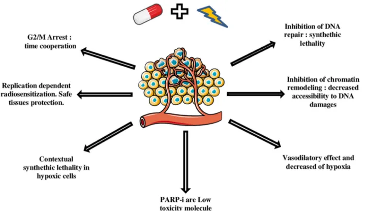
Background: Poly-(ADP-Ribose)-Polymerase (PARP) inhibitors are becoming important actors of anti-neoplasic agents landscape, with recent but narrow FDA's approvals for ovarian BRCA mutated cancers and prostatic cancer. Nevertheless, PARP inhibitors are also promising drugs for combined treatments particularly with radiotherapy. More than seven PARP inhibitors have been currently developed. Central Role of PARP in DNA repair, makes consider PARP inhibitor as potential radiosensitizers, especially for tumors with DNA repair defects, such as BRCA mutation, because of synthetic lethality. Furthermore the replication-dependent activity of PARP inhibitor helps to maintain the differential effect between tumoral and healthy tissues. Inhibition of chromatin remodeling, G2/M arrest, vasodilatory effect induced by PARP inhibitor, also participate to their radio-sensitization effect.
Materials and Methods: Here, after highlighting mechanisms of PARP inhibitors radiosensitization we methodically searched PubMed, Google Scholar, Cochrane Databases and meeting proceedings for human pre-clinical and clinical studies that evaluated PARP inhibitor radiosensitizing effect. Enhancement ratio, when available, was systematically reported.
Results: Sixty four studies finally met our selection criteria and were included in the analysis. Only three pre-clinical studies didn't find any radiosensitizing effect. Median enhancement ratio vary from 1,3 for prostate tumors to 1,5 for lung cancers. Nine phase I or II trials assessed safety data.
Conclusion: PARP inhibitors are promising radiosensitizers, but need more clinical investigation. The next ten years will be determining for judging their real potential.
INTRODUCTION
Poly-(adenosine diphosphate-ribose)-polymerase (PARP)
is a family of enzymes involved in a wide number of cellular
processes, including DNA replication, transcription, repair
and cell death. PARP proteins have been studied for decades
for notably their roles in DNA repair. PARP1 is the most
abundant and active enzyme of the PARP family, but roles
of other members including PARP2 and PARP3 in DNA
damage responses is emerging. In fact, they play an important
role by detecting the presence of damaged DNA and then by
activating signalization pathways that promote appropriate
cellular responses. PARP is involved in base excision repair
(BER) by allowing the recruitment and activation of BER
www.impactjournals.com/oncotarget
actors and consequently make easier the DNA single strand
break (SSB) reparation. Further studies have shown that
PARP-1 and PARP-2 were also implicated in DNA breaks repair by
Non-Homologous End Joining (NHEJ) and Homologous
Recombination (HR).
Increased PARP activity has been observed in
numerous cancers, and has been sounded to be one
possible mechanism of resistance to cell-death by
DNA-damaging therapeutics. Then progressively, PARP proteins
became a very interesting target for oncologic treatments,
and first PARP inhibitors (PARPi) were designed at the
end of the eighties. Indeed, 2005 year has been a real
turning point for the development of PARPi as two
high-value publications showed that dysfunction of homologous
recombination such as in BRCA1 and BRCA2 mutated
cells triggered a high sensitization to PARPi [1, 2].
PARPi leads to the persistence of DNA lesions normally
repaired by homologous recombination. Then, such
accumulation of non-repaired DNA lesions leads to cell
death. Synthetic lethality was then confirmed as a serious
avenue of therapeutic development. PARP inhibitors have
been evaluated in clinical trials either as single agents or
in combination with DNA damaging agents. Olaparib, the
most developed PARPi, has been approved by the FDA for
treatment of Ovarian and Prostate BRCA mutated cancers.
Many others molecules are being developed such as
rucaparib, talazoparib, niraparib, veliparib, or simmiparib.
Classically, radiosensitivity is described as function
of the tumor intrinsic radiosensitivity, the tumor repair
capacity, the reoxygenation process, the cell cycle
redistribution, and the tumoral tissue repopulation. More
recently, with development of stereotactic radiotherapy,
two additional characteristics appeared: the tumor
immunity and the vascular endothelial damage process [3].
So, a radiosensitizer has to impact one or more of these
processes without worsening the treatment toxicity. The
combination of ionizing radiation with radio-enhancing
agents represents an opportunity to increase the efficacy
of radiotherapy as a treatment modality, and at the same
time minimize toxic side effects and potential damages to
healthy surrounding tissues. PARPi have many qualities
required for radio sensitizing effects (Figure 1) and since
the last ten years, they have become interesting molecules
as radiosensitizers. There is evidence that the absence of
PARP-1 and -2, which are both activated by DNA damage
and facilitate DNA repair, produces an hypersensitivity
to ionizing radiation. Therefore, the inhibition of
PARP-mediated DNA damage repair can help to sensitize cells
to radiation by prolonging strand breaks and by leading
to a cell-death signaling pathway. This effect could be
enhanced in cancers harboring defects in homologous
recombination and by synthetic lethality mechanism.
Nevertheless modulating DNA damage repair is not the
only way of the radiosensistizing effect of PARPi.
The scope of this systematic review is to make a
state of the art about advancements of PARPi use as
radiosensitizer in human tumor cells and to provide
food for clinician’s thought who would wish to study
radiosensitization methods. Each organs group will be
treated separately. Before results of the systematic review,
principal mechanisms of PARPI radiosensitization will be
reminded.
Mechanisms of radiosensitization of parp
inhibitors
Inhibition of DNA repair
PARPs are involved in several DNA repair
mechanisms such as: Base Excision repair (BER),
homologous recombination (HR), conventional (c-NHEJ)
or alternative non-homologous end joining repair
(alt-NHEJ) [4–6].
PARP is one of the first actors of DNA single strand
break (SSB) repair because of its role in BER. PARP is a
part of the BER complex, described as a sensor of SSB.
PARP-1 and 2 are the two most important PARP family
enzymes implicated in BER. In fact, PARP detects SSB
and leads to PARylation, meaning accumulation of
poly-ADP-ribose (PAR) chain around the SSB [7, 8]. The local
PARylation allows the recruitment of XRCC1 for break
stabilization, DNA Polymerase β for the complementary
base synthesis, and DNA ligase III for final ligation [9].
Auto-PARylation of PARP releases PARP from the SSB
site. In case of PARP inhibition, PARP stays set on SSB
site, BER machinery is not recruited, and SSB persists.
Associated with PARPi, radiation therapy would
induce DNA damages, such as SSB or DSB which couldn’t
be repaired, and driving to DNA replication fork collapse.
SSB is then converted in potentially lethal double strand
break (DSB). PARPi are also trapped to DNA and induce
“mechanical” replication fork collapse and consequently
DSB [10]. It explains why PARP inhibition is more
effective than PARP suppression [11]. That’s the first and
main radio sensitizing effect of PARPi. If homologous
recombination deficient cells are considered, such as
BRCA mutated or BRCAness ones [12], the effect is more
potent: it is an example of synthetic lethality mechanism.
HR is an error free DNA repair mechanism in which
PARP1-2 play again a sensor role, by making easier the
recruitment of MRE11, DNA resection, and replication
restart [10, 13]. Furthermore PARP-1 regulate RH, and
can promote RH instead of error prone NHEJ. PARP
inhibition prevents the use of efficient and reliable DNA
repair system [5, 13].
There has been evidence, that an alternative
end-joining (Alt- NHEJ) operates in the absence of the
core components of classical NHEJ [14]. Alt-NHEJ is
efficient, but highly mutagenic. PARP-1 is even known
for his central role in alternative NHEJ [4]. PARPi
suppress again this way of repair and lead to more DNA
radiation damages.
As a conclusion the use of PARPi allow to target
most of DNA repair system, so as to potentiate the
radiotherapy effect by accumulating SSB and above all
DSB.
Replication-dependent radiosensitization
PARPi exercise their radiosensitizing effect,
more specifically, during the cell cycle S phase [15].
For example with olaparib, in Dungey’s glioblastoma
cells study, radiosensitization was significantly more
pronounced in S-phase (enhancement ratio at 50%
survival: SER50 = 1.60) than in G1 (SER50 = 1.27) or
G2 (SER50 = 1.33) enriched populations [16]. Moreover
it is well known that tumor have a higher proliferating
cell compartment in comparison to surround safe tissues.
It means that PARPi may radiosensitize tumor tissue,
while saving non tumoral tissue, which is one of the most
important qualities of a radiosensitizing agent.
Modulation of chromatin remodeling
PARP-1 dependent PARylation event directs the
recruitment of helicase, such as ALC1 (amplified in liver
cancer 1) to chromatin and nucleosomes [17, 18]. The
helicase activity promotes the unwinding of DNA double
helix and unblocks access to DNA of the machinery
responsible for transcription, replication and DNA repair
[17, 19]. After treatments by DNA damage agents just as
irradiation, inhibition of PARP-1 could delay DNA double
strand opening and DNA repair. It could be considered as
another way of PARPi potential radiosensitization.
Impact on microenvironnement and role of hypoxia
PARPi association with radiotherapy could help
to bypass the hypoxia induced radioresistance. On the
first hand, few PARPi, like rucaparib for example, have
structural similarities with nicotinamide, a vaso-dilatory
component. This specificity could enhance tumor growth
delay after radiotherapy, by increasing tumor blood
flow, enhancing drug penetration, and increasing oxygen
concentrations to offset hypoxic cell radioresistance [20].
It could explain why sometimes PARP efficiency is higher
in vivo than in vitro [20] . PARPi radiosensitize hypoxic
tumor thanks to an oxygen effect. Ionizing radiation
depends heavily on the presence of molecular oxygen to
produce cytotoxic effect. The molecular oxygen O2 is
absolutely necessary to chemically fix DNA free radicals
produced by ionizing radiation [21] . In the absence of O2,
DNA radicals are repaired by abstracting hydrogen from
sulfhydryl (SH) group present in protein [21]. It has been
reported that three times higher ionizing radiation dose is
required to kill hypoxic cancer cells, compared to
well-oxygenated cells, in order to achieve the equivalent level
of cell kill [22, 23].
On the other hand, even without any improvement
of the vasculature, PARPi exibit a radiosensitizing effect
in hypoxic cells. In fact hypoxia generates a genetic
instability by a mutator phenotype effect [24] linked
to the decreased transcription of proteins involved in
homologous recombination [25]. When PARPi and
radiotherapy are combined in hypoxic conditions, we
www.impactjournals.com/oncotarget
could observe contextual synthetic lethality. HR is altered
by hypoxia and carries out an increased death ratio [26].
G2/m arrest
With DNA repair, cell cycle regulation is perhaps
the most important determinant of ionizing radiation
sensitivity. A common cellular response to DNA-damaging
agents is the activation of cell cycle checkpoints,
leading to cell cycle arrest [27]. The concomitant
radio-chemotherapy induces temporo-spatial cooperation.
Spatial cooperation means that chemotherapy allows
to cure overfield micro metastatic disease, whereas
radiotherapy goal is to treat local invasion. Temporal
cooperation means that chemotherapy synchronizes,
and arrests cells in the radiosensitive phases of the cell
cycle: G2 and M. In this context of temporal cooperation,
chemotherapy could be considered as a radiosensitizer.
PARPi could take part into the radiosensitization process
in the same way as a result of the G2/M arrest induced,
secondary to chromosomic aberrations generated by
PARPi [1].
Low toxicity molecule
Most used radiosensitizers, such as Cisplatin or
Cetuximab, induce major systemic secondary effects,
which could limit their use in clinical practice particularly
for elderly patients such as: neuropathy, cytopenia,
nephropathy, cutaneous toxicity. In phase II-III clinical
trials studying PARPi monotherapy, toxicity remains
manageable and consists most of the time of anemia,
thrombocytopenia, neutropenia , asthenia and nauseas
rarely upper than grade II (23–26).
This low toxicity lets suggest that PARPi use as
radiosensitizer shouldn’t worsen treatment safety.
PARPi available or being developed
First PARPi were born at the beginning of the
eighties and were derived from 3-aminobenzamide. Due
to its lack of potency and specificity, 3-AB is not clinically
useful. Therefore, a number of third-generation PARP
inhibitors, some derived from the 3-AB structure, have
been developed in recent years and tested in pre-clinical
and clinical studies. Their development has been faster
during the second half of 2000’s, corresponding to the
discover of anti tumoral response in BRCA mutated cells
by Bryant and Farmer [1, 2]. PARPi suppress activity of
PARP catalytic domain explaining synthetic lethality in
HR defective cells. Nevertheless, PARP inhibition, delays
SSB repair to a greater extent than PARP depletion [11].
To explain these results, a PARP-1 trapping has been
proposed based on the idea that PARP1 is trapped on
DNA by PARP inhibitors, and PARP1-DNA complexes
can interfere with DNA fork replication [32, 33].
Actually seven PARPi are being developped by
pharmaceutical industry in clinical trials: Olaparib,
Rucaparib, Niraparib, talazoparib, veliparib, CEP 9722,
Simmiparib. They are all oral drugs. Among them only
Rucaparib, Olaparib, Niraparib and Veliparib have been
used as radiosensitizers. Others PARPi such as LT626,
PJ34, GPI 21016, 3-Aminobenzamide or
4-amino-1,8-naphthalimide have been less employed, and only in
pre-clinical studies (Table 1).
MATERIALS AND METHODS
We have led our bibliographic research in
accordance with PRISMA guidelines [34]. We looked
for all clinical or pre-clinical studies related to the use of
PARPi as radiosensistizers. We have requested PubMed,
Cochrane and Google Scholar databases without any date
limite and thus all the archives of meeting abstracts or
posters of ASTRO, ESTRO, ESMO and ASCO congresses
from 2010 to 2016. For PubMed Database, the key
words research strategy was: “(((radiosensitization) OR
radiotherapy)) AND (((((((niraparib) OR talazoparib) OR
rucaparib) OR veliparib) OR olaparib)) OR
“Poly(ADP-ribose) Polymerase Inhibitors” [Mesh])” We have selected
all research papers published in English language until
June 2017 7th. Studies dealing with animal cells were not
included in the systematic review. Then we have excluded
reviews, and articles which finally didn’t treat about
radiotherapy, such as, for example, use of radionuclides.
After manuscripts reviewing and application of inclusion
criteria, we have sorted the articles function of research
stage: in vitro, in vivo or clinical studies. For each selected
article we have extracted an enhancement ratio which
refers to the enhancement effect of radiation due to the
addition of PARPi. Enhancement ratio is classically a ratio
between doses associated with surviving fractions of 10%,
37% or 50% with or without the PARPi. For example:
SER37 = D37(no drug)/D37(PARPi). When enhancement
ratio (ER) wasn’t communicated as for in vivo studies,
clinical studies or few in vitro studies, we have supplied a
significant data such as survival, tumor growth delay. Then
data are assembled and discussed by organ groups.
RESULTS
After applying our selection criteria, 60 studies have
been included from Pubmed and Google scholar analysis.
Four more clinical studies have been added after reviewing
European, and American congress meeting abstracts, since
2010. Fifty five of the 64 selected studies are pre-clinical
in vitro or in vivo studies. For 49% of pre-clinical studies,
it has been possible to extract an enhancement ratio. For
others studies, a significant result about the efficacy of
the association has been given. Studies about brain or
digestive tumors represent 52% of studies dealing with
PARPi as radiosensitizer. Median enhancement ratio
vary from 1,30 for prostate tumors to,5 for lung cancers.
Until June 2017 7th, there was no data with Talazoparib,
Simmaparib or CEP 9722 use with radiotherapy. It is
worth noting that all studies meeting our review selection
criterias, are presented in the tables (Figure 2).
PARPi + radiotherapy for brain tumors
Glioblastoma is probably the tumor which could
benefit he most from radiosensitizing drugs. Indeed,
glioblastoma is considered as a radioresistant tumor
with a high level of intra-field recurrence. Furthermore
the target is surrounded of high risk complications
healthy tissues. Contrary to glioblastoma cells, neurons
don’t seem to express PARP-1 [49] and are low
replication cells: this aids to protect healthy tissues by
radiosensitizing only tumor cells. Glioblastoma stem
cells promote radioresistance and could be the source
of tumor recurrence after radiation [50]. Results from
Venere et al. studies highlight constitutive activation
of PARP1 in glioblastoma stem cells that can be
therapeutically exploited. They showed that PARPi plus
radiation compromises the stem cell phenotype in vivo
and inhibits glioblastoma stem cells enrichment [51].
Concurrent Temozolomide (TMZ) + Irradiation followed
by adjuvant TMZ is standard-of-care for patients
suffering from glioblastoma [52]. In that case PARPi –
inh could be considered as a radio and chemosensitizing
drugs [53, 54]. PARPi lead to glioblastoma cells
radiosensitization with most of in vitro/in vivo studies
which are promising with radiosensitizing enhancement
ratio comprised between 1,05 and 1,93 (Table 2).
Thanks to inhibition of BER, PARPi prevent repair of
N7-guanine and N3-adenine methylation induced by
TMZ, increase DNA strand breaks and is responsible
of a TMZ chemosensitization [53, 54] even on
glioblastoma stem cell [55]. Data concerning the role of
PARPi as TMZ chemosensitizer depending on MGMT
status stay debated [40, 56–58]even if some phase
II-III clinical trials recruits only patients with methylated
MGMT (NCT0215298). Moreover, PARPi could induce
synthetic lethality in PTEN deleted glioblastoma (36%
of Glioblastoma) suggesting that it might be logical to
treat PTEN-deficient glioblastomas with PARP inhibitors
in the future [59]. In summary, PARPi, in combination
with radiation therapy induce a median enhancement
ratio of 1,30.
It is worth noting that the ability for some PARP
inhibitors to cross the blood–brain barrier has been
validated like Veliparib [60]. Promising results are
www.impactjournals.com/oncotarget
thus expected in clinical studies. A phase I study has
found unsafe association of veliparib with radiotherapy
and TMZ for glioblastoma because of hematologic
complications [45]. Concerning brain metastases, whom
radiobiological characteristics are quite different of
primary tumors, the association with radiotherapy seems
to be safe but results are for the moment disappointing
[46, 48].
PARPi + radiotherapy for digestive system
tumors
Like for glioblastoma, radiation therapy plays
an important role in the treatment of locally advanced
pancreatic cancers, but its effect is limited by the sensitivity
of adjacent normal tissues (Duodenum, small bowel…),
and by the innate radioresistance of these cancers.
Germline mutations in BRCA1/2 define a molecular
subgroup of pancreatic cancers which in some populations
has a prevalence as high as 17% [61]. According to these
arguments, logically strategies of radiosensitization with
PARPi have been developped. In vitro enhancement ratios
vary from 1,2 to 1,5 with conventional radiotherapy until
1,98 with protontherapy(55–59) .
Moreover, the effect of radiosensitization was
greater for high LET [66].
Authors from Strasbourg laboratory are working
on the combination of PARPi (olaparib) and gemcitabine
after conventional and high dose photon radiation in
order to radiosensitize pancreatic cancer cell lines.
The first data have been obtained on three BRCA-wild
type pancreatic cancer cell lines (PANC-1, AsPC-1
and MIA PaCa-2). The clonogenic survival responses
showed that all cell lines could be radiosensitized
with olaparib through an increase of unrepaired DSBs
and a block in G2 phase. The radiosensitization with
olaparib was higher than with gemcitabine. Furthermore,
radiosensitization was higher with high dose per fraction.
Finally, in radioresistant cell line PANC-1, olaparib and
gemcitabine have a synergistic radiosensitization effect
(Waissi et al. [67]). Those data are promising and need
further investigations.
In vivo results are quite surprising with no increase
of growth tumor delay [64, 68], and no differences
between BRCA wild type and mutated tumors
responses were detected. Nevertheless a phase I study
is ongoing and evaluates the association of Veliparib
with gemcitabine and radiotherapy for locally advanced
pancreatic cancer [69]. First results should be published
in 2018. Olaparib is actually investigated alone as
maintenance therapy for metastatic pancreatic cancer in
phase III studies [70].
Others studies concern mainly colo-rectal cancer
with interesting results for in vivo studies. Combination
of radiotherapy with Veliparib and Irinotecan leads to a
threefold increase of growth tumor delay in Shelton et al.
study [71]. The phase I study LARC, assessing association
of Veliparib, Capecitabine and radiotherapy for stage
II-III rectal cancer, shows comfortable safety results with
promising anti-tumor activity with respectively 72%, 28%
and 70% for tumor downstaging, pathologic complete
response and sphincter sparing surgery [72]. In conclusion,
pre-clinical data show an enhancement ratio median value
for pancreatic cancer equal to 1,4 and 1,45 for colo-rectal
cancer (Table 3).
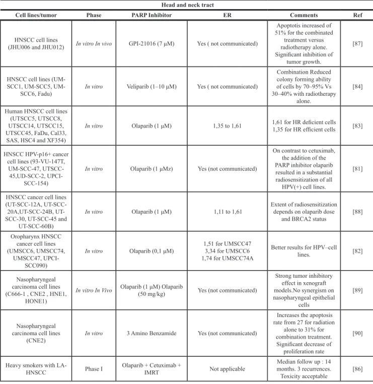
PARPi + radiotherapy for head and neck cancers
Current treatment regimen consists in a combination
of radiotherapy with chemotherapeutics agents such as
Cisplatin, 5FU, or Cetuximab and offers great effectiveness
but often with unacceptable levels of toxicity [80]. Then
we need studies about new radiosensitizers. Güster et al.,
has showed that addition of Olaparib to irradiation, for
HPV positive tumors, caused substantial radiosensitization
[81]. On the opposite Nickson et al., have described the
best radiosensitizing effect for HPV negative cell lines with
enhancement ratio reaching 3,34 [82]. PARP inhibition
Table 1: PARP inhibitors and their use as radiosensitizers in pre-clinical and clinical research
Name
PARP Targeted
Development as
radiosensitizer
EAM /FDA
approved
Indication
Olaparib
PARP1-2
Phase I
No
None
Rucaparib
PARP1-2
Pre-Clinical
No
None
Veliparib
PARP1-2
Phase II
No
None
LT 626
PARP-1
Pre-clinical
No
None
PJ34
PARP1-2
Pre-clinical
No
None
GPI 21016
PARP1
Pre-Clinical
No
None
4-amino-1,8-naphthalimide
PARP1
Pre-Clinical
No
None
3-Aminobenzamide
PARP1
Pre-Clinical
No
None
could serve as a substitute of Cisplatin, in the context of
de-intensified protocols, for HPV positive head and neck
tumors, so as to avoid systemic toxicity of conventional
chemotherapy [81]. As for other types of tumor, HR
defective cells lines are more sensitive to the combination
with PARPi [83]. PARPi seem to be able to attenuate the
nuclear translocation of EGF-R normally induced by DNA
damages. This could lead to lower nuclear interaction
between EGFR and DNA PK, and consequently lower
DNA repair by c-NHEJ [84, 85]. Median enhancement
ratio obtained from pre-clinical data is 1,35 for head and
neck tumors. The first phase I study, for locally advanced
tumors in heavy smockers, showed promising results but
follow-up is for the moment quite short [86] (Table 4).
PARPi + radiotherapy for urologic cancers
In the case of urologic cancer, PARPi are today
mainly developed for prostate cancer. Relevant studies
have identified genomic defects in DNA repair in 20–30%
of advanced castration-resistant prostatic cancer cases. A
proportion of them are germline aberrations and heritable
[91]. This is the first argument for investigating PARP
inhibitors or prostate cancer treatments. After publication
of high response rate in a phase II study Olaparib has been
approved by FDA for the treatment of metastatic castration
resistant prostate carcinoma. In this study, 88% of patients
with alteration of DNA repair genes (BRCA1&2, ATM,
Fanconi anemia genes) had an objective response [91].
The second argument depends on the prevalence of
TMPRSS2-ERG gene fusion in prostate cancer which has
been reported to range from 40% to 70%, depending on
the clinical cohorts investigated [92]. This gene fusion
TMPRSS2–ERG interferes with the assembly of c-NHEJ
factors at DSBs on the chromatin. By inhibiting c-NHEJ
via defective recruitment of XRCC4 and impaired
DNA-PKcs phosphorylation, TMPRSS2–ERG rearrangement
may reveal a “synthetic lethal” interaction with HR,
blocking repair of lesions at collapsed DNA replication
forks induced by PARPi [93]. According to the different
cell line, radiation enhancement ratio is comprised
between 1,05 and 2,2, with much higher effects for
TMPRSS2-ERG gene fusion or PTEN deficient cells. It is
worth noting that for chronic hypoxia condition, which is a
way of radioresistance of prostate cancer, radiosensitizing
effect seems better [94]. There is not any clinical trial
combining PARPi and conventional radiotherapy for the
moment, but a phase Ib with Radium 223 and Niraparib
will open soon (NCT03076203). To sum up pre-clinical
studies show a median enhancement ratio of 1,3 for
prostatic cancer (Table 5).
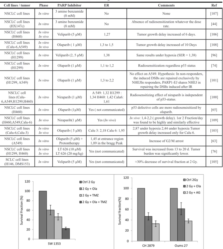
PARPi + radiotherapy for lung cancers
A third of non small cell lung cancers (NSCLC)
patients are diagnosed at a locally advanced stage.
Radiation therapy instead of surgery is so a standard
of care t for these patients. Despite technical advances
in radiation therapy, the local tumor control remains
suboptimal due to radioresistance. Multiple in vitro and
in vivo studies have been realized, assessing potential role
of PARPi as radio sensitizer. Enhancement ratio values
are comprised between 1,1 and 1,62 with modern PARP
inhibitors under oxia (21%O
2) conditions for NSCLC.
Under hypoxia conditions, which is closer to the clinical
reality, enhancement ratio reaches 2,87 suggesting that
hypoxia (1%O
2) induces contextual synthetic lethality
with PARP inhibition in vitro [103]. Small cell lung cancer
(SCLC) cell lines, which show a high expression level of
PARP-1 [104], are also radio sensitized by PARPi [105].
Albert et al., showed a decrease in in vitro endothelial
tubule formation with Veliparib/radiation combination
treatment, and revealed decreased vessel formation
in vivo, suggesting that, this strategy may also target tumor
angiogenesis [106]. Few phase I clinical trials are actually
recruiting patients. In conclusion pre-clinical data show a
median enhancement ratio of 1,5 for lung cancer (Table 6).
PARPi + radiotherapy for breast and gynaecologic
cancers
With ovary cancer, breast cancer is the most
known tumor to get BRCA1 or 2 mutations, and is thus
a candidate for PARPi radiosensitization. Nevertheless,
surprisingly, PARP1 inhibition improves the therapeutic
index of radiotherapy independent of breast cancer
subtype or BRCA1 mutational status. Among breast cancer
subtypes, HER2 and luminal cancer cells seem to get
better enhancement ratio [110]. While it remains dogma
that IR and genotoxic agents mediate their lethal effects
via enhanced apoptosis, necrosis or mitotic catastrophe,
Efimova et al. showed that for Breast cancer cell lines
PARP inhibitors may have a significant impact by inducing
senescence [111]. Median value of enhancement ratio
induced by PARPi for breast cancer is 1,36. The safety
datas of phase I study of Jagsi et al. about loco regional
radiotherapy for local recurrence of Triple negative breast
cancer are comforting, with one unexpected grade IV
toxicity on 30 patients [112] (Table 7).
PARPi + radiotherapy for rare cancers
Few authors expressed an interest in rare tumors
such as Ewing sarcoma and have obtained promising
pre-clinical results, but data stay poor [107, 114]. It is also
the case for chondrosarcoma which is however considered
as one of the most radioresistant tumor. Our laboratory is
working on combination of PARPi (AG15361, Olaparib,
and Talazoparib) and conventional radiotherapy,
protontherapy or hadrontherapy in order to radiosensitize
chondrosarcoma cells. First data with conventional
radiotherapy are very interesting and deserve further
www.impactjournals.com/oncotarget
Table 2: Studies concerning PARPi radiosensitization for brain tumors
Brain systemCell lines / tumor Phase PARP Inhibitor ER Comments Ref
Glioma cell lines (T98G
and U373-MG) In vitro PJ34 (3 μM) (not communicated)Yes
T98G sensitized by PJ34 to a range of low doses of radiation. U373-MG cells showed no increase of
radiosensitivity for low-dose range (1 Gy).
[35]
Glioma cell lines (T98G, U373-MG,
U87-MG,UVW) In vitro Olaparib (1 μM) 1,08 to 1,38
Radiosensitization is replication dependent and greater for fractionated than for single-dose
treatments
[16]
Glioma cell lines
(M059J and M059K) In vitro 4-amino-1,8-naphthalimide (30 μM) No No significant increase of radiosensistivity [15] Glioma cell line
(U251) In vitro In vivo
In vitro GPI-21016(3 μM). In Vivo GPI-21016 (40 mg/kg) +TMZ(3
mg/kg) 1,6
In vivo: Absolute growth delay 31d (1,53x) [36] Glioma cell lines
(SF188 and KNS42) In vitro Olaparib (0–8 μM) (not communicated)Yes Regression of cell Proliferation [37] Glioma cell lines
(T98G, LN18, U87 and
U251) In vitro Veliparib (5 μM) + TMZ (5–10 μM)
1,13 to 1,37 (Veliparib). 1,25 to 1,44 (Veliparib+TMZ)
Also measured in one of the MGMT-unmethylated cell lines
with a SER50 value of 1.30 [38] Glioma cell lines
(UVW/NAT) In vitro Rucaparib(1 μM) / Olaparib (1 μM) Rucaparib 1,33 Olaparib 1,91
PARP-1 inhibition in combination with X-irradiation
promoted G2/M arrest [39] Glioblastoma cell lines
derived from patients tumors (MGMT
unmethylated)
In vitro
In vivo Veliparib (12,5 mg/kg 2x/d)Veliparib (10 μM) (not communicated)Yes Significant increase of mouse survival (+ 10days). [40]
Primary patient-derived
glioblastoma cell lines In vitro Olaparib (not available)
1,93 for Cancer Stem cells 1,34 for bulk cells
No significant difference
between bulk and stem cells. [41] Pediatric Glioma cell
lines (SJG2, SF188, KNS42 )
In vitro
In vivo Niraparib (50 mg/kg)Niraparib (1 μM) (not communicated)Yes
Significant decrease of survival fraction. In vivo survival : 1,6x
longer [42] DIPG cell lines
(DIPGM36 DIPG58, and
SU-DIPG-IV) In vitro Niraparib (1 μM)
Yes
(not communicated) Significant decrease of survival fraction. [42] Ependymoma cell line
(Res196) In vitro Olaparib (0–8 μM) (not communicated)Yes Decrease of clonogenic survivalRegression of cell Proliferation. [37] Medulloblastoma cell lines
(D283-med, D556-med
and UW228-2) In vitro Olaparib (0–8 μM)
Yes (not communicated)
Persistance of γH2AX foci up to 72 hours after radiation. Regression of cell proliferation [37] Neuroblastoma cell
lines (SH-SY-5Y, Kelly, NB1691luc and Tet 21)
In vivo
In vitro Nirabarib (50 mg/kg) (not communicated)Yes In vivo Survival: ≈ 1,08 x longer [43] Neuroblastoma cell lines
(SK-N-BE(2c)) In vitro Rucaparib(1 μM) Olaparib (1 μM) Rucaparib 1,05 Olaparib 1,09
PARP-1 inhibition in combination with X-irradiation
promoted G2/M arrest [39] Neuroblastoma cell
(HX142c) In vitro 3 amino benzamide (6 mM) 1,18 No impact of dose rate. [44] Glioblastoma Phase I Veliparib (10 mg bid) + TMZ(75 mg/m2) not applicable Only safety results. [45] All histologies Brain
metastases Phase I
Veliparib (escalating doses of 10–300 mg bid ) + Whole brain
radiotherapy not applicable OS: 10 m Vs 3,5 m [46] Diffuse intrinsic pontine
glioblastoma Phase I–II
Veliparib 65 mg/m2 + radiotherapy followed with adjuvant TMZ +
veliparib (25 mg/m2) not applicable
OS: 1 year 29%, 2 year 11%. Well tolerated. No benefit. [47] NSCLC Brain metastases Randomized phase II Veliparib(50 mg/200 mg/placebo) + WBRT not applicable OS: 209d Vs 185d ( NS) [48]
Table 3: Studies concerning PARPi radiosensitization for digestive system tumors
Digestive tractCell lines / tumor Phase PARP Inhibitor ER Comments Ref
Colorectal cancer cell
lines (LoVo, SW620) In vitro In vivo AG14361 15 mg/kg/dayAG14361 0,4 μM Yes (not communicated) Until additional 18d of delay in tumor growth [73] Colorectal cancer cell
lines (HCT116) In vivo Veliparib (25 mg/kg/day) Not applicable increased from 23 to 36 daysMedian survival time [60] Colorectal cancer cell
lines (HCT116) In vitro Olaparib (1 μM) Yes (not communicated)
No radiosensitization of normal intestinal cell line.
Increased effect with Combined Chk1
inhibitor
[65] Colorectal cancer cell
line (DLD-1) In vitro Olaparib (1 μM) +/– Camptothecine 1,20 (alone) to 1,45 (combination) Increase of G2/M phase cells and reduced S phase cells. [74] Colorectal cancer cell
lines (HCT116) In vitro In vivo
Veliparib (5 μM) + Irinotecan or oxaliplatine
or 5FU
In vitro: enhanced ratio
1.60 to 1,82 Until additional 11d of delay in tumor growth [71] Colorectal cancer cell
lines (HCT116) In vitro Olaparib and Niraparib (1 μM) Yes (not communicated)
Reduction of clonogenic survival. Increased autophagy
and senescence, but not apoptosis.
[75] Pancreatic cancer cell
lines (Miapaca-2) In vitro In vitro GPI-21016 (3 μM) 1,4 None [36]
Pancreatic cancer cell lines (Miapaca-2,
MPanc-96) In vitro Olaparib (1 μM) 1,5
Increased effect with
Combined Chk1 inhibitor [65] Pancreatic cancer cell
lines (Miapaca-2) In vitro Olaparib (1 μM)
Low LET (γ): 1,4 High LET (Carbon 13keV/um): 1,2 High LET
(Carbon 70keV/um) : 1,4
Enhancement ratio reaches 2,5 with 5μM and High LET
radiation [63]
Pancreatic cancer cell lines (MiaPaCa-2,
Panc-1,Capan-1, AsPC-1)
In vitro Rucaparib (1 μM) + Gemcitabine Enhanced ratio: present Best ratio for BRCA2 mutated Capan-1 cell line. [62] Pancreatic
cancer cell lines (MiaPaCa2,AsPC-1)
In Vitro
In Vivo Olaparib (60 mg/kg)Olaparib (1 μM) 1,2 for MiaPaCa2 1,2 for AsPC-1. No difference for tumor growth delay. [64] Pancreatic cancer cell
lines (OCIP 23, OCIP
28) In vivo Olaparib (150 mg/kg) Not applicable
No difference for tumor growth delay. No radiosensitization observed in
the BRCA2 germline mutant tumor
[68] Pancreatic cancer cell
lines (Miapaca2, PDA) In vitro In vivo. LT 626 (10 μM) Yes (not communicated) Synergic effect on Isobolograms [76] Pancreatic cancer cell
lines (Miapaca-2) In vitro Olaparib (5 μM) + Protontherapy 1,59 at entrance region 1,98 in the bragg Peak Increased G2/M arrest [66] Hepatocarcinoma cell lines (HepG2, PLC-PRF-5) In vitro Veliparib (10 μM) 1,48 for HepG2 1,17 for PLC-PRF-5 None [77] Locally advanced
rectal cancer Phase Ib Veliparib + Capecitabine + RT Not applicable
Tumor downstaging 71% of 31 evaluable pts; Pathologic complete response 29%. Acceptable safety profile.
[78] Locally advanced
pancreatic cancer Phase I Veliparib + Gemcitabine+ RT Not applicable Safety data [69]
Advanced solid tumors and
carcinomatosis Phase I
Veliparib + whole
www.impactjournals.com/oncotarget
investigations. All clonogenic survival data with olaparib
showed radiosensitization of Oums 27 or CH 2879 cell
lines (PTCOG 2016, Chevalier et al. [115]).There are still
on going investigations (Figure 3).
DISCUSSION
Radiation therapy plays a central role in cancer
therapeutics, as an adjuvant therapy and above all as
first treatment, alone or with radiosensitizer, for
low-operable tumors such as glioblastoma or advanced lung
cancers. Nevertheless, cure rates stay disappointing, and
concomitant chemotherapy generate important systemic
toxicity, requiring search for new radiosensitizers. Ideal
radiosensitizing agents should have two main qualities:
on the first hand to offer better protection to surround
healthy tissues, on the other hand, to increase anti
tumoral efficiency, with hope to improve therapeutic
ratio. Since the last ten years PARP seems to be one of
the most interesting protein to target, in association with
radiotherapy because particularly of its role in DNA
repair. We have reported more than fifty in vitro or in vivo
studies which have investigated PARPi as radiosensitizer
with attractive enhancement ratio comprised
Table 4: Studies concerning PARPi radiosensitization for head and neck tumors
Head and neck tractCell lines/tumor Phase PARP Inhibitor ER Comments Ref
HNSCC cell lines
(JHU006 and JHU012) In vitro In vivo GPI-21016 (7 μM) Yes ( not communicated)
Apoptotis increased of 51% for the combinated
treatment versus radiotherapy alone. Significant inhibition of
tumor growth.
[87]
HNSCC cell lines (SCC1, SCC5,
UM-SCC6, Fadu) In vitro Veliparib (1–10 μM) Yes ( not communicated)
Combination Reduced colony forming ability of cells by 70–95% Vs 30–40% with radiotherapy
alone.
[84]
Human HNSCC cell lines (UTSCC5, UTSCC8, UTSCC14, UTSCC15, UTSCC45, FaDu, Cal33,
SAS, HSC4 and XF354)
In vitro Olaparib (1 μM) 1,35 to 1,61 1,61 for HR deficient cells 1,35 for HR efficient cells [83]
HNSCC HPV-p16+ cancer cell lines (93-VU-147T,
UM-SCC-47, UTSCC-45,UD-SCC-2,
UPCI-SCC-154)
In vitro Olaparib (1 μMz) Yes (not communicated)
On contrast to cetuximab, the addition of the PARP inhibitor olaparib resulted in a substantial radiosensitization of all
HPV(+) cell lines.
[81]
HNSCC cancer cell lines (12A, SCC-20A,SCC-24B, UT-SCC-30, UT-SCC-45 and
UT-SCC-60B)
In vitro Olaparib (1 μM) 1,11 to 1,61 Extent of radiosensitization depends on olaparib dose and BRCA2 status [88] Oropharynx HNSCC
cancer cell lines (UMSCC6, UMSCC74,
UMSCC47, UPCI-SCC090)
In vitro Olaparib (0,1 μM) 1,51 for UMSCC47 3,34 for UMSCC6 1,74 for UMSCC74A
Better results for HPV–cell lines. [82]
Nasopharyngeal carcinoma cell lines (C666-1 , CNE2 , HNE1,
HONE1)
In vitro In Vivo Olaparib (1 μM) Olaparib (50 mg/kg) Yes (not communicated)
Strong tumor inhibitory effect in xenograft models.No synergism on nasopharyngeal epithelial cells [89] Nasopharyngeal carcinoma cell lines
(CNE2) In vitro 3 Amino Benzamide Yes (not communicated)
Increases the apoptosis rate from 27 for radiation
alone to 31% for combination treatment. Significant decrease of
proliferation rate
[90]
Heavy smokers with
LA-HNSCC Phase I Olaparib + Cetuximab + IMRT Not applicable
Median follow up : 14 months. 3 recurrences.
between 1,04 and 2,87 for in vitro data (Tables 2–7),
PARPi could even bypass some radioresistant
mechanisms such as hypoxia [96, 103]. Nevertheless,
in vivo results are often less promising, and only 9 phase
I-II studies get published results, sometimes negative.
Nine other phase I-II clinical trials (clinicaltrials.gov)
are actually recruiting, all using Olaparib or Veliparib in
combination with radiotherapy or radio-chemotherapy
for soft tissue sarcoma, breast cancer, lung cancer, rectal
cancer or esophageal cancer. During the 2 or 3 next years,
they should bring some new information concerning the
potential of PARPi as radiosensitizer.
It is important to note that, combination treatment
appears to promote a state of growth arrest instead of cell
death, it is legitimate to speculate that radiosensitization
could be a consequence of an increase in the extent of
senescence [75, 97, 111]. The next critical question is
whether the growth arrest is sustained and irreversible or
transient and reversible, and consequently if this combination
approach is so useful for improving long-term endpoints.
A current interrogation remains that radiosensitizing
effect on high proliferating non tumoral tissues, like
mucosa or bone marrow, is quite unknown. Effectively
in vitro studies including investigations with normal cells
Table 5: Studies concerning PARPi radiosensitization for prostate tumors
Urologic cancerCell lines / tumor Phase PARP Inhibitor ER Comments Ref
Prostate carcinoma cell
lines (DU145) In vitro 4-amino-1,8-naphthalimide (ANI) (20 μM) 1, 3 None [95] Prostate carcinoma cell
lines (DU145) In vitro In vitro GPI-21016 (5 μM) 1, 7 None [36] Prostate carcinoma cell
lines (DU145, 22RV1) In vitro Veliparib (2, 5 μM) 1, 25 ( Under Hypoxia)
SF2 of DU145 and 22RV1 cells decreased from 0.44 and 0.36 to 0.27 and 0.20,
respectively.
[96]
Prostate carcinoma cell
lines (PC3, DU145) In vitro In vivo Veliparib (10 μM) Veliparib (25 mg/kg) Yes ( Not communicated)
More Significant decrease of survival fraction for PC3 thant DU-145 in vitro.
Significant delay in tumor regrowth only for PC3.
[97]
Prostate carcinoma cell lines (PC3, DU145,
LNCaP,VCaP) In vitro Rucaparib (≤ 2, 5 μM) Yes ( Not communicated)
More effective for PTEN deficient cells or with
TMPRSS2-ERG gene fusion [98]
Prostate carcinoma cell
lines (PC3, DU145) In vitro In vivo Olaparib (1 μM) Olaparib (100 mg/kg) 1, 12 to 1, 52
1,12 for ERG-, 1, 52 for Erg+. In vivo radioresistance
in ERG+ can be overcome through inhibition of PARP1.
[99]
Prostate carcinoma cell
lines (LNCaP) In vitro Niraparib (1 μM) 1, 43
Did not radiosensitize human cells derived from
normal tissues [100]
Prostate carcinoma cell lines (PC3, DU145,
LNCaP) In vitro Olaparib (1 μM)
1, 05 for DU 145 (NS) 1, 3 to 2, 2 for PC3,
LNCAP.
In non-responders, the induced DSBs are repaired
exclusively by NHEJIn responders, PARP1-EJ shares
NHEJ in repairing the DSBs induced after IR
[101]
Prostate carcinoma cell
line (22Rv1) In vitro In vivo Olaparib (1 μM) Olaparib (100 mg/kg) 1, 7
1, 2 under acute Hypoxia, 1, 8 under chronic hypoxia. In vivo : Growth delay increased of 6,06 days ( ns)
[94]
Prostate carcinoma cell
lines (PC3, LNCaP) In vitro Rucaparib (2, 5 μM) Yes ( Not communicated)
Most effective for TMPRSS2-ERG gene fusion
cells [93] Prostate carcinoma cell
lines (PC3, VCaP) In vitro Rucaparib (3 μM), Olaparib (3 μM) Yes ( Not communicated)
SF2 from 0, 50 to 0,12 for olaparib SF2 from 0, 45 to 0,
www.impactjournals.com/oncotarget
Table 6: Studies concerning PARPi radiosensitization for lung tumors
Lung cancerCell lines / tumor Phase PARP Inhibitor ER Comments Ref
NSCLC cell lines In vitro 3 amino benzamide (8 mM) No None [107] NSCLC cell lines
(HX147c) In vitro 3 amino benzamide (8 mM) No Absence of radiosensitization whatever the dose rate. [44] NSCLC cell lines
(H460) In vitro In vivo Veliparib (5 μM) 1,27 Tumor growth delay increased of 6 days. [106] NSCLC cell lines
(Calu-6,A549) In vitro In vivo Olaparib ( 1 μM) 1,3 to 1,5 Tumor growth delay increased of 10 Days [108] NSCLC cell lines
(H1299) In vitro Veliparib (2, 5 μM) 1,38 Same results under hypoxia (SER = 1,38) [96] NSCLC cell lines
(H1299) In vitro Olaparib (1 μM) 1,1 to 1,2 Radiosensitization regardless p53 status [74] NSCLC cell lines
(H1299, A549) In vitro Olaparib (1 μM) 1,3 to 2,2
No effect on A549. Hypothesis: In non-responders, the induced DSBs are repaired exclusively by NHEJIn responders, PARP1-EJ shares NHEJ in
repairing the DSBs induced after IR
[101]
NSCLC cell lines
(Calu-6,A549,H1299,H460) In vitro Niraparib (1 μM)
A 549: 1,32 H1299 : 1,34 H460: 1,42 Calu6:
1,61
Radiosensitizing effect of niraparib is independent of p53-status. [100] NSCLC cell lines
(H460) In vitro Olaparib (1μM) Yes ( not communicated) p53 defective cells are more radiosensisitized by olaparib. [65] NSCLC cell lines
(H460,A549,Calu-6) In vivo Niraparib(1 μM) Yes (In vivo) In vivo: 1,4-2,2 ( growth delay). 1or 2 Fraction/day was found to be highly and similarly effective [109] NSCLC cell lines
(Calu-6,Calu-3) In vitro In vivo Olaparib ( 5 μM) Calu 3: 2,18 Calu 6: 1,95 2,87 under hypoxia 2,44 under hypoxia Tumor growth delay increased only for Calu 6. [103] NSCLC cell lines
(A549) In vitro Olaparib (5 μM) + Protontherapy 1,45 at entrance region 1,89 in the bragg Peak Increase of G2/M arrest [63] NSCLC cell lines
(H1299, H460) In vitroIn vivo LT 626 (20 mg/kg)LT 626 (10 μM) Yes (not communicated) Survival was increased from 13 to 20 d. Tumor burden was significantly lower. [76] SCLC cell lines
(H146, DMS153) In vitro Veliparib (5 μM) Yes (not communicated) ≈30% decrease of survival fraction at 2 Gy. [105]
Figure 3: Clonogenic survival rate of chondrosarcoma cell after protontherapy sensitization with alkylant agents and
PARPi.
Chondrosarcoma cells were first cultured with Temozolomide and /or PARPi for 2 hours, then irradiated with proton beam at 2 Gy (62 MeV.u-1, SOBP, 1,1 keV u-1 at LNS, Catania, Italy) and then left overnight at 37°C. Cells were then seeded at low density for subsequentclonogenic assay, as previously described [116]. Left: survival fraction of SW1353 chondrosarcoma cells after 2 Gy proton alone, and with olaparib (2 µM), temozolomide (Sigma-Aldrich ref T2577) (20 µM) and olaparib with temozolomide (2 µM + 20 µM respectively). Right: survival fraction as a function of different chondrosarcoma cell lines. CH 2879 and Oums 27 are two other chondrosarcoma cell lines, showing that the variability of the response to the treatment is related to the cell line used. Cells were irradiated alone or with Olaparib (Focus biomolecules Ref 102154) at 2 µM, or AG (AG14361, Tebu-Bio, ref 27602-3) at 0.4 µM. Each treatment is estimated as a percentage of the control sample, repeated 3 times.
lines are very limited [63, 117] and there are too few of
phase I studies to bring some conclusions. Nevertheless as
single agent, in phase III study, Olaparib increased the risk
of myelodysplasia or acute myeloid leukemia [118]. These
secondary effects could be enhanced if large field spine
or pelvic radiotherapy was associated. Waiting for more
data, and to avoid unexpected late toxicity, we should
pay attention to the use of hadrontherapy, stereotactic
radiotherapy or radionuclides in combination with PARPi.
These techniques offer better ballistic and superior
biological efficiency, bringing a promise of improvement
of the therapeutic ratio [119].
If BRCA and PTEN mutations are actually known
for being predictive of high efficiency of PARP inhibitor
[120], accurate biomarkers for response and resistance to
PARPi are still needed. Intratumoral levels and activity
of the PARP does not correlate with clinical responses
in patients and shouldn’t be considered as predictive
of response [121]. On the contrary, homologous
recombination assay, targeted multiplex sequencing,
mutational signatures and alterations in genome structure,
functional assay, are potential solutions to identify
biomarkers [122]. For example Olaparib as single agent
for patients suffering from prostate cancer with genetic
aberrations in DNA-repair genes has showed significantly
higher efficacy with 88% overall response rate. In this
study, 12 genes were evaluated: BRCA1, BRCA2, ATM,
FANCA, CHK2, PALB2, HDAC2, RAD 51, MLH3,
ERCC3, MRE 11 and NBN [91]. Future phase I or II
studies should systematically include tumoral DNA Repair
capacity status, in order to better select patient who will
benefit from the treatment.
Finally we come to the conclusion that the
permanent development of new targeted therapies
makes radiotherapist hope for new interesting pair with
radiotherapy. Combinations of PARPi with radiotherapy
seem very promising but clinical data are still lacking.
Radiotherapists and searchers should pay attention to
include ancillary studies, in addition to clinic ones, in
order to develop biomarkers. The next ten years will
be crucial for judging the real potential of PARPi as
radiosensistizer.
CONFLICTS OF INTEREST
None.
Table 7: Studies concerning PARPi radiosensitization for breast and gynecologic tumors
Gynaecologic and breast cancerCell lines / tumor Phase PARP Inhibitor ER Comments Ref
Breast Cancer cell line
(MCF7) In vitro In vivo Veliparib (10 uM, 0,5 mg Bid) Yes ( Not communicated)
Colony formation following 2 Gy decreased
from 29.7 to 11.3% with veliparib. The antiproliferative effects of 3 Gy enhance from 41
to 27%
[111]
Breast Cancer cell line
(MDA-MB-231) In vivo Niraparib ( 25 to 50 mg/kg) Yes (In vivo) In vivo: increase growth delay x 1,9 [109] Breast Cancer cell lines
(MB-231, MDA-MB-436)
In vitro
In vivo Niraparib (1 μM) 1, 25 to 1,36
Normal breast cell line MCF 10A only slightly
radiosensitized. [100] Breast Cancer cell line
(ZR75-1, MDA-MB-231, MB-468, MDA-MB-453,HCC 1954, HCC
1937)
In vitro
In vivo Veliparib (2,5 uM) Veliparib 100 mg/Kg 1,22 to 2,31 In vivo : Tumor volume doubling time ≈ x2 [110] Triple negative breast
cancer Phase I Veliparib + 50 Gy Not applicable 30 patients. Safety Data [112] Cervix carcinoma cell
line (Hela) In vitro Olaparib (1 μM) 1,3 to 2,2
PARP inhibition significantly increased the number ofpersistent gH2AX foci at 24h but
not at 1h
[101]
Cervix carcinoma cell line
(HX 155, 156, 160) In vivo 3-Amino-benzamide (8 mM) 1,02 to 1,37 None [113] Cervix carcinoma cell
line (Hela) In vitro 4-amino-1,8-naphthalimide (30 μM) No No significant increase of radiosensitivity [15] Cervix carcinoma cell line
(HX 156c) In vitro 3-Amino-benzamide (8 mM) 1,16 to 1,18
Absence of radiosensitization
www.impactjournals.com/oncotarget
REFERENCES
1. Farmer H, McCabe N, Lord CJ, Tutt ANJ, Johnson DA, Richardson TB, Santarosa M, Dillon KJ, Hickson I, Knights C, Martin NMB, Jackson SP, Smith GCM, et al. Targeting the DNA repair defect in BRCA mutant cells as a therapeutic strategy. Nature. 2005; 434:917–21. https://doi. org/10.1038/nature03445.
2. Bryant HE, Schultz N, Thomas HD, Parker KM, Flower D, Lopez E, Kyle S, Meuth M, Curtin NJ, Helleday T. Specific killing of BRCA2-deficient tumours with inhibitors of poly(ADP-ribose) polymerase. Nature. 2005; 434:913–7. https://doi.org/10.1038/nature03443.
3. Brown JM, Carlson DJ, Brenner DJ. The tumor radiobiology of SRS and SBRT: are more than the 5 Rs involved? Int J Radiat Oncol Biol Phys. 2014; 88:254–62. https://doi. org/10.1016/j.ijrobp.2013.07.022.
4. Mansour WY, Rhein T, Dahm-Daphi J. The alternative end-joining pathway for repair of DNA double-strand breaks requires PARP1 but is not dependent upon microhomologies. Nucleic Acids Res. 2010; 38:6065–77. https://doi.org/10.1093/nar/gkq387.
5. Schultz N, Lopez E, Saleh-Gohari N, Helleday T. Poly(ADP-ribose) polymerase (PARP-1) has a controlling role in homologous recombination. Nucleic Acids Res. 2003; 31:4959–64. https://doi.org/10.1093/nar/gkg703. 6. Cheng Q, Barboule N, Frit P, Gomez D, Bombarde O,
Couderc B, Ren GS, Salles B, Calsou P. Ku counteracts mobilization of PARP1 and MRN in chromatin damaged with DNA double-strand breaks. Nucleic Acids Res. 2011; 39:9605–19. https://doi.org/10.1093/nar/gkr656.
7. Gradwohl G, Murcia JMM de, Molinete M, Simonin F, Koken M, Hoeijmakers JH, Murcia G de. The second zinc-finger domain of poly(ADP-ribose) polymerase determines specificity for single-stranded breaks in DNA. Proc Natl Acad Sci. 1990; 87:2990–4. https://doi.org/10.1073/ pnas.87.8.2990.
8. Ikejima M, Noguchi S, Yamashita R, Ogura T, Sugimura T, Gill DM, Miwa M. The zinc fingers of human poly(ADP-ribose) polymerase are differentially required for the recognition of DNA breaks and nicks and the consequent enzyme activation. Other structures recognize intact DNA. J Biol Chem. 1990; 265:21907–13.
9. Li M, Yu X. The Role of Poly(ADP-ribosyl)ation in DNA Damage Response and Cancer Chemotherapy. Oncogene. 2015; 34:3349–56. https://doi.org/10.1038/onc.2014.295. 10. Saintigny Y, Delacôte F, Varès G, Petitot F, Lambert S,
Averbeck D, Lopez BS. Characterization of homologous recombination induced by replication inhibition in mammalian cells. EMBO J. 2001; 20:3861–70. https://doi. org/10.1093/emboj/20.14.3861.
11. Ström CE, Johansson F, Uhlén M, Szigyarto CA-K, Erixon K, Helleday T. Poly (ADP-ribose) polymerase (PARP) is not involved in base excision repair but PARP inhibition traps a single-strand intermediate. Nucleic
Acids Res. 2011; 39:3166–75. https://doi.org/10.1093/nar/ gkq1241.
12. Cerrato A, Morra F, Celetti A. Use of poly ADP-ribose polymerase [PARP] inhibitors in cancer cells bearing DDR defects: the rationale for their inclusion in the clinic. J Exp Clin Cancer Res CR. 2016; 35:179. https://doi.org/10.1186/s13046-016-0456-2.
13. Bryant HE, Petermann E, Schultz N, Jemth AS, Loseva O, Issaeva N, Johansson F, Fernandez S, McGlynn P, Helleday T. PARP is activated at stalled forks to mediate Mre11-dependent replication restart and recombination. EMBO J. 2009; 28:2601–15. https://doi.org/10.1038/ emboj.2009.206.
14. Grabarz A, Barascu A, Guirouilh-Barbat J, Lopez BS. Initiation of DNA double strand break repair: signaling and single-stranded resection dictate the choice between homologous recombination, non-homologous end-joining and alternative end-joining. Am J Cancer Res. 2012; 2:249–68. 15. Noël G, Godon C, Fernet M, Giocanti N, Mégnin-Chanet F,
Favaudon V. Radiosensitization by the poly(ADP-ribose) polymerase inhibitor 4-amino-1,8-naphthalimide is specific of the S phase of the cell cycle and involves arrest of DNA synthesis. Mol Cancer Ther. 2006; 5:564–74. https://doi. org/10.1158/1535-7163.MCT-05-0418.
16. Dungey FA, Löser DA, Chalmers AJ. Replication-dependent radiosensitization of human glioma cells by inhibition of poly(ADP-Ribose) polymerase: mechanisms and therapeutic potential. Int J Radiat Oncol Biol Phys. 2008; 72:1188–97. https://doi.org/10.1016/j.ijrobp.2008.07.031. 17. Gottschalk AJ, Timinszky G, Kong SE, Jin J, Cai Y,
Swanson SK, Washburn MP, Florens L, Ladurner AG, Conaway JW, Conaway RC. Poly(ADP-ribosyl)ation directs recruitment and activation of an ATP-dependent chromatin remodeler. Proc Natl Acad Sci USA. 2009; 106:13770–4. https://doi.org/10.1073/pnas.0906920106.
18. Ahel D, Horejsí Z, Wiechens N, Polo SE, Garcia-Wilson E, Ahel I, Flynn H, Skehel M, West SC, Jackson SP, Owen-Hughes T, Boulton SJ. Poly(ADP-ribose)-dependent regulation of DNA repair by the chromatin remodeling enzyme ALC1. Science. 2009; 325:1240–3. https://doi. org/10.1126/science.1177321.
19. Gorbalenya AE, Koonin EV, Donchenko AP, Blinov VM. Two related superfamilies of putative helicases involved in replication, recombination, repair and expression of DNA and RNA genomes. Nucleic Acids Res. 1989; 17:4713–30. 20. Ali M, Telfer BA, McCrudden C, O’Rourke M, Thomas HD, Kamjoo M, Kyle S, Robson T, Shaw C, Hirst DG, Curtin NJ, Williams KJ. Vasoactivity of AG014699, a clinically active small molecule inhibitor of poly(ADP-ribose) polymerase: a contributory factor to chemopotentiation in vivo? Clin Cancer Res Off J Am Assoc Cancer Res. 2009; 15:6106–12. https://doi.org/10.1158/1078-0432.CCR-09-0398.
21. Brown JM, Wilson WR. Exploiting tumour hypoxia in cancer treatment. Nat Rev Cancer. 2004; 4:437–47. https:// doi.org/10.1038/nrc1367.
22. Hong B-J, Kim J, Jeong H, Bok S, Kim Y-E, Ahn G-O. Tumor hypoxia and reoxygenation: the yin and yang for radiotherapy. Radiat Oncol J. 2016; 34:239–49. https://doi. org/10.3857/roj.2016.02012.
23. Brown JM, Wilson WR. Exploiting tumour hypoxia in cancer treatment. Nat Rev Cancer. 2004; 4:437–47. https:// doi.org/10.1038/nrc1367.
24. Loeb LA. Human Cancers Express a Mutator Phenotype: Hypothesis, Origin, and Consequences. Cancer Res. 2016; 76:2057–9. https://doi.org/10.1158/0008-5472.CAN-16-0794.
25. Bristow RG, Hill RP. Hypoxia and metabolism. Hypoxia, DNA repair and genetic instability. Nat Rev Cancer. 2008; 8:180–92. https://doi.org/10.1038/nrc2344.
26. Chan N, Pires IM, Bencokova Z, Coackley C, Luoto KR, Bhogal N, Lakshman M, Gottipati P, Oliver FJ, Helleday T, Hammond EM, Bristow RG. Contextual Synthetic Lethality of Cancer Cell Kill Based on the Tumor Microenvironment. Cancer Res. 2010; 70:8045–54. https://doi.org/10.1158/0008-5472.CAN-10-2352.
27. Pawlik TM, Keyomarsi K. Role of cell cycle in mediating sensitivity to radiotherapy. Int J Radiat Oncol Biol Phys. 2004; 59:928–42. https://doi.org/10.1016/j.ijrobp.2004.03.005. 28. Ledermann J, Harter P, Gourley C, Friedlander M, Vergote I,
Rustin G, Scott CL, Meier W, Shapira-Frommer R, Safra T, Matei D, Fielding A, Spencer S, et al. Olaparib maintenance therapy in patients with platinum-sensitive relapsed serous ovarian cancer: a preplanned retrospective analysis of outcomes by BRCA status in a randomised phase 2 trial. Lancet Oncol. 2014; 15:852–61. https://doi.org/10.1016/ S1470-2045(14)70228-1.
29. Drew Y, Ledermann J, Hall G, Rea D, Glasspool R, Highley M, Jayson G, Sludden J, Murray J, Jamieson D, Halford S, Acton G, Backholer Z, et al. Phase 2 multicentre trial investigating intermittent and continuous dosing schedules of the poly(ADP-ribose) polymerase inhibitor rucaparib in germline BRCA mutation carriers with advanced ovarian and breast cancer. Br J Cancer. 2016; 114:723–30. https://doi.org/10.1038/bjc.2016.41.
30. Mirza MR, Monk BJ, Herrstedt J, Oza AM, Mahner S, Redondo A, Fabbro M, Ledermann JA, Lorusso D, Vergote I, Ben-Baruch NE, Marth C, Mądry R, et al. Niraparib Maintenance Therapy in Platinum-Sensitive, Recurrent Ovarian Cancer. N Engl J Med. 2016; 375:2154–64. https:// doi.org/10.1056/NEJMoa1611310.
31. Domchek SM, Aghajanian C, Shapira-Frommer R, Schmutzler RK, Audeh MW, Friedlander M, Balmaña J, Mitchell G, Fried G, Stemmer SM, Hubert A, Rosengarten O, Loman N, et al. Efficacy and safety of olaparib monotherapy in germline BRCA1/2 mutation carriers with advanced ovarian cancer and three or more lines of prior therapy. Gynecol Oncol. 2016; 140:199–203. https://doi.org/10.1016/j. ygyno.2015.12.020.
32. Helleday T. The underlying mechanism for the PARP and BRCA synthetic lethality: clearing up the
misunderstandings. Mol Oncol. 2011; 5:387–93. https://doi. org/10.1016/j.molonc.2011.07.001.
33. Kedar PS, Stefanick DF, Horton JK, Wilson SH. Increased PARP-1 association with DNA in alkylation damaged, PARP-inhibited mouse fibroblasts. Mol Cancer Res MCR. 2012; 10:360–8. https://doi.org/10.1158/1541-7786.MCR-11-0477.
34. Moher D, Liberati A, Tetzlaff J, Altman DG, PRISMA Group. Preferred reporting items for systematic reviews and meta-analyses: the PRISMA statement. J Clin Epidemiol. 2009; 62:1006–12. https://doi.org/10.1016/j.jclinepi.2009.06.005. 35. Chalmers A, Johnston P, Woodcock M, Joiner M,
Marples B. PARP-1, PARP-2, and the cellular response to low doses of ionizing radiation. Int J Radiat Oncol Biol Phys. 2004; 58:410–9.
36. Russo AL, Kwon H-C, Burgan WE, Carter D, Beam K, Weizheng X, Zhang J, Slusher BS, Chakravarti A, Tofilon PJ, Camphausen K. In vitro and In vivo Radiosensitization of Glioblastoma Cells by the Poly (ADP-Ribose) Polymerase Inhibitor E7016. Clin Cancer Res. 2009; 15:607–12. https:// doi.org/10.1158/1078-0432.CCR-08-2079.
37. van Vuurden DG, Hulleman E, Meijer OLM, Wedekind LE, Kool M, Witt H, Vandertop PW, Würdinger T, Noske DP, Kaspers GJL, Cloos J. PARP inhibition sensitizes childhood high grade glioma, medulloblastoma and ependymoma to radiation. Oncotarget. 2011; 2:984–96.
38. Barazzuol L, Jena R, Burnet NG, Meira LB, Jeynes JCG, Kirkby KJ, Kirkby NF. Evaluation of poly (ADP-ribose) polymerase inhibitor ABT-888 combined with radiotherapy and temozolomide in glioblastoma. Radiat Oncol Lond Engl. 2013; 8:65. https://doi.org/10.1186/1748-717X-8-65. 39. Nile DL, Rae C, Hyndman IJ, Gaze MN, Mairs RJ. An evaluation
in vitro of PARP-1 inhibitors, rucaparib and olaparib, as
radiosensitisers for the treatment of neuroblastoma. BMC Cancer. 2016; 16:621. https://doi.org/10.1186/s12885-016-2656-8. 40. Jue TR, Nozue K, Lester AJ, Joshi S, Schroder LBW,
Whittaker SP, Nixdorf S, Rapkins RW, Khasraw M, McDonald KL. Veliparib in combination with radiotherapy for the treatment of MGMT unmethylated glioblastoma. J Transl Med. 2017; 15:61. https://doi.org/10.1186/s12967-017-1164-1.
41. Ahmed SU, Carruthers R, Gilmour L, Yildirim S, Watts C, Chalmers AJ. Selective Inhibition of Parallel DNA Damage Response Pathways Optimizes Radiosensitization of Glioblastoma Stem-like Cells. Cancer Res. 2015; 75:4416–28. https://doi.org/10.1158/0008-5472.CAN-14-3790.
42. Chornenkyy Y, Agnihotri S, Yu M, Buczkowicz P, Rakopoulos P, Golbourn B, Garzia L, Siddaway R, Leung S, Rutka JT, Taylor MD, Dirks PB, Hawkins C. Poly-ADP-Ribose Polymerase as a Therapeutic Target in Pediatric Diffuse Intrinsic Pontine Glioma and Pediatric High-Grade Astrocytoma. Mol Cancer Ther. 2015; 14:2560–8. https:// doi.org/10.1158/1535-7163.MCT-15-0282.
43. Mueller S, Bhargava S, Molinaro AM, Yang X, Kolkowitz I, Olow A, Wehmeijer N, Orbach S, Chen J,