HAL Id: hal-00297601
https://hal.archives-ouvertes.fr/hal-00297601
Submitted on 12 Feb 2007
HAL is a multi-disciplinary open access
archive for the deposit and dissemination of
sci-entific research documents, whether they are
pub-lished or not. The documents may come from
teaching and research institutions in France or
abroad, or from public or private research centers.
L’archive ouverte pluridisciplinaire HAL, est
destinée au dépôt et à la diffusion de documents
scientifiques de niveau recherche, publiés ou non,
émanant des établissements d’enseignement et de
recherche français ou étrangers, des laboratoires
publics ou privés.
cloud and rain water produce biosurfactants but do not
cause ice nucleation
H. E. Ahern, K. A. Walsh, T. C. J. Hill, B. F. Moffett
To cite this version:
H. E. Ahern, K. A. Walsh, T. C. J. Hill, B. F. Moffett. Fluorescent pseudomonads isolated from
Hebridean cloud and rain water produce biosurfactants but do not cause ice nucleation. Biogeosciences,
European Geosciences Union, 2007, 4 (1), pp.115-124. �hal-00297601�
Biogeosciences, 4, 115–124, 2007 www.biogeosciences.net/4/115/2007/ © Author(s) 2007. This work is licensed under a Creative Commons License.
Biogeosciences
Fluorescent pseudomonads isolated from Hebridean cloud and rain
water produce biosurfactants but do not cause ice nucleation
H. E. Ahern1, K. A. Walsh2, T. C. J. Hill2, and B. F. Moffett2
1University of East London, Romford Road, Stratford, London, UK 2Environment Agency, Wallingford, UK
Received: 1 September 2006 – Published in Biogeosciences Discuss.: 4 October 2006 Revised: 11 January 2007 – Accepted: 9 February 2007 – Published: 12 February 2007
Abstract. Microorganisms were discovered in clouds over 100 years ago but information on bacterial community struc-ture and function is limited. Clouds may not only be a niche within which bacteria could thrive but they might also in-fluence dynamic processes using ice nucleating and cloud condensing abilities. Cloud and rain samples were collected from two mountains in the Outer Hebrides, NW Scotland, UK. Community composition was determined using a com-bination of amplified 16S ribosomal DNA restriction analy-sis and sequencing. 256 clones yielded 100 operational tax-onomic units (OTUs) of which half were related to bacteria from terrestrial psychrophilic environments. Cloud samples were dominated by a mixture of fluorescent Pseudomonas spp., some of which have been reported to be ice nucleators. It was therefore possible that these bacteria were using the ice nucleation (IN) gene to trigger the Bergeron-Findeisen process of raindrop formation as a mechanism for dispersal. In this study the IN gene was not detected in any of the iso-lates using both polymerase chain reaction (PCR) and differ-ential scanning calorimetry (DSC). Instead 55% of the total isolates from both cloud and rain samples displayed signif-icant biosurfactant activity when analyzed using the drop-collapse technique. All isolates were characterised as fluo-rescent pseudomonads. Surfactants have been found to be very important in lowering atmospheric critical supersatura-tions required for the activation of aerosols into cloud con-densation nuclei (CCN). It is also known that surfactants in-fluence cloud droplet size and increase cloud lifetime and albedo. Some bacteria are known to act as CCN and so it is conceivable that these fluorescent pseudomonads are us-ing surfactants to facilitate their activation from aerosols into CCN. This would allow water scavenging, countering desic-cation, and assist in their widespread dispersal.
Correspondence to: T. C. J. Hill
1 Introduction
There has been a resurgence of interest in microorganisms in the atmosphere due, in part, to heightened awareness of disease epidemiology. Health issues however may be less important than their role in cloud and rainfall processes and link to climate change. Recently it has been reported that there are between 1500 and 355 000 bacteria per millilitre of cloud water (Sattler et al., 2001, Bauer et al., 2002 and Am-ato et al., 2005). Therefore, as with other aerosol particles, their numbers are highly variable in both time and space. It has been known for more than a century that bacteria are present in clouds (Bujwid, 1888, cited in Mandrioli et al., 1973) and despite the fact that clouds cover about 50% of the Earth at any one time, there is only minimal and fragmen-tary information about the diversity, structure and function of their microbial communities.
To date, research into atmospheric bacteria has typically used conventional methods which are biased towards the cul-tivable fraction (Brown et al., 1964; Casareto et al., 1996; Fuzi et al., 1997; Mandrioli et al., 1973). The full degree of bias has been highlighted in a culture-independent study of bacterial 16S rRNA gene diversity of air samples collected over Salt Lake City, Utah, USA. When the 16S rRNA re-sults were compared with microscopic and viable counts it was found that only 0.08% of the bacteria in the sample were cultivable (Radosevich et al., 2002). In addition, Amato and colleagues (2005) investigated the microorganisms in clouds on Puy de Dˆome in south-central France using 16S DNA se-quencing to identify cultured bacterial isolates. They noted that the cultured fraction represented only 1% of the total count, and highlighted the need to use direct molecular meth-ods to fully characterise the microbial diversity of the total community.
Fully investigating diversity and testing for metabolic ac-tivity (e.g. Sattler et al., 2001) could help to ascertain whether microbes have a transient presence in the clouds or if they are
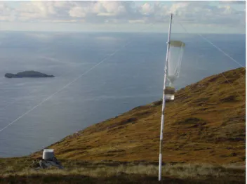
Fig. 1. Cloud collector set up on Caepabhal. Collector was erected
onto the end of a 2 m pole and secured with guy ropes.
residents, having adapted to exploit this diffuse but vast envi-ronmental niche. That is, are they scavenging nutrients from cloud water and using wind and rain for dispersal? In partic-ular, the role of some species of bacteria with heterogeneous ice nucleation ability may be highly significant in raindrop formation (Morris et al.; 2005 and Vali, 1996).
Liquid water can be “supercooled” to well below the solid phase melting point of 0◦C without freezing. The formation of ice involves the initial ordering of water molecules into or onto a hexameric ice-like lattice embryo critical for further water deposition and ice crystal growth. Freezing catalysts are known as ice nuclei and freezing is either homogeneous or heterogeneous. Homogeneous freezing involves the ran-dom assembly of water molecules into the lattice which can only occur at temperatures below –37.5◦C. For freezing at higher temperatures - heterogeneous freezing – ice nuclei are required to catalyse the process. Ice nuclei are struc-tures which encourage water molecules to align and form the ice embryo (Lindow, 1983; Vali, 1996). There are sev-eral nonbiological sources of ice nuclei and these include minerals such as silver iodide and kaolinite which can nu-cleate between –8◦C and –15◦C (Lindow, 1983). However, the ice nuclei which operate at the highest temperatures are bacterial proteins. Examples of ice-nucleating bacteria in-clude strains of Erwinia herbicola, Erwinia ananas,
Pseu-domonas flourescens and PseuPseu-domonas syringae which are
able to freeze water at temperatures as high as –2◦C (Mor-ris et al., 2005). This is due to an IN protein on the surface of the bacterial cell membrane, which contains a repeated central domain of 48 amino acids, serving as a template for alignment of water molecules for ice crystallisation (Wolber and Warren, 1989; Lindow, 1983; Szyrmer and Zawadski, 1997). A model proposed to explain the evolution of IN genes in these bacteria postulated that it offered a selective advantage to the bacteria expressing the IN phenotype
(Hi-rano and Upper, 1995). Frost-sensitive plants cannot tolerate ice formation within their tissues because when ice forms it spreads quickly both inter and intracellularly disrupting cell membranes. To avoid frost damage these plants are able to supercool to temperatures down to –14◦C (Lindow, 1983). Bacteria capable of expressing the IN phenotype are often abundant in the phyllosphere and can override this frost re-sistant strategy and cause frost injury on these plants at high temperatures, thus providing access to nutrients (Hirano and Upper, 1995).
The IN ability of these bacteria may have an additional function by serving as a means of water scavenging and dispersal when airborne (Kieft, 1988). Both would exploit the mechanism of raindrop growth known as the Bergeron-Findeisen process (Pruppacher and Klett, 1997). When a cloud contains a mixture of supercooled droplets and ice crystals the difference in saturation vapour pressure between the two causes transfer of water molecules from droplets to ice crystals. As the process continues, collisions between the growing ice crystals and cloud droplets accelerate further growth. Eventually they are heavy enough to overcome the convective currents in the clouds and fall to earth as rain. It has long been suggested that IN bacteria contribute to this process and this could be particularly important at the higher temperatures (Vali, 1995; Morris et al., 2005). The principal aim of this study was to use direct DNA based methods to characterise the bacterial community in cloud and rain sam-ples free of recent anthropogenic inputs. A secondary aim was to perform a preliminary screening for presence and ac-tivity of an ice nucleating gene in bacterial isolates. Samples were collected from two coastal mountains in the Outer He-brides (islands off the North West coast of Scotland).
2 Methods
2.1 Sample collection and processing 2.1.1 Sample location
Sampling sites were two mountains (Caepabhal and An Clisean) located on the Isle of Harris on the Western Isles of the Outer Hebrides, Scotland, UK. Caepabhal is a 365 m high coastal mountain and is located on the south western peninsula of the island (Lat. 57.8361◦N, Long. 7.1377˙ ◦W, National grid reference, NF951946). An Clisean, is 799 m high, located inland in North Harris (Lat. 57.9641◦N, Long. 6.81.7◦W, NGR, NB155074). Vegetation cover was a mix of upland heather peat bog and sheep grazed pasture. Clouds were predominantly present on the summits at night. 2.1.2 Sample collection
Samples were collected between 23 October 2003 and 25 Oc-tober 2003. Meteorological conditions over the sampling pe-riod are given in Table 1.
H. E. Ahern et al.: Cloud bacteria: diversity, ice nucleation and biosurfactants 117
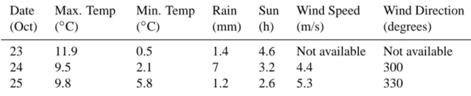
Table 1. Meteorological conditions in the region on the sampling dates (in October 2003), recorded at the Stornoway Meteorological Office
∼50 km north west of the sampling mountains.
Date Max. Temp Min. Temp Rain Sun Wind Speed Wind Direction (Oct) (◦C) (◦C) (mm) (h) (m/s) (degrees)
23 11.9 0.5 1.4 4.6 Not available Not available
24 9.5 2.1 7 3.2 4.4 300
25 9.8 5.8 1.2 2.6 5.3 330
Table 2. Sampling periods and sample details.
Sample Collection started Collection finished volume (ml) pH Conductivity (µS cm−1) Caepabhal 23/10/2003 (16:00:00) 25/10/2003 (13:00:00) Rain 150 6.4 54 Cloud 300 5.8 620 An Clisean 24/10/2003 (15:00:00) 25/10/2003 (13:00:00) Rain 240 5.8 37 Cloud 150 5.4 206
Both cloud and rain samples were collected at each sam-pling location. Cloud samples were collected using passive string cloud collectors. These intercepted cloud water that dripped into a plastic bottle via a funnel (Fig. 1). The cloud collectors were placed at the summit of Caepabhal (there is around a kilometre of upwind foreland before the ocean) and near the summit of An Clisean which has around 10 km of upwind foreland. Rain samples were collected using a fun-nel fixed into a plastic bottle. Within two hours of collection the samples were stored at 4◦C until processing.
All equipment was soaked in 3% H2O2, washed in
0.22 µm filtered deionised water before autoclaving. The string collectors were sterilised using gas plasma H2O2
ster-ilisation (University College Hospital, London, UK). Sam-pling details and sample parameters are given in Table 2. 2.2 Nucleic acid extraction and purification
To isolate the bacterial cells, cloud and rain water was trans-ferred into 300 ml sterile centrifuge buckets and centrifuged at 10 000×g for 20 min at 5◦C. The pellet was re-suspended in 2 ml of residual sample and transferred to a 2 ml tube and centrifuged at 12 000×g for 10 min at 5◦C. The pellet was then re-suspended in 500 µl TE (10 mM Tris-buffer, 1 mM disodium EDTA, pH 8.1), lysozyme added to a final concen-tration of 3 mg ml−1 and incubated at 37◦C for 30 min. A mini bead beater (Glenn Mills®, Clifton, New Jersey) was used to disrupt cells and the suspension transferred to a fresh 2 ml tube. SDS (sodium dodecyl sulphate) and Proteinase K was added at a final concentration of 0.5% and 70 µg/ml
re-spectively and incubated at 37◦C with shaking at 150 rpm for 30 min. 100 µl 5M NaCl was added and mixed. To this 80 µl CTAB reagent (CTAB 2%, NaCl81.8 g/l, EDTA 5.8 g/l, Tris 12.1 g/l adjusted to pH 8.0 with NaOH) was added, mixed and incubated at 56◦C for 10 min. An equal volume of CHCl3: iso amyl alcohol (24:1) was added and
inversion mixed for 2 min then centrifuged at 12 000×g for 5 min. The upper aqueous phase containing the DNA was transferred to a fresh tube. To this an equal volume of cold iso-propanol was added and inversion mixed for 2 min. The DNA was visible as a precipitate and collected by centrifu-gation for 5 min at 12 000×g. Excess alcohol was removed and the pellet washed with 1 ml ice-cold 70% ethanol and centrifuged at 12 000×g for 5 min. Residual ethanol was re-moved and the tube inverted for 20 min to air dry the pellet in a sterile cabinet. The DNA was then dissolved in 50 µl deionised water.
2.3 Total DNA analysis using 16S rDNA
2.3.1 PCR amplification and cloning of the 16S rDNA gene PCR amplification was undertaken using the method de-scribed by Moffett et al. (2000) with the following modifica-tions. Universal primers 530f (5’ GTGCCAGCAGCCGCGG 3’) and 1390 r (5’ GACGGGCGGTGTGTACAA 3’) were used to amplify a ∼860 bp product. REDTaq™ (Sigma, Dorset, UK) was used for PCR amplification under the fol-lowing conditions: initial denaturation at 95◦C for 3 min; 25 cycles of 95◦C for 30 s, 58◦C for 45 s and 72◦C for 2 min; and a final extension at 72◦C for 15 min. Amplicons were
purified using Microspin S400 HR columns according to the manufacturer’s instructions (Amersham Pharmacia Biotech inc., Piscataway, NJ). Purified amplicons were cloned using the TOPO TA Cloning Kit® (Invitrogen, Groningen, Nether-lands) according to the manufacturer’s guidelines. Trans-formants were selected on ImMedia™ Amp Blue (for lacZ AmpR recombinant E. coli strains) agar plates. Approxi-mately 100 clones from each sample were picked at random, transferred into 200 µl of sterile distilled water and frozen immediately at –20◦C.
Clones were amplified with vector-specific primers M13f (5’ GTTTTCCCAGTCACGAC 3’) and M13r (5’ GGAAACAGCTATGACCATG 3’). Each 25 µl reaction mix contained 1.25 U of REDTaq™, 1X PCR buffer (providing 1.1 mM MgCl2), 0.5 µM of each primer, 0.2 mM of each
d’NTP, 2 µl clone suspension (template DNA), and deionised water. The cycling conditions used were a single cycle of 96 oC for 3 min followed by 30 cycles of 94◦C for 1 min, 57◦C for 1 min and 72◦C for 1 min 30 s. A final two minute exten-sion at 72◦C completed the programme.
2.3.2 Restriction digestion, analysis and grouping of clones PCR products underwent a double digest with Hpa II and
EcoR I. EcoR I was included in each to ensure that the
orien-tation of the cloned sequence did not affect the results. Digest contained 6.5 U Hpa II, 3.5 U EcoR I, 1.5 µl of 10X MULTI-CORE™ Buffer, 0.82 µl of 50% glycerol and deionised wa-ter to a total volume of 10 µl (endonucleases and buffers from Promega, UK). Two to ten microlitres (depending on PCR band intensity under UV light) of PCR product was then added to the reaction mixture and incubated for 2 h at 37◦C in 60 well Terasaki plates (Nalge Nunc International, Rochester, NY). Patterns were visualised under transillumi-nation on 3% NuSieve® GTG® agarose gels (FMC Bioprod-ucts, Rockland, Maine). Restriction patterns were analysed and clustered into operational taxonomic units (OTUs) us-ing Bionumerics 2.0 software (Applied Maths, Kortrijk, Bel-gium) using the Dice similarity coefficient with hierarchi-cal tree-clustering using UPGMA. The criteria for clones to be grouped into the same OTU was a >95% similarity. A position tolerance and optimisation tolerance of 0.05% and 0.36%, respectively, was chosen. Following clustering the largest OTU was subsequently digested with 4.5 U of an ad-ditional enzyme, Hae III.
2.3.3 Sequencing
Representative clones from each OTU were sequenced using dideoxynucleotide chain termination chemistry to generate fragments. Purified PCR products were amplified in a lin-ear PCR reaction using the DYEnamic ET Terminator Cy-cle Sequencing Kit (Amersham Biosciences UK Ltd., Little Chalfont, UK). Each reaction comprised 1.5 µl PCR prod-uct, 0.38 µl of the appropriate primer (5 µM), 3.0 µl of the
sequencing mix and 2.63 µl deionised water. Cycling con-ditions were 25 cycles of 95◦C for 20 s, 50◦C for 15 s and 60◦C for 60 s. Precipitation and clean-up of the product was carried out according to the manufacturer’s instructions.
Each DNA pellet was re-suspended in 4 µl formamide loading buffer and loaded into alternate lanes on a 20 cm long, 75 µm thick 5% polyacrylamide gel (Long Ranger Sin-gel Pack, Cambrex, Rockland, MN). Electrophoresis was performed on a BaseStation 51 DNA fragment Analyzer (MJ GeneWorks Inc., Sauk City, WI) in 1×TAPS buffer (MJ Re-search, Waltham, MA) using a 2 min pre-run at 1900 V, a ramped injection increasing from 200 to 4000 V over 36 s and a 3 h collection at 1800 V. Sequences were generated us-ing Cartographer software (version 1.2.6sg, MJ GeneWorks Inc.) and analysed by comparing them to known sequences in GenBank (http://www.ncbi.nlm.nih.gov/BLAST). Multi-ple alignments of sequences were performed using ClustalW (http://www.ebi.ac.uk) and unrooted phylograms generated using Treeview software (Page, 1996).
2.4 Testing for Ice Nucleation
2.4.1 Growth and storage of bacteria isolated from cloud and rain samples
Within 48 h of collection 200 µl of each sample was spread in duplicate onto nutrient agar containing 20 mg ml−1
cyclo-hexamide to inhibit fungal growth and incubated at 20◦C. Isolated colonies were purified on nutrient agar and stock cultures prepared in both 20% glycerol and sterile deionised water and frozen at –20◦C. All isolates were subsequently streaked onto cetrimide agar and incubated at 20◦C for 48 h. This determined which belonged to the genus Pseudomonas. 2.4.2 Growth and storage of IN positive (IN+)
pseudomon-ads
Ice nucleation positive strains of Ps. fluorescens (26 & 26C) were cultured on nutrient agar with 2.5% glycerol and incu-bated at 20◦C for three days. Isolated colonies were picked using sterile pipette tips and inoculated into sterile deionised water. These were frozen, thawed and vortexed to release the cell contents and used directly as the template for PCR. 2.4.3 IN primer design
InaW gene primers were designed based on published se-quences of the InaW gene (Warren et al., 1986). The open reading frame of the IN gene comprises three domains; a repetitive core region flanked by unique C and N termini. The repetitive core region can be subdivided into blocks 1 to 4 with descending levels of internal homology, block 2 having the highest and block 4 the lowest (block 1 is too small for self homology to be significant) (Warren et al., 1986). A variety of primers were designed to target
H. E. Ahern et al.: Cloud bacteria: diversity, ice nucleation and biosurfactants 119 each domain and those targeting block 4 of the central
do-main were the most successful. Several primer combinations were tested, using PCR (methodology as per Sect. 2.3.3) on the IN+ controls, and two forward and one reverse selected: INAW F1 (5’ AACCAGATTGCGAGTCATAAG 3’), INAW F2 (5’-AGCAACAGTTATCTGACTGC 3’) and INAW R3 (5’ CATGGCTGAATCTGAGACTGG 3’). PCR products were sequenced (using the protocol described in section 2.3.3), to increase confidence in the data, and sub-mitted to GenBank for verification of identity.
2.4.4 PCR amplification of IN gene
Eighty cloud and rain pseudomonads isolated from the two sites were tested for the IN gene. PCR reaction mixture con-sisted of 0.5 U Supertherm Gold Taq polymerase (Labmas-ter, Kimbolton, UK), 1X PCR buffer, 0.5 µM of each primer, 0.2 mM d’NTPs, 1.1 mM MgCl2, and 2 µl of cell suspension
in a 25 µl total volume. PCR amplification was performed on a RoboCycler 96 Gradient Cycler (Stratagene, La Jolla, CA) under the following conditions: 95◦C for 13 min; 35 cycles of 95◦C for 40 s, 56◦C for 1 min and 70◦C for 1 min. PCR products were visualised on 1.8% agarose gels.
2.4.5 Freezing point
The IN+ Pseudomonas isolates were incubated at 20◦C to en-sure expression of the IN gene (Marcia Lee, personal com-munication) and grown to early log phase in nutrient broth with 2.5% glycerol. Viable counts and optical density moni-toring of the cultures were used to ensure that consistent con-centrations of bacteria were analysed.
Differential scanning calorimetric (DSC) measurements of the freezing point of the bacterial suspensions were made using a differential scanning calorimeter (DSC822e, Mettler Toledo, Leicester, UK). Two microlitres of bacterial culture was pipetted into a crucible and DSC scans obtained accord-ing to the followaccord-ing protocol: holdaccord-ing temp at 5◦C for 3 min; temperature lowered at a rate of –1◦C min−1 from 5◦C to –30◦C; temperature held at –30◦C for 5 min; temperature raised from –30◦C to 25◦C at a rate of 10◦C min−1. Freezing
events were displayed as exothermic peaks. The tempera-ture at which the cultempera-ture froze was recorded for the 80
Pseu-domonas isolates and positive and negative control cultures
2.5 Biosurfactants
2.5.1 Biosurfactant quantification
The biosurfactant production of the isolates was measured using the drop-collapse method originally described by Jain et al. (1991). A modified version of Bodour and Miller-Maier (1998) was incorporated to make it semi-quantitative. Briefly, stock solutions of SDS in deionised water were pre-pared over a range of 0 to 2.0 mg ml−1 in 0.5 mg ml−1
in-crements. Above 2 mg ml−1 SDS reaches critical micellar
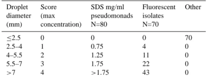
Table 3. Biosurfactant production by cloud and rain bacteria.
Iso-lates are grouped according to their level of production.
Droplet Score SDS mg/ml Fluorescent Other diameter (max pseudomonads isolates
(mm) concentration) N=80 N=70 ≤2.5 0 0 0 70 2.5–4 1 0.75 4 0 4–5.5 2 1.25 11 0 5.5–7 3 1.75 22 0 >7 4 >1.75 43 0
concentration and so surfactant level above this cannot be quantified using this method.
The 8 mm circular wells in the lid of a microtitre tray were coated in 2 µl of mineral oil and left to equilibrate for 2 h. Five microlitres of SDS standards were pipetted onto an oil coated well. After one minute the diameter of the droplet was measured using a RID plate reader (The Binding Site, Birmingham, UK). This was carried out for each of the stock solutions of SDS and drop diameter was plotted against con-centration to produce a standard curve.
To determine the level of biosurfactant production nutrient broth was inoculated with a single colony taken from a nutri-ent agar plate and incubated at 20◦C with shaking at 200 rpm. The drop-collapse method was applied to the cultures after 30 d incubation. The droplet diameter was measured using the plate reader and scored between 0 and 4, 0 indicating no spreading of the droplet and 4 as complete collapse (as per Jain et al., 1991). Using the standard curve, quantitative pa-rameters were set on droplet scoring (see Table 3).
3 Results and discussion 3.1 Cloud bacterial communities.
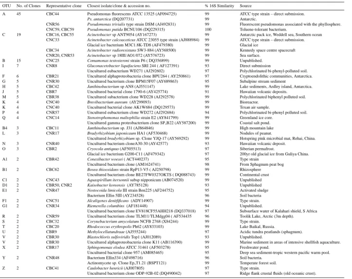
High molecular weight DNA was successfully extracted from the cloud and rain samples and the amplified 16S rDNA sequences cloned. Sixty four clones from each sample were re-amplified using M13 primers; digested using restriction enzymes and the fragment patterns clustered using Bionu-merics. This yielded 100 OTUs of which 31 contained two or more clones. Representative clones from each OTU were sequenced and their phylogenetic affiliation shown in Table 4.
The largest OTU (OTU A) contained 45 clones, 42 of which occurred in the cloud samples. Sequencing of all the clones from this OTU revealed that all were pseudomon-ads. Approximately 45% were within the Ps. fluorescens group (e.g. Ps. fluorescens and Ps. trivialis); 35% within the Ps. putida group (including Ps. putida) and 10% were
Ps. syringae. The Ps. fluorescens group (member of the
gamma (γ ) subclass Proteobacteria) is a remarkably
Table 4. Sequence similarity of representative clones from OTUs represented by two or more clones. Some OTUs possessed notable
intra-OTU variability (e.g. A and C).
OTU No. of Clones Representative clone Closest isolate/clone & accession no. % 16S Similarity Source
A 45 CBC44 Pseudomonas fluorescens ATCC 13525 (AF094725) 99 ATCC type strain – direct submission.
Ps. antarctica (DQ207731) 99 Antarctic.
CNR56 Pseudomonas trivialis type strain DSM (AJ492831) 99 Fluorescent pseudomonas associated with the phyllosphere.
CNC59, CBC59 Pseudomonas putida BCNU106 (DQ229315) 100 Toluene-tolerant bacterium.
C 19 CBC16, CBC55 Acinetobacter sp ANT9054 (AY167273) 99 Antarctic pack ice, Weddell sea, Southern ocean
CNC33 Acinetobacter calcoaceticus ATCC 23055 type strain (AJ888984) 99 ATCC type strain – direct submission.
Glacial ice bacterium M3C1.8K-TD8 (AF479380) 99 Glacial ice
CBC34 Acinetobacter radioresistans SW3-884 (AY568500) 99 Kennedy space centre spacecraft
CNR20, CNR53 Acinetobacter sp 18III/AO1/072 (AY576723) 99 Sea surface.
B 15 CNC25 Comamonas testosteroni strain P6 ( DQ356899) 99 Unpublished.
I 7 CNR8 Gluconacetobacter liquifaciens SRI 244 ( AF127391) 93 Direct submission
Uncultured eubacterium WD271 (AJ292602) 97 Polychlorinated bi-phenyl-polluted soil.
F 6 CBR21 Uncultured alphaproteobacteria clone BPU264 ( AY250861) 97 Cryptoendolithic communities, Antarctica.
G 5 CNR30 Uncultured bacterium clone BPM15F07 (AY689863) 95 Subalpine stream sediment
H 5 CBC42 Janthinobacterium sp AN8 (AJ551147) 99 Lake sediments, Ardley island, Antarctica.
J 5 CBR7 Uncultured bacterial clone 1790-6 (AY425774) 91 Hawaiian volcanic deposits.
M 5 CBR38 Uncultured subacterium clone WD228 (AJ292578) 99 Polychlorinated biphenyl polluted soil.
K 4 CNC40 Brevibacterium aureum (AY299093) 99 Bioreactor.
K 4 CNC40 Uncultured bacterial clone AK1W684 (DQ129573) 99 Texan air sample.
P 4 CNR57 Uncultured eubacterium clone WD272 (AJ292684) 94 Polychlorinated bi-phenyl polluted soil.
Q 4 CNC14 Stenotrophomonas maltophilia strain E2 (AY841799) 99 Greenland ice core.
Uncultured gamma proteobacterium clone SP B22 (AY587200) 99 Coastal salt pond.
B4 3 CBC11 Janthinobacterium sp. J31 (AJ864846) 99 High mountain lake
L 3 CNR17 Bradyrhizobium japonicaum HA1 (AF530468) 99 Nodules of peanut.
Uncultured bradyrhizobium sp. Clone YJQ-17 (AY569292) 99 Hotspring pink microbial mat, Rehai, China.
N 3 CNR40 Uncultured bacterium cloneA30-30 (AY42577) 93 Hawaiian volcanic deposit.
O 3 CBR2 Cryocola antiquus (AF505513) 97 Siberian permafrost.
Glacial ice bacterium G200-C11 (AF479342) 97 200yr old glacial ice from Guliya China.
A1 2 CBR42 Conexibacter woesei ( ACT440237) 95 Type strain
Uncultured bacterium clone (AM1624741) 98 From Sphagnum peat bog
B1 2 CBC62 Bosea thiooxidans strain RpP13-V5 ( AJ250798) 99 Rhizosphere
Uncultured bacterium clone BE27FW0327OKTS ( DQ088743) 99 Continental crust
C1 2 CNC43 Aquaspirillum itersonnii subsp nipponicum (AB074520) 99 Unpublished
D1 2 CBR50, CNR2 Kaisobacter koreensis (AY785128) 93 Unpublished
E1 2 CNR47 Nostocoida limicola III strain Ben225 (AF244752) 95 Activated sludge
Bacterium Ellin 5III (AY234528) 96 Soil bacteria
F1 2 CNC51 Alcaligenes denitfificans (ADY14907) 99 Type strain.
G1 2 CNR34 Riemerella columbins (AF181448) 94 Unpublished.
Uncultured bacterium clone EV818CF55AHH218 (DQ337018) 97 Subsurface water of Kalahari shield, S Africa
R 2 CNR59 Uncultured bacterium clone TLM11/TLMdgg04 ( AF534435 96 Toolik Lake, Arctic (3m depth).
S 2 CBC32 Corynebacterium amycolatum NCFB 2768 (X84244) 99 Type strain.
T 2 CBC20 Rhodococcus erythropolis Ph62 (AY833103) 99 Lake Baikal, Russia.
U 2 CBR9 Methylocellatundraae (AJ555244) 97 Acidic tundra peatlands (sphagnum).
V 2 CBR30 Blastochloris sulfoviridis Top1 (AJ012089) 93 Unpublished.
V 2 CBR30 Uncultured alphaproteobacteria clone K11 (AB116390) 99 Marine sediment in areas of intensive shellfish aquaculture.
X 2 CBR17 Sphingomonas elodea ATCC 31461 (AF503278) 95 Freshwater pond.
Uncultured bacterial clone 957 (AM085465) 95 Deep sea sediment-tropic western pacific warm pool.
Y 2 CNR48 Bacterium Ellin334 (AF498716) 96 Soil bacteria.
Actinomycete sp. Clone Ep T1.21 (BSPT121) 99 Temperate forest soil.
Z 2 CBC41 Caulobacter henricii (AJ007805) 97 Type strain.
Uncultured bacterium clone ODP-92B-02 (DQ490042) 97 Ridge flank crustal fluids (old oceanic crust).
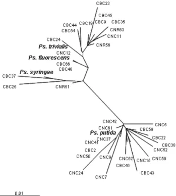
geneous group which displays both inter and intra-species heterogeneity. Ps. fluorescens for instance can be subdivided by various taxonomic criteria into many subspecies (Bossis et al., 2000). As a result of this the 16S rRNA gene has been found to have insufficient resolution to discriminate intra-generically within this group (Yamamoto et al., 2000) which explains the lack of hierarchical clustering in the phylogram (Fig. 2).
Gamma Proteobacteria have recently been found in air collected in Utah (Radosevich et al., 2002) and Northern France (Maron et al., 2005) where they comprised 6% and 12% of the total clones sequenced respectively. They have also been isolated from clouds sampled at the Puy de Dˆome (Amato et al., 2005). Indeed the γ -Proteobacteria, partic-ularly the fluorescent pseudomonads, were the single com-mon feature in all the samples reviewed. This may reflect the ubiquitous nature of the pseudomonads (Yamamoto et al.,
2000). Alternatively, if clouds foster a microbial ecosystem, the same constituent species should recur in diverse samples. By supporting growth and aiding dispersal, cloud processes may contribute to their ubiquity.
The second largest OTU (OTU C) comprised 19 clones which again were dominated by clones from cloud sam-ples. Representative clones had a 99% sequence similarity to Acinetobacter spp. of psychrophilic marine origin. Amato et al. (2005) also found that psychrophilic bacterial species had a significant presence in their samples. Interestingly, in the Hebridean samples, 80% of OTUs with two or more clones were found only in cloud or only in rain water. This could reflect an intrinsic difference in bacterial composition between clouds and rain. That each rain sample was also distinct may reflect the spatial and temporal variability of aerosols in different air masses captured when scrubbed by the rain. Ion species and concentrations are very
depen-H. E. Ahern et al.: Cloud bacteria: diversity, ice nucleation and biosurfactants 121 dent on the air mass from which the cloud derives. In areas
of anthropogenic activity ions are predominately related to pollution, such as SO2−4 , NO3− and NH+4. Clouds derived from air masses of marine origin are high in Na+and Cl− ions (Gioda et al., 2006). The high conductivity of the cloud samples reflects the marine origin of the winds, and the ex-ceptionally high conductivity of the sample collected from Caepabhal (Table 2) could additionally have been caused by dry salt deposition on the wires of the collector. The con-ductivities of the rain samples were much lower than the cloud samples because rain is formed higher in the atmo-sphere, a cleaner environment (Blas et al., 2004). This fur-ther raises the question of the origin of the bacteria in the cloud samples. It is possible that the clouds were enriched with locally-derived phyllosphere bacteria, which may have been activated into CCN (Sect. 3.3). Local enrichment, how-ever, seems less likely because in subsequent sampling of heath vegetation in the region (samples shaken vigorously in water then aliquots plated on cetrimide agar) pseudomonads were recovered from only two out of nine samples and were abundant in only one. Also, the fact that almost half of the OTUs were related to bacteria from psychrophilic, polar en-vironments suggests that this was their primary source. We are currently developing a quantitative reverse transcriptase PCR methodology to assess bacterial metabolic activity in future samples. This should indicate which, if any, are capa-ble of using clouds as an environmental niche.
3.2 Ice nucleation
Ice nuclei are an essential prerequisite of all natural freezing in temperate climates and even in polar regions at temper-atures not far below zero. IN gene-targeted PCR was ap-plied to cloud bacterial isolates to screen for its presence. Because members of the Ps. fluorescens group were found to be the dominant group within the samples the InaW gene from Ps. fluorescens (Warren at al., 1986) was selected for amplification. Ps. putida has been reported to possess IN ac-tivity but since its gene is yet to be sequenced it could not be targeted despite also being prevalent in the samples (Cas-trillo et al., 2000). The repetitive core region within the open reading frame of the IN gene is highly conserved between all IN+ species of bacteria, ensuring the hexameric folding es-sential for the ice nucleation protein to mimic an ice crystal (Edwards et al., 1994). Therefore this central region, par-ticularly block 4 (Warren et al., 1986), was selected for the design of Ina W primers. In all cases these efficiently ampli-fied the positive controls. However, when applied to the 80
Pseudomonas isolates from the Hebrides it failed to amplify
any IN gene. When the positive control amplicons were se-quenced, they were found to have 87% homology with the published sequence and when the corresponding amino acid sequence was analysed the homology was 95%. This de-generacy and missense mutations observed in the nucleotide sequence may reflect general intra-species diversity in the
Fig. 2. Unrooted phylogram of an 860 bp region of the 16S rDNA
gene of the fluorescent pseudomonads in OTU A. Sequences of representative type cultures are included for cluster identification. Multiple alignments of sequences were performed using ClustalW (http://www.ebi.co.uk) and then Neighbour Joining generated the unrooted phylogram in Treeview. The scale bar indicates a 1% sequence difference. NB: CNC & CNR denote clones from An Clisean cloud and rain respectively. CBC & CBR clones from Caepabhal cloud and rain.
InaW gene. It could have compromised priming success, thus preventing amplification of the IN gene within the cloud bacterial isolates. It was therefore necessary to test for IN ac-tivity of the cloud and rain isolates. A differential scanning calorimeter (DSC) was used to measure the freezing points. Both positive controls froze at mean temperatures of –4.75◦C +/-0.5◦C. By contrast the Pseudomonas isolates froze be-tween 16◦C and 24◦C lower than this, indicating the absence of appreciable ice nucleation activity in any isolates under the experimental conditions. Morris et al. (2005) described 3 types of bacterial ice nuclei; type I catalyses the freezing of water between –2◦C and –5◦C, type II between –5◦C and –7◦C and type II >–7◦C. They also reiterated that IN pro-tein expression was very sensitive to growth conditions and so, in addition to the observed degeneracy, the growth con-ditions may have been unsuitable for IN protein production or detection of activity. Although this study did not detect IN bacteria in oceanic clouds it does not demonstrate their absence. It has been argued that only a few ice nuclei can have a disproportionate influence on ice nucleation events in the clouds due to the production of secondary ice crystals
rived from the primary crystals (produced by heterogeneous nucleation). Secondary ice crystal production can occur via the “riming-splintering” mechanism whereby the outer shell of a droplet freezes and then shatters forming ice crystals which themselves can initiate ice crystal production. This could explain why ice particle concentrations appear to be much higher than can be explained by conventional ice nu-cleus measurements. (Hallett and Mossop, 1974). This also may suggest that only one or two bacteria in a pocket of air need possess the IN gene in order to influence cloud pro-cesses and these may have been missed in the sampling. 3.3 Biosurfactants
While using DSC to assess the freezing point of the He-bridean pseudomonads it was noted that manipulating small volumes (≤2 µl) of broth culture proved troublesome. When pipetted into the DSC crucible the droplets of bacterial sus-pension failed to maintain the convex shape attributable to surface tension. Instead they spread very quickly over the surface into a thin film. This was suggestive of biosurfac-tant production which is known to reduce surface tension. Therefore the drop-collapse technique developed by Jain et al. (1991) was applied to all Hebridean bacterial isolates. It was found that all of the fluorescent pseudomonads were bio-surfactant producers to varying degrees (see Table 3). There was no significant difference in the numbers of producers or level of biosurfactant production between the four sam-ples but a significant difference between the fluorescent pseu-domonads and other species (Mann Whitney U test).
The presence of biosurfactant producing bacteria in the air has a number of potentially important consequences. Aerosol particles can be activated into cloud condensation nuclei (CCN) onto which water vapour condenses to form cloud droplets (Kohler, 1936). There is a critical atmospheric su-persaturation at which an aerosol particle can be activated. Conditions which determine whether an aerosol will over-come this and activate depends on the size of the particle and its response to water. It has been long understood that at-mospheric surfactants play an important role in aerosol acti-vation (Brimblecombe and Latif, 2004) by lowering surface tension or altering the bulk hygroscopicity of the particles (Abdul-Razzak and Ghan, 2004) and thus reducing critical supersaturation (Bullrich and Hanel, 1978). Models of cloud formation suggest that reducing surface tension has numer-ous impacts affecting cloud droplet number, size, lifespan and the formation of precipitation (Shulman et al., 1996). The “Twomey” effect is the increase in cloud albedo due to an increase in aerosol concentration producing a larger num-ber of small cloud droplets. Facchini et al. (1999) reported a large difference (33%) in surface tension between cloud wa-ter collected from the Po Valley, Italy, and pure wawa-ter. Indeed they also noted that the aerosol population is generally made up of small particles and so a decrease in their surface ten-sion would activate a large proportion of smaller CCN and
increase the cloud lifespan. It has also been recognised that in a cloud made up of many small droplets the efficiency of droplet growth by collisions is reduced which exerts a degree of precipitation suppression (Ferek et al., 2000). If precipi-tation is suppressed, water in the atmosphere would remain aloft and be transported to other locations before it is de-posited to the surface; a characteristic that could be exploited by microorganisms. Several bacteria have been identified as effective CCNs including Ps. syringae, Erwinia herbicola and Micrococcus agilis (Franc and DeMott, 1998; Snider et al., 1985; Bauer et al., 2003). It has been suggested that the chemical composition, structure and hydrophilicity of the outer cell surface could play important roles in CCN activ-ity but the exact process is unknown (Sun and Ariya, 2006; Bauer et al., 2003) as is the source of cloud surfactants (Fac-chini et al., 2000; Fac(Fac-chini et al., 2001).
Considering the level of pseudomonad surfactant produc-tion in the Hebridean bacterial isolates it is conceivable that they are one of the sources and activate atmospheric aerosols (including themselves) into CCN. This could be useful in scavenging water and nutrients and utilising the cloud as a mechanism for their widespread dispersal, aiding ubiquity.
4 Conclusions
To our knowledge this is the first study characterising the to-tal microbial communities targeting the non-cultivable frac-tion using 16S rDNA analysis in cloud samples. It is perhaps significant that the genus Pseudomonas occurred in this and other similar studies. Further research, using RNA, will be pursued to investigate if the fluorescent pseudomonads could be using the clouds as an environmental niche. That all the fluorescent pseudomonads in the cultivable fraction of the sample were biosurfactant producers could also be particu-larly significant. The highly disproportionate relative abun-dance of Pseudomonads in the cloud sample, from 3/128 clones in rain samples to 42/128 in cloud samples, may be empirical evidence that their surfactant production is enhanc-ing their role as CCNs. Finally, these observations may not only indicate the origins of some cloud-derived surfactants but also opens up a new avenue into the study of the contri-bution bacteria may make to cloud processes.
Acknowledgements. Research was principally supported by NERC
(NER/A/S/2001/01097) and EPSRC (GR/P00338/01). We are also very grateful to M. Lee for the kind donation of isolates.
Edited by: T. J. Battin
References
Abdul-Razzak, H. and Ghan, S.: Parameterization of the influence of organic surfactants on aerosol activation, J. Geophys. Res., 109(D3), D03205, doi:10.1029/2003JD004043, 2004.
H. E. Ahern et al.: Cloud bacteria: diversity, ice nucleation and biosurfactants 123
Amato, P., Menager, M., Sancelme, M., Laj, P., Mailhot, G. D., and Delort, A.: Microbial population in cloud water at the Puy de Dome: Implications for the chemistry of clouds, Atmos. Env., 39, 4143–4153, 2005.
Bauer, H., Giebl, R., Kasper-Giebl, A., Loflund, H., Giebl, H., Hitzenberger, H, Zibuschka, F., and Puxbaum, H.: The contri-bution of bacteria and fungal spores to the organic carbon con-tent of cloud water, precipitation and aerosols. Atmos. Res., 64, 109—119, 2002.
Bauer, H., Giebl, H., Hitzenberger, R., Kasper-Giebl, A., Reis-chl, G., Zibuschka, F., and Puxbaum, H.: Airborne bacteria as cloud condensation nuclei, J. Geophys. Res., 108(D21), AAC 2– 1, 2003.
Blas, M., Sobik, M., and Twarowski, R.: Changes of cloud water chemical composition in the Western Sudety Mts., Poland, in: Proceedings of the third International Conference on Fog, Fog Collection and Dew, NetSys International, 2004.
Bodour, A. and Miller-Maier, R.: Application of a modified drop-collapse technique for surfactant quantitation and screening of biosurfactant-producing microorganisms, J. Microbiol. Methods, 32, 273–280. 1998.
Bossis, E., Lemanceau, P., Latour, X., and Gardan, L.: The taxon-omy of Pseudomonas fluorescens and Pseudomonas putida: cur-rent status and need for revision, Agronomie, 20, 51–63, 2000. Brimblecombe, P. and Latif, M.: Rediscovering atmospheric
sur-factants, Env. Chem., 1, 11–12, 2004.
Brown, M., Larson, D., and Bold, H.: Airborne algae: Their abun-dance and heterogeneity, Science, 143, 583–585, 1964. Bullrich, K. and H¨anel, G.: Effects of organic aerosol constituents
on extinction and absorption coefficients and liquid water con-tents of fog and clouds, Pure Appl. Geophys., 116, 293–301, 1978.
Casareto, B., Suzuki, Y. Okado, K., and Morita, M.:Biological micro particles in rain water, Geophys. Res Lett. 23, 173–176, 1996.
Castrillo, L., Lee, R., Wyman, J., Lee, M., and Rutherford, S.: Field persistence of ice-nucleating active Pseudomonas fluorescens strains for biological control of overwintering Colorado potato beetles (Coleoptera: Chrysomelidae), J. Econ. Entomol. 93, 226– 233, 2000.
Edwards, A., Van Den Bussche, R., Winchman, H., and Orser, C.: Unusual pattern of bacterial ice nucleation gene evolution, Mol. Biol. Evol., 11(6), 911–920, 1994.
Facchini, M., Mircea, M., Fuzzi, S., and Charlson, R.: Cloud albedo enhancement by surface-active organic solutes in grow-ing droplets. Nature, 401, 257–259, 1999.
Facchini, M., Decasari, S., Mircea, S., Fuzzi, S., and Loglio, G.: Surface tension of atmospheric wet aerosol and cloud/fog droplets in relation to their organic carbon content and chemical composition, Atmos. Environ., 34, 4853–4857, 2000.
Facchini, M., Mircea, M., Fuzzi, S., and Charlson, R.: Influence of soluble surfactant properties on the activation of aerosol particles containing inorganic solute, J. Atmos. Sci. 58, 1465–1467, 2001. Ferek, R., Garrett, T., Hobbs, P., Strader, S., Johnson, D., Taylor, J., Ackerman, K., Kogan, Y., Liu, Q., Albrecht, B., and Babb, D.: Drizzle suppression in ship tracks, J Atmos. Sci., 57, 2707–2728, 2000.
Franc, G. and DeMott, P.: Cloud activation characteristics of air-borne Erwinia carotovora cells, J. Appl. Meteorol., 37, 1293–
1300, 1998.
Fuzi, S., Mandrioli, P., and Perfetto, A.: Fog droplets – an atmo-spheric source of secondary biological aerosol particles, Atmos. Env., 31, 287–290, 1997.
Gioda, A., Mayol-Bracero, O., Rodriguez, A., Morales-Garcia, F., and Morales R.: Chemical characterization of cloud water at the East Peak, Puerto Rico, during the rain in cumulus over the ocean experiment (RICO), in: Proceedings of 12th Conference on Cloud Physics, American Meteorological Society, 2003. Hallet, J. and Mossop, S.: Production of secondary ice particles
during the riming process, Nature, 249, 23–28, 1974.
Hirano, S. and Upper, C.: Ecology of ice nucleation-active bacte-ria, in: Biological Ice Nucleation and Its Applications, edited by: Lee Jr., R., Warren, G., and Gusta, L., APS Press, St. Paul, Min-nesota, 41–62, 1995.
Jain, D., Collins-Thompson, D., Lee, H., and Trevors, J.: A drop-collapsing test for screening surfactant producing microorgan-isms, J. Microbiol. Methods, 13, 271–279, 1991.
Kieft, T.: Ice nucleating activity in Lichens, Appl. Env. Micro., 54, 1678–1681, 1988.
K¨ohler, H.: The nucleus in the growth of hygroscopic droplets, Trans. Far. Soc., 32, 1152–1161, 1936.
Lindow, S.: The role of Bacterial ice nucleation in frost injury to plants, Ann. Rev. Phytopathol., 21, 363–384, 1983.
Mandrioli, P., Puppi, G., Bagni, N., and Prodi, F.: Distribution of microorganisms in Hailstones, Nature, 246, 416–417, 1973. Maron, P., Lejon, D., Carvalho, E., Bizet, K., Lemanceau, P.
Ran-jard, L., and Mougel, C.: Assessing genetic structure and di-versity of airborne bacterial communities by DNA fingerprinting and 16S rDNA clone library, Atmos. Env., 39, 3687–3695, 2005. Moffett B., Walsh, K., Harris J., and Hill T.: Analysis of bacte-rial community structure using 16S rDNA analysis, Anaerobe 6, 129–131, 2000.
Morris, C., Georgakpoulos, D., and Sands, D.: Ice nucleation ac-tive bacteria and their potential role in precipitation, J. Phys. IV France, 121, 87–103, 2005.
Page, R.: TREEVIEW: An application to display phylogenetic trees on personal computers, CABIOS 12, 357–358, 1996.
Pruppacher, H. and Klett, J.: Microphysics of Clouds and Precipi-tation. 2d ed, Kluwer Academic Publishers, 974, 1997.
Radosevich, J., Wilson, W., Shinn, J., DeSantis, T., and Anderse, G.: Development of a high-volume aerosol collection system for the identification of air-borne micro-organisms, Lett. Appl. Mi-crobiol., 34, 162–167, 2002.
Sattler, B., Puxbaum, H., and Psenner, R.: Bacterial growth in supercooled cloud droplets, Geophys. Res. Lett., 28, 239–242, 2001.
Shulman, M., Jacobson, M., Carlson, R., Synovec, R., and Young, T.: Dissolution behaviour and surface tension effects of organic compounds in nucleating cloud droplets, Geophys. Res. Lett. 23, 277–280, 1996.
Snider, J., Layton, R., Caple, G., and Chapman, D.: Bacteria as condensation nuclei, J. Rech. Atmos., 19, 139–145, 1985. Sun, J. and Ariya, P.: Atmospheric organic and bio-aerosols as
cloud condensation nuclei (CCN): A review, Atmos. Env., 40, 795–820, 2006.
Szyrmer, W. and Zawadski, I.: Biogenic and anthropogenic sources of ice-forming nuclei: A review, BAMS, 78, 209–228, 1997. Vali, G.: Principles of ice nucleation, in: Biological Ice Nucleation
and Its Applications, editied by: Lee Jr., R., Warren, G., and Gusta, L., APS Press, St. Paul, Minnesota, 1–28, 1995.
Vali, G.: Ice Nucleation a review, in: Nucleation and Atmospheric Aerosols, Proc. 14th Int. Conf. Nucleation and Atmospheric Aerosols, edited by: Kulmala, M. and Wagner, P., Elsevier Sci-ence, Ltd. Oxford, UK, 89–92, 1996.
Warren, G., Corotto, L., and Wolber, P.: Conserved repeats in di-verged ice nucleation structural genes from two species of
Pseu-domonas, Nucleic Acids Res., 14, 8047–8060, 1986.
Wolber, P. and Warren, G.: Bacterial ice-nucleation proteins, Trends Biochem. Sci., 14, 179–182, 1989.
Yamamoto, S., Kasai, H., Arnold, D., Jackson, R., Vivian, A., and Harayama, S.: Phylogeny of the genus Pseudomonas: in-trageneric structure reconstructed from the nucleotide sequences of gyrB and rpoD genes, Microbiology, 146, 2385–2394, 2000.