HAL Id: tel-03161030
https://tel.archives-ouvertes.fr/tel-03161030
Submitted on 5 Mar 2021HAL is a multi-disciplinary open access
archive for the deposit and dissemination of sci-entific research documents, whether they are pub-lished or not. The documents may come from teaching and research institutions in France or abroad, or from public or private research centers.
L’archive ouverte pluridisciplinaire HAL, est destinée au dépôt et à la diffusion de documents scientifiques de niveau recherche, publiés ou non, émanant des établissements d’enseignement et de recherche français ou étrangers, des laboratoires publics ou privés.
toolkit for the simulation of radiobiological effects at the
sub-cellular scale
Wook Geun Shin
To cite this version:
Wook Geun Shin. Development and application of the Geant4-DNA toolkit for the simulation of radiobiological effects at the sub-cellular scale. Astrophysics [astro-ph]. Université de Bordeaux; Yonse Taehakkyo, 2020. English. �NNT : 2020BORD0310�. �tel-03161030�
THÈSE EN COTUTELLE PRÉSENTÉE
POUR OBTENIR LE GRADE DE
DOCTEUR
DE L’UNIVERSITÉ DE BORDEAUX
ET DE L’UNIVERSITÉ DE YONSEI
ÉCOLE DOCTORALE UNIVERSITÉ DE BORDEAUX
ÉCOLE DOCTORALE UNIVERSITÉ DE YONSEI
SPÉCIALITÉ : Astrophysique, Plasmas, Nucléaire
Par Wook-Geun SHIN
Development and application of the Geant4-DNA toolkit for
the simulation of radiobiological effects
at the sub-cellular scale
Sous la direction de Sebastien INCERTI
et de Chul Hee MIN
Soutenue le 22 décembre 2020
Membres du jury :
M. INCERTI, Sébastien Directeur de recherche CENBG / CNRS Directeur de thèse M. MIN, Chul Hee Professeur associé Yonsei University Codirecteur de thèse M. EL BITAR, Ziad Directeur de recherche IPHC / CNRS Rapporteur
M. CHO, Kihyeon Professeur UST / KISTI Rapporteur
M. TAKASHI, Sasaki Professeur KEK Examinateur
Résumé :
Prévoir les effets biologiques induits par les rayonnements ionisants est
un défi scientifique majeur de la radiobiologie actuelle, en particulier pour essayer de
mieux comprendre les effets des faibles doses sur le milieu vivant ainsi que la
cancérogénèse. L'approche computationnelle basée sur les codes de simulation des
structures de traces dans le milieu biologique par la technique Monte Carlo est
aujourd'hui la méthode la plus fiable pour calculer les effets précoces des radiations
ionisantes sur l'ADN, la cible cellulaire principale des effets des radiations. Parmi les
codes existants, l'extension Geant4-DNA de la boîte à outils généraliste Geant4 est la
première entièrement ouverte et librement accessible à la communauté. Geant4-DNA
peut simuler non seulement l'étape physique mais aussi les étapes physico-chimique
et chimique de la radiolyse de l'eau. Ces étapes peuvent être combinées avec des
modèles géométriques simplifiés de l'ADN afin d'évaluer les dommages précoces
directs et indirects à l'ADN. Dans cette thèse, je propose (1) d'améliorer dans
Geant4-DNA la modélisation de la diffusion élastique des électrons dans l'eau liquide pour
simuler plus précisément la distribution spatiale des dépôts d'énergie et des espèces
moléculaires. Ensuite, (2) l'étape physico-chimique de la radiolyse de l'eau est
également améliorée en se basant sur des approches décrites dans la littérature
(modélisation, mesures), cette étape affectant fortement l'étape chimique en modifiant
les rendements initiaux et la concentration des espèces. (3) La méthode du temps de
réaction indépendant (IRT) est en outre implémentée dans Geant4-DNA afin de
réduire le temps de calcul pour simuler la cinétique chimique de la radiolyse de l'eau.
Enfin, j'évalue (4) les dommages biologiques induits à l'échelle subcellulaire en
utilisant une géométrie de l'ADN cellulaire développée dans une étude précédente, en
incluant dans la simulation toutes les améliorations développées au cours de cette
thèse, jusqu'à la réparation des dommages précoces. Ces développements sont
regroupés au sein d'une chaine de simulation complète destinée aux utilisateurs de
Geant4 et de son extension Geant4-DNA.
Abstract :
Predicting the biological effects induced by ionizing radiation is a major
scientific challenge of current radiobiology, in particular to try to better understand the
effects of low doses on living beings as well as carcinogenesis. The computational
approach based on codes to simulate trace structures in the biological medium using
the Monte Carlo technique is today the most reliable method to calculate the early
effects of ionizing radiation on DNA, the main cellular target of radiation effects. Among
the existing codes, the Geant4-DNA extension of the Geant4 general purpose
simulation toolkit is the first one fully open and freely available to the community.
Geant4-DNA can simulate not only the physical but also the physico-chemical and
chemical stages of water radiolysis. These stages can be combined with simplified
geometric models of DNA to assess direct and indirect early DNA damage. In this
thesis, I propose (1) to improve in Geant4-DNA the modeling of the elastic scattering
of electrons in liquid water in order to simulate more precisely the spatial distribution
of energy deposits and molecular species. Then, (2) the physico-chemical stage of
water radiolysis is also improved based on approaches described in the literature
(modeling and measurements), this step strongly affecting the chemical stage by
modifying the initial yields and the concentration of species. (3) In addition, the
Independent Reaction Time (IRT) method is implemented in Geant4-DNA in order to
reduce the computational time to simulate the chemical kinetics of water radiolysis.
Finally, I evaluate (4) the biological damage induced at the subcellular scale using a
cellular DNA geometry developed in a previous study, including in the simulation all
the improvements developed during this thesis, up to the repair of early DNA damage.
These developments are grouped in a complete simulation chain for users of the
Geant4-DNA extension of Geant4.
Keywords :
Radiobiology, Damage, DNA, Monte Carlo simulation, Geant4-DNA
Unité de recherche
Table of contents
Chapter 1 Introduction 1
1.1.Context……….……...2
1.2.Monte Carlo track structure simulation………...………..…….4
1.3.The Geant4-DNA project………9
1.3.1. Physical stage………..…………...…...10
1.3.2. Pre-chemical and chemical stages………..…….…..14
1.3.3. Geometrical models and DNA damage scoring………...….16
1.4.Purpose of this thesis……….18
References………...…19
Chapter 2 Physical stage 28
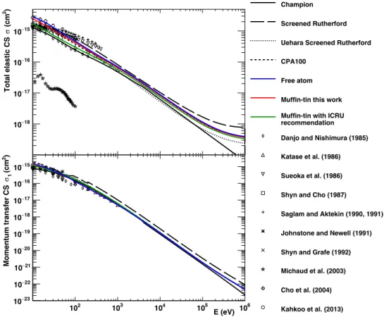
2.1.Development of a new electron elastic scattering cross-section model for Geant4-DNA
using ELSEPA for liquid-phase water………..………....31
2.1.1. Description of potentials and selected options………...…...33
2.1.2. Verification of validity by comparing calculated cross-section with experiments
and other cross-section models……….………...…..38
2.1.3. Results for determination of optimal options and parameters…………..……40
2.1.4. Plausibility of the electron elastic scattering models………46
2.2. The impact of the ELSEPA electron elastic scattering model on Geant4-DNA
simulations………...…………...………....48
2.2.1. Determination of optimal angle and energy binning in ELSEPA……….48
2.2.2. Geant4-DNA examples……….49
2.2.3. Results for the implementation of the elastic scattering cross-section into
Geant4-DNA………...55
2.2.4. Results for the Geant4-DNA simulations……….59
2.3. Conclusions……….…66
radiolysis simulations using Geant4-DNA………..…….………...78
3.1.1. Principles of Geant4-DNA simulation of water radiolysis…….…………79
3.1.2. Geant4-DNA elastic scattering models for electrons in liquid water…….81
3.1.3. Geant4-DNA electron thermalization model………..83
3.1.4. Chemistry parameters……….88
3.1.5. The new “chem6” example……….…90
3.1.6. Radiochemical yield simulation in water………....93
3.1.7. LET calculations……….94
3.1.8. Influence of electron elastic scattering models………...95
3.1.9. Influence of electron thermalization models………...97
3.1.10. Influence of chemistry parameters………101
3.2.Evaluation of the impact of the pre-chemical processes………...103
3.2.1. Physical and physico-chemical processes…….………103
3.2.2. Dissociation channels………....108
3.2.3. Displacement of hot fragments……….111
3.2.4. Validation study by comparing simulation data with literature data……114
3.2.5. Results for ionization and excitation……….117
3.2.6. Results for electron attachment process………119
3.2.7. Results for electron-hole recombination and dissociation channel……...122
3.2.8. Influence of the pre-chemical processes on water radiolysis simulation…124
3.3.Conclusions………...…....126
References………...………..128
Chapter 4 Chemical stage 135
4.1.Green Function Diffusion Equation……….………….136
4.1.1. Totally diffusion-controlled reactions (Types I and III) ………..138
4.1.2. Partially diffusion-controlled reactions (Types II and IV) ………...140
4.1.3. First-order reactions and background reactions (Type VI reactions) …..146
4.1.4. Contact reactions………...147
4.2.The independent reaction time technique……….148
4.2.3. Sampling of type VI reactions and contact reactions………153
4.2.4. Reaction site and position of secondaries……….154
4.3.Implementation of IRT in the chemistry framework of Geant4-DNA………….155
4.4.Radiochemical yield simulation in water………..156
4.5.Validation of implementation………...157
4.6.Results for G-values versus time………..160
4.7.Results for G-values versus LET………..163
4.8.Conclusions………..……….164
References……….165
Chapter 5 DNA damage 169
5.1.DNA geometry………..………171
5.2.DNA damage and scoring………..…………..………172
5.2.1. Source classification……….173
5.2.2. Complexity of breaks………176
5.3.Implementation of IRT method into “molecularDNA” example………..177
5.4.Verification of the applicability of the IRT approach (“cylinders” approach) …178
5.5.Evaluation of DNA damage in a simplified human cell (“human_cell”
approach)………...179
5.6.Cell repair model………...181
5.7.Results for verification of the applicability of the IRT approach……….182
5.8.Results for human cell nucleus……….183
5.9.Conclusions………..……….191
References………..…...………192
Conclusions & perspectives………...…….197
Chapter 1
Introduction
Table of contents
1.1. Context……….………...2
1.2. Monte Carlo track structure simulation………..…….4
1.3. The Geant4-DNA project………9
1.3.1. Physical stage………...…...10
1.3.2. Pre-chemical and chemical stages……….…..14
1.3.3. Geometrical models and DNA damage scoring……….….16
1.4. Purpose of this thesis……….18
1.1. Context
Figure 1.1: Percentage contribution of radiation sources reported by the National Council on Radiation Protection and Measurements (NCRP, 2009) based on collective effective dose S (person-sievert) and effective dose per individual in the U. S. population Eus (millisievert).
Human-beings are exposed to various sources of ionizing radiation during their life. According to the NCRP report 160 (NCRP, 2009), the average annual radiation dose in the U. S. is about 6.2 mSv, 50% originating from background radiation and 50% from human-made sources, as illustrated in Figure 1.1. The major contribution of human-made sources is for medical purpose.
Since X-rays were discovered in 1895 and researchers started to investigate the medical uses of ionizing radiation, the deleterious effects on human body have been of global interest for more
than a century (Nias, 1998).
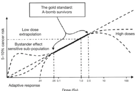
Figure 1.2: The cellular damage risk induced by ionizing radiation as a function of annual dose rate from Hall (2004).
It is possible to epidemiologically predict the radiation risks on humans by following up the atomic bomb survivors (UNSCEAR, 2000, Hall, 2004) at high doses, however the ionizing radiation hazards below the covered dose range (generally a few hundreds of mSv) suffer large uncertainties as illustrated in Figure 1.2. Several risk models in the so called "low-dose" region have been proposed: the Linear-No Threshold (LNT) model, which assumes that the stochastic effect has a linear relationship with dose and no lower dose threshold. This model forms the modern concept of radiation safety, "as low as reasonably achievable" (ALARA) (ICRP, 1977, Prasad et al., 2004), even though it is reported that the linear extrapolation cannot accurately predict the radiation risks (Hooker et al., 2004). Alternatively, the adaptive response model, well-known as radiation hormesis, is a hypothesis that any toxin below threshold stimulates a protective biological response, even in ionizing radiation (UNSCEAR, 1994, Wolff, 1998). There is another interesting experimental observation that unirradiated cells in an irradiated population of cells receive a signal from neighbor irradiated cells and mimic them, the so-called radiation-induced "bystander" effects (Nagasawa and Little, 1992, Seymour and Mothersill, 2004). However, experimental validation of those risk models remains today a scientific challenge.
The mechanistic evaluation of biological effects induced by ionizing radiation is necessary, in order not only to understand low-dose carcinogenesis in many domains but also to improve existing and develop, innovative therapeutic approaches that use ionizing radiation. For example, accurate calculation of relative biological effect (RBE) is important in radiation therapy, especially for charged particles (e.g. proton and carbon therapy) (Paganetti et al., 2002, Frese et al., 2012). Recently, biological effects revealed for nanoparticle-aided, FLASH or mini/microbeam radiotherapies still need to be elucidated (Engels et al., 2016, Dos Santos et al., 2020,
Ramos-Méndez et al., 2020). Also, the low-dose irradiation to patients undergoing radiology and nuclear medicine exams is an important subject of research (Fazel et al., 2009). In addition, in the case of radiation industry and space science, the influence of chronic exposure in the industry (Howe et al., 2004) or during space missions (Mortazavi et al., 2003) should be evaluated for radioprotection of workers.
In order to elucidate the mechanisms involved in ionizing radiation damage, the structure of human cells and their characteristics have been studied for a long time. Mammalian cells, including human cells, are complex biological systems consisting of a nucleus and surrounding cytoplasm. The cells contain several cytoplasmic organelles such as mitochondria, ribosomes, Golgi vesicles, centrioles, and lysosomes. However, it is still admitted today that the most sensitive target to ionizing radiation is the cell nucleus and its deoxyribonucleic acid (DNA) content, which can critically impact the fate of the cell after irradiation (Nias, 1998).
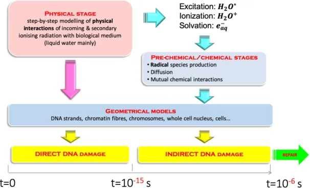
Figure 1.3: The time frame for effects of ionizing radiation reproduced from Turner (2007). It is classically reported that the radiobiological mechanisms consist of physical, pre-chemical, chemical and biological stages (Turner, 2007) as shown in Figure 1.3. At first, the physical stage takes place in attosecond scale (< 10-15 s) and corresponds to excitations and ionizations of molecules which lead to energy deposition. The resulting excited and ionized molecules can fragment into molecular species (< 10-12 s) which can chemically react with biomolecules (e.g. DNA and RNA) present in the cell and induce early indirect radiobiological effects (< 10-6 s). Nowadays, it is recognized that Monte Carlo (MC) simulations are the most reliable approach in order to estimate early radiobiological effects induced by ionizing radiation (Dingfelder, 2012).
1.2. Monte Carlo track structure simulation
The MC technique is based on random number generation and enables to simulate the stochastic nature of particle-matter interactions. In particular, it is used for the simulation of the transportation of radiation through matter (Metropolis, 1987). However, many general-purpose MC codes and toolkits, such as MCNP (Pelowitz, 2011) and Geant4 (Agostinelli et al., 2003, Allison et al., 2006, Allison et al., 2016), use a "condensed-history" approach, which approximates the multiple particle collisions as one single step accumulating them (Larsen and Tolar, 2001). The step limit is usually too large to simulate accurately particle transportations below the micrometer, which
is typically the sub-cellular scale (Lazarakis et al., 2018). Moreover, it is reported that the contribution of indirect damage is dominant at low linear energy transfer (LET) (Hirayama et al., 2009). Thus, the simulation of physical interactions is not sufficient and a careful modeling of radiolysis is required, including diffusions and chemical reactions of molecular species with biological medium, for the evaluation of indirect DNA damage (O'Neill and Wardman, 2009). In order to overcome such limitations of the MC technique, a number of Monte Carlo Track Structure (MCTS) codes and toolkits have been developed.
In brief, the “MCTS method” represents the MC method simulating every interaction without condensed-history approximation, using a "discrete" approach, which simulates particle transportation step-by-step. Most of the MCTS tools approximate the target as liquid water which composes more than 60% of human body (Mitchell et al., 1945).
The modeling of pre-chemical and chemical stages is more complicated. In the same spirit as for the physical stage, the water radiolysis simulation is performed with the assumption that human biological medium consists of liquid water. The water molecules ionized and excited during the physical stage undergo dissociation processes during the pre-chemical stage. For example, Figure 1.4 shows an overview of dissociation channels proposed by Kreipl et al. (2009).
Figure 1.4: Dissociation approach proposed by Kreipl et al. (2009). The figure is reproduced from Buck et al. (2012).
The molecular species (eaq-, H•, •OH, OH-, H3O+, H2, H2O2) generated by the dissociation process in the pre-chemical stage diffuse following a Brownian motion (Knight, 1962) during the chemical stage. In most MCTS tools, the Brownian transportation of the species is typically modeled using step-by-step (SBS) method (Turner et al., 1983, Michalik et al., 1998, Kreipl et al.,
2009, Karamitros et al., 2011), which diffuses all these molecules at every single time step. However, water radiolysis simulation using the SBS method has a huge computational burden due to the necessity to diffuse all the molecular species and calculate interparticle distances. Due to such limitation, several MCTS tools implemented the independent reaction time (IRT) method (Clifford et al., 1986), which approximates that the reaction probability depends on the initial separation distance and is independent from the diffusion trajectory (Plante and Devroye, 2017).
Simplified geometrical models of biological targets such as DNA, chromatin fibers, cell nuclei are fully tool-specific and usually do not provide a variety of such geometries. For instance, the KURBUC code uses a simplified cylindrical chromosome model developed by Charlton et al. (1989), Nikjoo et al. (1994), and the geometry is extended up to a cell nucleus (Nikjoo and Girard, 2012). In the case of the PARTRAC code (Friedland et al., 2003), more complex rosette structure is modeled up to human fibroblast cell nucleus (Friedland et al., 2011). Both geometrical models can be used to estimate the damages from cell nucleus scale down to chromatin fiber, base pairs, and even biomolecules.
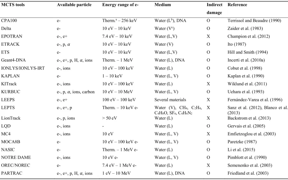
Table 1.1 from Tang (2019) shows the available MCTS simulation tools existing today, the list of particles they can transport, the electron energy range covered, the available biological media, and the capacity of simulating chemical stage. A more detailed review of some of these MCTS codes can be found in Nikjoo et al. (2006). In this thesis, we will focus exclusively on the Geant4-DNA MCTS extension of the Geant4 general purpose and open source MC toolkit.
Table 1.1: List of Monte Carlo track structure codes from Tang (2019).
MCTS tools Available particle Energy range of e- Medium Indirect
damage
Reference
CPA100 e- Therm.a – 256 keV Water (Lb), DNA O Terrissol and Beaudre (1990)
Delta e- 10 eV – 10 keV Water (Vc) O Zaider et al. (1983)
EPOTRAN e-, e+ 7.4 eV – 10 keV Water (L,V) X Champion et al. (2012)
ETRACK e-, p, ! 10 eV – 10 keV Water (V) O Ito (1987)
ETS e- 10 eV – 10 keV Water (L,V) O Hill and Smith (1994)
Geant4-DNA e-, e+, p, H, !, ions Therm. – 1 MeV Water (L), DNA O Incerti et al. (2010a)
IONLYS/IONLYS-IRT e-, ions 10 eV – 100 keV Water (L) O Cobut et al. (1998)
KAPLAN e- 1 – 10 keV Water (L, V) O Kaplan et al. (1990)
KITrack e-, ions 10 eV – 100 keV Water (L) X Wiklund et al. (2011)
KURBUC e-, p, !, ions, carbon 10 eV – 10 MeV Water (L, V) O Uehara et al. (1993)
LEEPS e-, e+ 100 eV – 100 keV Several materials X Fernández-Varea et al. (1996)
LEPTS e-, e+, p Therm. – 10 keV e- Water (V), CH4, C2H4,
C4H8O, SF6, C4H4N2
X Sanz et al. (2012), Blanco et al. (2013)
LionTrack e-, p, ions > 50 eV Water (L) X Backstrom et al. (2013)
LQD e-, ions - Water (L) O Gervais et al. (2005)
MC4 e-, ions 10 eV Water (L, V) X Emfietzoglou et al. (2003)
MOCA8B e- 10 eV – 100 keV e- Water (L, V) O Paretzke (1987)
NASIC e- Therm. – 1 MeV e- Water (L) O Li et al. (2015)
NOTRE DAME e-, ions 10 eV e- Water (L, V) O Pimblott et al. (1990)
OREC/NOREC e- 7.4 eV – 1 MeV e- Water (L) X Semenenko et al. (2003)
PITS04 e-, ions 10 eV Water (L) X Wilson et al. (2004)
PITS99 e-, ions 10 eV Water (V) O Wilson and Nikjoo (1999)
Ptra e-, p, ! 1 eV – 10 keV Water (L, V), DNA X Grosswendt and Pszona (2002)
RADAMOL
(TRIOL/STOCHECO) e-, ions 7.4 eV – 2 MeV Water (L) O Bigildeev and Michalik (1996)
RETRACKS/RITRACKS e-, ions 0.1 eV – 100 MeV Water (L, V) O Plante and Cucinotta (2009)
SHERBROOKE e-, ions 10 eV Water (L, V) O Cobut et al. (2004)
STBRGEN e-, ions 10 eV Water (L, V) O Chatterjee and Holley (1993)
TILDA-V e-, p, H, ions 7.4 eV Water (L, V), DNA X Champion et al. (2005)
TOPAS-nBio e-, e+, p, H, !, ions Therm. – 1 MeV Water (L), DNA O Schuemann et al. (2019)
TRAX/TRAX-CHEM e-, e+, p, ions 1 eV – several MeV Water (V) O Krämer and Kraft (1994)
TRION e-, ions 10 eV Water (L, V) X Lappa et al. (1993)
TRACEL e-, ions 10 eV Water (L, V) O Tomita et al. (1997)
a Therm. indicates the thermalization energy of electron b Liquid phase of water
1.3. The Geant4-DNA project
GEometry ANd Tracking4 (Geant4 - https://geant4.web.cern.ch) is an open source Monte Carlo toolkit developed in C++ language (Agostinelli et al., 2003, Allison et al., 2006, Allison et al., 2016) initiated in 1994 by an international collaboration for the simulation of high energy physics experiments at the CERN Large Hadron Collider, in Switzerland. Thanks to its object-oriented architecture, Geant4 has been progressively extended over the years for various research fields such as astrophysics, nuclear physics, medical physics, and radiation protection. Today, many international groups collaborate and contribute to the development of the toolkit for these various research topics, as illustrated in Figure 1.5.
Figure 1.5: Examples of Geant4 applications. The ATLAS project (left upper), a superficial brachytherapy device and corresponding dose distribution (right upper), a modeling of the CLAS12 detector at Jefferson Lab (left below), and the LISA science module spacecraft (right below). All figures are available in the Geant4 website (https://geant4.web.cern.ch).
The Geant4-DNA project (http://geant4-dna.org), fully included in Geant4, was initially launched in 2001 by the European Space Agency (ESA) in order to provide the community with an open access toolkit to evaluate the biological damage induced by ionizing radiation at the subcellular scale (Incerti et al., 2010a, Incerti et al., 2010b, Bernal et al., 2015, Incerti et al., 2018), in the context of space radiation protection studies. It was the first fully open access MCTS code available freely to the community without considering now the TOPAS-nBio extension of TOPAS (Perl et al., 2012, Schuemann et al., 2019), which in particular wrap Geant4-DNA and Geant4 respectively.
Figure 1.6: Geant4-DNA approach for the simulation of radio-induced biological effects.
Figure 1.6 shows the approach adopted by Geant4-DNA for evaluating DNA damage according to the time evolution and stage. This is a classical approach adopted by other codes (e.g. PARTRAC, KURBUC, TRAX/TRAX-CHEM, etc.). As described above, all the radiobiological stages are available, and the biological damages can be scored according to the source of the damage, direct damage from physical interactions and indirect damage from chemical reactions, respectively. Each stage of Geant4-DNA will be further described in the following paragraphs.
1.3.1. Physical stage
Accurate cross-sections models and descriptions of physical interaction final state (e.g. creation of secondary particles, energy loss and angular deviation of incident particle, etc.) are required for an accurate simulation of step-by-step particle tracking. Especially, low-energy secondary electrons dominantly affect the induction of sub-cellular scale damages (Nikjoo et al., 2016). For instance, inelastic interactions lead to direct energy deposition in the irradiated medium, and elastic interactions influence the energy deposition pattern. One of the main advantages of Geant4-DNA is the possibility to implement several alternative or complementary physics models describing such interactions. This is useful to evaluate the impact of physics models on simulation results. The selection of physics models can be done easily through the usage of "physics constructors" which contain all physics models associated to each particle that Geant4-DNA can handle (e.g. electrons, protons, etc.) for each physical interaction (also called "process", such as ionization, excitation, elastic scattering, etc). Geant4-DNA provides three recommended reference physics constructors
for MCTS simulations in liquid water. These constructors differ only by their electron models (all models for other particles are identical) as shown in Table 1.2. We will describe further the various theoretical or empirical approaches used to calculate such models in Chapter 2, associated references are given for further reading.
Table 1.2: Elastic and inelastic models of electrons employed in Geant4-DNA physics constructors and energy limits of applicability.
Physics constructor Elastic Excitation Ionization Vibrational excitation Attachment
G4EmDNAPhysics_ option2* Champion model (7.4 eV – 1 MeV) (Champion, 2003) Emfietzoglou dielectric model (9 eV – 1 MeV) (Incerti et al., 2010b) Emfietzoglou dielectric model (11 eV – 1 MeV) (Incerti et al., 2010b) Sanche cross-section (2 eV - 100 eV) (Michaud et al., 2003) Melton cross-section (4 eV – 13 eV) (Melton, 1972) G4EmDNAPhysics_ option4* Uehara screened Rutherford model (9 eV – 10 keV) (Uehara et al., 1993) Emfietzoglou-Kyriakou dielectric model (8 eV – 10 keV) (Emfietzoglou et al., 2005) Emfietzoglou-Kyriakou dielectric model (10 eV – 10 keV) (Emfietzoglou et al., 2005) - - G4EmDNAPhysics_ option6* CPA100 model (11 eV – 256 keV) (Bordage et al., 2016) CPA100 model (11 eV – 256 keV) (Bordage et al., 2016) CPA100 model (11 eV – 256 keV) (Bordage et al., 2016) - -
In addition to electrons, Geant4-DNA can simulate physical interactions (processes) for protons and alpha particles including their charged states (H0, H+, He0, He+, He2+). The models for protons and alpha particles are based on the models of Dingfelder et al. (2000). Below 500 keV, the Miller and Green excitation model and Rudd ionization model described in Rudd et al. (1985), Dingfelder et al. (2000) are used. The Born and Bethe theories are used above 500 keV for Rudd et al. (1985), Dingfelder et al. (2000). The model for the charge exchange process (gain or loss of electrons) is also obtained by applying the analytical model of Dingfelder et al. (2000). These models allow to perform simulations in the energy range of 100 eV-100 MeV and 1 keV-400 MeV for protons and alpha particles, respectively. In the case of heavy ions, such as Li, Be, B, C, N, O, Si, Fe, only the discrete ionization model of Booth and Grant (1965) is available (Francis et al., 2011a). Geant4-DNA uses the Livermore physics models for photons, based on the Evaluated Photon Data Library (EPDL97), which is available in Geant4 for the low energy domain (Cullen et al., 1997). All details of Geant4-DNA physics models are well-described in Incerti et al. (2010b), Incerti et al. (2018).
Moreover, Geant4-DNA provides also several examples for evaluating physical quantities which can be used to reproduce previous Geant4-DNA literature results. The list and associated references are shown in Table 1.3.
Table 1.3: List of Geant4-DNA examples available for MCTS simulations in liquid water, taken from Incerti et al. (2018).
Physics
example Purpose Reference
dnaphysics Details of tracking, automatic combination with Geant4 standard electromagnetic physics models Bernal et al. (2015) microdosimetry Combination of Geant4 standard electromagnetic and Geant4-DNA processes and models in different regions Bernal et al. (2015)
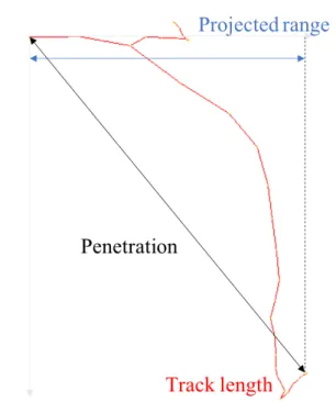
range Range, projected range, penetration Kyriakou et al. (2016)
spower Stopping power Incerti et al. (2017)
mfp Mean-free-path (MFP) Incerti et al. (2018)
wvalue Mean energy required for the creation of an ion pair in liquid water (the so-called "W-value") Kyriakou et al. (2015) svalue Dose to a liquid water target per unit of cumulated activity in a source region (the so-called "S-value") Bernal et al. (2015), André et al. (2014),
Sefl et al. (2015)
slowing Slowing-down electron spectra Incerti et al. (2017)
microyz Microdosimetric distributions (lineal energy y, specific energy z) and related quantities Kyriakou et al. (2017)
TestEm12 Dose point kernel Bernal et al. (2015), Kyriakou et al. (2016),
Bordes et al. (2017) TestEm5 Identification of atomic de-excitation products for Geant4-DNA processes -
1.3.2. Pre-chemical and chemical stages
As the simulation of physical interactions, Geant4-DNA provides a "chemistry constructor" which contains the dissociation probabilities of the ionized and excited water molecules, the list of molecular species, their diffusion coefficients, and the chemical reaction rates. In Geant4-10.3, there was only one chemistry constructor available, "G4EmDNAChemistry", based on the chemistry model of PARTRAC (Kreipl et al., 2009, Karamitros, 2012, Karamitros et al., 2014).
In the pre-chemical stage, the dissociations of molecular species and their probabilities are given according to the excitation and ionization levels of the water molecule as shown in Figure 1.4. And then, the initial positions of molecular species are determined by momentum conservation and empirical root-mean-square distance.
The modeling of chemistry (radiolysis) in Geant4-DNA is based on the SBS approach combining Smoluchowski Brownian diffusion equation (Berg, 1993) describing Brownian motion
and Brownian bridge technique developed by Karamitros (2012), Karamitros et al. (2014). However, the chemistry aspects in reality are continuous. In order to avoid the distortion induced by using discrete steps and to reasonably reduce calculation time, Geant4-DNA dynamically calculates time steps (using the "G4DNAMoleculeEncounterStepper" class) based on an idea initially proposed by Michalik et al. (1998). This technique evaluates the probability of a chemical reaction within the selected statistical confidence (95% confidence level by default). For each time step, the SBS algorithm should find the closest reactant to verify whether the reaction has happened. However, this process requires lots of separation assessments, of order N2 (square of the reactants number). K-d tree algorithm (de Berg et al., 2008) allows to decrease the time complexity from N2 to N×ln(N) based on a space-partitioning technique for organizing points in a k-dimensional space. Chemical reactions occur when two molecular species are closer than the specific reaction radius. After all possible chemical reactions have been processed, molecular species are diffused based on their diffusion coefficients and Brownian diffusion equation. However, due to the discretization of time steps, we should carefully consider possible reactions that can occur during a discrete step. For example, when the two reactants are separated by more than reaction radius at pre-step and post-step point which are the initial and final times of a time step, respectively. It is indeed possible that the separation is less than reaction radius during a time step. In order to account for these possible reactions, Brownian bridge technique is implemented in Geant4-DNA (in the "G4DNASmoluchowskiReactionModel" class).
Several Geant4-DNA examples are provided in order to test water radiolysis simulation and also to evaluate radiochemical yields, so called "G-values" which are the number of molecular species normalized to a deposited energy amount of 100 eV, using SBS approach, as shown in Table 1.4.
Table 1.4: List of Geant4-DNA examples available for water radiolysis simulations updated from Bernal et al. (2015).
Chemistry
example Purpose Reference
chem1 Activation of chemical module Karamitros et al. (2011)
chem2 Selection of time steps Karamitros et al. (2011)
chem3 Visualization of chemical stage as a function of time Karamitros et al. (2011)
chem4 Calculation of G-values as a function of time Karamitros et al. (2011)
chem5 Calculation of G-values as a function of time using specific constructors (released later in Geant4.10.5) Ramos-Mendez et al. (2018)
1.3.3. Geometrical models and DNA damage scoring
Several approaches to calculate DNA damages induced by ionizing radiation using Geant4-DNA have been proposed so far, as shown in Table 1.5. However, those examples (when released in version 10.3 of Geant4) oversimplify the damage scoring, as for example clustering of energy deposition without considering the simulation of full chemistry for the simulation of indirect damage.
In order to more accurately evaluate DNA damages, two geometrical approaches have been later proposed based on two dedicated examples: the dnadamage1 and molecularDNA examples. The dnadamage1 example was developed using the external tool DNAfabric, a C++ software generating complex DNA geometrical models from the nucleotide scale to cell nucleus (Meylan et al., 2016). This example can simulate not only physical interactions of sub-micrometer scale but also chemistry aspects of biomolecules (Meylan et al., 2017). The version released in Geant4 is limited to a segment of chromatin fiber and uses the step-by-step modelling of chemistry. Alternatively, the molecularDNA example developed by Lampe (2017) is also able to simulate both physical and pre-chemical/chemical stages (Lampe et al., 2018a, Lampe et al., 2018b) based on a private version of the IRT approach developed by Karamitros et al. (2020); in this example the geometrical model can be generated using python script, bringing easiness to model different types of geometries such as E. coli bacterium (Lampe et al., 2016). This example has not been released in Geant4. One of the objectives of this thesis is to finalize the development of this example for its future release.
Regarding the simulation of late damage, beyond a few ns, a repair model has not been released yet. Belov et al. (2015) were the first to propose a repair model for human fibroblast cells that can be implemented in Geant4-DNA. The model well-validated by Sakata et al. (2020) will be soon released with the molecularDNA example.
Table 1.5: List of Geant4-DNA examples available for geometrical and damage simulation reproduced updated from Bernal et al. (2015).
Damage example Purpose Reference
wholeNuclearDNA Geometry of the DNA contained in a eukaryotic cell nucleus Dos Santos et al. (2013)
pdb4dna Interface to the Protein Data Bank for the implementation of realistic molecular geometries Delage et al. (2015)
clustering Pattern of energy deposition Francis et al. (2011b)
microbeam Cellular irradiation in single ion mode Incerti et al. (2007)
neuron Simulation of a neural network Belov et al. (2016)
dnadamage1 Geometry generated by DNAfabric tool (Released in Geant4.10.6) Meylan et al. (2017)
molecularDNA Geometry generated by python script (Not released yet) Lampe (2017)
Geant4-DNA is available in open access to the community since 2010, being fully included in Geant4, and every year new developments are distributed in open access. It allows to simulate all the radiobiological stages described above, which can be reused by other simulation tools such as TOPAS-nBio (Ramos-Mendez et al., 2018) or GATE (Pham et al., 2015). However, Geant4-DNA has still important limitations that must be improved:
1) The elastic model, especially for low energy electrons, is difficult to validate because elastic cross-section measurements in liquid water are a technical challenge. Many MCTS tools including Geant4-DNA have taken into account the phase influence between vapour and liquid phase, however, the default elastic scattering model (Champion, 2003) shows deviations with experimental data.
2) The pre-chemical stage of Geant4-DNA is mainly based on that of PARTRAC, which adjusts parameters for matching with experimental G-values (Kreipl et al., 2009). However, Geant4-DNA takes into account additional pre-chemical interactions, such as electron molecular attachment and electron-hole recombination processes, and this leads to disagreements with experimental data.
3) Simulation of radiochemical yields using SBS method is a huge computational burden. For instance, a few days are needed for the proton case performed in the previous study (Meylan et al., 2017).
4) A fully integrated damage simulation chain easily usable at nucleus scale is still missing in Geant4-DNA.
1.4. Purpose of this thesis
The aim of this thesis is to improve Geant4-DNA for a more accurate simulation of DNA damage induced by ionizing radiation, up to late effects. This thesis includes (1) the development of more accurate Geant4-DNA physical and chemical models, and (2) the development of a fully integrated simulation "chain", simultaneously simulating all the radiobiological stages.
For that, at first, we propose to develop a new elastic scattering model for electrons in liquid water in order to accurately simulate the spatial distribution of secondary electrons. The developed model is verified and validated by comparing several physical quantities with experimental and computational results. This is described in Chapter 2. The new elastic scattering model may not impact much on physical quantities because it does not lead to any energy deposition, however, this model could influence the concentration of DNA damages, which has direct correlation with double strand breaks (DSBs), one of the most critical damages (Khanna and Jackson, 2001).
In Chapter 3, the pre-chemical stage of water radiolysis simulation is improved based on the approaches described in the literature. The pre-chemical models such as electron thermalization models, pre-chemical interactions, and dissociation channels employed by several MCTS tools are compared. The influence of the adjustable settings is evaluated for the initial radiochemical yields (which significantly affect the chemical stage), as a function of LET and time, using the new chem6 example developed in this thesis.
Next, in order to reduce the computational burden in the chemical stage, a new implementation of the independent reaction time (IRT) method is proposed in Chapter 4. The validation of this implementation is performed by comparing predictions with experimental data on G-values. This development is a key component of the simulation chain since it will allow to reach sufficient statistics in reasonable calculation time.
Finally, in Chapter 5, the impact of the above developments is evaluated with the prediction of biological damage using the not-yet-released molecularDNA example. Not only SSB and DSB yields but also other quantities, including repair tendency, are calculated using a simplified geometry of a human cell nucleus model. The results are compared with experimental data.
References
Agostinelli, S., Allison, J., Amako, K., et al. 2003, Geant4 - a simulation toolkit. Nucl. Instrum.
Meth. A, 506, 250-303.
Allison, J., Amako, K., Apostolakis, J., et al. 2006, Geant4 developments and applications. IEEE
T. Nucl. Sci., 53, 270-278.
Allison, J., Amako, K., Apostolakis, J., et al. 2016, Recent developments in Geant4. Nucl.
Instrum. Meth. A, 835, 186-225.
André, T., Morini, F., Karamitros, M., et al. 2014, Comparison of Geant4-DNA simulation of S-values with other Monte Carlo codes. Nucl. Instrum. Meth. B, 319, 87-94.
Backstrom, G., Galassi, M. E., Tilly, N., et al. 2013, Track structure of protons and other light ions in liquid water: applications of the LIonTrack code at the nanometer scale. Med.
Phys., 40, 064101.
Belov, O. V., Batmunkh, M., Incerti, S., et al. 2016, Radiation damage to neuronal cells:
Simulating the energy deposition and water radiolysis in a small neural network. Phys.
Med., 32, 1510-1520.
Belov, O. V., Krasavin, E. A., Lyashko, M. S., et al. 2015, A quantitative model of the major pathways for radiation-induced DNA double-strand break repair. J. Theor. Biol., 366, 115-30.
Berg, H. C. 1993. Random walks in biology, New Jersey, Princeton University Press.
Bernal, M. A., Bordage, M. C., Brown, J. M. C., et al. 2015, Track structure modeling in liquid water: A review of the Geant4-DNA very low energy extension of the Geant4 Monte Carlo simulation toolkit. Phys. Med., 31, 861-874.
Bigildeev, E. A. & Michalik, V. 1996, Charged particle tracks in water of different phases. Monte Carlo simulation of electron tracks. Radiat. Phys. Chem., 47, 197-207.
Blanco, F., Muñoz, A., Almeida, D., et al. 2013, Modelling low energy electron and positron tracks in biologically relevant media. Eur. Phys. J. D, 67.
Booth, W. & Grant, I. S. 1965, The energy loss of oxygen and chlorine ions in solids. Nucl. Phys., 63, 481-495.
Bordage, M. C., Bordes, J., Edel, S., et al. 2016, Implementation of new physics models for low energy electrons in liquid water in Geant4-DNA. Phys. Med., 32, 1833-1840.
Bordes, J., Incerti, S., Lampe, N., et al. 2017, Low-energy electron dose-point kernel simulations using new physics models implemented in Geant4-DNA. Nucl. Instrum. Meth. B, 398, 13-20.
Buck, E. C., Wittman, R. S., Skomurski, F. N., et al. 2012. Radiolysis process modeling results for scenarios. PNNL-21554.
Champion, C. 2003, Theoretical cross sections for electron collisions in water: structure of electron tracks. Phys. Med. Biol., 48, 2147-68.
Champion, C., L’Hoir, A., Politis, M. F., et al. 2005, A Monte Carlo code for the simulation of heavy-ion tracks in water. Radiat. Res., 163, 222-231.
Champion, C., Le Loirec, C. & Stosic, B. 2012, EPOTRAN: a full-differential Monte Carlo code for electron and positron transport in liquid and gaseous water. Int. J. Radiat. Biol., 88, 54-61.
Charlton, D. E., Nikjoo, H. & Humm, J. L. 1989, Calculation of initial yields of single- and double-strand breaks in cell nuclei from electrons, protons and alpha particles. Int. J.
Radiat. Biol., 56, 1-19.
Chatterjee, A. & Holley, W. R. 1993, Computer simulation of initial events in the biochemical mechanisms of DNA damage. Adv. Radiat. Biol., 17, 181-226.
Clifford, P., Green, N. J. B., Oldfield, M. J., et al. 1986, Stochastic models of multi-species kinetics in radiation-induced spurs. J. Chem. Soc. Faraday Trans. 1, 82, 2673-2689. Cobut, V., Cirioni, L. & Patau, J. P. 2004, Accurate transport simulation of electron tracks in the
energy range 1 keV–4 MeV. Nucl. Instrum. Meth. B, 215, 57-68.
Cobut, V., Frongillo, Y., Patau, J. P., et al. 1998, Monte Carlo simulation of fast electron and proton tracks in liquid water-I. Physical and physicochemical aspects. Radiat. Phys.
Chem., 51, 229-243.
Cullen, D. E., Hubbell, J. H. & Kissel, L. 1997. EPDL97: the evaluated photo data library `97 version. Lawrence Livermore National Lab., CA (United States).
de Berg, M., van Kreveld, M., Overmars, M., et al. 2008. Computational geometry, Berlin, Heidelberg, Springer.
Delage, E., Pham, Q. T., Karamitros, M., et al. 2015, PDB4DNA: Implementation of DNA geometry from the Protein Data Bank (PDB) description for Geant4-DNA Monte-Carlo simulations. Comput. Phys. Commun., 192, 282-288.
Dingfelder, M. 2012, Track-structure simulations for charged particles. Health Phys., 103, 590-5. Dingfelder, M., Inokuti, M. & Paretzke, H. 2000, Inelastic-collision cross sections of liquid water
for interactions of energetic protons. Radiat. Phys. Chem., 59, 255-275.
Dos Santos, M., Delorme, R., Salmon, R., et al. 2020, Minibeam radiation therapy: A micro- and nano-dosimetry Monte Carlo study. Med. Phys., 47, 1379-1390.
Dos Santos, M., Villagrasa, C., Clairand, I., et al. 2013, Influence of the DNA density on the number of clustered damages created by protons of different energies. Nucl. Instrum.
Meth. B, 298, 47-54.
Emfietzoglou, D., Cucinotta, F. A. & Nikjoo, H. 2005, A complete dielectric response model for liquid water: a solution of the Bethe ridge problem. Radiat. Res., 164, 202-211. Emfietzoglou, D., Karava, K., Papamichael, G., et al. 2003, Monte Carlo simulation of the energy
loss of low-energy electrons in liquid water. Phys. Med. Biol., 48, 2355.
Engels, E., Corde, S., McKinnon, S., et al. 2016, Optimizing dose enhancement with Ta2O5 nanoparticles for synchrotron microbeam activated radiation therapy. Phys. Med., 32, 1852-1861.
Fazel, R., Krumholz, H. M., Wang, Y., et al. 2009, Exposure to low-dose ionizing radiation from medical imaging procedures. New Engl. J. Med., 361, 849-857.
Fernández-Varea, J., Liljequist, D., Csillag, S., et al. 1996, Monte Carlo simulation of 0.1–100 keV electron and positron transport in solids using optical data and partial wave methods. Nucl. Instrum. Meth. B, 108, 35-50.
Francis, Z., Incerti, S., Karamitros, M., et al. 2011a, Stopping power and ranges of electrons, protons and alpha particles in liquid water using the Geant4-DNA package. Nucl.
Instrum. Meth. B, 269, 2307-2311.
Francis, Z., Villagrasa, C. & Clairand, I. 2011b, Simulation of DNA damage clustering after proton irradiation using an adapted DBSCAN algorithm. Comput. Meth. Prog. Bio., 101, 265-70.
Frese, M. C., Yu, V. K., Stewart, R. D., et al. 2012, A mechanism-based approach to predict the relative biological effectiveness of protons and carbon ions in radiation therapy. Int. J.
Radiat. Oncol., 83, 442-50.
Friedland, W., Dingfelder, M., Kundrat, P., et al. 2011, Track structures, DNA targets and radiation effects in the biophysical Monte Carlo simulation code PARTRAC. Mutat.
Res., 711, 28-40.
Friedland, W., Jacob, P., Bernhardt, P., et al. 2003, Simulation of DNA Damage after Proton Irradiation. Radiat. Res., 159, 401-410.
Gervais, B., Beuve, M., Olivera, G. H., et al. 2005, Production of HO2 and O2 by multiple ionization in water radiolysis by swift carbon ions. Chem. Phys. Lett., 410, 330-334. Grosswendt, B. & Pszona, S. 2002, The track structure of alpha-particles from the point of view
of ionization-cluster formation in "nanometric" volumes of nitrogen. Radiat. Environ.
Bioph., 41, 91-102.
Hall, E. J. 2004, Henry S. Kaplan Distinguished Scientist Award 2003. The crooked shall be made straight; dose-response relationships for carcinogenesis. Int. J. Radiat. Biol., 80, 327-37.
Hill, M. A. & Smith, F. A. 1994, Calculation of initial and primary yields in the radiolysis of water. Radiat. Phys. Chem., 43, 265-280.
Hirayama, R., Ito, A., Tomita, M., et al. 2009, Contributions of direct and indirect actions in cell killing by high-LET radiations. Radiat. Res., 171, 212-8.
Hooker, A. M., Bhat, M., Day, T. K., et al. 2004, The linear no-threshold model does not hold for low-dose ionizing radiation. Radiat. Res., 162, 447-52.
Howe, G. R., Zablotska, L. B., Fix, J. J., et al. 2004, Analysis of the mortality experience amongst U.S. nuclear power industry workers after chronic low-dose exposure to ionizing radiation. Radiat. Res., 162, 517-526.
ICRP 1977. Recommendations of the ICRP. Annals of the ICRP.
Incerti, S., Baldacchino, G., Bernal, M., et al. 2010a, The Geant4-DNA project. Int. J. Model.
Simul. Sci. Comput., 1, 157-178.
Incerti, S., Ivanchenko, A., Karamitros, M., et al. 2010b, Comparison of GEANT4 very low energy cross section models with experimental data in water. Med. Phys., 37, 4692-4708.
Incerti, S., Kyriakou, I., Bernal, M., et al. 2018, Geant4-DNA example applications for track structure simulations in liquid water: a report from the Geant4-DNA Project. Med.
Phys., 45, e722-e739.
Incerti, S., Kyriakou, I. & Tran, H. N. 2017, Geant4-DNA simulation of electron slowing-down spectra in liquid water. Nucl. Instrum. Meth. B, 397, 45-50.
Incerti, S., Zhang, Q., Andersson, F., et al. 2007, Monte Carlo simulation of the CENBG microbeam and nanobeam lines with the Geant4 toolkit. Nucl. Instrum. Meth. B, 260, 20-27.
Ito, A. 1987. Calculation of double strand break probability of DNA for low LET radiations based on track structure analysis. Nuclear and Atomic Data for Radiotherapy and Related
Radiobiology. International Atomic Energy Agency (IAEA).
Kaplan, I. G., Miterev, A. M. & Sukhonosov, V. Y. 1990, Simulation of the primary stage of liquid water radiolysis. Radiat. Phys. Chem., 36, 493-498.
Karamitros, M. 2012. Extension de l’outil Monte Carlo généraliste Geant4 pour la simulation de
la radiolyse de l’eau dans le cadre du projet Geant4-DNA. Ph.D. thesis, Université
Bordeaux 1.
Karamitros, M., Brown, J. M. C., Lampe, N., et al. 2020, Implementing the independent reaction time method in Geant4 for radiation chemistry simulations. arXiv preprint
arXiv:2006.14225.
Geant4-DNA. J. Comput. Phys., 274, 841-882.
Karamitros, M., Mantero, A., Incerti, S., et al. 2011, Modeling radiation chemistry in the Geant4 toolkit. Prog. Nucl. Sci. Tech., 2, 503-508.
Khanna, K. K. & Jackson, S. P. 2001, DNA double-strand breaks: signaling, repair and the cancer connection. Nat. Genet., 27, 247-254.
Knight, F. B. 1962, On the random walk and Brownian motion. T. Am. Math. Soc., 103, 218-228. Krämer, M. & Kraft, G. 1994, Calculations of heavy-ion track structure. Radiat. Environ. Bioph.,
33, 91-109.
Kreipl, M. S., Friedland, W. & Paretzke, H. G. 2009, Time- and space-resolved Monte Carlo study of water radiolysis for photon, electron and ion irradiation. Radiat. Environ.
Bioph., 48, 11-20.
Kyriakou, I., Emfietzoglou, D., Ivanchenko, V., et al. 2017, Microdosimetry of electrons in liquid water using the low-energy models of Geant4. J. Appl. Phys., 122.
Kyriakou, I., Incerti, S. & Francis, Z. 2015, Technical Note: Improvements in Geant4 energy-loss model and the effect on low-energy electron transport in liquid water. Med. Phys., 42, 3870-6.
Kyriakou, I., Šefl, M., Nourry, V., et al. 2016, The impact of new Geant4-DNA cross section models on electron track structure simulations in liquid water. J. Appl. Phys., 119. Lampe, N. 2017. The long term impact of ionising radiation on living systems. Ph.D. thesis,
Université Clermont Auvergne, France.
Lampe, N., Biron, D. G., Brown, J. M. C., et al. 2016, Simulating the impact of the natural radiation background on bacterial systems: Implications for very low radiation biological experiments. PLoS ONE, 11, e0166364.
Lampe, N., Karamitros, M., Breton, V., et al. 2018a, Mechanistic DNA damage simulations in Geant4-DNA part 1: A parameter study in a simplified geometry. Phys. Med., 48, 135-145.
Lampe, N., Karamitros, M., Breton, V., et al. 2018b, Mechanistic DNA damage simulations in Geant4-DNA Part 2: Electron and proton damage in a bacterial cell. Phys. Med., 48, 146-155.
Lappa, A. V., Bigildeev, E. A., Burmistorv, D. S., et al. 1993, “Trion” code for radiation action calculations and its application in microdosimetry and radiobiology. Radiat. Environ.
Bioph., 32, 1-19.
Larsen, E. W. & Tolar, D. R. A “Transport” Condensed History Method. In: KLING, A., BARÄO, F. J. C., NAKAGAWA, M., TÁVORA, L. & VAZ, P., eds. Advanced Monte Carlo for Radiation Physics, Particle Transport Simulation and Applications,
2001 Berlin, Heidelberg. Springer, 49-54.
Lazarakis, P., Incerti, S., Ivanchenko, V., et al. 2018, Investigation of track structure and condensed history physics models for applications in radiation dosimetry on a micro and nano scale in Geant4. Biomed. Phys. Eng. Express, 4.
Li, J., Li, C., Qiu, R., et al. 2015, DNA strand breaks induced by electrons simulated with Nanodosimetry Monte Carlo Simulation Code: NASIC. Radiat. Prot. Dosim., 166, 38-43.
Melton, C. E. 1972, Cross sections and interpretation of dissociative attachment reactions producing OH−, O−, and H− in H2O. J. Chem. Phys., 57, 4218-4225.
Metropolis, N. 1987, The beginning of the Monte Carlo method. Los Alamos Science, 15, 125-130.
Meylan, S., Incerti, S., Karamitros, M., et al. 2017, Simulation of early DNA damage after the irradiation of a fibroblast cell nucleus using Geant4-DNA. Sci. Rep., 7, 11923. Meylan, S., Vimont, U., Incerti, S., et al. 2016, Geant4-DNA simulations using complex DNA
geometries generated by the DnaFabric tool. Comput. Phys. Commun., 204, 159-169. Michalik, V., Begusova, M. & Bigildeev, E. A. 1998, Computer-aided stochastic modeling of the
radiolysis of liquid water. Radiat. Res., 149, 224-236.
Michaud, M., Wen, A. & Sanche, L. 2003, Cross sections for low-energy (1–100 eV) electron elastic and inelastic scattering in amorphous ice. Radiat. Res., 159, 3-22.
Mitchell, H. H., Hamilton, T. S., Steggerada, F. R., et al. 1945, The chemical composition of the adult human body and its bearing on the biochemistry of growth. J. Biol. Chem., 158, 625-637.
Mortazavi, S. M. J., Cameron, J. R. & Niroomand-rad, A. 2003, Adaptive response studies may help choose astronauts for long-term space travel. Adv. Space Res., 31, 1543-1551. Nagasawa, H. & Little, J. B. 1992, Induction of sister chromatid exchanges by extremely low
doses of α-particles. Cancer Res., 52, 6394-6396.
NCRP 2009. Ionizing radiation exposure of the population of the United States. NCRP Report
160. (Bethesda, MD:NCRP).
Nias, A. H. W. 1998. An introduction to radiobiology, Chichester ;New York :, Wiley. Nikjoo, H., Charlton, D. E. & Goodhead, D. T. 1994, Monte Carlo track structure studies of
energy deposition and calculation of initial DSB and RBE. Adv. Space Res., 14, 161-180.
response-a review. Rep. Prog. Phys., 79, 116601.
Nikjoo, H. & Girard, P. 2012, A model of the cell nucleus for DNA damage calculations. Int. J.
Radiat. Biol., 88, 87-97.
Nikjoo, H., Uehara, S., Emfietzoglou, D., et al. 2006, Track-structure codes in radiation research.
Radiat. Meas., 41, 1052-1074.
O'Neill, P. & Wardman, P. 2009, Radiation chemistry comes before radiation biology. Int. J.
Radiat. Biol., 85, 9-25.
Paganetti, H., Niemierko, A., Ancukiewicz, M., et al. 2002, Relative biological effectiveness (RBE) values for proton beam therapy. Int. J. Radiat. Oncol., 53, 407-421.
Paretzke, H. G. 1987, Radiation track structure theory. Kinetics of Non-homogeneous Processes. Pelowitz, D. B. 2011, MCNPX user’s manual version 2.7.0-LA-CP-11-00438. Los Alamos
National Laboratory.
Perl, J., Shin, J., Schumann, J., et al. 2012, TOPAS: an innovative proton Monte Carlo platform for research and clinical applications. Med. Phys., 39, 6818-37.
Pham, Q. T., Anne, A., Bony, M., et al. 2015, Coupling of Geant4-DNA physics models into the GATE Monte Carlo platform: Evaluation of radiation-induced damage for clinical and preclinical radiation therapy beams. Nucl. Instrum. Meth. B, 353, 46-55.
Pimblott, S. M., LaVerne, J. A., Mozumder, A., et al. 1990, Structure of electron tracks in water. 1. Distribution of energy deposition events. J. Phys. Chem., 94.
Plante, I. & Cucinotta, F. A. 2009, Cross sections for the interactions of 1 eV–100 MeV electrons in liquid water and application to Monte-Carlo simulation of HZE radiation tracks.
New J. Phys., 11.
Plante, I. & Devroye, L. 2017, Considerations for the independent reaction times and step-by-step methods for radiation chemistry simulations. Radiat. Phys. Chem., 139, 157-172. Prasad, K. N., Cole, W. C. & Haase, G. M. 2004, Radiation protection in humans: extending the
concept of as low as reasonably achievable (ALARA) from dose to biological damage.
Brit. J. Radiol., 77, 97-9.
Ramos-Méndez, J., Domínguez-Kondo, N., Schuemann, J., et al. 2020, LET-Dependent Intertrack Yields in Proton Irradiation at Ultra-High Dose Rates Relevant for FLASH Therapy.
Radiat. Res., 194, 351-362.
Ramos-Mendez, J., Perl, J., Schuemann, J., et al. 2018, Monte Carlo simulation of chemistry following radiolysis with TOPAS-nBio. Phys. Med. Biol., 63, 105014.
by 7-4000-keV protons. Phys. Rev. A, 31, 492-494.
Sakata, D., Belov, O., Bordage, M. C., et al. 2020, Fully integrated Monte Carlo simulation for evaluating radiation induced DNA damage and subsequent repair using Geant4-DNA.
Sci. Rep., 10, 20788.
Sanz, A. G., Fuss, M. C., Munoz, A., et al. 2012, Modelling low energy electron and positron tracks for biomedical applications. Int. J. Radiat. Biol., 88, 71-6.
Schuemann, J., McNamara, A. L., Ramos-Mendez, J., et al. 2019, TOPAS-nBio: An Extension to the TOPAS Simulation Toolkit for Cellular and Sub-cellular Radiobiology. Radiat.
Res., 191, 125-138.
Sefl, M., Incerti, S., Papamichael, G., et al. 2015, Calculation of cellular S-values using Geant4-DNA: The effect of cell geometry. Appl. Radiat. Isotopes, 104, 113-23.
Semenenko, V., Turner, J. & Borak, T. 2003, NOREC, a Monte Carlo code for simulating electron tracks in liquid water. Radiat. Environ. Bioph., 42, 213-217.
Seymour, C. B. & Mothersill, C. 2004, Radiation-induced bystander effects-implications for cancer. Nat. Rev. Cancer, 4, 158-164.
Tang, N. 2019. Évaluation, à partir de modélisations nanodosimétriques, de l’influence de la
compaction de la chromatine sur les effets radio-induits précoces et extension aux effets tardifs (réparation des dommages à l’ADN et mort cellulaire). Ph.D. thesis,
Université de Bordeaux, France.
Terrissol, M. & Beaudre, A. 1990, Simulation of space and time evolution of radiolytic species induced by electrons in water. Radiat. Prot. Dosim., 31, 175-177.
Tomita, H., Kai, M., Kusama, T., et al. 1997, Monte Carlo simulation of physicochemical processes of liquid water radiolysis. Radiat. Environ. Bioph., 36, 105-116. Turner, J. E. 2007. Chemical and Biological Effects of Radiation. Atoms, Radiation, and
Radiation Protection. Wiley.
Turner, J. E., Magee, J. L., Wright, H. A., et al. 1983, Physical and chemical development of electron tracks in liquid water. Radiat. Res., 96, 437-449.
Uehara, S., Nikjoo, H. & Goodhead, D. T. 1993, Cross-sections for water vapour for the Monte Carlo electron track structure code from 10 eV to the MeV region. Phys. Med. Biol., 37, 1841-1858.
UNSCEAR 1994. Sources and effects of ionizing radiation. UNSCEAR Report 1994. New York. UNSCEAR 2000. Sources and effects of ionizing radiation. UNSCEAR Report 2000. New York. Wiklund, K., Fernandez-Varea, J. M. & Lind, B. K. 2011, A Monte Carlo program for the
analysis of low-energy electron tracks in liquid water. Phys. Med. Biol., 56, 1985-2003.
Wilson, W. E., Miller, J. H., Lynch, D. J., et al. 2004, Analysis of low-energy electron track structure in liquid water. Radiat. Res., 161, 591-596.
Wilson, W. E. & Nikjoo, H. 1999, A Monte Carlo code for positive ion track simulation. Radiat.
Environ. Bioph., 38, 97-104.
Wolff, S. 1998, The adaptive response in radiobiology: evolving insights and implications.
Environ. Health Persp., 106, 277-283.
Zaider, M., Brenner, D. J. & Wilson, W. E. 1983, The applications of track calculations to radiobiology I. Monte Carlo simulation of proton tracks. Radiat. Res., 95, 231-247.
Chapter 2
Physical stage
Table of contents
2.1. Development of a new electron elastic scattering cross-section model for Geant4-DNA using ELSEPA for liquid-phase water………...31 2.1.1. Description of potentials and selected options……….……...33 2.1.2. Verification of validity by comparing calculated cross-section with experiments and other cross-section models……….……..38 2.1.3. Results for determination of optimal options and parameters…………..………40 2.1.4. Plausibility of the electron elastic scattering models………...46 2.2. The impact of the ELSEPA electron elastic scattering model on Geant4-DNA simulations………..48 2.2.1. Determination of optimal angle and energy binning in ELSEPA………48 2.2.2. Geant4-DNA examples………...49 2.2.3. Results for the implementation of the elastic scattering cross-section into
Geant4-DNA………...55 2.2.4. Results for the Geant4-DNA simulations………59 2.3. Conclusions………66 References………68
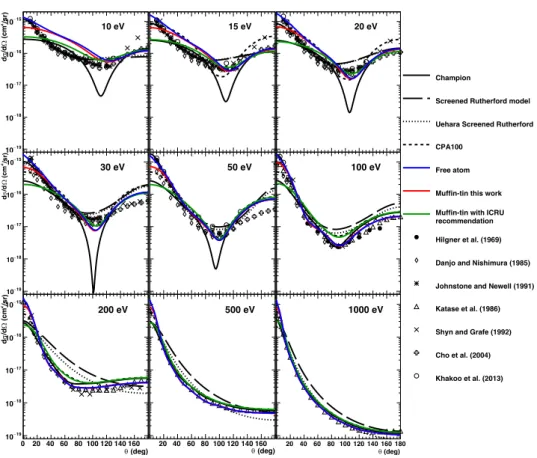
In order to investigate the induction of biological damage from ionizing radiation at the sub-cellular scale, MCTS simulation codes have been developed for several decades (Nikjoo et al., 1997, Uehara et al., 1999, Semenenko et al., 2003, Wilson et al., 2004, Nikjoo et al., 2006, Tung et al., 2007, Incerti et al., 2010a, Friedland et al., 2011, Alloni et al., 2012). These codes usually approximate the biological medium as liquid water, which composes more than 60% of human body (Mitchell et al., 1945). Since most physical damages are caused by secondary electrons, many sets of electron cross-section models for liquid water have been developed so far, for example, see a selection in Dingfelder et al. (1998), Emfietzoglou et al. (2005), Champion et al. (2009), Bordage et al. (2016), Garcia-Molina et al. (2017). In particular, it was reported that the interactions of low energy electrons below 100 eV should be carefully modeled for the prediction of damages to the DNA molecule, which is induced mainly through ionization (Nikjoo et al., 2016). At such low energy, elastic scattering also plays a key role; even if this process is not associated with significant energy loss, it allows to accurately describe the spatial distribution of electrons.
To accurately calculate the elastic scattering cross-section of electrons in liquid water, three main approaches have been proposed: the Born collision model (Mott and Massey, 1965), the non-relativistic (Schrödinger) partial wave model (Schiff, 1968), the non-relativistic (Dirac) partial wave analysis (Vanderpoorten, 1975), and other methods such as the Schwinger multichannel method with pseudopotentials at even lower energies (Varella et al., 1999). It has been reported that the Dirac partial wave method is today the most accurate to calculate such cross-section (Staszewska et al., 1984). Unfortunately, the validation of these calculations, especially elastic scattering in liquid water for low energy electrons, is still not possible due to the scarcity of reliable scattering cross-section data (IAEA, 1995, Incerti et al., 2010b).
However, at least over an incident energy of 60 eV, the calculated differential cross-sections (DCSs) between liquid water and vapour water show good agreement in the entire angle range with discrepancies of up to 1.4 times at 0 deg (Aouchiche et al., 2008). For this reason, the plausibility of calculated DCSs is typically verified by comparison with experimental data in the vapour-phase of water.
In this work, we propose to improve the default electron elastic scattering model of Geant4-DNA, initially developed by Champion et al. (2009), which presents several limitations (Champion, 2003): - This model does not include relativistic corrections;
- At low incident energies, especially below 60 eV, the DCS present too pronounced minima at intermediate scattering angles comparing with experimental data;
- At small scattering angle below 20 deg, the DCS appears underestimated compared with experimental data;