HAL Id: tel-00413972
https://tel.archives-ouvertes.fr/tel-00413972
Submitted on 7 Sep 2009HAL is a multi-disciplinary open access
archive for the deposit and dissemination of sci-entific research documents, whether they are pub-lished or not. The documents may come from teaching and research institutions in France or abroad, or from public or private research centers.
L’archive ouverte pluridisciplinaire HAL, est destinée au dépôt et à la diffusion de documents scientifiques de niveau recherche, publiés ou non, émanant des établissements d’enseignement et de recherche français ou étrangers, des laboratoires publics ou privés.
Control of epileptic seizures by the basal ganglia:
clinical and experimental approaches
Feddersen Berend
To cite this version:
Feddersen Berend. Control of epileptic seizures by the basal ganglia: clinical and experimental ap-proaches. Neurons and Cognition [q-bio.NC]. Université Joseph-Fourier - Grenoble I, 2009. English. �tel-00413972�
1 Ecole Doctorale Chimie
et Sciences du Vivant
U
NIVERSITEJ
OSEPHF
OURIERG
RENOBLEThèse
Neuroscience - Neurobiologie
Berend Feddersen
Control of epileptic seizures by the basal ganglia:
clinical and experimental approaches
Soutenue publiquement le: 10.07.2009
Membres du jury : Franck Semah (Rapporteur 1)
Stephane Charpier (Rapporteur 2)
Philippe Kahane
Soheyl Noachtar
Antoine Depaulis (Directeur de thèse)
ACKNOWLEDGEMENTS
Many fantastic people were inolved in this thesis, whom I want to thank deeply for all their help and support.
I want to thank my supervisors Soheyl Noachtar, Antoine Depaulis and Colin Deransart for all their help and fruitfull discussions in every kind of situation.
Sohyel Nochtar teached me in a perfect structured manner clinical epileptology and gave me always all the support I needed, especially for my stay in Grenoble.
Antoine Depaulis and Colin Deransart were at the origin of the enthusiasm for animal studies in epilepsy. They showed me how exciting stereotactic surgery in small animals can be, and were always present for discussions and helpfull comments on any sort of problems, not only related to my thesis. Sorry for the numerous “last minute corrections”. It is wonderful to have you as friends, thanks a lot also to your families for all the hospitality you gave me.
I thank the reporters of this jury for having agreed to judge this work and for all the comments to improve this thesis.
I wanted to thank also all members of the lab for such a wonderful year in Grenoble, especially Karine Bressand.
Merci beaucoup aussi aux cliniciens et techniciens du laboratoire EEG à Grenoble und in München, für die gute Atmosphäre und Hilfe.
Ralph Meier is thanked for his help and co-work in the field of coherence analysis and EEG-signaling-post-processing.
I want to thank the European Neurological Society (ENS) for the 12 months fellowship and financial support for my stay in Grenoble.
Ein ganz besonderer Dank geht an Pia, meine Frau, die mit viel Verständnis und Unterstützung nicht nur diese Arbeit begleitet hat. Vielen Dank für alles!
3 Bei Edgar, Emil und Anton möchte ich mich für die Zeit entschuldigen, die ich nicht mit Ihnen, sondern am Computer verbracht habe. Ein besonderer Dank geht an Gisela und Felix für das "einhüten" und die ständige Unterstützung zu Hause. Felix, Danke für die aufmunternden Worte und den Glauben daran, dass ich es schaffen kann.
Natürlich haben auch meine Eltern eine riesengrossen Dank verdient. Friddi und Jens, danke, dass ihr mich bei der Umsetzung all meiner Plänen unterstützt habt. Ausserdem habt ihr den Grundstein für die enge Verbundenheit nach Frankreich gelegt. Merci beaucoup à tous.
INDEX
ABBREVIATIONS 9
LIST OF FIGURES AND TABLES 10
RESUME EN FRANÇAIS 11 ENGLISH ABSTRACT 13 PROLOGUE 15 I. INTRODUCTION 17 A. The epilepsies 17 1. Epileptic seizures
1.1. Classification of epileptic seizures 18
1.2. Semiological seizure classification 19
1.3. The epileptogenic zone 23
1.4. Localizing significance of different seizure types 24
1.5. Localizing significance of different seizure evolutions 26
1.6. Ictal lateralizing phenomena 26
2. Epileptic syndromes 29
2.1. Mesial temporal lobe epilepsy (mTLE) 32
2.2. Neocortical temporal lobe epilepsy (nTLE) 32
2.3. Frontal lobe epilepsy (FLE) 32
2.4. Parietal lobe epilepsy (PLE) 33
2.5. Occipital lobe epilepsy (OLE) 33
3. Status epilepticus 33
4. Intractable epilepsy 34
5. Treatment of epilepsy 36
5.1. Pharmacological therapy 36
5.2. Resective surgical therapy 38
5.3. Multiple subpiale transsection 41
5.4. Callosotomie 41
5.5. Neurostimulation 41
5.5.1. Vagus Nerve Stimulation 41
5.5.2. Stimulation of the epileptic focus 42
5.5.3. Deep brain stimulation 44
Thalamus 45
5 B. The basal ganglia
6. Organisation of the basal ganglia 48
6.1. Organisation of basal ganglia circuits 51
6.2. Organisation of functional loop systems within the
basal ganglia 53
7. Involvement of the basal ganglia in the control of epileptic seizures 55
7.1. Animal studies 55 7.1.1. Pharmacology 55 Striatum 55 Subthalamic nucleus 56 Substantia nigra 57 7.1.2. Electrophysiology 58 Striatum 58 Subthalamic nucleus 59 Substantia nigra 60 7.1.3. Metabolism 60
7.1.4. Deep brain stimulation 61
Stimulation of the caudate nucleus 62
Stimulation of the subthalamic nucleus 62
Stimulation of the substantia nigra pars reticulata 63
7.2. Clinical studies 64
7.2.1. Pharmacology 64
7.2.2. Electrophysiology 65
7.2.3. Imaging 66
7.2.4. Deep brain stimulation 68
Stimulation of the caudate nucleus 68
Stimulation of the subthalamic nucleus 69
II. QUESTIONS AND OBJECTIVES 70
1. Does seizure spread to the basal ganglia inhibits secondary
generalization in focal epilepsies? 70
seizure interruption? 71 3. What are the optimal repeated stimulation parameters for sustained
seizure suppression? 71
4. Is it possible to characterize markers that heralds epileptic seizures
into the basal ganglia? 72
The GAERS model 73
III. RESULTS 75
Main findings
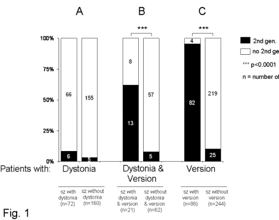
1. B. Feddersen, J. Remi, M. Kilian, L. Vercueil, C. Deransart, A. Depaulis, S. Noachtar, Does ictal dystonia have an inhibitory effect on seizure propagation in focal epilepsies?
Submitted to Epilepsia as a full-length original research article. 76 2. B. Feddersen, L. Vercueil, S. Noachtar, O. David, A. Depaulis, C.
Deransart. Controlling seizures is not controlling epilepsies: a parametric study of deep brain stimulation for epilepsy.
Neurobiology of Disease 2007; 27: 292-300. 77
3. B. Feddersen, R. Meier, A. Depaulis, C. Deransart. EEG Changes Between Left and Right Substantia Nigra Heralds the Occurrence of Generalized Seizures in a model of GAERS.
In preparation, to be submitted to Epilepsia as a short communication.78 4. M. Langlois, S. Saillet, B. Feddersen, L Minotti, L Vercueil,
S Chabardès, O. David, A. Depaulis, P. Kahane, C. Deransart. Deep brain stimulation in epilepsy: experimental and clinical data. In: Deep Brain Stimulation. Mark H. Rogers and Paul B. Anderson
Eds. Nova Science Publishers, Inc. 2009, in press. 79
IV. DISCUSSION 80
1. Question 1: Which epileptic syndromes and seizures are optimal
for deep brain stimulation in epilepsy? 80
Can epileptic candidates for deep brain stimulation be selected
7
- According to the seizure focus? 82
- According to their deficit in dopaminergic functions? 83
2. Question 2: What is the optimal target for deep brain stimulation in
epilepsy? 87
2.1. Anatomical considerations 87
2.1.1. Stimulation of the focus 87
2.1.2. Thalamus stimulation 88
2.1.2.1. Anterior nucleus 88
2.1.2.2. Centromedian nucleus 89
2.1.3. Stimulation of the basal ganglia 89
2.1.3.1. Caudate nucleus 89
2.1.3.2. Subthalamic nucleus 90
2.1.3.3. Substantia nigra pars reticulata 90
2.2. Considerations according to the seizure onset zone 91 2.3. Considerations according to the mechanism of action
of deep brain stimulation 92
3. Question 3: Which are the optimal parameters in deep brain stimulation
3.1. for acute seizure interruption? 93
3.2. for repeated seizure interruption? 95
4. Seizure aggravation by substantia nigra pars reticulata stimulation 97 5. How can seizures be predicted to release a seizure triggered
closed-loop stimulation? 98 V. CONCLUSION 100 VI. PERSPECTIVES 101 1. Animal studies 101 2. Human studies 102
REFERENCES 104 SCIENTIFIC PRODUCTION 121 Article 1 121 Article 2 138 Article 3 148 Article 4 158 Original publications 183 Book chapters 186 Abstracts 187 Letters 191 Oral presentation 191
Grants and Awards 194
9 ABBREVIATIONS
AN - Anterior nucleus (of the thalamus)
CM - Centromedian Thalamus
CN - Caudate nucleus
DBS - Deep brain stimulation
GABA - Gamma-Amino-Butyric-Acid
GAERS - Genetic Absence Epilepsy Rats from Strasbourg
GP - Globus pallidus
GPe - Globus pallidus externus
GPi - Globus pallidus internus
HFS - High-frequency stimulation
ID - ictal dystonia
ILAE - International League Against Epilepsy
SN - Substantia nigra
SNr - Substantai nigra pars reticulata
SNc - Substantia nigra pars compacta
STN - Subthalamic nucleus
MRI - Magnet Resonance Imaging
PET - Positron emission tomography
SPECT - Single photon emission computed tomography
TLE - Temporal lobe epilepsy
mTLE - mesial temporal lobe epilepsy
nTLE - neocortical temporal lobe epilepsy
PLE - Parietal lobe epilepsy
OLE - Occipital lobe epilepsy
RNS - Responsive neuro-stimulation system
SUDEP - Sudden Unexpected Death in Epilepsy
VM - ventro medial (thalamus)
LIST OF FIGURES AND TABLES
Figures :
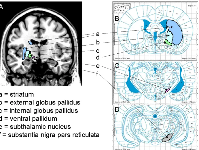
Figure 1: Overview of the basal ganglia in humans and rodents 49
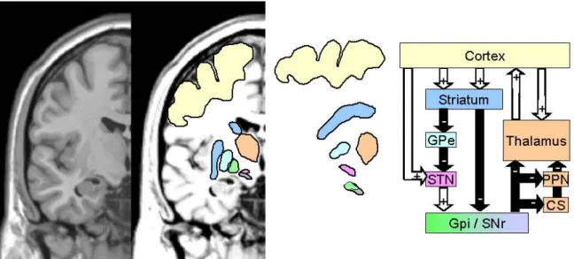
Figure 2: Classical model of the basal ganglia 50
Figure 3: Typically triphasic excitatory-inhibitory-excitatory responses 53 evoked in SNr cells following cortical stimulation
Tables:
Table 1: Classification of seizures by the ILAE 1981 19
Table 2: Semiological seizure classification 22
Table 3: Localizing significance of different seizure types 25
Table 4: Lateralizing seizure phenomena 28
Table 5 : Classification of epileptic syndromes by the ILAE 30
Table 6 : First choice of antiepileptic drugs 37
Table 7 : Overview of the presurgical proceedings 40
Table 8: Recommendation for the selection of patients for 85
DBS in epilepsy
11 RESUME EN FRANÇAIS
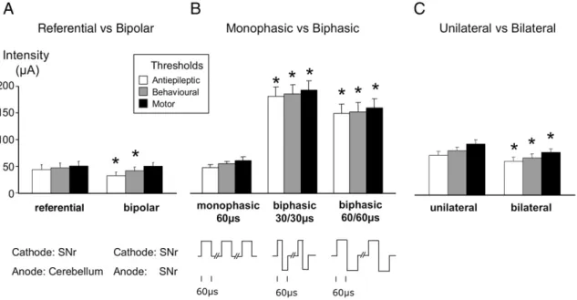
Près de 30% des patients qui souffrent d´une épilepsie sont résistants aux traitements pharmacologiques et seuls 30% de ces patients peuvent bénéficier d’une alternative thérapeutique par résection chirurgicale. La recherche de cibles et stratégies thérapeutiques innovantes constitue un enjeu majeur pour la prise en charge de ces patients. De nombreuses études expérimentales chez l’animal indiquent que les ganglions de la base, et en particulier la substance noire, exercent un contrôle sur la survenue des crises d’épilepsie. Des arguments cliniques, obtenus par électrophysiologie ou imagerie médicale, sont également en faveur de la mise en jeu des ganglions de la base dans certains syndromes épileptiques. Chez des patients souffrant d’épilepsie focale, l’influence de la propagation des crises au travers des ganglions de la base a été examinée en rapport avec le taux de généralisation secondaire. Chez ces patients l’activation des ganglions de la base semble associée à une influence inhibitrice sur la propagation des crises lorsque celles-ci envahissent le lobe frontal. L’exploration de ces mécanismes inhibiteurs des crises est susceptible d’ouvrir de nouvelles perspectives thérapeutiques comme celle portant sur la stimulation intracérébrale profonde. Les premières études de cas explorant les effets de la stimulation intracérébrale des ganglions de la base chez quelques patients ont permis d’obtenir des résultats encourageants chez certains d’entre eux. Cependant de nombreuses études précliniques devraient permettre de préciser les paramètres de stimulation à appliquer. Une approche expérimentale chez l’animal nous a permis de déterminer les paramètres optimaux à appliquer pour controler la survenue de crises spontanées dans un modèle d’épilepsie-absence chez le rat. Dans ce modèle les paramètres optimaux à appliquer à la substance noire réticulée consistent en des stimulations bilatérales, bipolaires, monophasiques, de 60 Hz en fréquence et de 60 µs en largeur d’impulsion. Appliqués de façon répétée, ces paramètres ne permettent cependant pas de supprimer durablement la survenue des crises et ont même tendance à augmenter le nombre de crises enregistrées. Un délais d’au moins 60 seconde entre l’application de deux stimulations consécutives est à respecter pour interrompre les crises. Dans nos conditions, bien qu’une stimulation haute-fréquence de la substance noire réticulée appliquée de façon aigue puisse interrompre une crise en cours, des stimulations répétées semblent inefficaces. Ceci est en faveur du développement en cours, dans de nombreux laboratoire à travers le monde, de procédures de stimulation des crises
asservie à leur détection afin de les supprimer de façon chronique dans le cadre d’applications thérapeutiques. De tels systèmes, dits « adaptatifs », seront particulièrement pertinents s’ils sont couplés à des modifications détectables, signalant l’arrivée d’une crise. Dans le modèle d’épilepsie-absence chez le rat, de telles modifications ont été identifiées au niveau de la cohérence entre signaux électroencéphalographiques issus des deux substances substances noires réticulées. Ces modifications pourraient etre utilisées comme signature spécifique de l’imminence d’une crise dans le couplage de la stimulation à la détection des crises. Toutefois, rien ne permet de dire si ces modifications sont spécifiques du modèle etudié ou encore si de telles modifications existent dans certains syndromes épileptiques en clinique. De nombreux arguments existent pour dire que l’épilepsie n’est pas une pathologie restreinte au seul cortex en tant que circuit générateur de crises, mais implique également des structures sous-corticales susceptibles d’exercer un contrôle à distance sur les circuits générateurs de crises. Cette conception de l’épilepsie permet d’envisager le développement de nouvelles stratégies thérapeutiques pour les patients pharmaco-résistants et qui ne peuvent pas bénéficier d’une intervention chirurgicale.
13 ENGLISH ABSTRACT
As about one third of epileptic patients are resistant to antiepileptic drugs, and only 30% of them are candidates for resective surgery, it exists a great demand for the development of alternative surgical therapies. It has been shown in animal studies, that the basal ganglia and especially the substantia nigra (SN) are involved in the control of epilepsy. Clinical evidence, using either electrophysiological or imaging approaches, also supports the involvement of the basal ganglia in some epileptic syndromes. The influence of seizure spread into the basal ganglia in patients with focal epilepsies was investigated on the rate of secondary generalization. We showed that activation of the basal ganglia was associated with an inhibitory effect on seizure propagation, when seizures spread into the frontal lobe. The elucidation of inhibitory mechanisms in epilepsy may open a new approach for therapeutic
strategies such as electrical deep brain stimulation. First open case series, investigating deep brain stimulation of the basal ganglia to suppress epileptic seizures, showed encouraging results in some patients. However, more preclinical studies are mandatory to investigate the optimal stimulation parameters. The aim of our experimental approach was to determine the optimal stimulation parameters to control spontaneous seizures in a genetic model of absence epilepsy in the rat. In this model, the optimal parameters of single substantia nigra pars reticulata (SNr) stimulation were determined as bilateral, bipolar, monophasic, 60 Hz frequency and 60 µs pulse width. When these parameters were used for repeated stimulations, no long-term suppression and even increase of the number of seizures was observed. A delay of at least 60 sec was necessary between stimulations to be fully effective. Although single high-frequency stimulation of the SNr can be used to suppress ongoing seizures, repeated stimulation are ineffective and could even aggravate seizures, thus supporting the need of closed-loop stimulation procedures to chronically suppress seizures in therapeutical applications. Such an adaptative device would be effective only when detectable changes heralds the seizure onset. In a genetic model of absence epilepsy such changes in the EEG-coherence between the left and right SNr could be identified. Such changes might be used as an hallmark for adaptative procedures like triggered single stimulation to avoid the occurrence of the presumed seizures. To date it remains unknown, if such changes in coherence between left and right SNr, are specific to the model of GAERS and if such changes occur also in other animal models or humans with different epileptic syndromes.
Accumulating evidences support that epilepsy is not a pathology restricted to the cortex as a seizure generator, but that subcortical structures are also involved, which might open new therapeutic options for patients who are pharmacoresistant and no candidates for a resective surgical treatment.
15 PROLOGUE
About one third of epileptic patients are resistant to antiepileptic drugs (AED) (Kwan and Brodie, 2000), and only 30% of them are candidates for resective surgery (Hauser et al., 1990; Semah et al., 1998; Wiebe et al., 2001). Such a therapeutic options is only considered in patients who are suffering from focal seizures, and in whom the epileptic zone can be removed safely. Therefore, there is a great need and interest for “alternative” or “novel” therapeutic options for patients who are
pharmacoresistant and whose seizures arise from eloquent cortices, from multifocal or bilateral seizure onset zones, or who have generalized seizures (Polkey, 2003).
Since the pioneering work of Iadarola and Gale (1982), suggesting a possible
anticonvulsive influence of the SNr (Iadarola and Gale, 1982), the role of structures of the basal ganglia in the pathophysiology of epileptic seizures (Deransart and
Depaulis, 2002) has regained increased interest. This interest was amplified by the encouraging results of deep brain stimulation in different forms of movement
disorders. For more than twenty years, stimulation of different deep brain targets has been shown to be feasible, safe and effective. This has led to its development and application in an increasing number of different neurological and psychiatric
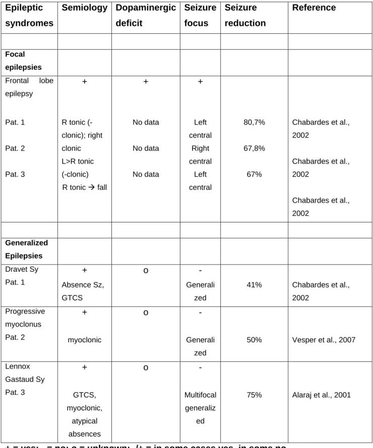
diseases, including epilepsy (Theodore and Fisher, 2004). However, first open case series were only partly effective in some patients (Benabid et al., 2000; Benabid et al., 2002; Chabardes et al., 2002; Fisher et al., 1992; Hodaie et al., 2002; Kerrigan et al., 2004; Loddenkemper et al., 2001; Velasco et al., 2001a; Vesper et al., 2007). This may be due to the often inhomogeneous population investigated with different seizure types and epileptic syndromes, different stimulated targets and different stimulation parameters. Therefore, it appears necessary to address these questions experimentally, before deep brain stimulation may become a real “alternative” or “new” therapy for epileptic patients. The aim should be an appropriate seizure reduction but also an increase in quality of life, which should be superior when compared to other “palliative” therapies such as vagus nerve stimulation.
As it may be important to understand the underlying circuits of epileptic propagation and its involved brain areas, with its posible different targets for acute seizure interruption, a review of different forms of epileptic seizures and syndromes is given in the introduction. Especially, the semiological seizure classification is addressed as
potential tool to choose candidates for deep brain stimulation. Indeed, different
seizure semiology reflects involvement of different activated brain areas and cerebral circuits. This knowledge may be as important as the understanding of the
organization of the basal ganglia, with its different structures involved in a system of endogenous control of epilepsy.
To understand better the possible control of epileptic seizures by the basal ganglia three main questions are presented and discussed in this thesis:
1. Is it possible to confirm the inhibitory role of the basal ganglia in human epilepsies, with impact on the selection of optimal candidates for deep brain stimulation in epilepsy ?
2. What are the optimal deep brain stimulation parameters in epilepsy for interruption of epileptic seizures ?
3. Are there electrophysiological hallmarks that heralds the onset of epileptic seizures into the basal ganglia ?
17 I. INTRODUCTION
A. The epilepsies
Epilepsy is defined by the occurrence of recurring seizures. An epileptic seizure is characterized by sudden onset of rhythmic and synchronized discharges of several neurons, in restricted brain areas or the whole brain. The semiology of seizures is diverse and reflects activation of different cerebral circuits. It depends of: (i) the dimension, (ii) the localization and (iii) the seizure spread to other brain areas. The seizure leads typically to an activation (hallucinations, motor symptoms) and seldom to inhibition of different areas (scotoma, dysphasia, paresis). The seizure frequency is very variable and can range from one in a lifetime to several hundreds a day or even evolve into status epilepticus, with lack of seizure termination (Hufnagel and Noachtar, 2003). However, all seizures are characterized by the hypersynchronous electrical neuronal activity, independent of the underlying etiology.
The prevalence of epilepsies range from 0,5 - 1%. After cerebrovascular disorders, epilepsy is the second most common neurological disorder. The incidence is 5-7 per 10.000 persons per year and is age dependent (Hufnagel and Noachtar, 2003). The rate of primary manifestation is highest in the first two years and declines to the end of the second decade. After the 40th year, it raises again with a second peak after the sixth decade. Idiopathic generalized epilepsies show a classical phase of manifestation in child- and adulthood. Focal epilepsies may become manifest at every age. In infants malformations, neurometabolic and perinatal brain damage are the main causes of epilepsies. Temporal lobe epilepsies manifest often between the age of 5-15 years. In the second to fifth decade, traumas, withdrawl of alcohol, infections and neoplasmas are the main causes. Stroke is the main etiology of new onset epilepsies after the sixth decade.
In human epilepsies, a differentiation is made of the ictal phase (up to several minutes), with alteration of neurological function due to the seizure activity. The postictal phase follows the ictal phase and is characterized by restitution of physiological function. The interictal phase is the time which preceeds the next seizure and may last from a few minutes to several years.
The International League Against Epilepsy (ILAE) recommends to distinguish between focal and primary generalized seizures. Seizures with a focal origin are mainly caused by structural lesions of the brain, whereas primary generalized seizures are often
determinated genetically. In seizures with focal origin, the seizure onset is restricted to a circumscribed brain area and propagation takes place through recruiting of surrounding neurons. Seizure spread by propagation to neighboured and distant brain areas may induce changes in seizure semiology.
Seizures, in which such propagation is limited to a restricted brain area are called simple partial seizures, when conciousness is not altered. The seizure becomes only recognizable, when symptomatic brain regions, with recognizable brain function, is affected.
When both hemispheres or the speech dominant hemisphere are involved,
concioussness is lost during these complex partial seizures. Propagation to the whole brain and involvement of motor areas resulting in secondary generalized tonic clonic seizures. Primary generalized seizures were not believed to have a focal origin and fast (10-20ms) involvement of synchronized discharges in both hemispheres occurs.
Conclusion: Epileptic seizures are the predomiant symptom of epilepsy. It is the second most common neurological disorder. Because of the unpredictability of epileptic seizures, it possess a great burden in daily and quality of life for the patients.
1. Epileptic seizures
1.1. Classification of epileptic seizures
The International League Against Epilepsy (ILAE) introduced a seizure classification in 1981 based on clinical semiology, interictal EEG findings, and ictal EEG patterns (Epilepsy, 1981) (see table 1). The assumption behind such a classification based on electroclinical features, is the existence of a strict one-to-one correlation between
clinical-ictal semiology and interictal EEG findings. However, detailed analysis of clinical semiology and EEG findings shows that this assumption is, particularly for infants
(Acharya et al., 1997), frequently incorrect (Manford et al., 1996). For this reason a classification on seizure semiology was proposed (Lüders et al., 1998; Noachtar et al., 1998). Such a semiological seizure classification stresses the differentiation between epileptic seizures and epileptic syndromes and provides common terms for typical ictal symptoms and types, that are independent of the underlying EEG pattern, as well as other laboratory information.
19 Table 1: Classification of seizures of the ILAE 1981 (Commission on classification 1981) I. Partial seizures
A. simple partial seizures (without alteration of cognition) 1. with motor symptoms
2. with somatosensory symptoms 3. with autonomic symptoms 4. with psychic symptoms
B. complex partial seizures (with alteration of conciousness)
1. simple partial seizures evolving into complex partial seizures
2. complex partial seizure (alteration of conciousness at the beginning) C. partial seizures (simple partial or complex partial) evolving into secondary generalized seizures.
II. Generalized seizures A. Absence seizures 1. typical absence 2. atypical absence B. Myoclonic seizures C. Clonic seizures D. Tonic seizures
E. Primary tonic-clonic seizures F. Atonic seizures
III. Unclassified seizures
adapted from (Epilepsy, 1981)
1.2. Semiological seizure classification
Careful clinical observations and detailed reports of seizure semiology by the patient or other observers have been used since the 18th century to classify epileptic seizures and epileptic syndromes. A detailed analysis of seizure semiology is still essential for the proper management of epileptic patients. Seizures comprise the main symptomatology of patients with epilepsy, and their control is the target of all treatments. The quality of a patient’s life depends in large part on the type of seizures, as well as the frequency. A clear definition of the seizure type is also important for classifying the correct epileptic
syndrome. Syndrome and etiology of the epilepsy are essential factors determining the prognosis, as well as the most effective pharmacological treatment (Benbadis and Lüders, 1996). Furthermore, selection of candidates for deep brain stimulation may be in the future dependent of seizure semiology, localization, seizure evolution and ictal phenomena.
Epileptic seizures are characterized by a variety of signs and symptoms. A seizure is analyzed according to the four categories of seizure symptomatology (Lüders et al., 1998):
(1) sensorial sphere; (2) autonomic sphere; (3) consciousness; (4) motor sphere.
Sensorial sphere: Only a few seizure types involve one of these categories exclusively. One example is an aura, which affects the sensorial sphere without any objective signs. Aura refers to an epileptic seizure during which exclusively subjective symptoms occur without objective signs that can be documented by an observer. Auras are the first clinical expression of a seizure, and therefore, they frequently provide extremely useful localizing information about the seizure-onset zone (Palmini and Gloor, 1992).
Autonomic sphere: Autonomic seizures refer to seizures, in which the predominant symptomatology is an objectively documented alteration of the autonomic system. Autonomic seizures are very rare. They show clear EEG seizure patterns but affect only the autonomic system, such as tachycardia or ictal pallor. Pure ictal tachycardia without any other clinical symptoms is highly correlated with a temporal, rather than
extratemporal, EEG seizure pattern (Weil et al., 2005).
Conciousness: Many seizures are associated with a disturbance of consciousness. However, only in some seizures this reflects the predominant feature of the seizure. The term “dialeptic seizure” for ictal episodes was proposed, in which the main manifestation is an alteration of consciousness, independently of whether the patient has focal or generalized epilepsy (Lüders et al., 1998; Noachtar et al., 1998). Dialeptic seizures consists of an episode of alteration of consciousness, during which a patient cannot react to external stimuli at all, or only to a limited extent, and which they do not recall later. The main feature is the behavioural arrest with starring. Dialeptic seizures occur in several generalized and focal epilepsies (Noachtar et al., 2000). In generalized
21 epilepsies, with the typical 3-Hz per second spike and wave discharges in the EEG, they are also often called “absence” seizures.
Motor sphere: Seizures in which the main manifestations are motor phenomena are called “motor seizures”. Motor seizures are divided into two major groups on the basis of the type of motor symptomatology: simple and complex motor seizures. Simple motor seizures are characterized by unnatural, relatively simple movements that can be
reproduced by electrical stimulation of the primary and supplementary sensorimotor areas. Complex motor seizures consist of motor seizures during which the patient performs movements that imitate natural movements, are relatively complex, and tend to involve different body segments, moving in different planes.
Others: Seizures that cannot be assigned to any of the four groups outlined above, are included in the group labeled “special seizures”. Most of these seizures
characteristically have a “negative” influence on motor (atonic, akinetic) or cognitive (aphasic) activity.
Table 2: Semiological seizure classification (adapted from (Lüders et al., 1998)) I. Epileptic
seizure
Aura Somatosensory aura a
Visual aura a Auditory aura a Olfactory aura Gustatory aura Autonomic aura Epigastric aura Psychic aura Autonomic seizure a Dialeptic seizure
Motor seizure a Simple-motor
seizurea Myoclonic seizure a Epileptic spasma Tonic-clonic seizure Tonic seizurea Clonic seizurea Versive seizurea Complex-motor seizureb Hypermotor seizureb Automotor seizureb Gelastic seizure
Special seizure Atonic seizurea
Astatic seizure Akinetic seizurea Negative myoclonic seizurea Hypomotor seizureb Aphasic seizureb II. Paroxysmal event a Left/right/axial/generalized/bilateral asymmetric.
23 The semiological seizure classification identifies in detail the somatotopic distribution of the ictal semiology as well as the seizure evolution. That gives important clues for the identification of the seizure onset zone as well as the brain areas which are involved during seizure propagation. This is a clinical approach which is not based on
concomitant EEG findings as it is the case in the seizure classification of the ILAE (Epilepsy, 1981). This might be important in the selection of optimal candidates for DBS in epilepsy, as the semiological seizure classifications gives clinical informations of different seizure spread patterns. Such semiological clues might overlap with basal ganglia circuits and therefore patients with different seizures might have different
optimal targets for DBS. The simple motor seizures reflect activation of the primary and premotor areas. Ictal phenomena of motor seizures could be reproduced using subdural stimulation of these motor areas (Lüders et al., 1998). Since the basal ganglia and especially the subthalamic nucleus (STN) and the Substantia nigra pars reticulata (SNr) have strong connections with the primary motor cortex it is possible that patients with seizures involving these frontal areas are likely to be better candidates for seizure suppression by stimulation of the STN or SNr than others.
1.3. The epileptogenic zone
At the cellular level, the primary mechanisms which lead to the synchronous repetitive depolarization of neuronal cells, is still not fully understood. It has been suggested that an imbalance of excitatory and inhibitory neuronal activation would be critical and cause of neuronal excitation evolving into epileptic seizures. Reality may be far more
complicated, even if it has been shown, that increase of excitatory neurotransmitters (like glutamate or aspartate) or reduced inhibitory transmitters (like gamma-amino-butyric-acid (GABA)) play an important role (Baulac et al., 2001; Klepper et al., 2001; Wallace et al., 2001).
The epileptogenic zone is the region from which the epileptic seizure originates (Lüders and Awad, 1992). An epileptic discharge that is limited to the seizure-onset zone does not necessarily lead to clinical symptoms, probably because the epileptogenic zone does not overlap with the symptomatogenic zone (Lüders and Awad, 1992). The term “symptomatogenic zone” refers to the area of the cortex that produces certain clinical symptoms as a result of epileptic activation. For example, seizures that originate in the frontal convexity remain asymptomatic as long as they do not spread into the
symptomatogenic zones. If the epileptic activation reaches the primary motor area, versive or focal clonic seizures occur (Noachtar et al., 2003).
1.4. Localizing significance of different seizure types
The knowledge of the seizure focus may be important not only for resective epilepsy surgery but also for alternative treatment strategies. To date, it is unknown which seizures would be best interrupted by electrical stimulation at different deep brain targets. It is possible that in the future, seizure propagation determines the optimal stimulation target. Therefore localization significance of different seizure types is
mandatory. Analysis of epileptic seizures documented by means of simultaneous video and EEG recordings have dramatically improved our knowledge of epileptic seizure semiology and its localizing significance. This knowledge is important for the definition of epileptic syndromes as well as for the presurgical investigations. Whether it plays also a crucial role in determining optimal candidates for alternative surgical treatments like deep brain stimulation needs to be further elucidated. Table 3 gives an overview of the localizing significance of different seizure types with emphasis on frequent and less frequent localizations:
25 Table 3: Localizing significance of different seizure types
SEIZURE TYPE FREQUENT LOCALIZATION LESS FREQUENT LOCALIZATION REF
Aura Somato-sensory
Contralateral
somatosensory cortex
Supplementary sensorimotor area or the secondary sensory area
a,b Visual Striate and parastriate
cortex
a Auditory Heschl Gyrus Temporal association cortex a,c
Olfactory Amygdala Orbitofrontal part of gyrus rectus d Gustatory Temporal lobe Frontal lobe e Psychic Neocortical temporal lobe Mesial temporal lobe f
Epigastric Insula g,h,i
Autonomic Basal frontal region and cingulate gyrus
a
Autonomic seizure Temporal lobe j
Dialeptic seizure Focal or generalized, involvement of the thalamus
k,l, m
Motor seizure
Simple motor
Myoclonic Primary motor cortex or premotor areas
n Epileptic spasm does not allow localization o
Tonic-clonic primary
motor and the supplementary sensorimotor areas
p
Tonic primary
motor and the supplementary sensorimotor areas
reticular formation of the brainstem and the thalamus (in Lennox Gastaud Syndrome)
q
Clonic Contralateral primary motor or premotor areas
r Versive Frontal eye field that is
contralateral to the side to which the eyes turn
a
Complex motor
Hypermotor Mesial frontal or supplementary sensorimotor area
s,t
Automotor Anterior cingulate gyrus, temporal lobe
Frontal lobe, especially orbitofrontal u,v, w Gelastic Hypothalamus x Special Seizure
Atonic nucleus reticularis gigantocellularis (brainstem)
Negative motor areas y, z Astatic See tonic clonic, myoclonic
and atonic
Akinetic Negative motor areas z
Negative myoclonic
Not yet been defined Hypomotor Mostly temporal and
generalized
Aphasic Cortical language areas in the speech dominan hemisphere
a (Penfield and Jasper, 1954) b (Morris et al., 1988)
c (Luders, 1991) d (Acharya et al., 1998) e (Palmini and Gloor, 1992) f (Ebner, 1994)
g (Rasmussen, 1982) h (Palmini and Gloor, 1992) i (Henkel et al., 2002) j (Weil et al., 2005) k (Kostopoulos, 2001) l (Noachtar et al., 2000) m (Lee et al., 2002)
n (Janz and Christian, 1957) o (Fogarasi et al., 2002) p (Trinka et al., 2002) q (Bleasel and Lüders, 2000) r (Noachtar and Arnold, 2000) s (Morris et al., 1988) t (Williamson et al., 1985) u (Ebner et al., 1995) v (Manford et al., 1996) w (Talairach et al., 1973) x (Berkovic et al., 1988) y (Lai and Siegel, 1988) z (Lüders et al., 1995)
1.5. Localizing significance of different seizure evolutions
Epileptic seizures frequently evolve from one seizure type into another. That reflects the seizure spread with involvement of different brain areas. It is a well-established fact that the initial seizure symptoms provide information on the location of the seizure-onset zone. The evaluation of seizure evolutions is important as there are typical seizure sequences, which point to different epilepsy syndromes. For example, early clonic seizures following manual hand automatisms occur significantly more frequently in patients with neocortical temporal lobe epilepsy than in patients with mesial temporal lobe epilepsy. In contrast, patients with mesial temporal lobe epilepsy showed hand dystonia significantly more often in the course of their seizures than patients with neocortical temporal lobe epilepsy (Pfander et al., 2002). These may be important information, as different distinct epileptic circuits may be sensitive to different new therapeutic approaches such as deep brain stimulation.
1.6. Ictal lateralizing phenomena
Most patients with medically intractable focal epilepsy who are considered for epilepsy surgery show ictal lateralizing phenomena such as dystonic hand posturing, version, ictal vomiting, unilateral clonic seizures, postictal aphasia, and preserved
27 responsiveness during automatisms (Table 4). These phenomena have often not only lateralizing values, but reflect also the spread of epileptic activity in different brain areas. Ictal dystonia
For example ictal limb dystonia (ID) is one of the best lateralizing signs and occurs contralateral to the seizure focus in TLE (Kotagal et al., 1989). It is regarded as a sign of involvement of the basal ganglia, as it was shown that ID is associated with a relative, short-term increase in the perfusion of putamen and caudate nucleus (striatum)
contralateral to the dystonic limb (Mizobuchi et al., 2004; Newton et al., 1992; Shin et al., 2002). Furthermore, in patients with ID, interictal hypometabolism was observed in the striatal region ipsilateral to the seizure focus and contralateral to the dystonic limbs evidenced by fluorodeoxyglucose positron emission tomography (FDG–PET) (Dupont et al., 1998).
Ictal version
Contralateral head versions were shown to typically occur prior to secondary
generalization in patients with focal (mostly temporal) epilepsy (O'Dwyer et al., 2007; Wyllie et al., 1986a). This appears to be due to the propagation of the seizure to the frontal eye field as electrical stimulation of this structure also elicits versive movements of the eyes and head to the opposite side (Godoy et al., 1990). If defined as forced and involuntary head movement resulting in unnatural positioning (Wyllie et al., 1986b), versive head movement displays a high positive predicitve value for localization of a contralateral seizure onset (Bleasel et al., 1997; Chee et al., 1993; Steinhoff et al., 1998). In seizures with secondary generalization, the most common headturning pattern consists of an ipsilateral head turn occurring early in the seizure evolution and before contralateral versive head movement with regard to the hemisphere of seizure onset (Abou Khalil and Fakhoury, 1996). The controversy over the lateralizing significance of head movements may be related to the observation that in many patients in addition to contralateral head version, there is also ipsilateral head turning and no systematic difference has been made as to the sequence of the occurrence and character of the movement (Bleasel et al., 1997; Chee et al., 1993; Ochs et al., 1984; Robillard et al., 1983; Steinhoff et al., 1998). In the study of O´Dwyer, utilizing a quantitative method of movement analysis shows that ipsilateral head movements followed by contralateral head movements, occurring before secondary generalization have an excellent PPV (100%) for lateralization of the seizure onset (O'Dwyer et al., 2007).
Table 4: Lateralizing seizure phenomena Lateralizing seizure
phenomena
Hemisphere Authors
Head and eye version Contralateral (Bleasel et al., 1997; Wyllie et al., 1986a) Dystonic hand posturing Contralateral (Bleasel et al., 1997; Kotagal et al.,
1989)
Figure 4 sign Contralateral (Kotagal et al., 2000)
Automatisms with preserved responsiveness
Nondominant (Ebner et al., 1995; Noachtar et al., 1992)
Ictal speech Nondominant (Gabr et al., 1989)
Postictal aphasia Dominant (Gabr et al., 1989)
Ictal vomiting Nondominant (Krämer, 1997; Kramer et al., 1988)
Ictal spitting Nondominant (Voss et al., 1999)
Periictal urinary urge Nondominant (Baumgartner et al., 2000)
Postictal nose rubbing Ipsilateral (Leutmezer and Baumgartner, 2002)
Postictal coughing Nondominant (Wennberg, 2001)
Unilateral clonic seizure Contralateral (Jackson, 1890) Unilateral tonic seizure Contralateral (Werhahn et al., 2000)
Unilateral eye blinking Ipsilateral (Benbadis et al., 1996; Henkel et al., 1999; Wada, 1980)
Conclusion: The semiological seizure classification of epileptic seizures, the course of seizure evolution and lateralizing signs takes into account the underlying affected brain regions and circuits which may be better localized and identified. This is mandatory for the selection of candidates for “novel” therapeutic approaches, like for deep brain stimulation and more generally for our understanding of the pathophysiology of seizures.
29 2. Epileptic syndromes
An epileptic syndrome is defined by the epileptic seizures occurring in a characteristic manner together with specific etiological, genetical, pathophysiological and
phenomenological factors. In 1989 the ILAE defined different epileptic syndromes, which were differentiated in focal, generalized, neither focal nor generalized and special syndromes. Another discrimination factor was the underlying etiology. Idiopathic
seizures are characterized by a genetic etiology with an age-specific onset.
Symptomatic seizures have a known underlying etiology. When such an etiology is absent and they are not idiopathic they are called kryptogenic seizures. Actually, a revised form of classification of epileptic seizures and syndromes is in process (see also table 5: Classification of epileptic seizures and syndromes by the ILAE (www.ilae.org). All available informations as imaging studies (MRI, PET, SPECT), seizure semiology and genetic analysis are included in the definition of epileptic syndromes.
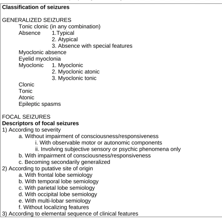
Table 5 : Classification of epileptic syndromes by the ILAE Classification of seizures
GENERALIZED SEIZURES
Tonic clonic (in any combination)
Absence 1.Typical
2. Atypical
3. Absence with special features Myoclonic absence Eyelid myoclonia Myoclonic 1. Myoclonic 2. Myoclonic atonic 3. Myoclonic tonic Clonic Tonic Atonic Epileptic spasms FOCAL SEIZURES
Descriptors of focal seizures 1) According to severity
a. Without impairment of consciousness/responsiveness i. With observable motor or autonomic components
ii. Involving subjective sensory or psychic phenomena only b. With impairment of consciousness/responsiveness
c. Becoming secondarily generalized 2) According to putative site of origin
a. With frontal lobe semiology b. With temporal lobe semiology c. With parietal lobe semiology d. With occipital lobe semiology e. With multi-lobar semiology f. Without localizing features
31 Electro-clinical syndromes and other epilepsies.
Electro-clinical syndromes Neonatal period
Benign familial neonatal seizures (BFNS) Early myoclonic encephalopathy (EME) Ohtahara syndrome
Infancy
Migrating partial seizures of infancy West syndrome
Myoclonic epilepsy in infancy (MEI) Benign infantile seizures
Dravet syndrome
Myoclonic encephalopathy in nonprogressive disorders Childhood
Febrile seizures plus (FS+) (can start in infancy)
Early onset benign childhood occipital epilepsy (Panayiotopoulos type) Epilepsy with myoclonic astatic seizures
Benign childhood epilepsy with centrotemporal spikes (BCECTS) Autosomal-dominant nocturnal frontal lobe epilepsy (ADNFLE) Late onset childhood occipital epilepsy (Gastaut type)
Epilepsy with myoclonic absences Lennox-Gastaut syndrome
Epileptic encephalopathy with continuous spike-and-wave during sleep (CSWS) including: Landau-Kleffner syndrome (LKS)
Childhood absence epilepsy (CAE) Adolescence - Adult
Juvenile absence epilepsy (JAE) Juvenile myoclonic epilepsy (JME)
Progressive myoclonus epilepsies (PME)
Autosomal dominant partial epilepsy with auditory features (ADPEAF) Other familial temporal lobe epilepsies
Epilepsy with generalized tonic-clonic seizures alone Less Specific Age Relationship
Familial focal epilepsy with variable foci (childhood to adult) Reflex epilepsies
Distinctive Constellations
Mesial temporal lobe epilepsy with hippocampal sclerosis (MTLE with HS) Rasmussen syndrome
Gelastic seizures with hypothalamic hamartoma
Epilepsies that do not fit into any of these diagnostic categories can be distinguished first on the basis of the presence or absence of a known structural or metabolic
condition (presumed cause) and then on the basis of the primary mode of seizure onset (generalized versus focal).
Epilepsies attributed to structural-metabolic causes (by cause) Malformations of Cortical development
Tuberous sclerosis complex Tumors Infections Trauma Peri-natal insults Stroke Etc
Epilepsies of unknown cause
Conditions with epileptic seizures that are traditionally not diagnosed as a form of epilepsy per se.
Benign neonatal seizures (BNS) Febrile seizures (FS)
In the following section, a short description of the focal epileptic syndromes which
appear in the paper “Does ictal dystonia have an inhibitory effect on seizure propagation in focal epilepsies?” and in the PhD work is given.
2.1. Mesial temporal lobe epilepsy (mTLE)
Auras occurs in 5% of all patients with mTLE. It is often followed by dialeptic seizures and / or automotor seizures with oral and manual automatisms. Unilateral ictal dystonia occurs when seizure propagates to the basal ganglia. Ictal or postictal aphasia results, when the speech dominant hemisphere is involved. Secondary generalization is seen after seizure spread in the frontal lobe.
2.2. Neocortical temporal lobe epilepsy (nTLE)
may be distinguished from mTLE when auras include complex visual hallucinations, simple or complex acoustic hallucinations, vertigo or speech arrest (by involvement of the speech dominant hemisphere). Secondary generalization may occur after seizure spread in the frontal lobe.
2.3. Frontal lobe epilepsy (FLE)
May originate from the central region resulting in contralateral clonic convulsions, tonic phenomena or negative motor phenomena (Noachtar 1998). The involved topographic region of the central gyrus determinates which body part is affected. The seizure
33 from the supplemental motor parts are short (5-3 s) and are leading to tonic posturing with sudden on- and offset. Hypermotor seizures with bizarre automatisms and heavy motor involvement are seldom but occurs often during sleep. Frontolateral onset leads to an isolated conjugated bulbus deviation to the contralateral side. Seizure duration is short, patients are amnestic for the episode. Premotor origin is charcterized by complex automatisms like body rocking, in some cases versive movements and bilateral
increase of muscle tonus occurs. Fronto orbital originating seizures have a diffuse beginning and ending, sometimes olfactory auras or epigastric auras occurs. Mostly complete amnesia for the event and high tendency of complex motor and whole body automatisms, early ictal urination and seizure spread to the temporal lobe is present.
2.4. Parietal lobe epilepsy (PLE)
have often versive movements, bilateral increase of the muscle tone, contralateral hyp- or paresthesias and acoustic sensations. They are charcterized by seizure spread to the occiptal lobe, temporal lobe or frontal lobe.
2.5. Occipital lobe epilepsy (OLE)
With a seizure onset zone in the occipital lobe results in visual hallucinations in form of flashlights or scotomoas. When associative visual fields are involved this results in more complex visual hallucinations like seeing colours, micropsia, macropsia or strange complex scenes.
Conclusion. Epileptic syndromes are defined by all available informations including
genetic data, etiology, imaging data and seizure semiology. In some of these syndomes
the involvement of the basal ganglia has been described (see section of the Basal Ganglia). This may give additional clues for the selection of patients for DBS in epilepsy (see Discussion).
3. Status epilepticus
Status epilepticus is a medical emergency associated with significant morbidity and mortality. Based on clinical studies showing that single seizures rarely last longer than 5 minutes (Theodore et al., 1994), and on experimental evidence of irreversible neuronal damage caused by prolonged seizures (Meldrum, 1983), it has been suggested that the
operational definition of generalized tonic-clonic status epilepticus should be more than 5 minutes of continuous seizures or two or more discrete seizures between which recovery of conciousness is not regained. Status epilepticus escapes the control of seizure termination. It is currently under debate if seizure termination is due to depletion of activity of the seizure focus or structures maintaining epileptic activity or due to
activation of an endogenous inhibitory system (Lado and Moshe, 2008; Ziemann et al., 2008). In principle, status epilepticus may occur in every epileptic syndrome and in every seizure type. Especially patients with ring chromosome 20 are suffering from many seizures often evolving into status epilepticus. In these patients a bilateral
decrease in 18[F]fluoro-l-DOPA uptake in both putamen and caudate nucleus has been shown and it was suggested that the dysfunction of the striatal dopaminergic
neurotransmission may impair seizure interruption (Biraben et al., 2004). If such involvement of the dopaminergic system are also responsible for the occurrence of status epilepticus in other patients or epileptic syndromes was to date not investigated.
Conclusion: Status epilepticus is a medical emergency, and can be considered as seizure activity that escapes endogenous control mechanisms. In patients with ring chromosome 20, a decrease in uptake of L-DOPA was described in the striatum. If such alterations of the basal ganglia are also involved in other forms of status epilepticus, is to date unknown.
4. Intractable epilepsy
Being able to define and study intractable epilepsy allows one to identify potential underlying causes, some perhaps modifiable, that could result in prevention of intractability and provide insight into the mechanisms and therefore help in the development of new therapeutic approaches. This is especially true for the development of new deep brain stimulation therapies. To date, candidates for such a new treatment are not only pharmacological intractable but also intractable for resective surgery treatments. Common components for a definition of intractability emerge from a number of recent studies in which intractability have been revised (Arts et al., 2004; Berg et al., 2003; Berg et al., 2001; Kwan and Brodie, 2000).
1. AED failure. The concept of “medically refractory” is defined with respect to a minimum number of administered drugs which were ineffective. As it is not
35 possible to test all available drugs in all possible combinations, some minimum threshold number of AED failure must be specified. There seems to be a growing consensus that failure of two drugs places a person in a category where it becomes highly unlikely that further AEDs will be successful to control seizures (Arts et al., 1999; Dlugos, 2001; Wiebe et al., 2001).
2. Seizure occurrence. It is under debate, if there is a minimum frequency at which seizures must occur to be considered intractable (Berg, 2001; Engel, 2004). Others define a minimum seizure remission in order not to qualify as intractable (Arts et al., 1999; Dlugos, 2001; Kwan and Brodie, 2000).
3. Time dimension. For seizure frequency, the seizures must be observed over some period of time. This period might be very variable. Another question is the timing during the course of the disorder. Evaluation is possible in a certain time of remission, or time after last follow up. Neither of these approaches considers the evolution of the seizure disorder up until that time.
An alternative approach might be to consider a constellation of criteria. Once those criteria will be met, the individual is considered intractable, regardless of when during the disorder the criteria are met and regardless of prior or subsequent outcome (Berg, 2001). It is important to note that epilepsy is a very heterogenous disorder with different clinical expressions, different underlying etiologies, different natural histories, and different implications for management and treatments (Berg and Kelly, 2006). For selection of candidates for “alternative” therapies, definition of “weak” criteria of intractability might result in a high probability of “false positive” selected patients.
Which patients are defined as intractable from a “classical” point of view and might be therefore a good candidate for new therapy options like DBS in epilepsy? Patient with focal epilepsies are considered as candidates for such therapies, when they are pharmacoresistant without any option of resective surgical therapy after a presurgical EEG-video-monitoring. This can be caused by multiple seizure onset zones or involvement of eloquent cortex. In generalized epilepsies this might be patients with underlying syndromes who are associated with epileptic encaphalopathies or catastrophic epilepsies, such as West, Lennox Gastaud, Dravet, and Landau-Kleffner syndromes. The seizures are highly refractory from the start and it is rare that these syndromes remit. If these patients are optimal candidates for deep brain stimulation and were they should be stimulated is unknown.
Conclusion: Patients with intractable epilepsies are a heterogenous population with different underlying epileptic syndromes. The selection of optimal candidates for new therapeutic approaches is mandatory, as patients with different syndromes may respond different successful to new therapeutic options.
5. Treatment of epilepsy
Regarding any form of treatments, primary therapy goals needs to be distinguished from secondary therapy goals. Primary therapy goals are to obtain seizure freedom. When this can not be achieved, a reduction of seizure frequency with as less as possible secondary side-effects should be obtained. Secondary therapy goals are the
improvement of the psychosocial situation, especially reintegration into work. Another important goal is the prevention of secondary burden through injuries during the
seizures and progression of illness with further decrease of memory function, as it is the case in hippocampal sclerosis, as well as the prevention of sudden unexpected death during a seizure (SUDEP). Treatment of associated disturbances such as psychiatric disorders is also mandatory. Nevertheless, all pharmacological and surgical treatments should be aimed at obtaining seizure freedom as this is crucial for an increase of the quality of life.
5.1. Pharmacological therapy
Initially, epilepsy therapy should be started as monotherapy according to a focal or generalized origin. Different medications can be choosen as first choice (see table 6).
37 Table 6: First choice of antiepileptic drugs (ordered alphabetically
When monotherapy fails, another antiepileptic drug (AED) should be initiated as monotherapy or combined with a second AED (see table 6). In the combination or “add-on” therapy,a drug should preferentially be choosen with a different mechanism of action. However, that may result in an increase of side effects (Schmidt, 1982). The chance of seizure freedom with the second AED in monotherapy decrease to 10-15% and with the third AED to 4% (Kwan and Brodie, 2000) and is less likely with every new drug independent of the mechanism of action.
Pharmacoresistance is defined by unsufficient seizure control with respect to a
minimum number of AEDs tested (Kwan and Brodie, 2001). In clinical practice, this is the case after failure of 2-3 AEDs in mono- or add-on therapy with maximal tolerable Epilepsies with focal origin
First choice: Carbamazepine Gabapentine Lamotrigine Levetiracetam Oxcarbazepine Phenytoine Topiramate Valproid acid Second choice: Clobazam Gabapentin Pregabalin Tiagabin Zonisamide
Idiopathic generalized epilepsies First choice: Lamotrigine Levetiracetam Phenytoine Topiramate Valproid acid
When one of these five AEDs are contraindicated or incompatible Phenobarbital can be used.
Second choice: Clobazam Phenytoine
dosages. Therefore an evaluation for resective surgical therapy should be initiated in patients with focal epilepsies in a specialized EEG-Video-Monitoring Unit.
5.2. Resective surgical therapy
The basic requirements for a surgical treatment is the exact diagnosis of epilepsy. This also includes that non epileptic seizures are ruled out. Furthermore, it must have been proven that pharmacological treatment is not successful, i.e., that the compliance of the patient was correct and that the AEDs used fit to the epileptic syndrome of the patient. An AED is regarded as not successful after the dosage was increased up to intolerable side effects. In general within two years, especially for TLE, it is possible to prove that a pharmacological treatment was not successful. It has been shown that in TLE the resective surgical treatment is more efficient than a pharmacological treatment (Wiebe et al., 2001). That implements that the presurgical evaluation should be initiated without further delay to avoid neuropsychological and psychosocial burden (Jokeit, 2001) Requirements for a resective surgical treatment are:
- proven diagnosis of epilepsy - pharmacoresistance - disability of the seizures - resectable focus - motivation of the patient
- no progressive etiology (exception Rasmussen encephalitis) - high probability that seizure freedom increases the quality of life.
It has been shown that the prognosis is better when seizure freedom is achieved early. Single seizures may lead to impaired cognition in patients with left sided TLE (Jokeit et al., 2001). Especially in children, it is well accepted that early surgical treatment may have the opportunity to limit neuropsychological and psychosocial disadvantages as it is the case when the time-course to achieve seizure freedom is delayed (Lindsay et al., 1984). The success of a resective surgical treatment depends on how good the epileptogenic zone can be identified and fully removed, without injuries of functional important cortex areas (Awad et al., 1991). If it is not possible to resect the
epileptogenic zone totally, a palliative improvement of seizure severity and frequency can be the aim, even if the results are less favorable (Wyllie et al., 1987).
Another conceptional approach of epilepsy surgery is to interrupt the seizure spread, although the results are less positive compared to a complete resection of the
39 epileptogenic zone (Fish et al., 1991). The transsection of the corpus callosum
(callosotomy) may be helpful to diminish seizures with severe falling as in the Lennox-Gastaud Syndrome. Similarly, subpiale transsections may interrupt seizure spread in Landau-Kleffner syndrome (Morrell et al., 1989). When non-invasive Video-EEG-Monitoring and the analysis of MRI, seizure semiology, EEG and neuropsychology reveals not congruent findings, an invasive EEG monitoring with subdural implanted electrodes or depth electrodes may help to identify the epileptogenic zone properly. Hereby, a cortical stimulation of the implanted electrodes is possible to distinguish seizure onset zones from eloquent cortex, whereas a resective surgical therapy would lead to functional impairment (Rosenow and Luders, 2001). Such overlap of eloquent cortex with seizure onset zones may be one of the reasons to consider new therapeutic approaches like deep brain stimulation. In focal epilepsies, other candidates for such therapies are patients in whom bilateral or multiple seizure onset zones have been detected by invasive EEG monitoring. Therefore, candidates for deep brain stimulation may suffer from different seizure types and epileptic syndromes and do not constitute a homogeneous population.
An overview of the presurgical proceedings is given in table 7.
41 5.3. Multiple subpiale transsection
The technique of multiple subpiale transsection was developped for patients in whom the epileptogenic zone is overlapping with eloquent cortices and resection of the focus is not possible (Morrell et al., 1989). The concept is to cut the horizontal cortical
connections which are responsible for seizure propagation and to preserve the vertical descending axons to minimize the postoperative functional deficit (Kaufmann et al., 1996). In patients with multiple subpiale transsection plus resection, excellent outcome (>95% reduction in seizure frequency) was obtained in 87% of patients for generalized seizures, 68% for complex partial seizures, and 68% for simple partial seizures. For the patients who underwent multiple subpiale transsection without resection, the rate of excellent outcome was only slightly lower, at 71% for generalized, 62% for complex partial, and 63% for simple partial seizures (Spencer et al., 2002).
5.4. Callosotomie
Transsection of the corpus callosum (callosotomy) may be considered for patients with medically intractable generalized epilepsies, suffering from a combination of different seizures. These include often tonic, atonic, generalized tonic clonic, absence and seldom focal seizures (Wyler, 1993). A realistic aim of that kind of surgery is the reduction of severe seizures, not the achievement of seizure freedom. Callosotomy often leads to a reduction of tonic and atonic seizures, known to be associated with falls and secondary injuries (Gates et al., 1984). Most of these patients suffer from mental retardation and have Lennox Gastaud Syndrome. A stepwise approach is considered, with a 2/3 transection of the anterior part of the corpus callosum in the first step. When lacking a clear benefit, a complete callosotomy can be performed without risk of a so-called “disconnection syndrome” (Wilson et al., 1982).
5.5. Neurostimulation
5.5.1. Vagus Nerve Stimulation (VNS)
Vagus nerve stimulation is to date the only approved stimulation therapy by the FDA (Amar et al., 2008) as it was proven to lead to a reduction in seizure frequency in generalized as well as in focal epilepsies. A seizure reduction up to 50% is reported in the 30 to 50% of the patients treated with VNS for several months or years (Ben Menachem et al., 1999). In about 10% of patients, a seizure reduction of more than 80% was observed (Ramsay et al., 1994). The antiepileptic effect is thought to be
transmitted through the nucleus tractus solitarius and parabrachial nucleus which
projects to the limbic, autonomic, and reticular structures of the forebrain (Henry, 2002). It has been correlated to changes in the the noradrenergic and serotoninergic
neurotransmission systems of the brain and spinal cord, the amygdala, insula, hypothalamus, periaqueductal grey matter, and the thalamus (Vonck et al., 2001). These widespread, bilateral, and multisynaptic projections account for the possible multiple therapeutic mechanisms leading to a change in neurotransmission systems which takes place over months. Therefore, this technique is not an acute interruption of ongoing seizures but implements a chronic change which leads to the described
changes. VNS is reasonable in patients who are not candidates for a resective surgical treatment. During the stimulation period hoarseness, local paresthesias, dyspnae and coughing may occur, but can be reduced by lowering the stimulation intensity. It has also been shown that VNS is leading to antidepressant and activation processes, which is probably due to the changes in serotinergic and noradrenergic neurotransmissions. This lead to initiate trials in pharmacoresistant depressive patients (Schlaepfer et al., 2008). As side effects are very low, it was proposed to perform VNS prior to
callosotomy. However, seizure freedom is rarely achieved with VNS probably because candidates are mainly patients suffering from severe epileptic syndromes with a long history of seizures (Amar et al., 2008). Because surgery and preclinical evaluation are easy for VNS and because this therapy has lower rates of complications, new
stimulation paradigms for deep brain stimulation which are certainly more risky, required better results than VNS. As VNS therapy is to date the only approved electrical
stimulation therapy for epilepsy by the FDA, new therapies should be compared with this “gold standard” and aim to lead to seizure freedom. Seizure freedom is achieved in VNS therapie in very few patients but is important as it would result in substantially increase of quality of life.
5.5.2. Stimulation of the epileptic focus
Stimulation of the epileptic focus has been performed in the hippocampus or the cortex. Hippocampal stimulation was carried out in 10 patients who had undergone implantation of subdural or depth electrodes in the temporal region for investigation prior to temporal lobectomy. Bipolar high frequency stimulation at 130 Hz was applied continuously for 2-3 weeks. In 7 patients, complex partial and secondarily generalized seizures were