HAL Id: dumas-02139246
https://dumas.ccsd.cnrs.fr/dumas-02139246
Submitted on 24 May 2019HAL is a multi-disciplinary open access archive for the deposit and dissemination of sci-entific research documents, whether they are pub-lished or not. The documents may come from teaching and research institutions in France or abroad, or from public or private research centers.
L’archive ouverte pluridisciplinaire HAL, est destinée au dépôt et à la diffusion de documents scientifiques de niveau recherche, publiés ou non, émanant des établissements d’enseignement et de recherche français ou étrangers, des laboratoires publics ou privés.
canal anal
Émilien Bertin
To cite this version:
Émilien Bertin. Efficacité et tolérance de la curiethérapie haut débit de dose en surimpression dans le carcinome épidermoïde du canal anal. Human health and pathology. 2018. �dumas-02139246�
THESE D’EXERCICE DE MEDECINE
Pour l’obtention du Diplôme d’Etat de Docteur en Médecine
Présentée et soutenue publiquement à la faculté de médecine de Nice le Jeudi 13 Décembre 2018
par
Emilien BERTIN
Né le 2 Février 1993 à Pointe-à-Pitre
Discipline : Oncologie Radiothérapie
Président du Jury :
Monsieur le Professeur Jean-Michel HANNOUN-LEVI
Assesseurs :
Madame le Docteur Karen BENEZERY Monsieur le Professeur Emmanuel BENIZRI Monsieur le Docteur Jérôme DOYEN
Monsieur le Docteur Eric FRANCOIS
Directeur de Thèse :
Monsieur le Docteur Alexander FALK
EFFICACITE ET TOLERANCE DE LA
CURIETHERAPIE
HAUT DEBIT DE DOSE EN SURIMPRESSION DANS
LE CARCINOME EPIDERMOIDE DU CANAL ANAL
Liste des enseignants au 1er septembre 2018 à la Faculté de Médecine de Nice
Doyen Pr. BAQUÉ
Patrick Vice-doyens
Pédagogie Pr. ALUNNI Véronique Recherche Pr DELLAMONICA jean Etudiants M. JOUAN Robin Chargé de mission projet Campus Pr. PAQUIS Philippe
Conservateur de la bibliothèque Mme AMSELLE Danièle
Directrice administrative des services Mme CALLEA Isabelle
Doyens Honoraires M. AYRAUD Noël M. RAMPAL Patrick M. BENCHIMOL Daniel
Liste des enseignants au 1er septembre 2018 à la Faculté de Médecine de Nice
PROFESSEURS CLASSE EXCEPTIONNELLE
M. AMIEL Jean Urologie (52.04)
M. BAQUÉ Patrick Anatomie - Chirurgie Générale (42.01) M. BERNARDIN Gilles Réanimation Médicale (48.02)
M. BOILEAU Pascal Chirurgie Orthopédique et Traumatologique (50.02) M. DARCOURT Jacques Biophysique et Médecine Nucléaire (43.01)
M. ESNAULT Vincent Néphrologie (52-03)
M. FENICHEL Patrick Biologie du Développement et de la Reproduction (54.05)
M. FUZIBET Jean-Gabriel Médecine Interne (53.01) M. GILSON Éric Biologie Cellulaire (44.03) M. GUGENHEIM Jean Chirurgie Digestive (52.02) M. HASSEN KHODJA Reda Chirurgie Vasculaire (51.04) M. HÉBUTERNE Xavier Nutrition (44.04)
M. HOFMAN Paul Anatomie et Cytologie Pathologiques (42.03)
Mme ICHAI Carole Anesthésiologie et Réanimation Chirurgicale (48.01) M. LACOUR Jean-Philippe Dermato-Vénéréologie (50.03)
M. LEFTHERIOTIS Geogres Chirurgie vasculaire ; médecine vasculaire (51.04) M. MARQUETTE Charles-Hugo Pneumologie (51.01)
M. MARTY Pierre Parasitologie et Mycologie (45.02)
M. MICHIELS Jean-François Anatomie et Cytologie Pathologiques (42.03) M. MOUROUX Jérôme Chirurgie Thoracique et Cardiovasculaire (51.03) Mme PAQUIS Véronique Génétique (47.04)
M. PAQUIS Philippe Neurochirurgie (49.02)
M. QUATREHOMME Gérald Médecine Légale et Droit de la Santé (46.03) M. RAUCOULES-AIMÉ Marc Anesthésie et Réanimation Chirurgicale (48.01) M. ROBERT Philippe Psychiatrie d’Adultes (49.03)
M. SANTINI Joseph O.R.L. (55.01)
M. THYSS Antoine Cancérologie, Radiothérapie (47.02) M. TRAN Albert Hépato Gastro-entérologie (52.01)
Liste des enseignants au 1er septembre 2018 à la Faculté de Médecine de Nice
PROFESSEURS PREMIERE CLASSE
Mme ASKENAZY-GITTARD Florence Pédopsychiatrie (49.04)
M. BARRANGER Emmanuel Gynécologie Obstétrique (54.03) M. BÉRARD Étienne Pédiatrie (54.01)
Mme BLANC-PEDEUTOUR
Florence Cancérologie – Génétique (47.02) M. BONGAIN André Gynécologie-Obstétrique (54.03) Mme BREUIL Véronique Rhumatologie (50.01)
M. CASTILLO Laurent O.R.L. (55.01)
M. CHEVALLIER Patrick Radiologie et Imagerie Médicale (43.02) M. DE PERETTI Fernand Anatomie-Chirurgie Orthopédique (42.01) M. DRICI Milou-Daniel Pharmacologie Clinique (48.03)
M. FERRARI Émile Cardiologie (51.02)
M. FERRERO Jean-Marc Cancérologie ; Radiothérapie (47.02) M. FONTAINE Denys Neurochirurgie (49.02)
M. GIBELIN Pierre Cardiologie (51.02)
M. HANNOUN-LEVI Jean-Michel Cancérologie ; Radiothérapie (47.02)
M. LEVRAUT Jacques Anesthésiologie et Réanimation Chirurgicale (48.01) M. LONJON Michel Neurochirurgie (49.02)
M. MOUNIER Nicolas Cancérologie, Radiothérapie (47.02) M. PADOVANI Bernard Radiologie et Imagerie Médicale (43.02) M. PICHE Thierry Gastro-entérologie (52.01)
M. PRADIER Christian Épidémiologie, Économie de la Santé et Prévention (46.01)
Mme RAYNAUD Dominique Hématologie (47.01) M. ROSENTHAL Éric Médecine Interne (53.01) M. SCHNEIDER Stéphane Nutrition (44.04)
M. STACCINI Pascal Biostatistiques et Informatique Médicale (46.04) M. THOMAS Pierre Neurologie (49.01)
Liste des enseignants au 1er septembre 2018 à la Faculté de Médecine de Nice
PROFESSEURS DEUXIEME CLASSE
Mme ALUNNI Véronique Médecine Légale et Droit de la Santé (46.03) M. ANTY Rodolphe Gastro-entérologie (52.01)
M. BAHADORAN Philippe Cytologie et Histologie (42.02) Mme BAILLIF Stéphanie Ophtalmologie (55.02)
Mme BANNWARTH Sylvie Génétique (47.04)
M. BENIZRI Emmanuel Chirurgie Générale (53.02) M. BENOIT Michel Psychiatrie (49.03)
M. BOZEC Alexandre ORL- Cancérologie (47.02) M. BREAUD Jean Chirurgie Infantile (54-02)
M. CHEVALIER Nicolas Endocrinologie, Diabète et Maladies Métaboliques (54.04)
Mme CHINETTI Giulia Biochimie-Biologie Moléculaire (44.01) M. CLUZEAU Thomas Hématologie (47.01)
M. DELLAMONICA Jean réanimation médicale (48.02) M. DELOTTE Jérôme Gynécologie-obstétrique (54.03) M. FOURNIER Jean-Paul Thérapeutique (48-04)
Mlle GIORDANENGO Valérie Bactériologie-Virologie (45.01) Mme GIOVANNINI-CHAMI Lisa Pédiatrie (54.01)
M. GUÉRIN Olivier Gériatrie (48.04)
M. IANNELLI Antonio Chirurgie Digestive (52.02)
M. ILIE Marius Anatomie et Cytologie pathologiques (42.03) M JEAN BAPTISTE Elixène Chirurgie vasculaire (51.04)
M. PASSERON Thierry Dermato-Vénéréologie (50-03)
M. ROGER Pierre-Marie Maladies Infectieuses ; Maladies Tropicales (45.03) M. ROHRLICH Pierre Pédiatrie (54.01)
M. ROUX Christian rhumatologie (50.01)
M. RUIMY Raymond Bactériologie-virologie (45.01) Mme SACCONI Sabrina Neurologie (49.01)
M. SADOUL Jean-Louis Endocrinologie, Diabète et Maladies Métaboliques (54.04)
Liste des enseignants au 1er septembre 2018 à la Faculté de Médecine de Nice
MAITRES DE CONFÉRENCES DES UNIVERSITÉS - PRATICIENS HOSPITALIERS M. M. Mm e M. AMBROSETTI Damien BENOLIEL José BERNARD-POMIER Ghislaine BRONSARD Nicolas Cytologie et Histologie (42.02)
Biophysique et Médecine Nucléaire (43.01) Immunologie (47.03)
Anatomie Chirurgie Orthopédique et Traumatologique (42.01)
Mme BUREL-VANDENBOS Fanny Anatomie et Cytologie pathologiques (42.03) M. DOGLIO Alain Bactériologie-Virologie (45.01)
M DOYEN Jérôme Radiothérapie (47.02) M FAVRE Guillaume Néphrologie (52.03)
M. FOSSE Thierry Bactériologie-Virologie-Hygiène (45.01) M. GARRAFFO Rodolphe Pharmacologie Fondamentale (48.03) Mme HINAULT Charlotte Biochimie et biologie moléculaire (44.01) M. HUMBERT Olivier Biophysique et Médecine Nucléaire (43.01) Mme LAMY Brigitte Bactérilogie-virologie ( 45.01)
Mme LONG-MIRA Elodie Cytologie et Histologie (42.02) Mme MAGNIÉ Marie-Noëlle Physiologie (44.02)
Mme MOCERI Pamela Cardiologie (51.02) M. MONTAUDIE Henri Dermatologie (50.03)
Mme MUSSO-LASSALLE Sandra Anatomie et Cytologie pathologiques (42.03) M. NAÏMI Mourad Biochimie et Biologie moléculaire (44.01) Mme POMARES Christelle Parasitologie et mycologie (45.02)
M. SAVOLDELLI Charles Chirurgie maxillo-faciale et stomatologie (55.03) Mme SEITZ-POLSKI barbara Immunologie (47.03)
M. TESTA Jean Épidémiologie Économie de la Santé et Prévention (46.01)
Liste des enseignants au 1er septembre 2018 à la Faculté de Médecine de Nice
PROFESSEUR DES UNIVERSITÉS
M. HOFLIGER Philippe Médecine Générale (53.03)
MAITRE DE CONFÉRENCES DES UNIVERSITÉS
M. DARMON David Médecine Générale (53.03)
PROFESSEURS AGRÉGÉS
Mme LANDI Rebecca Anglais
PRATICIEN HOSPITALIER UNIVERSITAIRE
M. DURAND Matthieu Urologie (52.04)
PROFESSEURS ASSOCIÉS
M. GARDON Gilles Médecine Générale (53.03) Mme MONNIER Brigitte Médecine Générale (53.03)
MAITRES DE CONFÉRENCES ASSOCIÉS
Mme CASTA Céline Médecine Générale (53.03) M. GASPERINI Fabrice Médecine Générale (53.03) M. HOGU Nicolas Médecine Générale (53.03)
Liste des enseignants au 1er septembre 2018 à la Faculté de Médecine de Nice
Constitution du jury en qualité de 4ème membre Professeurs Honoraires
M ALBERTINI Marc M. GÉRARD Jean-Pierre M. BALAS Daniel M. GILLET Jean-Yves M. BATT Michel M. GRELLIER Patrick M. BLAIVE Bruno M. GRIMAUD Dominique M. BOQUET Patrice M. HARTER Michel M. BOURGEON André M. JOURDAN Jacques M. BOUTTÉ Patrick M. LAMBERT Jean-Claude M. BRUNETON Jean-Noël M. LAZDUNSKI Michel Mme BUSSIERE Françoise M. LEFEBVRE Jean-Claude M. CAMOUS Jean-Pierre M. LE FICHOUX Yves M. CANIVET Bertrand Mme LEBRETON Elisabeth M. CASSUTO Jill-patrice M. LOUBIERE Robert M. CHATEL Marcel M. MARIANI Roger M. COUSSEMENT Alain M. MASSEYEFF René Mme CRENESSE Dominique M. MATTEI Mathieu M. DARCOURT Guy M. MOUIEL Jean M. DELLAMONICA Pierre Mme MYQUEL Martine M. DELMONT Jean M. ORTONNE Jean-Paul M. DEMARD François M. PRINGUEY Dominique M. DESNUELLE Claude M. SAUTRON Jean Baptiste M. DOLISI Claude M. SCHNEIDER Maurice Mme EULLER-ZIEGLER Liana M. TOUBOL Jacques M . FRANCO Alain M. TRAN Dinh Khiem
M. FREYCHET Pierre M VAN OBBERGHEN Emmanuel M. GASTAUD Pierre M. ZIEGLER Gérard
M.C.U. Honoraires
M. ARNOLD Jacques M. GIUDICELLI Jean M. BASTERIS Bernard M. MAGNÉ Jacques Mlle CHICHMANIAN Rose-Marie Mme MEMRAN Nadine Mme DONZEAU Michèle M. MENGUAL Raymond M. EMILIOZZI Roméo M. PHILIP Patrick M. FRANKEN Philippe M. POIRÉE Jean-Claude M. GASTAUD Marcel Mme ROURE Marie-Claire
Remerciements
A Monsieur le Professeur Jean-Michel HANNOUN-LEVI
Je vous remercie de m’avoir fait l’honneur de présider ce jury.
Merci pour vos enseignements, votre accompagnement quotidien et votre écoute qui contribuent à la qualité de notre formation.
A mon directeur de thèse, Monsieur le Docteur Alexander FALK
Je te remercie d’avoir accepté de diriger ma thèse. C’est grâce à toi que ce travail a vu le jour, merci pour ton aide et le temps que tu y as consacré. Ton implication, tes
connaissances et ta volonté de transmettre nous sont indispensables !
A Madame le Docteur Karen BENEZERY
Je te remercie d’avoir accepté d’être membre de mon jury. Je suis très heureux de pouvoir travailler et apprendre à tes côtés. Tes connaissances, ta bonne humeur et ta gentillesse sont des plus appréciables et essentielles au service !
A Monsieur le Professeur Emmanuel BENIZRI
C’est à vos côtés que j’ai découvert l’univers de la chirurgie, l’atmosphère du bloc opératoire, que j’apprécie retrouver en curiethérapie. Vous avez également accepté de juger ce travail, veuillez trouver ici l’expression de ma profonde reconnaissance.
A Monsieur le Docteur Jérôme DOYEN
Ich danke dir, an der Jury für meine Doktorarbeit teilgenommen zu haben. Merci pour tes enseignements et ton partage de connaissances, avec un grand S. Nous attendons ton retour avec impatience !
A Monsieur le Docteur Eric FRANCOIS
Vous m’avez encouragé à m’intéresser à l’oncologie digestive et continuez à cultiver ma curiosité pour cette spécialité, que j’étudie avec plaisir. Vous avez accepté d’être membre de mon jury, veuillez trouver ici l’expression de mes sincères remerciements.
Table des matières Première partie
Résumé de l’article Page 12
Deuxième partie
Article publié Page 13
Introduction Page 14
Matériel et méthode Page 14
Résultats Page 18
Discussion et conclusion Page 22
Références Page 24
Annexe Page 27
Abréviations Page 31
Résumé de l’article
Objectif de l’étude : Evaluer l’efficacité et la toxicité d’une surimpression par curiethérapie de haut débit de dose (HDD) dans les carcinomes épidermoïdes de l’anus (CEA).
Matériel et Méthode : Etude rétrospective monocentrique portant sur des patients traités par irradiation externe (+/- chimiothérapie), avec surimpression en curiethérapie HDD, pour un CEA localisé. La surveillance clinique a été réalisée tous les six mois. Les résultats oncologiques ont été analysés avec : les survies sans récidive locale (SSRL), sans
colostomie (SSC), sans métastase (SSM), sans évènement (SSE) et globale (SG). Les toxicités aigues et tardives ont été recueillies (CTCV4.0) et un questionnaire dérivé du score SOMA / LENT a été proposé aux patients.
Résultats : De mai 2005 à janvier 2018, 46 patients (pts) ont été analysés. La médiane de suivi était de 61 mois (10-145 mois), l’âge médian de 65 ans (34-84 ans) avec un sexe ratio M/F= 0,24. La classification TNM était : T1 13 pts (21,7%), T2 34 pts (73,9%), T3 deux pts (4,3%), N+ six pts (13,1%). La radiothérapie externe (RTE) a délivré une dose médiane de 45 Gy (36-50.4 Gy) en 25 fractions et la curiethérapie HDD de 12Gy (10-18 Gy) en trois fractions. L’étalement médian était de 58 jours (41-101 jours) avec un intervalle médian RTE / curiethérapie de 17 jours (4-60 jours). Les résultats carcinologiques
montraient des taux à cinq ans de SSRL 81,2%, SSM 88,7%, SSE 70% et SG 90%. Toutes les amputations abdomino-périnéales ont été réalisées en cas de rechute locale
(4 pts - 8,7%) conduisant à une SSC à cinq ans de 79,5%. Les toxicités urinaires aigues étaient fréquentes (Grade 1 (G1): 41,3%, G2: 4,3%). Les toxicités digestives aigues étaient : G1 71,7%, G2 6,5% et G3 2,2%. Les toxicités urinaires tardives étaient : G1 4,3%; G2 2,2%; G3 2,2%. Les toxicités digestives tardives étaient : G1 56,5%; G2 8,7%; G3 2,2% et G4 2,2%. Nous avons recueilli 27 scores dérivant du questionnaire SOMA / LENT prétraitement et 28 scores post traitement. Les moyennes, avant et après traitement étaient de,
respectivement, 0,097 et 0,5029; avec une différence statistiquement significative (p=0,001). Conclusion : Dans la prise en charge du CEA, la surimpression par curiethérapie HDD semble être un traitement présentant un profil de toxicité acceptable sur le long terme, plus court qu’une surimpression par radiothérapie externe avec une irradiation partielle du canal anal.
Article publié
Journal of Contemporary Brachytherapy
Efficacy and tolerance of high dose rate brachytherapy
boost after external radiotherapy in the treatment of
squamous cell carcinoma of the anal canal
Emilien Bertin1, 2 ; Karen Benezery1,MD ; Daniel Lam Cham Kee1,MD ; Eric François1,3,MD ; Ludovic Evesque1,3,MD ;Mathieu Gautier1 ;
Jean-Pierre Gerard1,MD, PhD ; Jean-Michel Hannoun-Levi1,2,MD, PhD ; Alexander T Falk1, 2 MD, MSc
1 Department of Radiation Oncology, Centre Antoine Lacassagne, Nice, France 2 Université Cote d’Azur, Nice, France
3 Department of Medical Oncology, Centre Antoine Lacassagne, Nice, France
Research was conducted in the department of Radiation Oncology, Centre Antoine Lacassagne
33 Avenue de Valombrose, 06189 Nice
Purpose
Anal squamous cell carcinoma (ASCC) is a rare cancer with an estimated 30 000 new cases per year worldwide (1).The increased incidence of ASCC reported in the last few decades (2–4) has been linked to the raise of human papilloma virus (HPV) infection, which is more prevalent in the human immunodeficiency virus positive (HIV+) population (5). Treatment modalities for ASCC have evolved from radical surgery with abdominoperineal resection (APR) (6) to chemoradiotherapy (CRT) which is now the standard treatment for localized forms of ASCC (7). Current international recommendations advocate for a total dose of 36 to 45Gy on the anal canal, mesorectum and prophylactic lymph nodes (8) with an additional irradiation of 15 to 25Gy on the anal tumor (9). Moureau-Zabotto et al. showed that complementary irradiation could be performed by brachytherapy (BT) with better local control and less toxicity compared to an external beam radiotherapy (EBRT) boost (10). According to the latest European recommendations (11), high dose rate brachytherapy (HDR-BT) can be used as a boost following standard chemoradiotherapy. A recent study showed that a boost with low dose rate brachytherapy (LDR-BT) seems to have same clinical results but less toxicity than EBRT (12). However, LDR-BT is not available anymore in Europe. HDR-BT is a recent technique, data on dose and toxicities are still scarce. The aim of this study is to report efficacy and toxicity of HDR-BT in the treatment of ASCC.
Material and methods
Patient selection:
This retrospective single-institution study included patients who had a histology proven squamous cell carcinoma and received EBRT followed by HDR-BT. Patients
underwent chemotherapy after a multidisciplinary consultation. Initial evaluation were made, by clinical examination with digital rectal examination and dated schema, followed by
computed tomography (CT) scan, and endorectal endoscopy, magnetic resonance imaging (MRI) or positron emission tomography CT (PET-CT). We used the UICC-AJCC TNM classification, 7e edition (2011) to classify the tumor. Eligible patients were non metastatic and the circumference of the initial tumor had to be less than 2/3 of the anal canal. Patients that could not undergo general anesthesia were excluded.
Radiotherapy:
EBRT was delivered by intensity modulated radiotherapy (IMRT) or 3-dimensional technique. Patients should receive between 45Gy to 46Gy in 1.8 or 2 Gy / fraction. Final dose prescription was left to radiotherapist’s discretionaccording to last recommendation and patient’s characteristics or side effects. As stated by the International Commission on
Radiation Units Measurements (ICRU), prescription was made, depending on tumor size and the risk of lymph node involvement, to the ICRU point and planning target volume (PTV) was defined as 0.7 to 1 centimeter margin around the Clinical Target Volume (CTV) in all
Chemotherapy:
According to our digestive tumor pluridisciplinary board, patients with a tumor classified T ≥ 2 or N ≥ 1 needed systemic treatment. They received concomitant chemotherapy with two cycles of a combination of 5-Fluoro-Uracil and mitomycine or cisplatin. The standard dose of 5-FU was 1000mg/m2/96 hours on day one to four and day
29 to 32. Mitomycine was delivered at the dose of 10mg/m2 on day one and 29 and cisplatin
60mg/m2 on day one and 29.
Brachytherapy:
Patients were hospitalized in a non-shielded room from the day before the
intervention to the day after the removal of the material. BT was planned seven to 14 days after the end of EBRT to allow healing of the post radiation perineal epithelitis. BT treatment could be rescheduled depending on the clinical condition of the patient or technical
constraints. Prior intestinal preparation consisted of a fiber free diet for five days and enema before the intervention. The implantation was realized under general anesthesia and
preceded by a clinical evaluation of local response after EBRT and chemotherapy. Complete response was defined as a complete clinical response with digital examination and
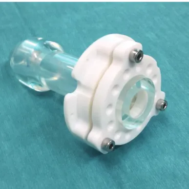
anuscopy, partial response was defined in case of tumor residue at the digital examination and anuscopy. Needles (Sharp Needles™; Nucletron, an Elekta company, Elekta AB, Stockholm, Sweden) were implanted, respecting the Paris system recommendations (13), according to the pre-treatment target volume (based on the initial schema) respecting a minimal distance of four to five mm from the needles to the anal canal mucosa. We used a dedicated circular perineal template punched by a total of ten holes (every 12 mm). A plastic tube (20 mm in external diameter, Figure 1) was placed into the anal canal and fixed to the perineal template which was finally sutured to the skin. A CT-scan was then realized and treatment plan was established (Figure 2) on Oncentra® Brachy software (V4.5.2, Elekta). Clinical evaluation of tumor bed and tumor residue at the time of implantation and also the pre-treatment volume were considered for the HDR boost CTV. If the clinical response was complete, only 12Gy in three fractions over two days were delivered. But, in case of partial response, 15Gy in three fractions over two days were delivered with HDR microSelectron® V2 Digital platform (Elekta) using Iridium192 sources. A minimum interval of six hours between
Figure 1 : Brachytherapy applicator
Clinical evaluation:
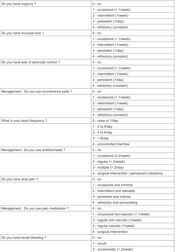
Each patient was examined by a radiotherapist or an oncologist every six months during five years. After five years, the follow-up was performed by the patient’s general practitioner. In this study, we specifically evaluated acute and late toxicities to estimate patient’s quality of life after BT. According to the Late Effects Normal Tissue (LENT) / Subjective, Objective, Management, Analytic (SOMA) score (14) for late radiation effects to the anus, we evaluated patients with this score to analyze the long-term side effects of this technique (15). We retrospectively called back patients, in January 2018, to propose them a LENT/SOMA adapted questionnaire (Annex - table 5). Their score was calculated and the results before and after treatment compared.
Endpoints:
The primary endpoint of this study was the overall survival (OS). Secondary endpoints were local relapse free survival (LRFS), colostomy free survival (CFS), metastatic free
survival (MFS) and disease free survival (DFS). We based ourselves on the LENT/SOMA score and the national cancer institute (NCI) Common Terminology Criteria for Adverse Events (CTCAE) version 4.0 (16) to evaluate toxicities.
Statistical analysis:
Population’s characteristics were described using minimum, maximum, mean and median values. Endpoints were defined as follows: OS was the interval between date of diagnostic (histologically proven on biopsies) and the date of death from any cause, LRFS was the interval between date of diagnostic and date of local recurrence, CFS was the interval between date of diagnostic and date of colostomy from any cause, MFS was the interval between date of diagnostic and date of first metastasis, DFS was the interval between date of diagnostic and date of local recurrence or distant recurrence, Overall treatment time (OTT) was the interval between date of the first day of treatment and last day of treatment. Dosimetric results included the dose delivered to 90% of the CTV (D90), the dose delivered to 100% of the CTV (D100), the volume receiving 100%, 150% and 200% of the prescribed dose (respectively V100, V150 and V200) and the dose homogeneity index (DHI : [V100-V150]/V100). We also compared LENT/SOMA scores before and after radiotherapy using Chi-2 test. All survivals were estimated according to the Kaplan-Meier method. They were realized with SPSS statistical software (20.0, IBM Corporation). This study was approved by a local institutional ethics committee (Ethical committee number : 2209548 ).
Results
Population:
A total of 46 patients were analyzed between May 2005 and January 2018 for a median follow-up of 61 months [9 – 145 months]. The median age was 65 years [34 – 84 years]. The male / female sex-ratio was 0.24. Forty-one patients (89.1%) had a performance status (PS) according to the Eastern Cooperative Oncology Group (ECOG) of zero and the other five patients (10.9%) had a PS of one. Patients’ characteristics are detailed in table 1.
Table 1 Patient, tumor and treatment characteristics
Characteristics Number (%) n = 46 Median (Interval) Age (years) 65.1 (34 – 84) Gender Men Women 9 (19.6%) 37 (80.4%) Tumor stage T1 T2 T3 T4 10 (21.7%) 34 (73.9%) 2 (4.3%) 0 Lymph node stage
N0 N1 N2 40 (87%) 5 (10.9%) 1 (2.2%) Tumor ulceration 19 (41.3%) Chemotherapy 33 (71.7%)
EBRT Dose (Gy) 45 (36 – 52)
BT boost Dose (Gy) 12 (10 – 18)
OTT (days) 58 (41 – 101)
Interval EBRT / BT (days) 17 (4 – 60)
Tumor and treatment:
The median initial tumor size was 3 cm [0.7 – 6 cm] and seven patients (15.2%) had anal margin invasion. The 16 biopsies (34.7%) tested for the HPV 16 serotype were positive. Thirty-three patients (71.7%) underwent chemotherapy, among which 26 patients (56.5%) received 5FU and mitomycine-C. Six patients (13%) received a combination of 5-Fluoro-Uracil and cisplatin.
Twenty-five patients (54.3%) were treated with IMRT.
Median dose of EBRT was 45 Gy [36 – 50.4] in 20 to 28 fractions. Median dose of BT boost was 12 Gy [10– 18 Gy] with 2.5 to 5 Gy in two to six fractions with most patients receiving three fractions of 4 Gy (73.9%). Four patients did not receive the standard dose of 45 Gy : one patient received 36 Gy because of previous radiotherapy for cervical cancer, two
patients stopped before 45 Gy due to digestive toxicities and one patient received 50.4 Gy at the physician’s discretion. One patient received a 10 Gy brachytherapy boost in two fractions because of hospitalization duration constraint, which correspond, in equivalent dose at 2 Gy per fraction, a dose similar to 12 Gy in three fractions. Another patient received 18 Gy in five fractions over three days because he only received 36 Gy in EBRT because of previous radiotherapy history. The median duration of brachytherapy was two days [2,3]. Median number of needles was five [4 – 18]. Details of dosimetric data are reported in table 2.
Table 2 Dosimetric data
Data Median Interval
CTV 18.2 [7.58 – 46.69] D90% EQD2 αβ10 (Gy) EQD2 αβ3 (Gy) 106 14.9 17.9 [32 – 117] [5.47 – 21.87] [6.86 – 26.45] D100% EQD2 αβ10 (Gy) EQD2 αβ3 (Gy) 78 11 13.2 [20 – 98] [3.35 – 18.7] [4.2 – 21.6] V100 % cc 96 17 [58 – 100] [7.1 – 45.8] V150 % cc 36 6.9 [26 – 57] [2.53 – 26.6] V200 % cc 17 3.2 [10 – 26] [1.2 – 11.7] DHI 0.62 [0.42 – 0.69]
CTV: Clinical target volume; D90: dose delivered to 90% of the CTV; EQD2 αβ10: equivalent dose at 2 Gy per fraction for αβ10 (tumor); EQD2 αβ3: equivalent dose at 2 Gy per fraction for αβ3 (normal tissues); V100: volume which received 100% of the prescribed dose; V150: volume which received 150% of the prescribed dose; V200: volume which received 200% of the prescribed dose; DHI: dose homogeneity index.
Clinical results:
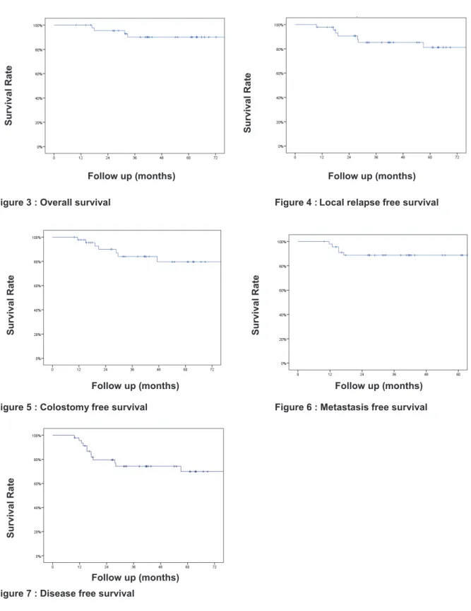
Respectively, the 5-years overall survival (OS), 5-years local relapse free survival (LRFS), cumulative rate of local recurrence (CRLR), 5-years colostomy free survival (CFS), 5-years metastatic free survival (MFS) and the 5-years disease free survival (DFS) were 90% [Standard Error (SE) 4.7%], 81.2% [ (SE) 6.6%], 15.2%, 79.5% [ (SE) 7.2%], 88.7% [ (SE) 4.8%] and 70% [ (SE) 7.6%]. At the end of the follow-up, 7 patients (15.2%) had a local recurrence. Figures 3 to 7 (Kaplan-Meier curves).
Figure 3 : Overall survival Figure 4 :Local relapse free survival
Figure 5 : Colostomy free survival Figure 6 : Metastasis free survival
Figure 7 : Disease free survival
Follow up (months) Survival Rat e Survival Rat e Survival Rat e Surviv al Rat e Follow up (months) Follow up (months) Follow up (months) Survival Rat e Follow up (months)
Colostomy :
Seven patients had a colostomy : one patient (2.2%) because of grade 4 ulceration but was able to benefit a restoration of continuity, one patient (2.2%) after grade 4 anal incontinence, four patients (8.7%) after APR because of a local recurrence and one patient (2.2%) had a local resection with a temporary colostomy because of a LR.
Toxicity:
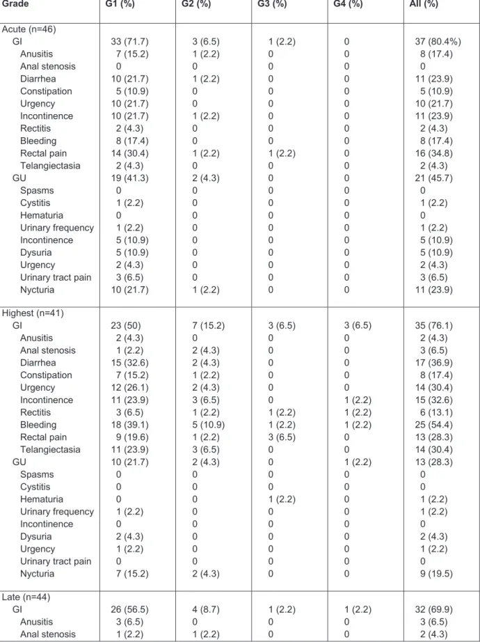
Gastro-intestinal (GI) acute side effects were frequent but not severe: 33 (71.7%) grade 1 (G1) and 3 (6.5%) G2. Acute genito-urinary (GU) side effects were also frequent but not severe: 19 (41.3%) G1, two (4.3%) G2. Only one patient (2.2%) had a G3 acute toxicity, a rectal pain, managed with medication.
Late GI toxicities were mainly G1 (56.5%) and we observed a few late GU toxicities: two (4.3%) G1 and one (2.2%) G2. Late high toxicities G3 and G4 remained very low: for GI toxicity one (2.2%) G3 and one (2.2%) G4; for GU toxicity only one (2.2%) G3. All toxicities are detailed in table 3 and 4 (annex).
Table 3 Toxicities Grade G1 (%) G2 (%) G3 (%) G4 (%) All (%) Acute (n=46) GI 3D IMRT GU 3D IMRT 33 (71.7) 16 (34.8) 17 (37) 19 (41.3) 5 (10.9) 14 (30.4) 3 (6.5) 3 (6.5) 0 2 (4.3) 1 (2.2) 1 (2.2) 1 (2.2) 0 1 (2.2) 0 0 0 37 (80.4) 19 (41.3) 18 (39.1) 21 (45.7) 6 (13) 15 (32.6) Highest (n=41) GI 3D IMRT GU 3D IMRT 23 (50) 11 (23.9) 12 (26.1) 10 (21.7) 6 (13) 4 (8.7) 7 (15.2) 3 (6.5) 4 (8.7) 2 (4.3) 1 (2.2) 1 (2.2) 3 (6.5) 3 (6.5) 0 0 3 (6.5) 2 (4.3) 1 (2.2) 1 (2.2) 0 1 (2.2) 36 (78.3) 19 (41.3) 17 (37) 13 (28.3) 7 (15.2) 6 (13) Late (n=44) GI 3D IMRT GU 3D IMRT 26 (56.5) 15 (32.6) 11 (23.9) 2 (4.3) 2 (4.3) 0 4 (8.7) 0 4 (8.7) 1 (2.2) 1 (2.2) 0 1 (2.2) 1 (2.2) 0 1 (2.2) 0 1 (2.2) 1 (2.2) 0 1 (2.2) 0 32 (69.9) 16 (34.8) 16 (34.8) 4 (8.7) 3 (6.5) 1 (2.2) G1: grade 1; G2: grade 2; G3: grade 3; G4: grade 4; GI: gastro-intestinal; GU: genito-urinary; IMRT : patients treated by intensity modulated radiotherapy; 3D : patients treated by 3-dimensional technique; Adverse events were graded with the common terminology criteria for adverse events version 4.0
Concerning LENT/SOMA scores, 27 pre-treatment and 28 post-treatment scores were collected. Mean score was 0.097 before treatment and 0.5029 after, with a statistically significant difference (p=0,001). Median score was 0 [0 – 0.46] before treatment and 0.46 [0 – 1.18] after.
Discussion with Conclusions
Brachytherapy boost for ASCC appears to be an effective treatment (10,17). Recently HDR-BT has been developed and several studies have demonstrated its feasibility and effectiveness (17). This study is based on the early results of Falk et al.(18) and presents late results. It also takes into account the quality of life with the LENT / SOMA score.
At 5 years the CRLR was 15.2%, 7 patients had a LR, which seems similar to data reported in the literature : Cordoba et al. described a 5-year CRLR of 10.1%, (11 patients) (12) and Moureau-Zabotto et al. a 5-year CRLR of 11% (18 patients) (10). However Gryc et al. had a 5-year CRLR of 24.8% (45 patients) for PDR-BT boost (19). Concerning EBRT boost series, Hannoun-Levi et al. found a LR rate of 12.3% (20 patients) (20) and Moureau-Zabotto et al.a year CRLR of 32% (13 patients) (10). In EBRT, Northover et al. found a 5-year CRLR of 32.3% (95 patients) (21) and Ajani et al. found a 5-5-year CRLR of 25% (81 patients) (22). The Kiel Group had a local control of 89% but no information concerning patient selection were available (23). A recent systematic review of brachytherapy boost, all techniques combined, found a median 5-year local control of 78.6% (7 studies) and a median 5-year CFS of 76.1% (5 studies) (24).
In our series, by analyzing the 7 patients who had a LR, we noticed that : one of these patients was infected by the human immunodeficiency virus (HIV) which is a risk factor of recurrence and complications (25). Two of these patients had an ulcerated tumor which is a negative prognostic factor. Moreover, tumor size and T stage have been recognized as pejorative prognostic factors (26). Of these patients, 4 were T2 (with an initial tumor size more than 3 centimeters). Five patients had a longer interval between EBRT and BT of 1 to 43 days more than the median interval of 17 days. One patient had an interval between RT / BT of 60 days because of acute toxicity during EBRT (stop at the dose of 37.8 Gy). As described in the literature (10), reducing OTT could improve local control, we will aim in our department to reduce OTT. Overall, patients who had a LR presented unfavorable prognostic factors that could explain these results.
Concerning the treatment tolerance, acute toxicities were frequent but not severe which is a similar profile compared to LDR or PDR-BT but late side effects seemed to be less frequent with HDR-BT (12,27). In fact, previous study found severe (at least G3) late
toxicities rate from 9% to 13% with LDR-BT (28,29), one study of HDR-BT by Kapoor et al. did not describe any G3 or more toxicities (30). In our study, only 6.5% severe late toxicities were observed. That could be explained by a DHI (median = 0.62, SE [0.42 – 0.69]) higher than Kapoor et al. (median DHI = 0.83, SE [0.55–0.98] -(30)). Modern external beam
radiotherapy with boost also seems to have more toxicities than HDR-BT, Kachnic et al. had 21% gastrointestinal / genitourinary G3 and G4 (31). We excluded patients with a
Only two patients had a colostomy due to toxicities : one ulceration (in HIV infected patient with high viral load at the time of diagnosis and treatment) and one anal incontinence for the 5 other patients it was because of local recurrence. Deniaud et al. had similar results with EBRT boost : 5% of G3 late toxicities, one colostomy due to incontinence (32). The patient who presented G4 incontinence had an epidermoid carcinoma histology with a stage T3 N1 with an initial tumor size of 4.5cm with ulceration. This patient received chemotherapy by cisplatin and a total dose in EBRT of 46 Gy with a 3-dimensional technic. A boost of 15Gy in 3 fractions of 5 Gy were delivered in BT, half of the circumference was treated. The patient who presented G4 ulceration had a stag T2 N1 with an initial tumor size of 3.5cm with ulceration. This patient received chemotherapy by cisplatin and a total dose in EBRT of 45 Gy by 3-dimensional technic. A boost of 12Gy in 3 fractions of 4 Gy were delivered in BT with less than half of the circumference was treated. New procedures using multiparametric imaging with MRI-compatible applicator to define the clinical target volume are feasible and could reduce the canal anal irradiation and toxicities (33).
We decided to evaluate patient before and after treatment with the LENT SOMA score to have an idea of the effect of the treatment on patient’s quality of life. Many patients had rectal symptoms before treatment therefore we found relevant to use this score to compare before and after treatment. The variation between LENT / SOMA score pre and post treatment seemed to be significantly different and clinically relevant. Most frequently, patients reported intestinal disorders such as : urgency, loss of sphincter control and diarrhea.
In addition, HDR-BT boost reduces OTT with a median duration of BT-boost of 2 days against 14 days for EBRT boost (10). It results in a reduction of OTT (58 days [41 - 101] in our study versus 85 days [45 - 141] for exclusive EBRT (10), and must be taken into consideration for a cost-effectiveness analysis and patients’ comfort. HDR allows less constraint than other BT techniques: the patient does not need to be hospitalized in a shielded-room; medical and paramedical staff are less prone to be exposed to radiation.
This study has some limitation; it was a retrospective monocentric study with a limited number of patients, but ASCC remains a rare disease and it is unlikely randomized clinical trials will be performed to compare HDR-boost. This database collects data since May 2005, at the beginning we did not have IMRT available in our department. Since 2010, all patients were treated with IMRT.
In ASCC management, HDR-BT boost after radiochemotherapy appears to be a feasible treatment with acceptable toxicities and good efficacy. It allows a partial anal canal irradiation with long term toxicities similar or lower than other boost techniques. While HDR brachytherapy is more and more used around the world, it is very unlikely that there will be randomized data to support HDR-BT in the near future. Retrospective and observational prospective studies are currently the highest level of evidence to support this technique.
References
1. Abramowitz L, Jacquard A-C, Jaroud F, et al. Human papillomavirus genotype distribution in anal cancer in France: The EDiTH V study. Int J Cancer.
2011;129(2):433–9.
2. Robinson D, Coupland V, Møller H. An analysis of temporal and generational trends in the incidence of anal and other HPV-related cancers in Southeast England. Br J Cancer. 2009;100(3):527–31.
3. Nielsen A, Munk C, Kjaer SK. Trends in incidence of anal cancer and high-grade anal intraepithelial neoplasia in Denmark, 1978-2008. Int J Cancer. 2012;130(5):1168–73. 4. Johnson LG, Madeleine MM, Newcomer LM, et al. Anal cancer incidence and survival:
The Surveillance, Epidemiology, and End Results experience, 1973-2000. Cancer. 2004;101(2):281–8.
5. Colón-López V, Shiels MS, Machin M, et al. Anal Cancer Risk Among People With HIV Infection in the United States. J Clin Oncol. 2017;36(1):68–75.
6. Nigro ND, Vaitkevicius VK, Considine B. Combined therapy for cancer of the anal canal: a preliminary report. Dis Colon Rectum. 1974;17(3):354–6.
7. Martin D, Balermpas P, Winkelmann R, et al. Anal squamous cell carcinoma – State of the art management and future perspectives. Cancer Treat Rev. 2018;65:11–21. 8. Lépinoy A, Lescut N, Puyraveau M, et al. Evaluation of a 36 Gy elective node irradiation
dose in anal cancer. Radiother Oncol J Eur Soc Ther Radiol Oncol. 2015;116(2):197– 201.
9. Epidermoid anal cancer: results from the UKCCCR randomised trial of radiotherapy alone versus radiotherapy, 5-fluorouracil, and mitomycin. UKCCCR Anal Cancer Trial Working Party. UK Co-ordinating Committee on Cancer Research. Lancet Lond Engl. 1996;348(9034):1049–54.
10. Moureau-Zabotto L, Ortholan C, Hannoun-Levi J-M, et al. Role of Brachytherapy in the Boost Management of Anal Carcinoma With Node Involvement (CORS-03 Study). Int J Radiat Oncol. 2013;85(3):e135–42.
11. Glynne-Jones R, Nilsson PJ, Aschele C, et al. Anal cancer: ESMO-ESSO-ESTRO clinical practice guidelines for diagnosis, treatment and follow-up. Eur J Surg Oncol EJSO. 2014;40(10):1165–76.
12. Cordoba A, Escande A, Leroy T, et al. Low-dose-rate interstitial brachytherapy boost for the treatment of anal canal cancers. Brachytherapy. 2017;16(1):230–5.
13. Pierquin B, Dutreix A, Paine CH, et al. The Paris system in interstitial radiation therapy. Acta Radiol Oncol Radiat Phys Biol. 1978;17(1):33–48.
14. Lent soma scales for all anatomic sites. Int J Radiat Oncol • Biol • Phys. 1995;31(5):1049–91.
15. Lund J-Å, Kaasa S, Wibe A, et al. Late radiation effects to the rectum and anus after treatment for prostate cancer; validity of the LENT/SOMA score. Acta Oncol.
2013;52(4):727–35.
16. NCI Term Browser [Internet]. [cited 2018 Sep 10].
17. Papillon J, Montbarbong JF, Gerard JP, et al. Interstitial curietherapy in the
conservative treatment of anal and rectal cancers. Int J Radiat Oncol. 1989;17(6):1161– 9.
18. Falk AT, Claren A, Benezery K, et al. Interstitial high-dose rate brachytherapy as boost for anal canal cancer. Radiat Oncol. 2014;9(1):240.
19. Gryc T, Ott O, Putz F, et al. Interstitial brachytherapy as a boost to patients with anal carcinoma and poor response to chemoradiation: Single-institution long-term results. Brachytherapy. 2016;15(6):865–72.
20. Hannoun-Levi J-M, Ortholan C, Resbeut M, et al. High-Dose Split-Course Radiation Therapy for Anal Cancer: Outcome Analysis Regarding the Boost Strategy (CORS-03 Study). Int J Radiat Oncol. 2011;80(3):712–20.
21. Northover J, Glynne-Jones R, Sebag-Montefiore D, et al. Chemoradiation for the treatment of epidermoid anal cancer: 13-year follow-up of the first randomised UKCCCR Anal Cancer Trial (ACT I). Br J Cancer. 2010;102(7):1123–8.
22. Ajani JA, Winter KA, Gunderson LL, et al. Fluorouracil, mitomycin, and radiotherapy vs fluorouracil, cisplatin, and radiotherapy for carcinoma of the anal canal: a randomized controlled trial. JAMA. 2008;299(16):1914–21.
23. Niehoff P, Kovács G. HDR brachytherapy for anal cancer. J Gastrointest Oncol. 2014;5(3):218–22.
24. Frakulli R, Buwenge M, Cammelli S, et al. Brachytherapy boost after chemoradiation in anal cancer: a systematic review. J Contemp Brachytherapy. 2018;10(3):246–53. 25. Oehler-Jänne C, Huguet F, Provencher S, et al. HIV-Specific Differences in Outcome of
Squamous Cell Carcinoma of the Anal Canal: A Multicentric Cohort Study of HIV-Positive Patients Receiving Highly Active Antiretroviral Therapy. J Clin Oncol. 2008;26(15):2550–7.
26. Das P, Crane CH, Eng C, et al. Prognostic Factors for Squamous Cell Cancer of the Anal Canal. 2008;2(1):5.
27. Bruna A, Gastelblum P, Thomas L, et al. Treatment of squamous cell anal canal carcinoma (SCACC) with pulsed dose rate brachytherapy: A retrospective study. Radiother Oncol. 2006;79(1):75–9.
28. Wagner JP, Mahe MA, Romestaing P, et al. Radiation therapy in the conservative treatment of carcinoma of the anal canal. Int J Radiat Oncol Biol Phys. 1994;29(1):17– 23.
29. Bartelink H, Roelofsen F, Eschwege F, et al. Concomitant radiotherapy and
chemotherapy is superior to radiotherapy alone in the treatment of locally advanced anal cancer: results of a phase III randomized trial of the European Organization for
Research and Treatment of Cancer Radiotherapy and Gastrointestinal Cooperative Groups. J Clin Oncol Off J Am Soc Clin Oncol. 1997;15(5):2040–9.
30. Kapoor R, Khosla D, Shukla AK, et al. Dosimetric and clinical outcome in image-based high-dose-rate interstitial brachytherapy for anal cancer. Brachytherapy.
2014;13(4):388–93.
31. Kachnic LA, Winter K, Myerson RJ, et al. RTOG 0529: A Phase 2 Evaluation of Dose-Painted Intensity Modulated Radiation Therapy in Combination With 5-Fluorouracil and Mitomycin-C for the Reduction of Acute Morbidity in Carcinoma of the Anal Canal. Int J Radiat Oncol. 2013;86(1):27–33.
32. Deniaud-Alexandre E, Touboul E, Tiret E, et al. Carcinomes épidermoïdes du canal anal traités par association concomitante de radiothérapie et de chimiothérapie. Évaluation des résultats fonctionnels. Cancer/Radiothérapie. 2006;10(8):572–82. 33. Tagliaferri L, Manfrida S, Barbaro B, et al. MITHRA – multiparametric MR/CT image
adapted brachytherapy (MR/CT-IABT) in anal canal cancer: a feasibility study. J Contemp Brachytherapy. 2015;7(5):336–45.
Acknowledgements
Annexe
Table 4All toxicities
Grade G1 (%) G2 (%) G3 (%) G4 (%) All (%) Acute (n=46) GI Anusitis Anal stenosis Diarrhea Constipation Urgency Incontinence Rectitis Bleeding Rectal pain Telangiectasia GU Spasms Cystitis Hematuria Urinary frequency Incontinence Dysuria Urgency
Urinary tract pain Nycturia 33 (71.7) 7 (15.2) 0 10 (21.7) 5 (10.9) 10 (21.7) 10 (21.7) 2 (4.3) 8 (17.4) 14 (30.4) 2 (4.3) 19 (41.3) 0 1 (2.2) 0 1 (2.2) 5 (10.9) 5 (10.9) 2 (4.3) 3 (6.5) 10 (21.7) 3 (6.5) 1 (2.2) 0 1 (2.2) 0 0 1 (2.2) 0 0 1 (2.2) 0 2 (4.3) 0 0 0 0 0 0 0 0 1 (2.2) 1 (2.2) 0 0 0 0 0 0 0 0 1 (2.2) 0 0 0 0 0 0 0 0 0 0 0 0 0 0 0 0 0 0 0 0 0 0 0 0 0 0 0 0 0 0 0 0 37 (80.4%) 8 (17.4) 0 11 (23.9) 5 (10.9) 10 (21.7) 11 (23.9) 2 (4.3) 8 (17.4) 16 (34.8) 2 (4.3) 21 (45.7) 0 1 (2.2) 0 1 (2.2) 5 (10.9) 5 (10.9) 2 (4.3) 3 (6.5) 11 (23.9) Highest (n=41) GI Anusitis Anal stenosis Diarrhea Constipation Urgency Incontinence Rectitis Bleeding Rectal pain Telangiectasia GU Spasms Cystitis Hematuria Urinary frequency Incontinence Dysuria Urgency
Urinary tract pain Nycturia 23 (50) 2 (4.3) 1 (2.2) 15 (32.6) 7 (15.2) 12 (26.1) 11 (23.9) 3 (6.5) 18 (39.1) 9 (19.6) 11 (23.9) 10 (21.7) 0 0 0 1 (2.2) 0 2 (4.3) 1 (2.2) 0 7 (15.2) 7 (15.2) 0 2 (4.3) 2 (4.3) 1 (2.2) 2 (4.3) 3 (6.5) 1 (2.2) 5 (10.9) 1 (2.2) 3 (6.5) 2 (4.3) 0 0 0 0 0 0 0 0 2 (4.3) 3 (6.5) 0 0 0 0 0 0 1 (2.2) 1 (2.2) 3 (6.5) 0 0 0 0 1 (2.2) 0 0 0 0 0 0 3 (6.5) 0 0 0 0 0 1 (2.2) 1 (2.2) 1 (2.2) 0 0 1 (2.2) 0 0 0 0 0 0 0 0 0 35 (76.1) 2 (4.3) 3 (6.5) 17 (36.9) 8 (17.4) 14 (30.4) 15 (32.6) 6 (13.1) 25 (54.4) 13 (28.3) 14 (30.4) 13 (28.3) 0 0 1 (2.2) 1 (2.2) 0 2 (4.3) 1 (2.2) 0 9 (19.5) Late (n=44) GI Anusitis Anal stenosis 26 (56.5) 3 (6.5) 1 (2.2) 4 (8.7) 0 1 (2.2) 1 (2.2) 0 0 1 (2.2) 0 0 32 (69.9) 3 (6.5) 2 (4.3)
Diarrhea Constipation Urgency Incontinence Rectitis Bleeding Rectal pain Telangiectasia GU Spasms Cystitis Hematuria Urinary frequency Incontinence Dysuria Urgency Tract pain Nycturia Tract obstruction Sexuals 8 (17.4) 6 (13.1) 7 (15.2) 5 (10.9) 2 (4.3) 19 (41.3) 5 (10.9) 7 (15.2) 2 (4.3) 0 0 0 0 1 (2.2) 1 (2.2) 1 (2.2) 0 2 (4.3) 0 1 (2.2) 3 (6.5) 1 (2.2) 1 (2.2) 1 (2.2) 0 0 1 (2.2) 1 (2.2) 1 (2.2) 0 0 0 0 0 0 0 0 1 (2.2) 0 0 0 0 0 0 0 0 1 (2.2) 0 0 0 0 1 (2.2) 0 0 0 0 0 0 0 0 0 0 0 0 1 (2.2) 1 (2.2) 0 0 1 (2.2) 0 0 0 0 0 0 0 0 0 0 0 11 (23.9) 7 (15.2) 8 (17.4) 6 (13.1) 3 (6.5) 20 (43.5) 7 (15.2) 8 (17.4) 4 (8.7) 0 0 1 (2.2) 0 1 (2.2) 1 (2.2) 1 (2.2) 0 3 (6.5) 0 1 (2.2) G1: grade 1; G2: grade 2; G3: grade 3; G4: grade 4; GI: gastro-intestinal; GU: genito-urinary;
Table 5SOMA / LENT questionnaire
Do you have urgency ? 0 - no
1 - occasional (< 1/week) 2 - intermittent (1/week) 3 - persistent (1/day) 4 - refractory (constant)
Do you have mucosal loss ? 0 - no
1 - occasional (< 1/week) 2 - intermittent (1/week) 3 - persistent (1/day) 4 - refractory (constant)
Do you have loss of sphincter control ? 0 - no
1 - occasional (< 1/week) 2 - intermittent (1/week) 3 - persistent (1/day) 4 - refractory (constant) Management : Do you use incontinence pads ? 0 - no
1 - occasional (< 1/week) 2 - intermittent (1/week) 3 - persistent (1/day) 4 - refractory (constant)
What is your stool frequency ? 0 - none or 1/day
1 - 2 to 4/day 2 - 4 to 8/day 3 - > 8/day
4 - uncontrolled diarrhea
Management : Do you use antidiarrheals ? 0 - no
1 - occasional (≤ 2/week) 2 - regular (> 2/week) 3 - multiple (> 2/day)
4 - surgical intervention / permanent colostomy
Do you have anal pain ? 0 - no
1 - occasional and minimal 2 - intermittent and tolerable 3 - persistent and intense 4 - refractory and excruciating
Management : Do you use pain medication ? 0 - no
1 - occasional non-narcotic (< 1/week) 2 - regular non-narcotic (1/week) 3 - regular narcotic (1/week) 4 - surgical intervention
Do you have rectal bleeding ? 0 - no
1 - occult
3 - persistent / daily 4 - gross hemorrhage Management : Do you need treatment against this
bleeding ? 0 - no
1 - stool softener, iron therapy 2 - occasional transfusion 3 - frequent transfusion
4 - surgical intervention / permanent colostomy
Do you have anal ulceration ? 0 - no
1 - superficial (≤ 1cm2) 2 - superficial (> 1cm2) 3 - deep ulcer
4 - perforation, fistulae Management : Do you need treatment against this
ulceration ? 0 - no
1 - stool softener, diet modification 2 - occasional steroids
3 - steroids per enema, hyperbaric oxygen 4 - surgical intervention / permanent colostomy
Do you have anal stricture ? 0 - no
1 - > 2/3 normal diameter with dilatation 2 - 1/3 to 2/3 normal diameter with dilatation 3 - < 1/3 normal diameter
4 - complete obstruction Management : Do you need treatment against this stricture
? 0 - no
1 - diet modification 2 - occasional dilatation 3 - regular dilatation
4 - surgical intervention / permanent colostomy Based on the SOMA / LENT score : calculated by adding the 14 items and dividing by 11
Abréviations
APR : Abdominoperineal resection ASCC : Anal squamous cell carcinoma BT : Brachytherapy
CEA : Carcinome épidermoïde de l’anus CFS : Colostomy free survival
CRT : Chemoradiotherapy
CTCAE : Common terminology criteria for adverse events DFS : Disease free survival
EBRT : External beam radiotherapy
ECOG : Eastern cooperative oncology group GI : Gastro-intestinal
HDD : Haut débit de dose
HDR-BT : High dose rate brachytherapy HIV : Human immunodeficiency virus HPV : Human papilloma virus
ICRU : International commission on radiation units measurements IMRT : Intensity modulated radiotherapy
LDR-BT : Low dose rate brachytherapy LRFS : Local relapse free survival MFS : Metastatic free survival NCI : National cancer institute OS : Overall survival
OTT : Overall treatment time
PET-CT : Positron emission tomography computed tomography Pts : Patients
RTE : Radiothérapie externe SG : Survie globale
SOMA / LENT : Subjective, Objective, Management, Analytic ./ Late Effects Normal Tissue SSC : Survie sans colostomie
SSE : Survie sans évènement SSM : Survie sans métastase SSRL : Survie sans récidive locale
Serment d’Hippocrate
En présence des Maîtres de cette Faculté, de mes chers condisciples et devant l’effigie d’Hippocrate,
Je promets et je jure d’être fidèle aux lois de l’honneur et de la probité dans l’exercice de la Médecine.
Je donnerais mes soins gratuitement à l’indigent et n’exigerai jamais un salaire au-dessus de mon travail. Je ne participerai à aucun partage clandestin d’honoraires.
Admis dans l’intimité des maisons, mes yeux n’y verront pas ce qui s’y passe ; ma langue taira les secrets qui me seront confiés et mon état ne servira pas à corrompre les mœurs, ni à favoriser le crime.
Je ne permettrai pas que des considérations de religion, de nation, de race, de parti ou de classe sociale viennent s’interposer entre mon devoir et mon patient.
Je garderai le respect absolu de la vie humaine.
Même sous la menace, je n’admettrai pas de faire usage de mes connaissances médicales contre les lois de l’humanité.
Respectueux et reconnaissant envers mes Maîtres, je rendrai à leurs enfants l’instruction que j’ai reçue de leurs pères.
Que les hommes m’accordent leur estime si je suis fidèle à mes promesses. Que je sois couvert d’opprobre et méprisé de mes confrères si j’y manque.