Université du Québec
Institut National de la Recherche Scientifique Institut Armand-Frappier
New densoviruses, comparison of transcription strategies,
recombination and contribution to 3D-structures
Par Qian Yu
Thèse présentée pour l’obtention du grade de Philosophiae doctor (Ph.D.) en Virologie et Immunologie
Jury d’évaluation
© Droits réservés de Qian Yu, 2015
Président du jury et Examinateur interne
Jean-François Laliberté
INRS-Institut Armand-Frappier
Examinateur interne Pierre Talbot
INRS-Institut Armand-Frappier
Examinatrice externe Selena M. Sagan
Department of Microbiology and Immunology
Mcgill University
Examinateur externe Just M. Vlak
Laboratory of Virology, Wageningen University
Directeur de recherche Peter Tijssen
ii
ACKNOWLEDGEMENTS
I would like to thank Prof. Peter Tijssen for giving me the opportunity to study at INRS-Institut Armand Frappier, and providing me precious advice and direction throughout my PhD study. He has created a warm and stimulating research environment allowing me explore a complex and fascinating scientific field. Thanks to him for his kindness, patience and support during my PhD path. I learned lots from him. Being able to study under his supervision is one of the most important achievements in my life.
I want to acknowledge Dr. Kaiyu Liu. He gave me invaluable help at the beginning of my PhD. I am grateful to Prof. Max Bergoin from the “Laboratoire de Pathologie Comparée, Université Montpellier II, France” and now as an invited professor in INRS- Institut Armand-Frappier for his important comments during my project research. Thanks to all the group members for their help throughout the work. I am grateful to have been part of such supportive, hardworking, and inspiring groups. Over the course of my research I have been fortunate enough to work with the truly outstanding groups. These people include: Dr. Maude Boisvert, Dr. Hanh Thi Pham, Dr. Sandra Fernandes, and Oanh T. H. Huynh.
I thank to my PhD committee (comité d’encadrement): Dr. Jean-François Laliberté and Dr. Jonathan Perreault, both professors at INRS-Institut Armand Frappier, for their kindly discussions and advice.
I thank the departmental and technical staff at INRS- Institut Armand-Frappier. They are very helpful, particularly Micheline Letarte for her EM technical support and Jessy Tremblay for his microscopy technical support.
iii
Sepcial thanks to Madame Anne Philippon from the administration unit of the INRS-Institut Armand-Frappier, who always help me with many patience for the applications and document works.
I am eternally grateful to my dearest parents, who have been a constant source of love, encouragement for me. I want to thank to my dear husband, Jianming Zhang, who loves me deeply and supports me selflessly. I want to say them, I love you. I thank my family and my friends for their continuing love and heartily support.
Finally, I wish to acknowledge the following organizations for their financial support: the Chinese Scholarship Council and Natural Sciences and Engineering Research Council of Canada.
iv
ABSTRACT
Parvoviruses are small, isometric viruses, with a diameter of 20–25 nm, containing a linear, single-stranded DNA genome of 4–6 kb, and have been classified in the family Parvoviridae. Densoviruses (DNVs) are parvoviruses of invertebrates, infecting at least insects, shrimp, and urchins, and form a separate subfamily (Densovirinae) within the Parvoviridae family. The nonenveloped icosahedral capsids consist of 60 structural proteins some of which have N-terminal extensions. Moreover, capsids of many vertebrate parvoviruses and DNVs have a phospholipase A2 (PLA2) activity that allows them to breach the endosomal membrane barrier during cell entry. During the past few decades, many new vertebrate parvoviruses and invertebrate densoviruses have been discovered. However, research on their molecular biology characteristics is still limited.
This thesis contains three different projects. The first objective, which is also the major project of this thesis, started with the discovery of three new iteradensoviruses. Since 2010, we got some infected Papilio polyxenes (black swallowtail butterfly) larvae and some infected monarch butterfly pupae from a lab of Cornell University and a farm in Granby (Quebec), respectively. Together with an older, stored Sibine fusca larvae sample, we identified three new viruses. The method we used for identification was named DNase&RNase-SISPA. Cloning and sequencing of the genomes of those three viruses revealed high identities with members of Iteradensovirus genus of
Densovirinae subfamily. These new iteradensoviruses have later been named as Papilio polyxenes iteradensovirus (PpDV), Sibine fusca iteradensovirus (SfDV) and Danaus plexippus plexippus iteradensovirus (DppIDV).
Together with the other two iteradensovirus Casphalia extranea iteradensovirus (CeDV) and Bombyx mori iteradensovirus (BmDV) which have been cloned and reported from our lab, in the second project we determined the transcription strategies of these five iteradensoviruses. We showed that all these monosense
v
iteradensoviruses genomes contain two large reading frames (ORFs) coding for two non-structural (NS) proteins. NS1 and NS2 that are generated from individual transcripts, which initiated from either side of NS1 ATG with only 10-15 nt distance between each other. The expression of the two NS proteins is controlled by two overlapping promoters. The two NS transcripts shared the same poly(A) motif downstream of the TATA-box of VP. All the structural proteins were generated from a single VP transcript by leaky scanning.
The third project contained different studies on cricket densoviruses, which included a contribution work on the transcription strategy of AdDNV and the discovery of two novel cricket densoviruses. AdDNV belongs to the Ambidensovirus genus and its expression strategy of NS proteins is identical with the classical ambidensoviruses. However, the structural proteins (VPs) were generated from two split ORFs and employed a unique alternative splicing mechanism for expression. So far, this expression strategy is not only unique among the other densoviruses, but also among the vertebrate parvoviruses. The first novel cricket densovirus reported in this project was Acheta domesticus Mini Ambidensovirus AdMADV. The novel AdMADV has a small ambisense genome of 4945 nts and the related 199 nt inverted terminal repeats (ITRs) fold into Y-shaped hairpin structure at both telomers. However, its genome organization is identical to classical ambidensoviruses. Another novel cricket densovirus consisted of a special segmented genome, the segments of which coded separately for the NS and VP proteins.. This new Acheta domesticus segmented densovirus, AdSDV, as the first bipartite parvovirus has been reported, arose from mosquitoes and through recombination with AdDNV to obtained phospholipase A2 activity that enabled it to infect crickets. Here, we also provided a hypothesis on the origin of this new virus.
Besides the main chapters for the three projects, the thesis also includes some published side projects in the appendix. The first project included in the appendix I aimed to determine the capsid structure for AdDNV and an adeno-associated virus
vi
isolated from snakes (serpine adeno-associated virus, sAAV). For AdDNV, the 3.5-Å resolution X-ray crystal structure of the mature virus particle has been determined. It was shown that raising the temperature of the AdDNV particles caused a loss of the ssDNA genome. A 5.5-Å resolution structure of the empty virion was obtained by cryoelectron microscopy and was found to be the same as that for the full, mature virus except for the absence of the three ordered nucleotides. So far, serpine AAV (sAAV) is the only known parvovirus to infect reptiles (although recently, we got the complete sequence of a second reptile AAV from bearded dragon lizards (Pogona), unpublished). In this study, we determined the capsid structure at 6.7-Å resolution. Compared to reported structures of different serotypes of AAVs, significant differences were caused by the conformational changes within the variable regions VR-IV, VR-VIII, and VR-IX. Structural comparisons show that vertebrate and invertebrate parvoviruses have evolved independently, although there are common structural features among all parvovirus capsid proteins. Appendix II contains publications concerning the genome sequences of three novel single-stranded DNA (ssDNA) viruses. These viruses include the Helicoverpa armigera Densovirus HaDNV, Pseudoplusia includens Densovirus PiDNV, and a novel Circo-Like virus isolated from Penaeus monodon shrimp (PmCV-1). PmCV-1 was isolated from PstDNV-infected shrimps, Penaeus monodon, and was the first circular ssDNA virus that has been identified in shrimp.
viii
RÉSUMÉ
Les Parvovirus sont de petit virus icosaédriques d’un diamètre d’environ 20-25nm, contenant un génome d’ADN simple brin de 4-6kb et sont classifiés dans la famille Parvoviridae. Les Densoviruses (DNVs) sont des parvovirus infectant les invertébrés, incluant des insectes, les crevettes et les oursins et forme la sous-famille
Densovirinae dans la famille des Parvoviridae. La capside des parvoviruses contient
60 protéines structurales dont certaines contiennent une extension N-terminale par rapport aux autres. De plus, la capside de plusieurs parvovirus des vertébrés et DNVs possède une activité enzymatique de phospholipase A2 (PLA2) qui permet l’évasion de la voie endosomale lors de l’entrée du virus dans la cellule, via des bris de la membrane endosomale. Lors des dernières décennies, plusieurs nouveaux parvovirus de vertébrés et invertébrés ont été découverts. Cependant les connaissances concernant leur biologie moléculaire demeurent limitées.
Cette thèse contient trois projets différents. Le premier objectif, qui était le projet majeur de cette thèse a débuté par la découverte de trois nouveaux Iteradensoviruses. Depuis 2010, nous avons reçu des échantillons de larves de papillons du céleri (black swallowtail butterfly, Papilio polyxenes) d’une ferme de Granby ainsi que des pupes de papillons monarques provenant de l’Université Cornell et Granby (Québec). Nous avions aussi en notre possession des échantillons de larves de Sibine fusca. À partir de ces échantillons, nous avons isolé et identifié trois nouveaux virus. La méthode utilisée pour leur identification est nommée « DNase&RNase-SISPA ». Le clonage et le séquençage des génomes de ces trois virus ont révélé une forte identité avec les membres du genre Iteradensovirus de la sous famille Densovirinae. Ces nouveaux
Iteradensovirus ont été nommés Papilio polyxenes iteradensovirus (PpDV), Sibine fusca iteradensovirus (SfDV) et Danaus plexippus plexippus iteradensovirus (DppIDV).
Avec deux autres Iteradensovirus qui ont été clonés précédemment dans notre laboratoire : Casphalia extranea iteradensovirus (CeDV) and Bombyx mori
ix
iteradensovirus (BmDV), la stratégie de transcription de ces 5 iteradensovirus est déterminée dans le second projet. Nous avons montré que ces virus sont tous monosens et contiennent deux grands cadres de lectures (ORF). Le premier permet l’expression de deux protéines non structurales (NS1 et NS2) qui sont générées par des transcrits indépendants, initiés à partir de séquences ATG à seulement 10-15 nucléotides de distance. L’expression des protéines NS est contrôlée par des promoteurs chevauchants. Les deux transcrits NS utilisent le même signal de polyadénylation (poly(A)), situé en aval du motif TATA-box des gènes des protéines structurales (VPs). Les protéines VPs sont toutes générées à partir d’un seul transcrit, par un mécanisme de « leaky scanning » (traduction par balayage fuite des codons d'initiation).
Le troisième projet contient différents études sur les densovirus de criquets, incluant un travail de contribution sur la stratégie de transcription d’AdDNV et la découverte de deux nouveaux densoviruses de criquets. AdDNV appartient au genre
Amibendensovirus et sa stratégie de transcription des protéines NS est identique aux
autres membres de ce genre. Cependant, la stratégie de transcription des protéines VPs est unique. Les protéines structurales sont générées à partir de deux cadres de lecture fendues et emploie une stratégie d’épissage alternatif unique. Cette stratégie est non seulement unique au sein des densovirus mais des parvoviruses de vertébrés aussi. Le premier nouvel densovirus de criquet rapporté de ce projet était Acheta
domesticus Mini Ambidensovirus AdMADV. Le nouvel AdMADV possède un petit
génome de 4945 nucléotides ainsi que des séquences terminales inversées (ITR) de 199 nucléotides, repliées en forme de Y aux deux extrémités. L’organisation génomique est similaire par rapport aux autres ambidensovirus classiques. Un autre nouvel densovirus de criquet contient une séquence nucléotidique spéciale, où les deux séquences ont codé les protéines de NS et VP séparément. La nouvelle séquence de densovirus AdSDV de Acheta domesticus qui est le premier parvovirus en deux parties, a été rapportée d’émerger de moustiques et par la recombinaison avec AdDNV obtenu d’une activité de la phospholipase A2 qui était capable de infecter
x
les criquets. En plus, une hypothèse de l’origine de ce nouvel virus est aussi présentée ici.
Sauf les chapitres principaux de ces trois projets, on a aussi conclu des travails de collaboration comme les projets secondaires dans l’annexe. Le premier projet inclus dans l’annexe I consistait à la détermination de la structure du densovirus AdDNV et d’un parvovirus isolés à partir de serpents (Serpine adeno-associated virus (sAAV)). Pour AdDNV, la structure de la particule mature a été déterminée par cristallographie à rayons X avec une résolution de 3.5-Å. Nous avons aussi montré que l’augmentation de la température des particules menait à l’éjection des génomes. Une structure du virion vide de résolution de 5.5 Å a été obtenue par microscopie cryo-électronique et est identique à la structure de la capside pleine, excepté pour les trois nucléotides ordonnés présent dans la structure de la capside pleine. sAAV est le seul parvovirus de reptile connu (bien que ces dernières années nous avons obtenu la séquence complète d’une seconde reptile AAV à partir de lézards dragons (Pogona), non publié). Nous avons déterminé sa structure à une résolution de 6.7-Å. En comparant cette structure avec les autres AAVs connus, certaines différences significatives ont été observées. Elles sont dues à un changement de conformation dans les régions variables VR-IV, VR-VIII et VR-IX. La comparaison des structures a montré que l’évolution des parvovirus des vertébrés et invertébrés a été effectuée indépendamment, malgré le fait que certaines caractéristiques structurales soient partagées chez tous les parvovirus. L’annexe II a contenu la séquence nucléotidique de trois nouveaux virus à ADN simple brin. Ces virus incluent Helicoverpa armigera Densovirus (HaDNV); Pseudoplusia includens Densovirus (PiDNV); et un nouveau virus à génome circulaire (Circo-Like) isolée à partir de crevettes Penaeus monodon (PmCV-1). .PmCV-1, isolées à partir de crevettes infectées par le virus PstDNV est le premier virus avec un génome d’ADN simple brin circulaire identifié chez les crevettes.
xii
TABLE OF CONTENTS
ACHNOWLEDGEMENTS... ii
ABSTRACT...……... iv
RÉSUMÉ... viii
TABLE OF CONTENTS... xii
LIST OF TABLES………... xviii
LIST OF FIGURES... xx
LIST OF ABBREVIATIONS…..……... xxii
Chapter I: Introduction ……….……...2
1 Viruses and biological control ……….…….4
2 Insect parvoviruses (Densoviruses) ………6
2.1 Genotype and Taxonomic structure history of Densovirus ………...6
2.2 Pathology caused by densoviruses ………..……..10
2.3 Genome organization of densoviruses ………10
2.3.1 Genome organization of the Ambidensovirus genus………...12
2.3.2 Genome organization of the Iteradensovirus genus………..……...15
2.3.3 Genome organization of the Brevidensovirus genus………..…..………16
2.3.4 Genome organization of the Hepandensovirus and Penstyldensovirus genus ……….16
2.4 Transcription strategy of densoviruses ………..….17
2.5 Terminal structures of densovirus genome ……….….. 24
xiii
2.6.1 Nonstructural protein and rolling cycle replication ………... 27
2.6.1.1 Rolling hairpin replication ……….……. 28
2.6.2 Structure protein ………..…………. 32
3 Atomic structures of viral particles ……….…. 35
Chapter II: Materials and Methods ………..…… 42
Chapter III: New iteradensoviruses………..……… 52
Preamble………...………..54
Article 1: Iteravirus-Like Genome Organization of a Densovirus from Sibine fusca Stoll ……….… 56 Contribution of authors……….. 56 Abstract……….……….. 56 Résumé………...……… 56 Results……….…… 57 Achnowledgments………...….. 59
Article 2: Papilio polyxenes Densovirus Has an Iteravirus-Like Genome Organization.. 60
Contribution of authors……….. 60
Abstract……….…….. 60
Résumé………...………… 60
Results……….………… 61
Achnowledgments………...…….. 62
Article 3: Iteradensovirus from the Monarch Butterfly, Danaus plexippus plexippus … 64 Contribution of authors……….. 64
Abstract……….…….. 64
Résumé………...……… 64
Results……….… 65
Achnowledgments………... 67
Chapter IV: Iteradensovirus transcription strategy………...………..….68
xiv
Article 4: Gene expression of five different iteradensoviruses: BmDV, CeDV, PpDV,
SfDV and DpIDV……….………..…… 72 Contribution of authors……….. 72 Abstract……….….. 72 Résumé………...… 72 Introduction………. 73 Results……….…… 76 Achnowledgments………...….. 83
Chapter V: Cricket densoviruses.……….…… 84
Preamble………...………..86
Article 5: The Acheta domesticus densovirus, isolated from the European house cricket, has evolved an expression strategy unique among parvoviruses ……… 88
Contribution of authors……….. 88
Abstract……….…….. 89
Résumé………...… 90
Introduction………. 91
Materials and methods……….…. 93
Results……….…… 98
Discussion………...……. 111
Achnowledgments………... 113
Supplemental materials………...114
Article 6: A Novel Ambisense Densovirus, Acheta domesticus Mini Ambidensovirus, from Crickets………...………. 120 Contribution of authors……….……….. 120 Abstract………...……….. 120 Résumé……….……… 120 Results………..……….121 Achnowledgments……….……….. 123
In progress: Novel densoviruses from crickets include a segmented densovirus….…124 Abstract………...………124
xv
Results………...……….125
Discussion………...………...129
Chapter VI: Discussion …..……….. 132
1. The discovery of new iteradensoviruses………..……….… 134
2. Cloning and sequencing of new iteradensoviruses genome……….….. 135
3. Expression strategy of iteradensoviruses………...…. 138
4. The unique transcription strategy of AdDNV………...…. 141
5. Comparison of AdDNV and sAAV capsid structures………...…….. 142
6. The cricket densoviruses and other novel single-stranded DNA viruses of insects ……….145
7. Horizontal transmission of cricket densovirus……….146
Conclusions and Perspectives………...………..…….. 150
References ……….……. 154
Appendix I: The Structure of Densovirus and Parvovirus……….…………..….………... 174
Article 7: The Structure and Host Entry of an Invertebrate Parvovirus………... 176
Contribution of authors………..………..…... 176
Abstract………..……….…….. 177
Résumé……….……… 177
Introduction………..………. 178
Materials and methods……….……….………. 182
Results and Discussion ……….……… 188
Achnowledgments……….……….. 198
Article 8: The structure of Serpine AAV Conserves the Capsid Topology of the Vertebrate AAVs……….…….………..……. 200
Contribution of authors……….……….. 200
Abstract………..…….……….. 201
xvi
Results……….………….………… 202
Appendix II: Novel single-stranded DNA viruses ………...206
Article 9: Organization of the Ambisense Genome of the Helicoverpa armigera Densovirus………..….208
Contribution of authors……….…….. 208
Abstract………...……….. 208
Résumé……….……… 208
Results………..……… 209
Article 10: Pseudoplusia includens Densovirus Genome Organization and Expression Strategy……….………212 Contribution of authors……….……….. 212 Abstract………...……….. 212 Résumé……….……… 212 Results………..……… 213 Achnowledgments……….……….. 215
Article 11: A Circo-Like Virus Isolated from Penaeus monodon Shrimps….…………. 216
Contribution of authors……….……….. 216
Abstract………...……….. 216
Résumé……….……… 216
Results………..……… 217
Achnowledgments……….……….. 218
xviii
LIST OF TABLES
Introduction
Table 1.1 New classification of Densovirinae subfamily……….... 9 Table 1.2 Five new classified genera of Densovirinae subfamily………... 11
Chapter IV
Table 4.1 Primers for the experiments………...…....….. 77
Chapter V
Table 5.1 PCR primers used in this study...103
Appendix I
Table a1.1 Structural studies of autonomous parvoviruses……….. 180 Table a1.2 X-ray data collection and structure refinement...…...184 Table a1.3 Sequence and structural comparisons of AdDNV capsid protein with other
autonomous parvovirus capsid proteins.……….…………..189 Table a1.4 Superposition of the AdDNV jelly roll core (70 residues) on other invertebrate parvovirus capsid proteins…….……….………. 189 Table a1.5 Results of a six-dimensional search on fitting of the AdDNV capsid protein structure into the 5.5-Å cryoEM density by using the EMfit program (55)a……….……….. 196 Table a1.6 AAV sequence identities……….……….……… 203
xx
LIST OF FIGURES
Introduction
Figure 1.1 Densovirus for biology control……….……. 6
Figure 1.2 ORFs of ambidensoviruses……… 13
Figure 1.3 Genome organization of iteradensoviruses……….. 15
Figure 1.4 Porcine parvovirus transcription map………..….. 18
Figure 1.5 Details of MlDNV, AalDNV and BgDNV transcription map……….….. 23
Figure 1.6 Hairpin structure of different densoviruses……….……. 26
Figure 1.7 Conserved regions between densoviruses and vertebrate parvoviruses………… 28
Figure 1.8 Parvoviral Rolling hairpin replication (RHR) scheme……….… 31
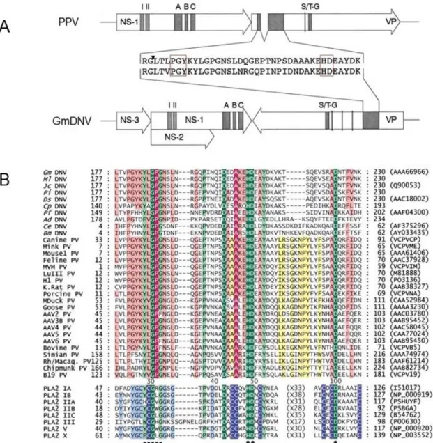
Figure 1.9 Conserved motifs within the viral genome and PLA2 motif………..….… 34
Figure 1.10 The architecture of parvovirus………. 36
Figure 1.11 Structure comparison of three densoviruses……….. 39
Figure 1.12 Adeno-associated virus (AAV) capsid structure………….………41
Chapter II
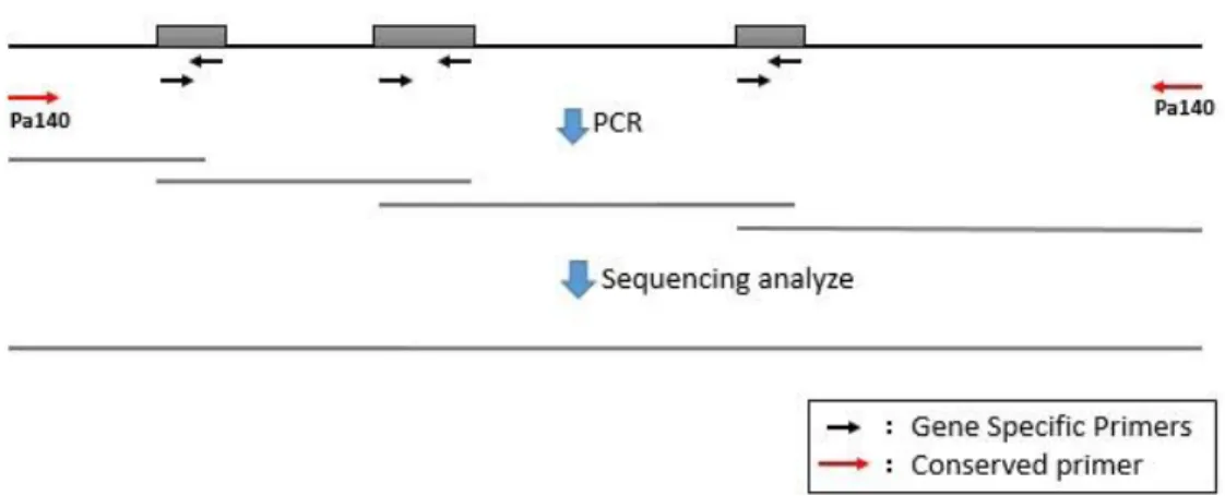
Figure 2.1 Model for identifying new viruses’ genome sequence from SISPA results……….. 46Chapter III
Figure 3.1 EM of SfDV virus particle and J-shaped telomere sequence of SfDV ITR….…… 59Figure 3.2 EM of PpDV virus particle and J-shaped telomere sequence of PpDV………….. 63
Figure 3.3 EM of DppIDV virus particle and J-shaped telomere sequence of DppIDV……… 67
Chapter IV
Figure 4.1 Iteradensoviruses and their transcription……….. 75Figure 4.2 Genome organization and transcription profiles of 5 iteradensoviruses………….. 79
xxi
Chapter V
Figure 5.1 Genome organization of AdDNV………...…100 Figure 5.2 SDS-PAGE, Northern-blotting and promoter activities………..102 Figure 5.3 Results for mRNA sequencing and mass spectrometry………... 105 Figure 5.4 Conformation of the splicing between ORF-A and ORF-B……….. 106 Figure 5.5 Expression of pFastbac VP constructs……….………...108 Figure 5.6 Potential VP1 proteins encoded by the VP gene cassette………..… 110 Figure S1: Annotated sequence of the AdDNV strain isolated in 1977... 116 Figure S2: MS results...…... 117 Figure S3: Similarities of AdDNV ORF sequences with those of other densoviruses…….. 118 Figure 5.7: Genome analysis of AdSDV………...126 Figure 5.8: Telomeres and gnome organization of AdSDV………...127 Figure 5.9: The new recombinant cricket aphrodisiac iridescent virus……….129 Figure 5.10: Hypothesis on the origin of AdSDV………...…..131
Chapter VI: Discussion
Figure 6.1 Genome comparison of iteradensoviruses………..………. 137 Figure 6.2 Structure comparison of AdDNV and sAAV...…... 144
Appendix I
Figure a1.1 Structure of the AdDNV capsid protein………..…. 181 Figure a1.2 Heat treatment and PLA2 activity of the virus particles…….………... 183 Figure a1.3 SDS-PAGE of heat-treated AdDNV particles……….…..…………. 186 Figure a1.4 Fourier shell correlation (FSC)……….……..….……. 187 Figure a1.5 Structure of AdDNV emptied particles………...……….… 191 Figure a1.6 Glycine-rich regions comparison……….…..192 Figure a1.7 Phylogenetic analysis………..………….…. 193 Figure a1.8 Structure of the three ordered ssDNA bases in AdDNV particles…….…….… 197 Figure a1.9 Phylogenetic Tree for AAV1-AAV13 and sAAV……….…....… 202 Figure a1.10 Pseudo-atomic model of sAAV ...……….………. 204 Figure a1.11 AAV capsid structures ...……….……… 205
xxii
LIST OF ABBREVIATIONS
3D Three-dimensional
Å
ångström
aa / a.a. Amino acid
AaeDNV Aedes aegypti densovirus
AalDNV Aedes albopictus densovirus
AAV Adeno-associated virus
AdDNV Acheta domesticus densovirus
AdMADV Acheta domesticus mini ambidensovirus
AdSDV Acheta domesticus segment densovirus
AgDNV Anopheles gambiae densovirus
AMV Avian myeloblastosis virus
ATP Adenosine triphosphate
B19 human parvovirus B19
BgDV1 Blattella germanica densovirus 1
BmDNV-1 (BmDV) Bombyx mori densovirus
CeDNV (CeDV) Casphalia extranea densovirus
CpDNV (CpDV) Culex pipens densovirus
CppDNV Culex pipiens pallens densovirus
DNVs (DVs) Densoviruses
xxiii
DPE Downstream promoter element
DppIDV Danaus plexippus plexippus iteradensovirus
DMSO Dimethyl sulfoxide
FBS Foetal bovine serum
FPV Feline parvovirus
g (μg,mg) Gram
GFP Green fluorescence protein
GmDNV Galleria mellonella densovirus
HeDNV Haemagogus equinus densovirus
hr Hour
ICTV International Committee on Taxonomy of Viruses
IF Immunoflourescence
IHHNV Infectious Hypodermal and Hematopoietic
Necrosis Virus
Inr Initiator
ITRs Inverted Terminal Repeats
JcDNV (JcDV) Junonia coenia densovirus
KDa Kilo Dalton
MlDNV (MlDV) Mythimna loreyi densovirus
MpDNV Myzus persicae densovirus
MS Mass spectrometry
xxiv
NS Non-structural protein
nt (nts) Nucleotide(s)
ORFs Open reading frames
PBS Phosphate-buffered saline
PcDV Planococcus citri densovirus
PfDNV (PfDV) Periplaneta fuliginosa densovirus
PiDNV Pseudoplusia includens densovirus
PLA2 Phospholipase A2
PpDNV (PpDV) Papilio polyxenes densovirus
PPV Porcine parvovirus
PstDNV (PstDV1) Penaeus stylirostris penstyldensovirus
RACE Rapid Amplification of cDNA Ends
RCR Rolling-circle replication
RCRE Rolling-circle replication initiation endonuclease
RF Replicative-form
RHR Rolling hairpin replication
SF3H Superfamily 3 helicase
SfDNV (SfDV) Sibine fusca densovirus
ssDNA Single-stranded DNA
wt Wild-type
VP Viral protein
VRs Variable regions
2
4
1
Viruses and biological control
Arthropods are undoubtedly the most widespread and diverse groups of animals, with an estimated 4–6 million species worldwide (Novotny et al., 2002). While only a small percentage of arthropods are classified as pest species, they nevertheless cause major devastation to crops, destroying around 18% of the world’s annual crop production (Oerke et al., 2004), contributing to losses of nearly 20% of stored food grains (Bergvinson et al., 2004), and causing around US$100 billion in damage each year (Carlini et al., 2002). In 2001, a total of US$7.56 billion was spent in order to protect crops from damage by invertebrate phytophagous pest species (Nicholson, 2007).
Pest control is a worldwide problem in agricultural and forest ecosystem management. Broad spectrum chemical pesticides have been used abundantly in the containment and eradication of pests of medical, veterinary, agricultural and environmental importance. Though chemical crop protection plays an important role in modern agricultural practices (Fest et al., 1982), it is still viewed as a profit-induced poisoning of the environment. The nondegradable chemical residues, which accumulate to harmful levels, are the root cause of health and environmental hazards and deserve most of the present hostility toward them. Meanwhile, the widespread use of classical agrochemical pesticides, along with their limited number of nervous system targets, has inevitably resulted in widespread resistance among arthropod populations. Moreover, synthetic pesticides often disrupt the balanced insect communities (Kurstak, 1982). This leads to the interest in biological control methods for vectors and plant pests (De Bach, 1964).
Recent advances have highlighted the potential of genetic engineering in the development of novel bio-insecticides which utilize natural organisms, or their products, in the production of transgenic plants or recombinant baculoviruses (Nicholson, 2007). Baculoviruses are arthropod-specific, unable to infect vertebrates or plants (Herniou et
5
al., 2003), and have been applied to protect crops in the agricultural sector since the
1930s (Inceoglu et al., 2001). However, they are limited by their slow ‘time-to-kill’: it may take days to weeks after infection from applying virus to stop feeding, with consequent further damage to the crop (Bonning et al., 1996). The development of recombinant baculoviruses has greatly improved the speed of action via the insertion of foreign gene products. However, the public reticence of the use of genetically modified organisms has contributed to their limited commercial development. Thus, viruses that act rapidly will be of great importance as viable alternatives to chemical pesticides. As an example, the polydnaviruses, which are introduced into insect larvae through parasitism to temporarily reduce or knock out insect immunity in favour of a parasitoid, are potential candidates for biological control with insect viruses (Lavine et
al., 1995).
The densoviruses are another group of viruses with great potential for biological control due to their high virulence and infectivity for their natural hosts, although their use as viral pesticides has so far been limited. Field trials to control different species of mosquitos with a Densovirus infecting the mosquito Aedes aegypti showed that the virus had a significant efficiency (Buchatsky et al., 1987). The insecticidal potential of
Galleria mellonella Densovirus on its host, the greater wax moth, has also been studied
(Tal et al., 1993). Densoviruses were also successfully used for controlling the pests in oil palm fields in Colombia and Ivory Coast infested with Sibine fusca and Casphalia
extranea (Fédière et al., 2002), respectively (Figure 1.1). In this case, application of as
few as 10-50 infected larvae per hactare was sufficient to achieve over 90% protection (Belloncik, 1989, Genty et al., 1975). The virus can spread rapidly to adjacent parcels, and the larvae stop feeding quite fast, usually after 1-2 days and start showing paralysis only 8-10 days after infection. Although it does not carry over very well during the wet and dry seasons in West Africa, this successful method of using densovirus for biological control showed great potential.
6
Figure 1.1 Densovirus for biology control.
Casphalia extranea densovirus (CeDNV) used for biological control in oil palm fields of Ivory
Coast. A) Oil palm leaves destroyed by CeDNV. B) Helicopters were used for spraying the virus.
2
Insect parvoviruses (Densoviruses)
2.1 Genotype and Taxonomic structural history of Densovirues
The first densovirus was discovered in 1964 when Meynadier and his colleagues observed a high mortality within an insect rearing facility of larvae of the greater wax moth G. mellonella in their research unit at St-Christol (France). They observed an outbreak caused by a new virus that was identified as a densonucleosis virus according to its cytopathological symptoms (Meynadier et al., 1964). A second densovirus,
7
BmDNV-1, was discovered just after the GmDNV isolation in sericultural farms in Ina City, Japan (Kawase, 1985, Kawase et al., 1976). Since then, an increasing number of densoviruses have been reported as a group of pathogens that are highly pathogenic and fatal to their hosts.
Densoviruses (DNVs) are parvoviruses of invertebrates, infecting at least insects, shrimps, and urchins, and form a separate subfamily (Densovirinae) within the the
Parvoviridae family (Cotmore et al., 2014a, Gudenkauf et al., 2014, Tijssen et al., 2011).
Nevertheless, DNVs share many propertyes with the Parvovirinae subfamily of vertebrates such as a single-stranded (ss), linear DNA genome of about 4-6 kb, terminal hairpins in the genomes which are for some viruses in the form of inverted terminal repeats (ITRs) and similar icosahedral capsid structures consisting of 60 proteins some of which have N-terminal extensions (Chapman et al., 2006, Cotmore
et al., 2006a, Tijssen et al., 2006a).
Together with the Parvovirinae subfamily of vertebrates, with which they share little sequence identity, the Densovirinae make up the family of Parvoviridae (Berns et
al., 2000). Thus far, a limited number of around thirty densoviruses were divided into
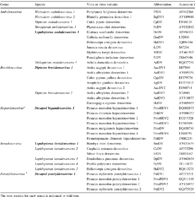
three genera, Densovirus, Iteravirus and Brevidensovirus (Anonyme, 2000). Due to the growing knowledge about these densoviruses, the taxonomy of Densovirinae subfamily was last modified in 2004, prior to publication of the 8th International Committee on Taxonomy of Viruses (ICTV) Report (Tattersall et al., 2005). According to this reclassification, the genus Densovirus was divided into three subgroups (subgroup A. B and C) and a single new genus, the Penfudensovirus was introduced.
In 2014, a set of proposals to update the taxonomy of the family Parvoviridae has been submitted by the ICTV Parvoviridae Study Group (Cotmore et al., 2014a), and have been approved by ICTV. The new taxonomy followed a novel viral definition rule: “In order for an agent to be classified in the family Parvoviridae, it must be judged to be an authentic parvovirus on the basis of having been isolated and sequenced or,
8
failing this, on the basis of having been sequenced in tissues, secretions, or excretions of unambiguous host origin, supported by evidence of its distribution in multiple individual hosts in a pattern that is compatible with dissemination by infection. The sequence must be in one piece, contain all the non-structural (NS) and viral particle (VP) coding regions, and meet the size constraints and motif patterns characteristic of the family” (Cotmore et al., 2014a). In the subfamily Densovirinae, proposed changes include the introduction of two new genera for shrimp viruses and the expansion of the existing genus names Iteravirus and Densovirus to Iteradensovirus and
Ambidensovirus, respectively. In both genera, species identity levels have been
lowered, numbered, binomial species names adopted, and new species introduced (Cotmore et al., 2014a). For viruses from the subfamily Densovirinae, viral names have typically been assembled from binomial host names plus the word ‘‘densovirus’’, for example, Junonia coenia densovirus, originally abbreviated to JcDNV (where the capitalized ‘‘N’’ harks back to a time when these viruses were called ‘‘densonucleosis viruses’’). The Study Group suggested eliminating the vestigial N from all abbreviations (Table 1.1). The new taxonomy of subfamily Densovirinae includes genera
Ambidensovirus, Iteradensovirus, Brevidensovirus and two new genera for shrimp
9
Table 1.1 New classification of Densovirinae subfamily. *
* According to the new classification of Densovirinae subfamily, the abbreviation names for viruses have also been changed. i.e. GmDNV->GmDV, MlDNV->MlDV, CeDNV->CeDV. In this thesis, we will use the new abbreviations in the research results obtained since 2014. For the viruses reported before the newly released classification of 2014, we will still keep the original abbreviation.
10
2.2 Pathology caused by densoviruses
The densoviruses are responsible for fatal diseases of their host. Mortality due to densovirus infections takes effect between two to twenty days after infection depending on the inoculating virus concentration and the insect larval stage (Meynadier et al., 1964, Shimizu, 1975). Generally the first symptoms are anorexia and lethargy followed by flaccidity and the inhibition of moulting and metamorphosis. During infection, larvae become whitish and progressively paralyzed, followed by a slow melanisation (Meynadier et al., 1964, Vago et al., 1966). Insect larvae usually develop anorexia a few days after infection and stop feeding (Kawase et al., 1990). Densoviruses of Sibine fusca and Casphalia extranea produce tumor-like lesions in the intestines of their hosts. The midgut epithelial cells of diseased larvae undergo intensive proliferation and the progressive thickening and opacity of the gut wall screens off the intestinal content (Fédière, 2000). In the case of C. extranea, the larval color changes from green to yellowish brown and the transparent gut becomes opaque (Fédière, 2000). With BmDNV-1 infected silkworm larvae, the alimental canal of the diseased larvae is pale yellow with little internal content and the larvae usually die after seven days showing body flaccidity (Shimizu, 1975).
2.3 Genome organization of densoviruses
Densoviruses package a monopartite linear single-stranded DNA molecule (ssDNA) of 4-6 kb within their virion of either a negative or a positive polarity. The proportion of encapsidated negative and positive strands of DNA varies according to the virus genus. For members of the Iteradensovirus genus, both strands are encapsidated in an equimolar proportion sharing that characteristic with vertebrate dependoviruses (AAVs) (Berns et al., 1972). In contrast, in Brevidensovirus, AaeDNV and AalDNV, the negative strand is preferentially encapsidated (90%) similarly to vertebrate parvovirus such as MVM and PPV (Muzyczka et al., 2001, Tijssen, 1995). Cloning of the complete genome of several densoviruses and determination of their sequence has provided a prediction of the genomic organization of this group of viruses.
11
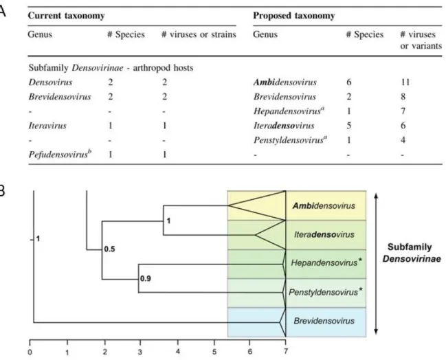
The emerging number of new densoviruses led to a reclassification of their taxonomy (Cotmore et al., 2014a) (Table 1.2).
Table 1.2 Five new classified genera of Densovirinae subfamily.
A) Summary of changes between the previous and new taxonomy. B) Phylogenetic tree showing genera in the subfamily Densovirinae. Phylogenetic analysis is based on the amino acid sequence of the viral replication initiator protein, NS1, which contains a conserved AAA+ helicase domain corresponding to the Parvo_NS1 Pfam domain:
http://pfam.sanger.ac.uk/family/Parvo_NS1. The size of the color block for each genus indicates the relative number of species currently recognized, as an indicator of its diversity (Cotmore et
12
2.3.1 Genome organization of the Ambidensovirus genus
This genus includes all densoviruses having an ambisense genome organization, with the VP (structural protein) and NS (non-structural protein or Rep protein) on the 5’ halves of the complementary strands, and with inverted terminal repeats (ITRs) (Tijssen et al., 2006a). Members were sub-divided into six host species. Blattodean
ambidensovirus 1 and Blattodean ambidensovirus 2 are two cockroach host species
include PfDV and BgDV1 (Mukha et al., 2006), respectively. Dipteran ambidensovirus 1 includes the mosquito ambidensovirus CpDV. PcDV (Thao et al., 2001) is a densovirus infecting mealybugs and belongs to Hemipteran ambidensovirus 1. The Lepidopteran ambidensoviruses contain the classical ambisense densoviruses usually isolated from lepidopteran insects such as JcDV. The densovirus isolated from cricket host Acheta domesticus first belonging to a separate genus Pefudensovirus was reclassified as the sixth host species group of Ambidensovirus genus. Figure 1.2 shows the comparison of the open reading frames (ORFs) for each of the classical members.
13
Figure 1.2 ORFs of ambidensoviruses.
The large open reading frames (ORFs) for the member from different group of Ambidensovirus genus. The graphical analysis tool ‘ORF Finder’ http://www.ncbi.nlm.nih.gov/gorf/orfig.cgi has been used for analyze virus genome sequence. The ‘+’ or ‘-’ with number indicate different frames of complement strands.
14
Both of the complementary DNA strands of ambisense densoviruses have the capacity to code for proteins through a limited number of large ORFs distributed within the 5’-half of the complementary strands. One strand codes for structural proteins, usually 4 (VP1-VP4) (Tijssen et al., 1976), whereas the complementary strand codes for the three non-structural proteins NS1-NS3. As exemplified by GmDNV, which is the first densovirus isolated from insects (Meynadier et al., 1964), the members from
Lepidopteran ambidensovirus 1 exhibit an identical organization of their genome. The
genome of GmDNV contains a long inverted terminal repeat (ITR) at both ends of about 550 nts. The palindromic sequence of 136 nts folds back to create a Y-shaped hairpin with flip and flop orientations (Tijssen et al., 2003). Minus and plus strands package in separate virions in a 1/1 ratio. Three ORFs coding for three NS proteins are present on one strand, the left-most ORF (near the 5' extremity) being in frame with, and separated from the largest NS ORF only by a TAA stop codon. The largest ORF codes for NS1 (Rep protein) and contains the NTPase motif that is typically found in NS1 of all parvoviruses. A single large ORF codes for the four structural polypeptides on the complementary strand. The VP gene containing phospholipase A2 (PLA2) motif is within the N-terminus of the VP protein. The genome of AdDNV is different in that (i) three (two of which overlap) ORFs are present on one strand, each likely coding for a single NS protein, and (ii) the VP gene coding for the four structural polypeptides on the complementary strand is split in two and, successively with two reading frames (Liu
et al., 2011) (Figure 1.2).
Other Lepidopteran densoviruses such as Diatraea saccharalis Densovirus DsDNV, Pseudoplusia includens Densovirus PiDNV and Junonia coenia Densovirus JcDNV share the same characteristics and high sequence identity with GmDNV. However, other ambidensoviruses isolated from orders other than Lepidoptera (Table 1.1), such as PfDNV and CpDNV are not homogenous in genome organization. These viruses have a shorter genome of around 5.5 kb and smaller structural protein molar masses as well as relative amounts that are distinct from the classical members (Figure 1.2). They also have shorter ITRs that are different in both size and structure.
15
2.3.2 Genome organization of Iteradensovirus genus
All members in the Iteradensovirus genus possess a monosense genomic organization, i.e. their sequences coding for NS and VP proteins are located in the left and the right halves of the same strand. Until a few years ago, only three iteradensoviruses were known: BmDNV-1 from Bombyx mori (Nakagaki et al., 1980), CeDNV from Casphalia extranea (Fédière et al., 2002) and DpDNV from Dendrolimus
punctatus (Wang et al., 2005). Their genomes are about 5 kb in length with 70-85%
sequence identity, particularly in their typical J-shaped ITRs. Recently, a second densovirus HaDNV-1 was reported in Helicoverpa armigera which was clustered into this genus by phylogenic analysis but complete ITRs sequences were not been obtained (Xu et al., 2012). All the iteradensovirus genomes contain three large open reading frames, two overlap in the left-hand half of the genome and encoding non-structural proteins (NS1 and NS2) and one in the right-hand half encoding capsid proteins (VP1-VP4) (Figure 1.3). NS1 protein contains replicator protein and helicase/ATPase motifs. The GPG and HD regions in VP1 capsid protein is conserved among most parvoviruses and contain the Ca2+ binding motif and phospholipase A2 (pLA2) motif.
Figure 1.3 Genome organizations of iteradensoviruses.
Comparison of genome organizations for Casphalia extranea densovirus CeDNV, Bombyx mori densovirus BmDNV-1 and Dendrolimus punctatus densovirus DpDNV.
16
2.3.3 Genome organization of the Brevidensovirus genus
Most members in this group are isolated from persistently or chronically infected mosquito cell lines: Aedes aegypti Densovirus AaeDNV (Afanasiev et al., 1991), Aedes albopictus Densovirus AalDNV (Boublik et al., 1994), Anopheles
gambiae Densovirus AgDNV (Ren et al., 2008), Culex pipiens pallens densovirus
CppDNV (Zhai et al., 2008), and Haemagogus equines Densovirus HeDNV (Paterson
et al., 2005). Their hosts are known as the epidemiologically essential vectors for
several dangerous human pathogens such as yellow fever virus, dengue fever virus, malaria, chikungunya virus, and West Nile virus. Therefore, mosquito brevidensoviruses are potential biological insecticides to control these paratransgenic pathogens. Members in Brevidensovirus genus contain the smallest genome (about 4 kb) in the Densovirinae subfamily. The genomic organization is also monosense, but this genus has some distinct characteristics from other genera. Their genomes do not have ITRs but contain distinct T-shaped hairpin telomeres. They have two large ORFs for NS genes instead of three, as in the case of the Ambidensovirus genus. In addation, there is only one large ORF for the VP genes, but it does not contain the PLA2 motif.
2.3.4 Genome organization of the Hepandensovirus and Penstyldensovirus genus
All densoviruses isolated from shrimp were reclassified into two new genera.
Hepandensovirus is the genus of viruses formerly known as hepatopancreatic
parvovirus [HPV] of shrimp, and Penstyldensovirus is a siglum for Penaeus stylirostris densovirus which formerly known as infectious hypodermal and hematopoietic necrosis virus (IHHNV) of shrimp. The genome of the classical shrimp densovirus (PstDNV = IHHNV) also contains three large ORFs as for the brevidensoviruses. The 5’ ORF sequence shares some identity with brevidensovirus NS1 with a rolling-circle replication motif at the N-terminus and an NTP-binding/helicase/NTPase motif at the C-terminus (Pham et al. unpublished data). The upstream, mid, and downstream.
17
ORFs of the PstDNV sequence (3873 nts, ~98% complete) have potential coding capacities of 666 amino acids (aa), 363 aa, and 329 aa, respectively.
2.4 Transcription strategy of densoviruses
Several models for vertebrate parvovirus transcription profiles have been determined at a detailed level. In each case, the parvoviruses have adapted a complex pattern of alternative splicing and polyadenylation to maximize the information from their small genomes. These complex patterns of expression generally result in a set of mRNAs that express two to three capsid proteins from overlapping ORFs, and one large and two to three small non-structural proteins. In addition, adeno-associated virus (AAV) gene expression has evolved to be dependent on helper-virus function (Qiu et
al., 2006). The core promoter element for gene expression usually contains a TATAA
consensus sequence and an upstream SP1 binding site with a GC rich motif (Lorson
et al., 1996). The activity of this promoter is NS1-dependent. Figure 1.4 illustrates a
summary of the porcine parvovirus (PPV) transcription profile. The PPV genome contains two open reading frames (ORFs). The left ORF allows the production of NS proteins from the P4 promoter. The single primary transcript (gray, top) contains two splice sites. The splice site of the small intron is located downstream of the stop codon of the NS protein, and therefore does not change their sequence. The production of the NS1 protein is from non-spliced ORF in the mRNA. Splicing within this ORF allows the production of the NS2 and the sequence after the splice is in a different reading frame relative to NS1. PPV uses a single polyadenylation signal (AAAn) located at the end of the coding regions providing mRNA considerably longer than the coding region for the transcripts of the NS proteins. The structural proteins are produced from a single primary transcript from the right ORF and are transcribed from the p40 promoter. There are two donor sites and one acceptor site for splicing. The first splice donor site removes the start codon of the protein VP1, resulting in the production of the VP2 protein. The second splicing donor site retains the initiation codon for VP1. The VP1 and VP2 proteins come from the same reading frame.
18
Figure 1.4 Porcine parvovirus transcription map.
D1 and D2 represent the first and second donor sites of VP transcripts, respectively. R represents the receptor sites for splicing (Bergeron et al., 1993).
19
The expression strategies of densoviruses vary significantly. Densoviruses have been accorded little attention for the gene expression of their genes compared to well-known strategies of the vertebrate parvoviruses. As for most parvoviruses in the
Parvovirinae subfamily, all densoviral genomes contain at least two distinct promoter
sets for non-structural (NS) and structural (VP) genes. Densoviruses also employ alternative splicing, leaky scanning, and polyadenylation mechanisms for producing different proteins from their limited genomes.
Insect gene expression requires transcription factors that are often different from those of vertebrate genes. For instance, insect cells lack Sp1 that recognizes GC-rich domains (Courey et al., 1988, Santoro et al., 1988). Although the TATA box is the most common core promoter element for insect gene expression, almost one half of studied Drosophila promoters can be classified as TATA-less promoters (Arkhipova, 1995). A conserved downstream core promoter element (DPE) has been recognized within TATA-less promoters in Drosophila, which is required for the sequence-specific binding of the transcription factor, TFIID. The DPE is located from +28 to +32 relative to the +1 transcription start site. Moreover, in Drosophila, the DPE appears to be about as common as the TATA box (Kadonaga, 2002). Arthropod initiator elements for transcription starts have a proposed consensus of ATCAG/TTC/T (often CAGT) which allows RNA polymerase II to function even in the absence of a clear TATA-box. The DPE, with the consensus A/G/T-C/G-A/T-C/T-A/C/G-C/T acts in conjunction with the initiator sequence (Inr) (CAGT), to provide a binding site for TFIID in the absence of a TATA box.
To date, the transcription strategies of ambidensoviruses are the most were described. The detailed RNA processing profiles for GmDNV, MlDNV, JcDNV and BgDNV have been reported (Fédière et al., 2004, Kapelinskaya et al., 2011, Tijssen et
al., 2003, Wang et al., 2013). Their ambisense nature dictates that transcription occurs
on both strands. Indeed, VP and NS genes are transcribed independently, each from one species of un-spliced mRNA, from promoters located within the ITRs (Bergoin et
20
al., 2000). The TATA-box motifs are within the terminal repeats and the CAGT-box in
the unique sequence. Figure 1.5A shows the detailed transcription map for MlDNV (Fédière et al., 2004). The NS and VP genes are located on the 5’-halves of the complementary strands and their transcripts have an overlap of 57 nucleotides in the middle of the genome. Two sizes of NS transcripts were detected that start 27 nts downstream of the 5’ ITRs. The NS cassette consists of three genes with NS1 and the overlapping NS2 downstream of NS3. Only one promoter, with the TATA-box located within the upstream ITR, drives the expression of the NS transcripts. The NS3 gene was spliced out from a fraction of the NS transcripts to allow leaky scanning translation of the downstream bicistronic NS1 and NS2 genes. The splicing donor site is immediately upstream of the initiation codon of NS3 and the acceptor site immediately upstream of NS1, so that both spliced and unspliced transcripts have an identical leader sequence except for the last two nts (GT) preceding NS3 (Fédière et al., 2004). One large ORF on the right-hand half of the complementary strand codes for approximately four VP proteins. No splicing has been detected within the VP transcripts, suggesting the four VPs were similarly generated by leaky scanning translation of an unspliced mRNA. The 5’-untranslated region of the VP transcript is only seven nucleotides long. The sequence context around the ATG start codons also seems to determine how well that ATG is recognized (Fédière et al., 2004). Thus, the untranslated 5'-terminal sequence of the mRNA coding for the structural polypeptides is very short, so that only a small percentage of the ribosomes initiate translation at the first AUG codon. As a result, VP1 is poorly expressed (Fédière et al., 2004). In contrast, the high level of expression of VP4 is due to the favorable environment of its two initiator codons. Unlike the situation with vertebrate parvoviruses whose mRNAs co-terminate at the 3' extremity of the genome, MlDNV possesses two polyadenylation (poly(A) signals (AATAAA) in the middle of its genome. The first two nts, AA, in the poly(A) sequence of the NS transcript overlap with the NS stop codon. Therefore, the VP and NS transcripts have 56 nts overlapping sequence. The termination of both the VP and NS transcripts occurs very close to the end of their coding sequences, so that only the 5'-halves of the complementary strands are transcribed. This probably prevents the meeting of the polymerase complexes when
21
the genes are only partially transcribed (Bergoin et al., 2000). GmDNV and JcDNV have similar genome organization to MlDNV and hence, their expression strategies have only a few differences. In GmDNV and JcDNV, the two sizes of NS transcripts start at 23 and 32 nts downstream of the left ITRs, respectively (Tijssen et al., 2003, Wang et al., 2013). In JcDNV, a single mRNA for expressing VP proteins is transcribed from the P9 promoter. While the P93 is the unique putative promoter on the strand bearing NS genes, a single transcription start for the two NS mRNAs was located 32 nt downstream of the P93 TATA box but 83 nt and 86 nt upstream of NS3 and NS1, respectively. With their TATA box just at the limit of the ITR and their regulatory sequences within the ITR, the P9 and P93 appear to be both structurally and functionally very similar (Wang et al., 2013). (Note: the authors used the original orientation of the JcDNV genome: in the new sequence P9 becomes P91 and P93 becomes P9 (Pham et al., 2013d)).
The Blattella germanica densovirus (BgDNV) is an autonomous parvovirus that infects the German cockroach. This virus belongs to the Ambidensovirus genus and possesses a 5,335-nt-long ambisense genome that contains five basic ORFs. Two of them (ORF1 and ORF2) on one strand code for VP, and three (ORF3 to ORF5) on another strand code for NS proteins (Kapelinskaya et al., 2008). Moreover, one additional ORF, ORF6, was found to be located on the same strand as ORF3 and ORF5 but within the VP-coding region.The reported expression strategy of BgDNV was unique among the other ambisense densoviruses (Kapelinskaya et al., 2008, Kapelinskaya et al., 2011). BgDNV possesses three mRNAs transcribed from the single P2 promoter for NS proteins, with spliced and unspliced transcript variants. The unspliced variant encodes NS3 which is similar to the classical ambidensoviruses (GmDNV, MlDNV), while the first splice product encodes NS1 and NS2. The second splice product encodes the C-proximal half of NS1. Long NS transcripts have approximately 1,600 nt of overlap with mRNAs for VP proteins. This overlap may lead to the formation of a relatively long region of double-stranded RNA (dsRNA) during the virus life cycle, which in turn can induce a cellular antiviral response by means of RNA interference (Kapelinskaya et al., 2011). BgDNV possesses three VP transcripts which
22
are transcribed from a single promoter P1, one of which (VP) is unspliced, while the other two (VPspl1 and VPspl2) are generated by alternative splicing. The unspliced VP transcript contains both ORF1 and ORF2, while in VPspl1, ORF1 and ORF2 are joined in frame (Figure 1.5 C). Like the majority of densoviruses, BgDNV possesses only one termination site for mRNAs for both capsid and NS proteins.
The brevidensoviruses with monosense genomic organization contain three large ORFs coding for NS and VP gene. Compared to other densoviruses, the transcription pattern of brevidensoviruses has not been well characterized. it was reported that PstDNV generates five transcripts: two spliced for NS1, two for NS2 and one for VP under the control of three promoters P2, P11, and P61 (Dhar et al., 2010). Those results have been corrected recently (Pham et al. unpublished data). Figure 1.5 B gives a detailed transcription map of AalDNV as an example (Pham et al., 2013a). AalDNV used one promoter region with closely overlapping elements to start transcription of NS1 and NS2 at positions that are just 14 nt apart at either side of ATGNS1. All NS mRNAs co-terminated with VP mRNA. No clear TATA-like motif sequences were found upstream of the initiator sequence CAGT of the VP of AalDNV and AaeDNV, suggesting that these promoters were under the control of DPE (Pham
et al., 2013a). This regulatory circuit is likely to be one means by which insect virus
networks can transmit transcriptional signals, such as those from DPE-specific and TATA-specific enhancers, via distinct pathways (Hsu et al., 2008), to regulate NS and VP expression.
23
Figure 1.5 Details of MlDNV, AalDNV and BgDNV transcription map.
A) MlDNV transcription map: two putative promoter regions within the left and right ITRs control the expression of NS and VP transcripts. The 5’-splicing donor and 3’-splice acceptor sites are upstream NS3 and NS1, respectively. The two poly(A) signals are in the middle of the MlDNV genome. The initiation and termination of NS and VP transcripts are also indicated (Fédière et
al., 2004). B) AalDNV transcription map: two putative Inr of NS1 and NS2 and one Inr of VP gene.
NS and VP gene expression share the same polyadenylation signal downstream of VP (Pham et
24
2.5 Terminal structures of Densovirus genomes
Priming a DNA molecule during the replication process is a key step. DNA viruses have evolved a number of unusual mechanisms to prime the replication to their genome such as RNA priming, DNA priming and even protein priming (Flint et al., 2009). Parvoviruses replicate their short (~ 5 kb) linear single-stranded DNA genomes using a unidirectional strand-displacement strategy called “rolling hairpin” replication, which is an evolutionary adaptation of rolling circle synthesis (Chapman et al., 2006). This adaptation is mediated by small imperfect palindromes positioned at each end of the viral genome, or so-called terminal hairpins, on their 3’ and 5’ extremities of both complementary strands. This terminal structural contain most of the cis-acting information required for viral DNA replication and packaging (Li et al., 2013). Despite these central roles in replication, the sizes, sequences, and predicted structures of the hairpins can vary remarkably between different genera of the Parvoviridae and even between the two ends of a single virus, suggesting they have been adapted to fulfill additional roles in the viral life cycle.
In densoviruses, the presence of these terminal palindromic structures has been proven by both direct observation using electron microscopy as well as by direct sequencing for several viruses and prediction of their secondary structures (Dumas et
al., 1992, Fédière et al., 2002, Li et al., 2001, Tijssen et al., 2006a). Those palindromic
sequences at the termini can fold into Y-shaped, J-shaped, T-shaped or I-shaped hairpins (Figure 1.6) and serve as primers for viral replication at the 3’-extremity. In addition to their role in viral DNA replication, they allow the rescue of the cloned genome during experimental transfections, both in vitro and in vivo (Jourdan et al., 1990).
Hairpin structure of ambidensoviruses, especially for those classical viruses belonging to the Lepidopteran ambidensovirus 1 group possess relatively long ITRs. JcDNV has ITRs of 517 nts long (Dumas et al., 1992), which exceeds the size of all
25
known parvovirus ITRs, such as B19 (383 nts) (Zhi et al., 2004) and AAV2 (145 nts) (Lusby et al., 1980). The distal 96 nucleotide form a Y-shaped hairpin which is typical for parvovirus termini with the flip and its complement flop configuration. Their terminal structure contain at least four important elements: (i) the origin of replication at the approximate position of nt 95; (ii) a site-specific nicking enzyme that recognizes only ds DNA at approximately the same site in the complementary strand (terminal resolution); (iii) a flip/flop region (nts 35 - 65) that is most likely important in positioning enzymes that act at the genome ends, and, (iv) for some members (i.e. GmDNV, JcDNV, MlDNV, PiDNV), promoter elements for both the NS and VP genes (Bergoin
et al., 2000). The other non-classical ambidensoviruses have shorter ITRs with a
different structure. For AdDNV, the ITRs are 144 nts, of which the distal 114 nts could fold into a perfect I-type palindromic hairpin (Liu et al., 2011).
The iteradensovirus genomes have ITRs that are only 225 - 230 nts long of which the terminal 150 - 160 nts may form a hairpin. This hairpin does not form a typical T- or Y-type structure as seen with the other parvoviruses but possesses a J-shaped structure with two imperfectly base-paired side arms and can be found in either form, flip or flop (Fédière et al., 2002, Li et al., 2001) (Figure 1.6 B). The origin of replication is therefore located about 70 nts into the ITR and the nicking site is predicted to be located at about 165 nts into the ITRs.
The brevidensoviruses have the smallest genomes among all parvoviruses. Their terminal structures lack ITRs and resemble the majority of vertebrate autonomous parvoviruses. This dissimilarity at the ends may also explain why different amounts of the complementary strands are encapsidated. This may be due to a difference in the packaging signal (probably in the ITRs, which in the other two genera result in equimolar packaging) or due to asymmetric strand displacement (Bergoin et al., 2000). However, these terminal hairpins can fold back on themselves into T-shaped structure with 134 nts and 182 nts at either ends (Figure 1.6 C).
26
Figure 1.6 Hairpin structure of different densoviruses.
A) JcDNV (Ambidensovirus genus) left and right hairpins(Pham et al., 2013d). B) CeDNV (Iteradensovirus genus) flip and flop (Fédière et al., 2002). C) AaeDNV (Brevidensovirus genus) 3’ and 5’ terminal hairpins. (Bergoin et al., 2000, Fédière et al., 2002)