HAL Id: tel-01059806
https://tel.archives-ouvertes.fr/tel-01059806
Submitted on 2 Sep 2014HAL is a multi-disciplinary open access archive for the deposit and dissemination of sci-entific research documents, whether they are pub-lished or not. The documents may come from teaching and research institutions in France or abroad, or from public or private research centers.
L’archive ouverte pluridisciplinaire HAL, est destinée au dépôt et à la diffusion de documents scientifiques de niveau recherche, publiés ou non, émanant des établissements d’enseignement et de recherche français ou étrangers, des laboratoires publics ou privés.
CD8 T cell differentiation during immune responses
Sara Sofia de Campos Pereira Lemos
To cite this version:
Sara Sofia de Campos Pereira Lemos. CD8 T cell differentiation during immune responses. Immunol-ogy. Université René Descartes - Paris V, 2014. English. �NNT : 2014PA05T009�. �tel-01059806�
1 UNIVERSITÉ PARIS DESCARTES
FACULTÉ DE MEDECINE-site Necker INSERM U1020 (ex U591)
_______________________________________________________________________
THÈSE
Pour obtenir le grade de
DOCTEUR
Sciences de la Vie et de la Santé
Discipline: Immunologie École Doctoral: Gc2iD
Présentée et soutenue publiquement par
Sara Sofia de Campos Pereira Lemos
Le 23 Mai 2014
CD8 T cell differentiation during immune responses
Jury:
Dr. Jacqueline MARVEL Rapporteur
Dr. Sylvie GUERDER Rapporteur
Dr. Jérôme DELON Examinateur
Dr. Nicolas BLANCHARD Examinateur
2
To a wonderful person, an extraordinary woman and an amazing mother...
my mother, Arlete Pereira
“The first precept was never to accept a thing as true until I knew
it as such without a single doubt.”
René Descartes(French philosopher and scientist)
“We live where no one knows the answer
and the struggle is to figure out the question”
Joshua Schimel(in “Writing Science” book).
“ To be great, be whole;
Exclude nothing, exaggerate nothing that is not you.
Be whole in everything. Put all you are
Into the smallest thing you do.
So, in each lake, the moon shines with splendor
Because it blooms up above.”
Fernando Pessoa (Portuguese poet)
“The beauty of life is not about finding something big enough,
but rather on finding something deep enough!
3
Acknowledgments
My very first and respectful acknowledgment I owe to an extraordinary scientist and remarkable person: my supervisor Benedita ROCHA. Thank you for accepting me in your lab, for supervising this work, for all the opportunities given to grow as a PhD student and as a person, for all the freedom at the bench, and yet for always pushing me forward. It was such an HONOR and CHALLENGE to work with you!
To all jury members, a special thank you for accepting to evaluate this thesis work. To Jacqueline MARVEL and Sylvie GUERDER a particularly thank you for your precise and critical analyze of my thesis work as rapporteurs. To Jérôme DELON and Nicolas BLANCHARD a special thank you for your patience in waiting for the final version and for revising this work as examinateurs. It was also an HONOR to have all of you judging this work.
From Institut Pasteur, I would like to thank Antonio FREITAS for providing us with OT-1 mice, Matthew ALBERT for MyD88-/- mice and Hélène DECALUWE for preparing LCMV and performing the corresponding immunizations. From Necker Institute, I would like to thank Alain CHARBIT at INSERM U1002 for his advice and bacteria expertise, and for using his bacteria lab facilities too. To his team members, thank you for always making me feel truly welcome in a “bacterial” lab (my 2nd lab/home!). Thank you to all members of INSERM U1020 (ex U591). Sophie EZINE for all the B6 Thy1.1 mice given, for pushing my organizational skill to top levels and for always being there to solve lab problems and to guide me in the thesis manuscript/defense procedures. Orly AZOGUI for truly understanding my complainings about FlowJo crashes and for always being available to discuss science! (a special thanks for precious help with my thesis summary in French too). Florence VASSEUR and Pierric PARENT for big help with mice during the lab moving from Necker Institut to Broussais Hospital. Agnes LEGRAND for precious help on single-cell multiplex RT-PCR experiments, for teaching me SDS program, for all your sincerity, efficiency and friendship. To all secretaries, for patience with my complicated orders of new products or mice. Chantal for helping me with DNA extractions for mice genotyping (a tremendous time consuming job to do alone!). Benoir for efficiently providing material disappeared at the last minute. Evelyn, for always reminding me that the world is round and that sometimes we need to say a big NO (I should practice it more!!!). To the new members of the new INSERM U1151-team 13, Flora ZAVALA and Sarantis KORNIOTIS, a big thank you for your kindness, happy “bonjours” and powerful “courage Sara, c’est bientôt la fin!”, during the last tough weeks of thesis writing!
To Cesar EVARISTO and Patricia SANTOS for being awesome "lab parents" to me and Miguel, for NEVER having denied a scientific discussion, for your truth friendship and for all the tuga's dinners around Paris! To Cesar, extra thanks for being ALWAYS available to teach me: at 4 am when dilacerating spleens to plate; at 2 am when fighting for a 3rd (not broken) Canto to not lose our incredible amount of work; and even at 7.000 Km away, for answering all my specific questions, or sending me your data to combine with mine (what a nightmare!). My gratefulness and respect to you are infinite! Thanks for the astonishing book “Writing Science”. To Patricia extra thanks for your objectivity, sincerity and CLEVER talks either about science or life! I particularly enjoyed the ones in the corridors or those seated on FACS flow boxes! To Miguel FERREIRA, a true "brother in science", thank you for the immense patience to discuss with me homologous recombination tricks (as well as a bunch of other scientific stuff) and for being always on
4 my right side (of the office) to listen, to talk, to share, or simply just to be there with a true friendship. An extra thanks for being the only one TRULY understanding the meaning of a back pain, or a cold feeling when huge experiments take more than 20h. (Contributions to my French metro-poetry posters’ collection, not forgotten!). To Vanessa ZEPPONI, my "French lab sister", an immense thank you for always being there, at my left side (of the bench), for teaching me new concepts and views about life, and for ALL FUNNY moments lived in the lab and around Paris! An extra thank you for beers or Japanese moments of friendship (Disneyland and a lot of not taken T3 tramways moments too!) and also for always helping me with French language, essential to survive in the lab and in the Paris-quotidian life!
To Hsueh SUNG, a big thank you for being always available to answer, at distance, my 1001 questions about practical details of the CD8 inflammatory project. Thanks a lot for all the true friendship, PATIENCE AND RESPECT (it was a pity that we couldn´t work together on the bench!).
To all current postdocs, an enormous thank you for the “mental support” during my thesis writing (advice, tricks and frustrations included! you know how hard it was to leave my lovely bench!). Alessia GALGANO for all the discussions about molecular biology and lab (dis)organization. Marie CHERRIER, for sharing with me much more than the office in this last year, for all your sincerity, advice, friendship, scientific talks, and good lab mood (once one is an OUTSTANDING person, one will be an outstanding group leader too. All the best!). To Gerald KRENN and Alexander BARINOV… to you “my” guys… My gratitude and respect are INFINITE. A truthful thank you for all your friendship, your sincerity, wise advice, men’s objectivity, confidence and beers that always comforted me! I’ll miss a lot our Austro-Russian-Luso “canteen-gang”!!! A special thanks for the multicultural "soirées" in the company of your wives too. To Ricarda KRENN, a special thanks for reviewing my English writing. To Pedro GONÇALVES for friendship and all the supporting: Write Sara!!! Write Sara!!! I’m still deeply grateful to Gerald, Alexander, Pedro and Vanessa for helping me when my computer broke down 3 days before sending my manuscript to evaluation! (what a panic attack!).
To the current students Victoria M. LOPEZ and Thomas HARBONNIER, thank you for all the good moments, friendship, and “student’s solidarity”! (good luck!!!).
To former members of the Necker lab with whom I also shared big moments around pipettes, reagents and laughs, a sincere thank you for being so nice with me: Sylvain MEUNIER, Laetitia PEAUDECERF, Lamina SKHIRI, Marie BÉDORA, Amine BOUDIL, and Kateryna KOZYRYTSKA. Special thanks to all the" petits" that were quite big friends: Paula MARTINS, Cédric DONATHELLO and Coraline CANIVET for the "on mange the hors!", Yann BECKER for "poker face" laughing moments and Laurent BEZIAUD for COMEDY moments and for being so present and supportive (and I am so sorry for being always late! - I’m a disaster in terms of orientation !!!) (good luck for your thesis writing!). An IMMEASURABLE thank you to my (maternal) FAMILY! My MOTHER, for unconditional love and support during every single day of my life! My UNCLES, those who welcomed me several times at the airport, or those who always made my favorite chocolate cake for departure! My COUSINS, those who always say to me “Sara wake up! You live like a nomad!”, those who never deny card games during our gatherings of family dinners, and yet, the ones who never let me sing alone (in particular Linkin Park songs!) and always gave me those sincere and powerful hugs! My maternal GRANDPARENTS and GREAT-UNCLES for always being there with open loving arms. To other RELATIVES who constantly
5 ask my mother “how is Sara, there? when is she back?” a special thanks too. When one grows up in a small village, your NEIGHBORS are also your family, thus a big thank you for all the Sangemil-Penalva-Viseu’s love and care.
To my true and UNFORGETTABLE Biochemistry friends since the University of Coimbra era, an immeasurable thank you too, for being always so present in my life with a sincere friendship, even if more than 1500Km away! Thank you for being ALWAYS there. It is splendid how we stay so “covalent bonded” after all these years (not even an electron moved out from their orbit/our friendship!). An infinite thank you for all lunches or dinners to feed friendship/“matar saudades”, specially the Christmas ones! Verinha, Anita, Clau, Jony, Ana Marisa, Isa, Rafa, Carol, Tânia, [e respectivo(a)s]… we definitely are a family! To Cité Universitaire Paris friends, for amazing friendships, picnics and “soirées”. Especially those who never denied our mussels dinners and its corresponding WALKS to feed our friendship! Cleopatra Silvana for being an outstanding friend in this day and age and also my “FAMILY” during my stay in Paris. Lucia M. Teixeira for unlimited immunology cross-talks, around pots and pans in the kitchen! (and for always pointing me the RIGHT DIRECTION when the hardness of a PhD overshadowed me!)
To Paris University Club volley-ball team members for electrifying and relaxing journeys of volleyball games (specially the “tournois”! and the ones “sur la pelouse CiteU” with Cyril, Flo, Caro, Gael et Pierre). A particular thanks to Marisa Baptista, for UNLIMITED discussions about Immunology, for a great friendship and for funny trips in the company of two other EXTRAORDINARY friends: Andreia Santos and Daniela Melo- thank you so much for keeping me away from “the dark hole of thesis writing depression” with an outstanding friendship (I’m also dedicating this thesis to both of you!); to Catarina Almeida, for more than 20 years of true and OUTSTANDING friendship; and to my high school Philosophy teacher (Ana Paula Agostinho), who cleverly taught me how to see “THE INVISIBLE” of objects and subjects, and their importance in life (with just a single question!).
A special thanks to my previous supervisors, at Karolinska Institut, for giving me the opportunity to experience a wonderful research lab environment and for teaching and guiding me in my first steps in the Immunology world. That infectious Immunology passion gained there was crucial to my being here today!
This PhD thesis was supported by:
International Individual Ph.D Scholarship -SFRH/BD/47001/2008-, (2009-2012), from Foundation of Science and Technology (FCT), Ministry of Science (Portugal).
“Aides Individuelles Jeunes Chercheurs - Aides doctorales”/ PhD Scholarship-DOC20121206103-, (2013), from Fondation ARC pour la Recherche sur le Cancer (France).
Extra support was given by Doctoral School Gc2iD (grant covering the inscription fee for the D.U. “Formation spéciale à l’expérimentation animale - Niveau 1”) and by LABEX (grant covering the registration fee for the “3rd
EFIS-EJI Summer School in Clinical Immunology”).
To ICBAS-University of Porto (Portugal) thank you for receiving me as PhD student at the starting date of my FCT-PhD scholarship.
6
Table of Contents
Abbreviations ... 8
List of figures and tables ... 10
Summary ... 11
Introduction ... 12
I. The Immune System ... 13
1. Innate Immune System... 13
2. Adaptive Immune System... 13
3. Cellular elements of immune system ... 14
3.1. Hemathopoiese ... 14
3.2. T cell development: thymocytes ... 15
3.2.1. Thymocytes as short-lived cells (a notion to reconsider) ... 18
3.3. Innate and adaptive cells of the immune system ... 19
II. Innate immune response ... 21
1. Inflammation: the key component of innate responses ... 21
2. Pattern recognition receptors (PRRs): innate immune recognition ... 23
3. Monocytes/Macrophages ... 29
4. Neutrophils ... 31
5. Natural killer cells ... 32
6. Dendritic cells ... 33
III. Acquired immune response ... 36
CD8 T cell response ... 36
1. TCRαβ structure ... 36
2. T-cell co-receptor: CD8αβ... 37
3. Immunological synapse and TCR triggering ... 38
4. TCR downstream signaling: MAPKs (Erk, p38, JNK), NF-kB, and AKT pathways 39 5. Negative regulation of TCR signaling ... 43
6. TCR downregulation ... 44
7. Cell surface phenotypic modifications after T cell activation ... 46
8. Lymphocyte traffic ... 50
9. Effector functions: cytolysis, chemokine and chemokine production ... 56
10. Transcription factors: the intrinsic controls ... 63
7
12. Memory CD8 T cells and secondary immune responses ... 72
IV. Methods and models to evaluate T-cell immune responses ... 78
1. Fluorencent-labeled pMHC multimers ... 78
2. TCR Transgenic T cells ... 80
3. Models of infection: LM and LCMV ... 82
Aims and experimental approaches ... 87
Results ... 96
Article I ... 97
Epitope specificity and relative clonal abundance do not affect CD8 differentiation patterns during Lymphocytic Choriomeningitis Virus infection Munitic I*, Decaluwe H*, Evaristo C, Lemos S, Wlodarczyk M, Worth A, Le Bon A, Selin LK, Rivière Y, Di Santo JP, Borrow P, Rocha B Journal of Virology, 2009, 83(22):11795-807 Article II ... 111
Equally efficient primary CD8 immune responses generate memory with different protection capacity Sara Lemos*, César Evaristo*, Ivana Munitic, Hélène Decaluwe, Iharilalao Dubail, Alain Charbit and Benedita Rocha Manuscript submitted Article III ... 142
Cognate antigen stimulation generates potent CD8+ inflammatory effector T cells Sung H-C*, Lemos S*, Ribeiro-Santos P, Kozyrytska K, Vasseur F, Legrand A, Charbit A, Rocha B and Evaristo C Frontiers in Immunology. 2013. 4:452. Discussion ... 158
Bibliography ... 174
Annexes ... 196
Article IV ... 197
Thymocytes may persist and differentiate without any input from bone-marrow progenitors Peaudecerf L, Lemos S, Galgano A, Krenn G, Vasseur F, Di Santo JP, Ezine S, Rocha B. Journal of Experimental Medicine, 2012, 209(8):1401-8 Abstract S068- 3rd European Congress Immunology. Immunology. 2012 Sep. Vol 137. ... 209
List of courses, conferences and congresses during the thesis ... 210
8
Abbreviations
AIRE Autoimmune regulator AP-1 Activator protein 1
APC Antigen presenting cell
ATP Adenosine triphosphate
BAFF B cell-activating factor Bcl-6 B-cell lymphoma 6
BCR B cell receptor
BIR Baculovirus inhibitor repeats Blimp1 B-lymphocyte induced maturation
protein
C Carboxyl terminal region
C3,4,5 Complement fragments 3,4,5 Ca2+ Calcium
CaMK Ca2+-camlodulin-dependent kinase CARD Caspase activation and recruitment
domains
CD Cluster of differentiation cDC conventional Dendritic Cell
CDRs Complementary-determining regions CFSE Carboxyfluorescein diacetate
succinimidyl ester Clb Casitas B-lymphoma
CLP Common myeloid progenitors CLR C-type lectin receptor CMP Common myeloid progenitors CpG cytidine-phosphate-guanosine CRAC Ca2+ release-activation Ca2+ channels Csk C-terminal Src kinase
cSMAC Central supramolecular activation cluster CTL Cytotoxic T lymphocyte
CTLA-4 Cytotoxic T lymphocyte antigen-4
DAG Diacylglycerol
DAMP Damage-associated molecular patterns
DC Dendritic cell
DN Double negative
DNA Deoxyribonucleic acid
ds double strand
EMC Extracellular matrixes Eomes Eomesodermin ER Endoplasmic reticulum
Erk Extracellular signal-regulated kinase ETP Early T-lineage progenitor
Fc Fragment crystallizable region FOXP3 Forkhead box protein 3 FPR1 N-formyl peptide receptor FRC Fibroblastic reticular cells
GM-CSF Granulocyte-macrophage colony
stimulating factor
GP33 Glycoprotein 33 (residues 33 to 41) GRK2 G protein-coupled receptor kinase2 Gzm Granzyme
HA Hyaluronic acid
HEV High endothelial venules HSC Hematopoietic stem cell HSV Herpes simplex virus
HY Male-specific antigen
ICAM Intercellular adhesion molecule-1 IEL Intraepithelial lymphocytes IFN Interferon
Ig Immunoglobulin
IkB Inhibitor of NF-kB
IKKi Inducible inhibitor of NF-kB [ikB] kinase
IL Interleukyne
iNOS inducible nitric oxide synthase IP-10 Interferon gamma-induced protein 10
(CXCL10)
IP3 Inositol 1,4,5-triphosphate
IRAK Interleukin-1 Receptor-Associated Kinase IRF Interferon regulatory transcription factor
IS Immunological synape
ITAM Immunoreceptor tyrosine-based activation motif
ITIM Immunoreceptor tyrosine-based inhibition motif
JNK c-Jun NH2-terminal kinase KLRG1 Killer cell lectin-like receptor G-1
KO Knock out
LAT Linker for activation of T cells Lck Lymphocyte-specific Tyrosine Kinase LCMV Lymphocytic choriomeningitis virus LFA-1 Lymphocyte function-associated antigen1 LM Listeria Monocytogenes
LNs Lymph nodes
LPS Lipopolysaccharide LY6C Lymphocyte antigen 6C
Lyve-1 Lymphatic vessel endothelial hyaluronan receptor 1
MAC1 Macrophage receptor 1
MAPK Mitogen-activated protein kinase MDP Muramyl depeptide
MHC Major histocompatibility complex MIP Macrophage Inflammatory Proteins MPECs Memory precursor effector cells MPP Multipotent progenitors
9
mTOR Mammalian target of rapamycin
MyD88 Myeloid differentiation primary response gene 88
NADPH Nicotinamide adenine dinucleotide phosphate-oxidase
NET Neutrophil extracellular traps NFAT Nuclear factor of activated T cells NFkB Nuclear factor
kappa-light-chain-enhancer of activated B cells NK Natural killer cell
NK T Natural killer-T cell NLR Nod-like receptor
NO Nitric oxide
NOD Nucleotide-binding oligomerization domain
OT-1 OVA-trangenic
OVA Ovalbumin
p38 P38 mitogen-activated protein kinases PAMP Pathogen associated molecular patterns PD-1 Programmed death-1
pDC plasmacytoid Dendritic Cell PDGF Platelet-derived growth factor PECAM1 Platelet endothelial cell adhesion
molecule
PGE/PGI Prostaglandins E and I PI3K Phosphoinositide 3-kinase
PIP2 Phosphatidylinositol 4,5-bisphosphate PKC Protein kinase C
PLC Phospholipase C
PMN Polymorphonuclear leucokytes PNAd Periheral node addressins poly I:C polyinosinic-polycytidycic acid PPs Peyer’ patches
Prf1 Perforin
PRR Pathogen recognition receptor PSGL1 P-selectin glycoprotein ligand 1 pSMAC Peripheral supramolecular activation
cluster
PTK Protein with tyrosine kinase activity pTα pre-TCRα chain
PYD Pyrin domain
RAG Recombination activating gene RANTES Regulated on activation, normal T cell
expressed and secreted (CCL5) RIP2 Receptor interacting protein-2 RLR RIG-I-like receptors
RNA Ribonucleic acid ROS Reactive oxygen species S1P Sphingosine-1-phospate
S1P1 S1P-receptor 1
SCID Severe combined immunodeficiency Ser Serine
SHP1 SH2 domain-containing protein tyrosine phosphatase
SLECs Short-lived effector cells SLO Secondary lymphoid organs SLP-76 Src homology 2 domain-containing
leukocyte phosphoprotein of 76 kDa
SP Single positive
SPHK Sphingosine kinases
ss single strand
STAT Signal transducer and activator of transcription
TAP Transporter associated with antigen processing
TBK TANK binding kinase-1 TCM Central memory T cell
TCR T cell receptor
TD T cell-dependent
TEC Thymic ephitelial cell TEM Effector memory T cell
Tfh Follicular helper T cell
Tg Transgenic
TGF Transforming growth factor
tGPI trypomastigote
glycosylphosphatidylinositol mucin Th T helper cell
Thr Threonine
TI T cell-independent
TIM Inflammatory memory T cell
Tip-DC TNF- and iNOS-producing DC TIRAP Toll-interleukin 1 receptor (TIR)
domain containing adaptor protein TLR Toll like receptor
TN Triple negative
TNFα Tumor-necrosis factor-α TRAM TRIF-related adaptor molecule Treg Regulatory T cells
TRIF TIR-domain-containing adapter-inducing interferon-β
TRM Resident memory T cell
Tyr Tyrosine
V amino-terminal variable region VLA4 very late antigen 4
WT wild type
10
List of figures and tables
Introduction I - The immune system:
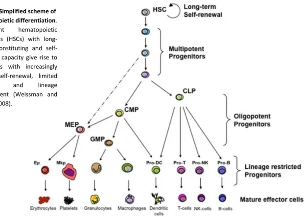
Figure 1. Simplified scheme of hematopoietic differentiation
Figure 2. Overview of T cell development in thymus: from an ETP to a mature CD8 T cell Introduction II - Innate immune response:
Figure 3. Simplified inflammatory pathway components
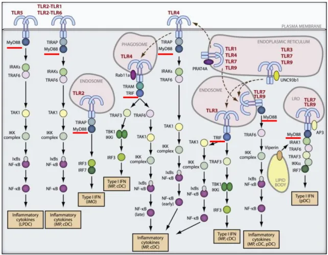
Table 1. Pattern recognition receptors (PRRs): TLRs, RLRs, NLRs, CLRs, and their ligands Figure 4. TLR trafficking and signaling
Table 2. Functional outputs of some of the genes upregulated by TLR4 Introduction III - Acquired immune response:
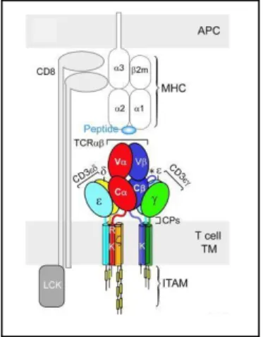
Figure 5. The TCRαβ/CD3 complex of T cells
Figure 6. TCR complex and CD8αβ heterodimer interactions with a pMHC class I molecule on an antigen presenting cell (APC): cooperative trimeric interaction
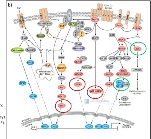
Figure 7. TCR-mediated signal transduction after pMHC interaction: proximal signaling complex and TCR downstream signaling pathways
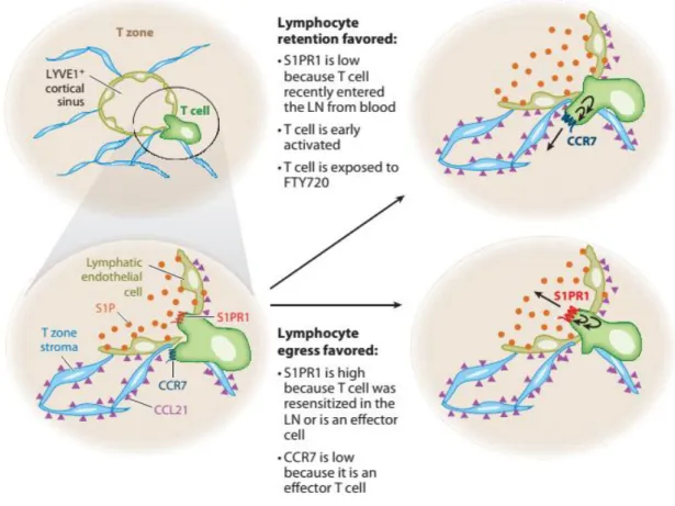
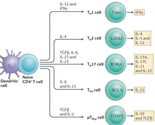
Figure 8. Model of events occurring during lymph node egress decision making. Figure 9. Differentiation of CD4+ T cells into different T helper (Th) subsets: instructed
cytokines, lineage-defining transcription factors, and signature expressed cytokines. Figure 10. Model of transcriptional programs controlling differentiation of antigen-specific
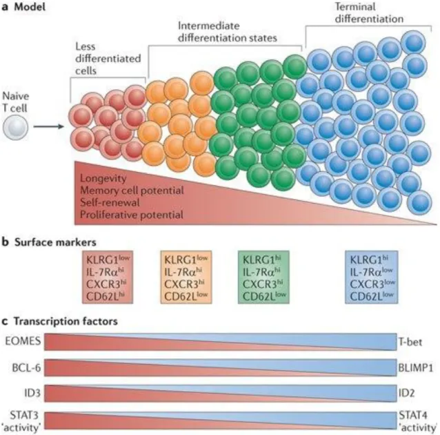
effector CD8 T cells: graded activity of transcription factors.
Figure 11. Different phases of a CD8 T cell response to an acute infection and heterogeneous CD8 T cell populations with different fates and memory potential
Table 3. Memory T cell subsets: TCM (T cell central memory), TEM (T cell effector memory) and TRM (tissue-resident memory T cells)
Figure 12. Models for generating effector and memory T cell heterogeneity Experimental approaches and aims of the thesis:
Figure 13. Outline of the quantitative single-cell multiplex RT-PCR technique
Figure 14. Used protocols: (A) to generate in vivo effector CD8 T cells, (B) to test cell recruitment capacity of pro-inflammatory effector.
37 37 41 54 69 71 75 75 77 93 95 15 18 23 24 27 28
11 Summary
CD8 T cells are essential for the elimination of intracellular pathogens and tumor cells. Understanding how naïve CD8 T cells differentiate into effector cells capable of eliminating pathogens and to generate adequate memory cells during immune responses is fundamental for optimal T cell vaccine design.
In this PhD thesis work we addressed two central questions:
1) What are the mechanisms by which early effector T cells could act as pro-inflammatory effectors? And what is their role in the immune response?
2) How heterogeneous are CD8 responses? Could different pathogens modulate CD8 T cell differentiation programs and be responsible for CD8 cell-to-cell heterogeneity? Could they also generate memory cells with different protection capacities?
To address these questions related to the diversity of CD8 T cell differentiation during immune responses, we used the single cell RT-PCR technique to detect ex vivo expression of mRNA in each individual cell, and Brefeldin A injected mice to detect ex vivo intracellular proteins. As experimental system to evaluate in vivo cell activation we used T cell receptor transgenic (TCR-Tg) CD8 T cells.
Since the use of TCR-Tg cells to study immune responses has been subjected to criticism (due to high frequency of naïve-precursor transfers), in a first Ms. we compared the behavior of TCR-Tg and endogenous (non-transgenic and present at low frequency) cells in the same mouse. We found fully overlapping behavior between these two cell populations, which reinforced the advantage of using TCR-Tg cells to study CD8 immune responses. In addition, we concluded that the frequency of naïve-precursors do not induce diversity on CD8 T cell differentiation patterns.
In a second Ms. we evaluated the impact of different pathogens in the diversity of CD8 T cell properties during two different immune responses: OT1 TCR-Tg cells (specific for OVA antigen) in the response to LM-OVA (Listeria
Monocytogenes expressing OVA) infection; and P14 TCR-Tg cells (specific for GP33 epitope) in the response to
Lymphocytic choriomeningitis vírus (LCMV) infection. We found that OT1 and P14 cells had different properties. As this difference could also be attributed to the different TCR avidity between OT1 and P14 cells, we then compared the behavior of P14 and OT-1 cells in the same mouse, co-injected with LM-OVA and LM-GP33. Since no differences were then detected, these results demonstrated that priming with different pathogens generates CD8 T cells with different characteristics that are not determined by TCR usage, but rather by the infection context. In addition, when looking for the protection capacity of endogenous CD8 memory cells generated in bacterial or viral context, we found that memory cells generated after LCMV priming were more efficient in responding to a second challenge, than memory cells generated after LM-GP33 priming. We also found that this better protection is associated with a T cell effector memory (TEM) phenotype associated with the LCMV infection, in contrast with a T
cell central memory (TCM) phenotype generated after LM-OVA infection. These results demonstrate that different
pathogens are responsible for diversity of CD8 T cell differentiation patterns and that even when distinct pathogens are efficiently eliminated during the primary immune response the quality of the memory generated may differ.
In a third Ms. we studied the mechanisms by which effector CD8 T cells attracted other cell types in the early days of an immune response. We used two experimental systems: the response of OT1 TCR-Tg cells to LM-OVA infection; and the response of anti-HY TCR-Tg cells to male cells (“sterile”-non infectious context). In both cases we found that immediately after activation, CD8 T cells expressed high levels of pro-inflammatory cytokines and chemokines (such as TNFα, XCL1, CCL3 and CCL4). We also confirmed the expression of these earlier mediators in a small fraction of activated endogenous cells, which could still be identified by pMHC multimers. A local injection of CD8 pro-inflammatory effectors in the ear induced: hypertrophy of the draining lymph node (DLN); recruitment of several leucokytes (B, T, NK, Monocytes, PMNs and DCs) into the DLN; and increased S1P levels in the DLN responsible for a cell egress block. This inflammatory potential was also detected after intranodal injection of a physiological number of CD8 pro-inflammatory effectors. In contrast with the classic cytotoxic CD8 T cell functions, the pro-inflammatory mediator’s expression declined with cell division and when antigen was still abundant. The rapid loss of CD8 inflammatory effector functions was correlated with an extensive TCR down-regulation at the cell surface, as well as with a down-down-regulation of the TCR signaling pathways (MAPkinases). These results demonstrated for the first time that CD8 responses involve two distinct effector phases with opposite rules (inflammatory and cytotoxic), and also that cognate antigen stimulation is sufficient to induce the CD8 inflammatory effector phase necessary for maximal screen of rare APCs first presenting the antigen.
In conclusion, our studies revealed diversity on CD8 T cell functions (inflammatory and cytotoxic) during immune responses and that different pathogens induce distinct CD8 T cell differentiation patterns. These results are thus crucial to predict and to efficiently evaluate CD8 T cell responses.
12
13
I. The Immune System
A key feature of the immune system is the ability to induce protective immunity against pathogens while maintaining tolerance towards self and innocuous environmental antigens, and it has evolved to protect organisms from diseases. It allows the host to detect and eliminate a diversity of pathogenic organisms that are themselves constantly evolving (virus, bacteria, and worms) and it also helps the host to eliminate toxic or allergic substances, and tumor cells. To mobilize a response against these threats, the host’s immune system needs to distinguish self from non-self, needs to distinguish foreign and harmful from the own healthy cells. This discrimination is essential in order to efficiently eliminate the threat without an excessive damage of self-tissues. Classically, the immune system has been divided in two categories: the innate immune system and the adaptive immune system.
1. Innate Immune System
Innate immune system acts as the first line of resistance against pathogen invasion and includes: i) physical barriers such as epithelial cells layers, secreted mucus overlaying the epithelium in the respiratory, gastrointestinal and genitourinary tracts, and the epithelial cilia that sweep away the mucus layer; ii) soluble proteins constitutively present in biological fluids (complement proteins, defensins and ficolins) or those released from activated cells (cytokines that regulate the function of other cells, chemokines that attract inflammatory leukocytes, lipid mediators of inflammation, reactive free radical species, and bioactive amines and enzymes that contribute to tissue inflammation); iii) membrane bound and cytoplasmic receptors that are expressed broadly on a large number of cells. These receptors are encoded, in their mature functional forms, by conserved and limited germ-line genes of the host, which enable the recognition of molecular patterns shared by many invading environmental signals (Rahman et al. 2008; Cui et al. 2011).
2. Adaptive Immune System
Unlike innate system, the adaptive immune system has restrict recognition for its target antigens, and is based primarily on antigen-specific receptors expressed on the surfaces of T- and B-lymphocytes. T cell receptors (TCR) and immunoglobulin (Ig) B cell receptors (BCR) are encoded by genes that are assembled by somatic rearrangement of germ-line gene elements. These rearrangements permit the formation of a vast diversity of receptors able to recognize virtually any type of antigen. The mechanisms governing the assembly and the selection of the B and T cell antigen receptors allows for a properly functioning repertoire of receptor-bearing cells. Besides the
14 specificity, another hallmark of adaptive immune responses is the production of long-lived (memory) cells that persist and can rapidly express effector functions after a second time encounter with their specific antigen.
In spite of being described as separate arms of the immune response, innate and adaptive systems act together with the innate response being the first line of host defense and the adaptive response becoming prominent after several days. Several components of the innate system contribute to acute inflammation induced by microbial infection or tissue damage, and also for the activation of adaptive antigen-specific cells essential for an effective immune response (Dutko and Oldstone 1983; Panus et al. 2000; Jang et al. 2009).
3. Cellular elements of immune system
3.1. Hemathopoiese
An immune response includes contributions from many subsets of leukocytes. Different leukocytes can be discriminated morphologically and by differentiation antigens on their membrane surfaces, also named as cluster of differentiation (CD).
All blood cells, including mature circulating lymphocytes, differentiate from the same progenitor cells, the hematopoietic stem cells (HSC), which are found in bone marrow, peripheral blood and placenta. These pluripotent HSCs are capable of self-renewal and multilineage differentiation, giving rise to all types of blood cells throughout an individual’s life by generating precursors of increasingly limited potential and lineage-bias. HSCs differentiate in multipotent progenitors (MPP), which have lost their self renewing capacity, and then further differentiate into common myeloid progenitors (CMP) or common lymphoid progenitors (CLP) (Fig. 1).
CMPs give rise to erythrocytes, platelets, macrophages and to distinct forms of granulocytes. Granulocyte lineage cells include neutrophils, eosinophils/mast cells and basophils. CLPs further differentiate into mature lymphocytes: T cells, B cells, natural killer (NK) cells (Weissman and Shizuru 2008).
15 3.2. T cell development: thymocytes
The development of functional T cells is essential for mounting a protective immune response against diverse threats. Whereas the majority of hematopoietic lineages mature in the bone marrow, T cell development takes place in a specialized organ: the thymus. This primary lymphoid organ is responsible for the generation and selection of T cells bearing a diverse T cell receptor (TCR) repertoire: restricted to self-major histocompatibility complexes (MHC) and tolerant to self-antigens. Besides the differentiation of distinct T cells: CD4, CD8α/β and γδ T cells, the thymus also supports the differentiation of NKT cells, regulatory T cells (Treg), and intraepithelial lymphocytes (IEL) (Weinreich and Hogquist 2008).
Due to the scope of this thesis in study CD8 T cell differentiation during immune responses (cells whose TCR is composed of an α and β chains: TCRαβ), only the TCRαβ-cell development will be described below, and in subsequent chapters, TCRαβ T cells will be simply designated as T cells.
The thymus is colonized by hematopoietic progenitors derived from HSC cells that then become committed towards a T cell differentiation program. T cell differentiation relies on multiple signals provided by the thymic stroma that is composed of dendritic cells, macrophages, endothelial cells, fibroblasts and thymic epithelial cells (TEC). Those signals include: growth factors (c-Kit ligand, FLT3L
Figure 1. Simplified scheme of hematopoietic differentiation. Pluripotent hematopoietic stem cells (HSCs) with long-term reconstituting and self-renewing capacity give rise to precursors with increasingly limited self-renewal, limited potential and lineage commitment (Weissman and Shizuru 2008).
16 and IL-7), chemokines (CCL25, CxCL12, CCL19 and CCL21) and cell surface receptor ligands (Notch ligand and peptide-MHC ligands) that sustain thymocyte survival, proliferation, migration and differentiation (Alves et al. 2009a). Among these, the cytokine IL-7, which is predominantly produced by TECs (Alves et al. 2009b), has a crucial role in promoting survival and expansion of early T cell precursors (Peschon et al. 1994).
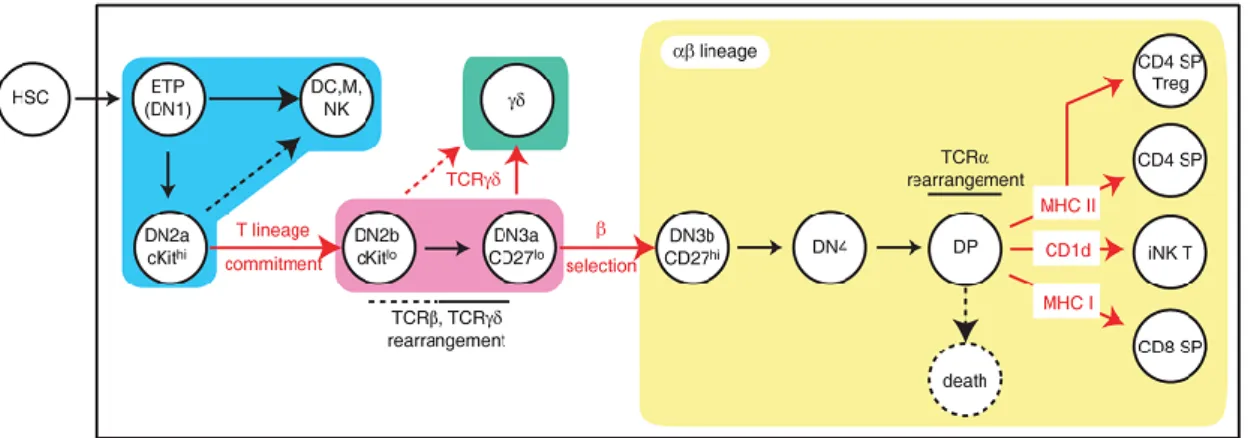
The identity of the cell progenitor, which initially seeds the thymus, is still open to debate, however it is established that the early T-lineage progenitor (ETP) is the most immature T-cell precursor within the thymus (Bhandoola et al. 2007; Benz et al. 2008). The majority of intrathymic precursors do not express CD4 nor CD8 surface markers, and hence, they are designated as double-negative (DN) thymocytes (in addition, as they do not express CD3 either, they are also nominated as triple-negative (TN) thymocytes). DN thymocytes are subdivided into 4 subpopulations according the surface expression profiles of CD44 (an adhesion receptor) and CD25 (the α chain of IL-2 receptor): DN1 (CD44+CD25-), DN2 (CD44+CD25+), DN3 (CD44-CD25+) and DN4 (CD44- CD25-) (Godfrey
et al. 1993). Early thymocytes possess multilineage potential, which is progressively restricted as cells
transit through the DN stages of T-cell development (Fig. 2).
DN1 thymocytes constitute a heterogeneous population with different potential to generate T cells and different maturation and proliferation capacities. This population can be further subdivided according to the IL-7Rα and c-Kit expression, where CD44+ CD25- c-Kithi IL-7Rα- are the most immature thymocytes, also known as ETPs (Allman et al. 2003). The ETPs retain yet the potential to develop NK, DCs, B and some myeloid cells. They do not express the recombination-activating gene (Rag) or the CD3ε genes at detectable levels, and they do not have D-J rearrangements of TCRβ locus. However, they still keep a strong proliferative capacity. In contrast, the most mature DN1 subpopulation expresses IL-7Rα and it expresses CD3ε and have D-J rearrangement of TCRβ locus (Porritt et al. 2004).
DN1 cells become committed to the T lineage upon Notch1-Delta-like four (Notch1-DL4) interactions on the thymic stroma, resulting in transition to the DN2 stage, which is characterized by the upregulation of CD25. DN2 population has lost the B cell potential but some cells retain yet NK and myeloid potential (Balciunaite et al. 2005). During the DN2- DN3 stages, lymphocyte and T-cell-specific factors such as Rag-1, Rag-2, pTα (pre-TCRα chain), CD3ε and IL-7R are up regulated (Taghon
et al. 2005).
DN3 thymocytes undergo intense V-DJ rearrangement of the TCRβ locus. Those cells that have generated a functionally rearranged TCRβ chain associate it with the invariant pre-TCRα and CD3 signaling molecules to assemble the pre-TCR on cell surface. This process is called β selection as thymocytes that fail to generate an in frame/functional TCRβ chain are not selected for further differentiation to αβ lineage and thus may die by apoptosis. Signaling through the pre-TCR promotes: proliferation, induce differentiation into DN4 cells, and inhibit further TCRβ rearrangement (allelic
17 exclusion) by negative regulation of Rag genes (reviewed in (von Boehmer et al. 1998; Ciofani and Zuniga-Pflucker 2007)).
In contrast with DN2 stage where T cell precursors express IL-7Rα and c-Kit, in late DN3 stage both of these receptors are downregulated, rendering DN3 cells dependent on pre-TCR and Notch1-DL signaling for survival (Ciofani and Zuniga-Pflucker 2005).
In mice lacking CD3ε or Rag-2 gene (TCRs-V(D)J recombination abrogated), thymocytes do not develop beyond the DN3 stage (Shinkai et al. 1992; DeJarnette et al. 1998). When IL-7 signaling is impaired (caused by loss of either α or common γ chains of the IL-7R) there is an arrest in the development at the DN2 stage (Peschon et al. 1994; Cao et al. 1995; Moore et al. 1996). Moreover, absence of IL-7 induces apoptosis of DN1, DN2 and DN3 thymocytes, and IL-7 assures survival of these thymocytes by increasing the intercellular levels of anti-apoptotic Bcl-2 and reduction of pro-apoptotic Bax (Kim et al. 1998).
Following β selection, DN3 cells express CD27 and progress to the DN4 stage, characterized by the loss of CD25 expression. The expression of CD4 and CD8 coreceptors is initiated and these thymocytes become designed as double positive (DP): CD8+CD4+ cells.
DP cells represent the majority of the thymocytes, they highly express Rag-1 and Rag-2, and they also initiate Tcrα gene rearrangements, resulting in the surface expression of TCRαβ/CD3 complexes. Due to the random nature of TCR α and β loci rearrangements, a vast diversity of TCRs is generated to cope with an immense variety of antigens. However, not all TCRs are capable to effectively interact with peptide-MHC (pMHC) complexes, while some will strongly recognize self-peptides. In order to generate useful and safe mature TCRαβ cells, DP thymocytes pass through a positive and negative selection processes. The fate of a DP cell is dependent on the signaling mediated by the interaction of the TCRαβ with self-peptides MHC class I and II, highly expressed by thymic ephitelial cells.
Positive selection: DP thymocytes bearing TCRαβ that fail to productively interact with self peptide-MHC class I or class II complexes die by neglect within few days. Thus, only thymocytes that receive signals from the TCR engagement are selected (positive selection). The TCR engagement is responsible not only for the survival but also for the maturation of double positive (DP) thymocytes on single positive (SP) cells. Based on the appropriate degree of interaction between the TCR and the pMHC complexes expressed on thymic epithelial cells, and depending on the class of MHC molecule recognized, thymocytes are positively selected either to a CD4+ or to a CD8+ single-positive (SP) cell fate. CD4 and CD8 molecules are coreceptors that bind to MHC class I and class II, respectively, favoring CD8+ and CD4+ T-cell MHC restriction. Their cytoplasmic domain binds to the tyrosine kinase Lck that, when in close vicinity to the TCR complex, initiates TCR signaling. Therefore, CD4 and CD8 coreceptors provide yet an extra guard against the selection of non-MHC reactive cells (coreceptors not engaged) as they sequester the Lck far from the TCR complex.
18 Negative selection: SP thymocytes carrying TCRαβ with high avidity for self pMHC (strong TCR signaling) undergo TCR-induced programmed cell death. Thus, only thymocytes that do not express TCR with high affinity for self antigens are selected (negative selection). This involves the exposure of thymocytes to peripheral tissue-specific antigens ectopically expressed by thymic epithelial cells under the control of the transcription factor AIRE (autoimmune regulator). This selection leads to the elimination of self-reactive T cells (reviewed in (Takahama 2006; Carpenter and Bosselut 2010)).
These selection processes ensure that only self-MHC-restricted and self-tolerant T cells survive and leave the thymus as mature T cells that continuously circulate between blood and lymph through secondary lymphoid organs in search of invading pathogens/cognate antigens interactions. Survival and homeostatic proliferation of individual naïve T cells in the periphery is dependent on continuous TCR signaling (interaction with self-pMHC ligands) (Tanchot et al. 1997) plus IL-7R signaling (Schluns et al. 2000), which occurs in secondary lymphoid organs.
Figure 2. Overview of T cell development in thymus: from an ETP to a mature CD8 T cell.
The earliest thymic precursors entering in the thymus derive from bone marrow hematopoietic stem cells (HSCs) and are designated as early T cell progenitors (ETPs). Expression of CD4 and CD8 defines DN (double negative) and DP (double positive) thymocytes. Expression of CD44 and CD25 defines four DN subtypes: DN1, DN2, DN3 and DN4. During the DN1-DN4 progression, thymocytes lose their potential to develop dendritic (DC), natural killer (NK) or myeloid (M) lineages and gain specific characteristics of T cell lineage commitment. β selection engages TCRαβ commitment of DN3 cells, and TCRαβ signaling upon MHC class I recognition induces differentiation of DPs into mature CD8+ single positive (SP) cells, which are released into the periphery and join the pool of naïve CD8 T cells (Carpenter and Bosselut 2010).
3.2.1. Thymocytes as short-lived cells (a notion to reconsider)
Surgical removal of the thymus demonstrated the requirement of this organ in the generation of T cells (Miller 1961). Likewise, thymus grafts have also been used to correct deficiencies of the thymus epithelium (Markert et al. 2007; Markert et al. 2011). However, they are not used to correct intrinsic T cell deficiencies, as it is believed that thymocytes are short-lived, and thus, continuous T cell differentiation in the thymus depends on constant supply of lymphocytes progenitors from bone
19 marrow (Berzins et al. 1998). This occurs even when the host is T cell deficient. Transplantation of wild-type thymi into hosts which are unable to generate mature T cells (SCID or Rag2-/- hosts) also show that, in the transplants, donor T cells are substituted by incompetent precursors from the host bone marrow within few weeks (Frey et al. 1992; Takeda et al. 1996).
3.3. Innate and adaptive cells of the immune system
Cellular elements of the innate system are: mast cells, basophiles, eosinophils, neutrophils, macrophages, dendritic cells (DC) and natural killer cells (NK). The major functions of the innate system cells include:
1) the initial recognition of foreign substances;
2) the recruitment of additional immune cells to the sites of infection and inflammation though the production of inflammatory mediators;
3) the elimination of microorganisms by phagocytosis, reactive oxygen species production, type I IFNs, or complement cascade activation;
4) triggering of the adaptive immune system through antigen presentation. These cells bear pattern recognition receptors (PRRs) that recognize a broad molecular patterns found on pathogens: PAMPs (pathogen associated molecular patterns).
Cellular elements of adaptive immune system are the T and B lymphocytes. Both of these cells recognize specific targets trough a vast diversity of receptors generated by genetic recombination of antigen receptor gene segments, associated to several other mechanisms, as reported above.
Activated B cells are responsible for antigen-specific antibodies secretion and they provide an important line of defense against infection through the neutralization and/or elimination of extracellular pathogens or foreign substances. In addition, B cells also function as antigen presenting cells, they produce multiple cytokines and they can suppress inflammatory responses that occur during autoimmune diseases or that can be caused by unresolved infection. B cells recognize and capture external antigens through their B cell receptor (BCR), a cell surface immunoglobulin (Ig) receptor which recognizes antigens directly without need for antigen processing. Upon binding to the BCR, the antigen is internalized by receptor-mediated endocytosis and it is processed by degradation into peptides. These antigenic peptides are displayed by B cells on their surface MHC class II molecules, where they can be recognized by antigen-specific T helper cells. This B and T cell interaction (T cell-dependent (TD) activation of B cells) provides the co-stimulatory signal required for B cells to differentiate into high-affinity antibody-producing plasma cells and to develop into memory B cell populations. B cells can also be activated and produce antibodies in a T-cell independent (TI) mode through: signaling of their TLRs (Toll like receptors), BCR crosslinking, and help signals provided by bone-marrow-derived myeloid cells. However, the nature of antigens
20 recognized and the outcome of antibody responses are distinct between TD and TI B cell activation (Mauri and Bosma 2012; Vinuesa and Chang 2013; Yuseff et al. 2013).
T cells are divided in two major subsets: CD8+ cytotoxic (CTLs) and CD4+ helper T (Th) cells. In general, CD8 T cells respond to intracellular infections with virus, protozoa and intracytoplasmatic bacteria, and also to tumor cells. Upon antigen recognition through interaction of the TCR and the processed pathogen/malignant-derived peptide bound on MHC class I of an APC, CD8 T cells specifically kill the infected or tumoral cells by production of cytotoxic molecules. They also produce cytokines, chemokines and microbicidial and anti viral molecules to combat infection (Harty et al. 2000).
CD4 T cells play an important role in coordinating acquired immune responses. There are several Th cell subsets (Th1, Th2, Th17, Th21, Treg), and each of which has specialized functions to control immune responses. These subsets emerge from naïve CD4 T cells after specific recognition of antigen-derived peptide displayed on MHC class II molecules of APCs and B cells. Both the TCR-mediated stimulation and the cytokine environment influence the fate decision of naïve CD4 T cells towards distinct Th subsets. Each Th cell subset expresses a unique set of transcription factors and produce hallmark cytokines that promote: CD8 T cell or B cell differentiation and memory establishment; enhance innate immune components’ action; or even suppress the immune response (Yamane and Paul 2013).
21
II. Innate immune response
The innate immune system not only monitors the host for microbes but also the health of the host’s own cells. In response to pathogens and cell injury, the innate immune system alerts the adaptive immune system to a potential problem in order to generate a specific response through antigen presentation. However, in parallel, the innate immune system also rapidly mobilizes innate defenses to the site of injury through the generation of an inflammatory response.
1. Inflammation: the key component of innate responses
Inflammation is an immediate response that is triggered by noxious stimuli and conditions, such as infection and tissue’s injury (classical acute or chronic inflammatory response), or such as tissue’s stress or malfunction (para-inflammatory response). The defining features of inflammation are redness, swelling, heat, pain, and loss of tissue function which have as physiological basis the local and transient: vasodilatation, leakage of plasma soluble molecules, and migration of leukocytes out of blood vessels into the surroundings of the affected tissue.
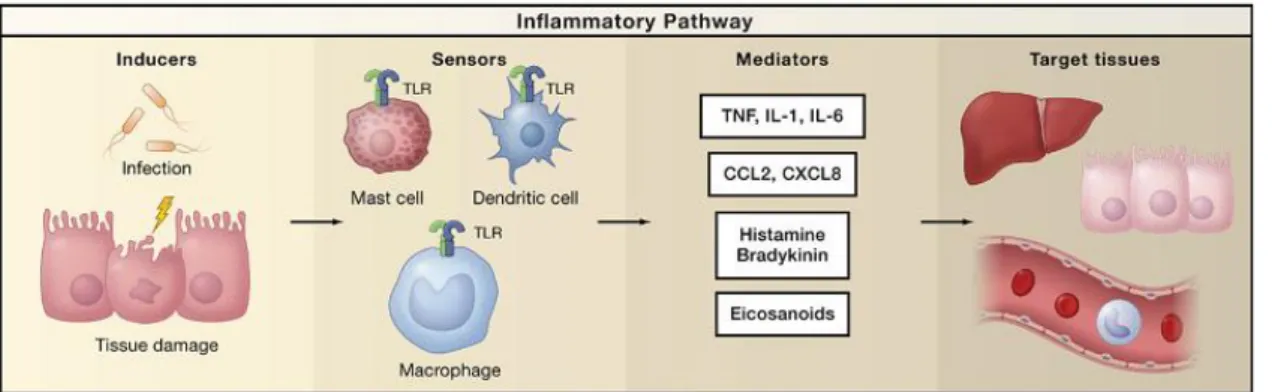
The physiologic purpose of inflammation is to restore the homeostasis, resulting in the elimination of the infectious agents and/or the repair of tissue injury. A typical inflammatory response consists of four components: i) the inflammatory inducers, ii) the sensors that detect them, iii) the inflammatory mediators induced by the sensors, and iv) the inflammatory effectors/target tissues that are affected by the inflammatory mediators (Fig. 3) (Nathan 2002; Medzhitov 2010).
Inflammation is triggered when innate immune cells residing in tissues (macrophages, mast cells and dendritic cells) or non professional immune cells (epithelial cells, endothelial cells and fibroblasts), as well as circulating monocytes and neutrophils, recognize pathogen invasion or cell damage through their intracellular or surface-expressed pattern recognition receptors (PRRs). These receptors (inflammatory sensors), directly or indirectly detect inflammatory inducers like pathogen-associated molecular patterns (PAMPs) or damage-pathogen-associated molecular patterns (DAMPs). DAMPs are nuclear or cytosolic host molecules that when released outside the cell, or exposed on cell surface following tissue injury or cell necrosis, can contribute to a sterile (noninfectious) inflammation. They include ATP, the cytokine IL1α, uric acid, calcium-binding proteins, DNA-binding nuclear proteins, amyloid β fibrils, heat shock proteins, defensins, phagocyte-specific proteins, etc. By contrast, PAMPs are exogenous molecules of both pathogenic and non-pathogenic origin; they are shared by a large group of microorganisms and are often conserved products essential for microbial survival. They include bacterial and viral nucleic acids, fungal β-glucan and α-mannan cell wall components, bacterial protein flagellin, components of the peptidoglycan bacterial cell wall, lipopolysaccharide (LPS) from Gram-negative bacteria, etc (Medzhitov 2008; Newton and Dixit 2012).
22 Inflammatory mediators are responsible for vascular alterations and for the recruitment of leukocytes.
Vasoactive amines (histamine and serotonin) are produced in an all-or-none matter by mast cells and platelets degranulation, and they are responsible for increased vascular permeability and vasodilatation. Vasoactive peptides can be stored in an active form in secretory vesicles (like substance P) or generated by proteolytic processing of inactive precursors in the extracellular fluid (for example, kinins, fibrinopeptide and fibrin degradation products). Substance P is released by sensory neurons and promotes itself mast-cell degranulation. Other vasoactive peptides are generated through proteolysis by the Hageman factor, thrombin or plasmin and cause vasodilatation and increased vascular permeability. The Hageman factor activates the kallikrein-kinincascate resulting in bradykinin production which has a pain-stimulating effect. The complement fragments C3a, C4a and C5a (known as anaphylatoxins) are produced via several pathways of complement activation. C5a, in a higher extension than C3a and C4a, promote granulocyte and monocyte recruitment and induce mast cell degranulation, therefore affecting the vasculature. Lipid mediators, (such as eicosanoids and platelet-activating factors) are derived from phosphatidylcholine, a phospholipid present in the inner leaflet of cellular membranes. After activation by intracellular Ca2+ ions, cytosolic phospholipase A2 generates arachidonic acid and lysophosphatidic acid. Arachidonic acid is metabolized to form eicosanoids either by cyclooxygenases, which generate prostaglandins and thromboxanes, or by lipoxygenases, which generate leukotrienes and lipoxins. The prostaglandins PGE2 and PGI2 cause vasodilatation and PGE2 induce high sensitivity to pain and fever, and can stimulate DCs and promote IL-12 production, which is necessary for efficient antigen presentation and T ell activation. Lipotoxins (and dietary ω3-fatty-acid-derived resolvins and protectins) inhibit inflammation and promote resolution of inflammation and tissue repair. Platelet-activating factors are generated by the acetylation of lysophospatidic acid and induce recruitment of leukocytes, vasodilatation and vasoconstriction, increase vascular permeability and platelet activation. Proteolytic enzymes (including elastin, chathepsins and matrix metalloproteinases) through degradation of extra cellular matrix and basement-membrane proteins are involved in host defense, tissue remodeling and leukocyte migration (reviewed in Medzhitov 2008).
Inflammatory cytokines (tumor-necrosis factor-a (TNF-a), IL-1, IL-6 and many others are produced by many cell types, most importantly by macrophage and mast cells. They promote leukocyte extravasation by increasing the levels of leukocyte adhesion molecules on endothelial cells. In addition, they can have systemic effects. They induce hepatocytes to produce acute phase proteins such as C-reactive protein and coagulation factors, and they activate brain endothelium to produce PGE2. Activated dendritic cells, macrophages, and neutrophils, remove foreign particles or host debris by phagocytosis and they also secrete cytokines that shape the lymphocyte-mediated adaptive immune response.
Depending on the type of infection (bacterial, viral, or parasitic), the sensors, mediators, and target tissues vary such that the appropriate type of inflammatory response is induced. For example, viral infections induce the production of type-I interferons (IFN-α and IFN-β) by infected cells and the
23 activation of cytotoxic lymphocytes, whereas infections with parasitic worms lead to the production of histamines, IL-4, IL-5, and IL-13 by mast cells and basophils.
Chemokines (e.g. CCL2 and CXCL8) are produced by many cell types in response to inducers of inflammation and they control leukocyte extravasation and chemotaxis towards the affected tissues (reviewed in Medzhitov 2010; Newton and Dixit 2012).
Figure 3. Simplified inflammatory pathway components.
Inducers (infection or tissue damage) initiate the inflammatory response and are detected by sensors like Toll-like receptors (TLRs) which are expressed on sentinel cells, such us tissue-resident macrophages, dendritic cells and mast cells. They induce the production of mediators, including cytokines, chemokines, bioactive amines, eicosanoids, and products of proteolytic cascades, such us bradykinin. These inflammatory inducers act on several target tissues to elicit vascular alterations and circulating leuckocyte recruitment to the site of injury (Medzhitov 2010).
2. Pattern recognition receptors (PRRs): innate immune recognition
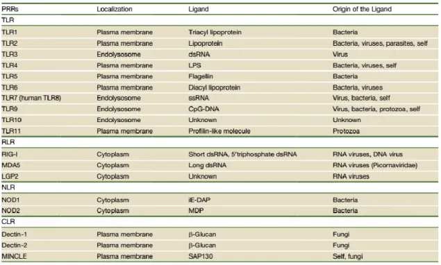
Pattern recognition receptors (PRRs) are not only involved in sensing pathogen invasion but also in sensing damaged cells. PPRs include Toll-like receptors (TLRs), Nod-like receptors (NLRs), RIG-I-like receptors (RLRs), and C-type lectin receptors (CLRs).
2.1. Toll-like receptors (TLRs)
TLRs were the first PPRs family members to be identified and are one of the best-characterized. They are type I transmembrane proteins expressed either on cell surface or associated with intracellular vesicles. TLRs are characterized by N-terminal leucine-rich repeats on their ectodomain that mediate the recognition of a wide range of PAMPs, and also by a cytoplasmic Toll/IL-1R homology (TIR) domain that activate downstream signaling pathways.
24 Ten TLRs have been identified in humans and 12 in mice, with TLR1-TLR9 being conserved in both species. Mouse TLR10 is not functional because of a retrovirus insertion, and TLR11, TLR12 and TLR13 have been lost from the human genome. Depending on their cellular localization and respective PAMP ligands, TLRs are divided in two subgroups: 1) TLR1, TLR2, TLR4, TLR5, TLR6 and TLR11, which are expressed on cell surfaces and recognize mainly microbial membrane components such as lipids, lipoproteins and proteins; 2) TLR3, TLR7, TLR8 and TLR9, which are exclusively expressed in intracellular vesicles (endoplasmic reticulum, endosomes, lysosomes and endolysosomes) and recognize microbial nucleic acids. (Kawai and Akira 2010).
Different TLRs recognize different molecular patterns of microorganisms and self-components (Table 1).
Table 1. Pattern recognition receptors (PRRs): TLRs, RLRs, NLRs, CLRs, and their ligands (Takeuchi and Akira 2010).
TLR4 forms a complex with MD2, and together they serve as the main LPS -binding component (lipopolysaccharide, a component of the outer membrane of Gram-negative bacteria known to be a cause of septic shock). The formation of a receptor homodimer composed of two copies of the TLR4-MD2-LPS complex (Park et al. 2009) initially transmits signals for the early-phase activation of NF-kB by recruiting the TIR domain-containing adaptors TIRAP (Mal) and MyD88 (MyD88-dependent pathway). The TLR4-MD2-LPS complex is then internalized and retained in the endosome, where it triggers signal transduction by recruiting TRAM and TRIF, which leads to the activation of IRF3 (for induction of type I interferon) and the late-phase NF-kB (TRIF-dependent pathway). Both early- and late-phase activation of NK-kB is required for the induction of inflammatory cytokines (Kawai and Akira 2010). In addition to binding LPS, TLR4 is involved in the recognition of respiratory syncytial
25 virus fusion proteins, mouse mammary tumor virus envelop proteins, Streptococcus pneumonia pneumolysin and the plant-derived cytostatic drug paclotaxel (Akira et al. 2006). TLR4 is also involved in the recognition of viruses by binding to viral envelope proteins, and it modulates pathogenesis of H5N1 avian influenza virus infection by recognizing a DAMP rather than the virus itself (Imai et al. 2008). Concerning host cell ligands, TLR4 recognizes fibrinogen, hyaluronic acid, and heparin sulfate fragments, as well as several heat shock proteins (also secreted by bacteria) (Akira and Takeda 2004).
TLR2 recognize lipopeptides from bacteria, peptidoglycan and lipoteichoic acid from Gram-positive bacteria, lipoarabinomannan from mycobacteria, zymosan from fungi, tGPI-mucin from Trypanossoma cruzi, the hemagglutinin protein from measles virus, and HSP70 from host cells (Akira
et al. 2006). TLR2 generally forms heterodimers with TLR1 or TLR6. Different lipid-binding pockets
formed with TLR1 or TLR6 are responsible for the discrimination between lipoproteins. TLR2-TLR1 heterodimer recognizes triacylated lipopeptides from Gram-negative bacteria and mycoplasma, whereas TLR2-TLR6 heterodimer recognizes diacylated lipopetides from Gram-positive bacteria and mycoplasma. Although it was believed that TLR2 agonists mainly induce the production of inflammatory cytokines and not type I interferon by macrophages and dendritic cells, it was also shown that it can trigger the production of type I interferon by inflammatory monocytes in response to infection with vaccinia virus (Barbalat et al. 2009). This suggests that cellular responses to TLR2 ligands differ depending on the cell types involved.
TLR2 and TLR4 engagement also results in recruitment of mitochondria to macrophage phagosomes and increased production of mitochondrial ROS that have been implicated in mouse macrophage bactericidal activity (West et al. 2011).
TLR5 recognizes the flagellin protein component of bacterial flagella. CD11c+CD11b+ lamina propia DCs (LPDCs) in the small intestine have high expression of TLR5. Lamina propia DCs are unique in promoting the differentiation of IL-17-producing helper T cells (Th17 cells) and T helper type 1 (Th1) cells, as well as the differentiation of naïve B cells into immunoglobulin A-producing plasma cells in response to flagellin (Uematsu et al. 2008).
TLR11 is a relative of TLR5, it is expressed in mouse’s kidney and bladder, it recognizes urophatogenic bacterial components, and TLR11-deficient mice are susceptible to infection with these bacteria (Zhang et al. 2004). TLR11 also recognizes the profilin-like molecule derived from the intracellular protozoan Taxoplasma gondii (Yarovinsky et al. 2005).
TLR3 recognizes a synthetic analog of double-strand RNA (dsRNA), poly I:C (polyinosinic-polycytidylic acid), which mimics viral infection and induces antiviral immune responses. TLR3 triggers antiviral immune responses through the production of type I interferon and inflammatory cytokines with an essential role in preventing virus infection. TLR3-deficient mice are susceptible to lethal infection with murine Cytomegalovirus (Tabeta et al. 2004), and TLR3 deficiency in humans is
26 associated with susceptibility to herpes simplex virus type 1 (HSV-1) (Zhang et al. 2007). In the endolysosome, TLR3 also recognizes the genomic RNA of reoviruses, dsRNA produced during replication of single strand RNA (ssRNA) and certain small interfering RNAs (Akira et al. 2006; Bell et
al. 2006). Ligand binding dimerizes two TLR3 molecules (Choe et al. 2005).
TLR7 was originally identified as recognizing imidazoquinoline derivates and guanine analogs such as loxoribine, which have antiviral and antitumor properties. It recognizes ssRNA derived from RNA viruses such as vesicular stomatitis virus, influenza A virus and human immunodeficiency virus (Kawai and Akira 2006). TLR7 also recognizes synthetic poly (U) RNA and certain small interfering RNAs (Hornung et al. 2005). TLR7 is highly expressed on plasmacytoid DCs (pDCs), which are able to produce large amount of type I IFN after virus infection, and cytokine induction that in response to RNA virus are totally dependent of TLR7 (Kawai and Akira 2006). In addition, TLR7 expressed on conventional DCs (cDCs) senses RNA from group B Steptococcus bacteria and induces type I IFN (Mancuso et al. 2009). TLR7 senses virus that are internalized and recruited to the endolysosomes, and also virus that enter the cytoplasm via autophagy (in which self-proteins and damaged organelles are degraded in double-membrane vesicles: autophagosomes). pDCs show constitutive autophagy formation, and pDCs lacking the autophagy-relate protein Atg5 show defects in the production of interferon-α after infection with vesicular stomatitis virus (Lee et al. 2007).
TLR9 recognizes unmethylated 2’-deoxyribo CpG (cytidine-phosphate-guanosine) DNA motifs that are frequently present in bacteria and viruses but are rare in mammalian cells. Synthetic CpG oligodeoxynucleotides function as TLR9 ligands and directly activate DCs, macrophages, B cells, and drive strong Th1 responses There is high expression of TLR9 in pDCs and it serves as a sensor of DNA virus infection (like murine cytomegalovirus, HSV-1 and HSV-2) (Akira et al. 2006). TLR9 also recognizes the insoluble crystal hemozoin, which is generated on the detoxification process after digestion of host hemoglobin by the malaria parasite Plasmodium falciparum (Coban et al. 2010).
Individual TLRs trigger specific biological responses. TLR3 and TLR4 generate both type I interferon and inflammatory cytokine responses, whereas cell surface TLR1-TLR2, TLR2-TLR6 and TLR5 induce mainly inflammatory cytokines (Fig. 4). These differences are due to TIR domain-containing adaptor molecules: MyD88, TIRAP, TRIF and TRAM, which are recruited by distinct TLRs and activate distinct signaling pathways. MyD88 was the first member of the TIR-family to be discovered and is universally used by all TLRs except TLR3, and activates the transcriptional factor NF-kB and mitogen-activated protein kinases (MAPKs) to induce inflammatory cytokines. In contrast, TRIF is used by TLR3 and TLR4, and induces alternative pathways that lead to activation of the transcription factor IRF3 and NF-kB and the consequent induction of type I interferon and inflammatory cytokines. TIRAP is an adaptor that recruits MyD88 to TLR2 and TLR4, whereas TRAM is an adaptor that recruits TRIF to TLR4.
TLR4 is the only TLR that recruits four adaptor proteins and activates two distinct signaling pathways: the MyD88-dependent and the TRIF-dependent pathways (Fig. 4). These two pathways