HAL Id: tel-03014043
https://pastel.archives-ouvertes.fr/tel-03014043
Submitted on 19 Nov 2020
HAL is a multi-disciplinary open access archive for the deposit and dissemination of sci-entific research documents, whether they are pub-lished or not. The documents may come from teaching and research institutions in France or abroad, or from public or private research centers.
L’archive ouverte pluridisciplinaire HAL, est destinée au dépôt et à la diffusion de documents scientifiques de niveau recherche, publiés ou non, émanant des établissements d’enseignement et de recherche français ou étrangers, des laboratoires publics ou privés.
Intestinal epithelial cells instruct myeloid cell-mediated
T-cell responses
Arunima Chatterjee
To cite this version:
Arunima Chatterjee. Intestinal epithelial cells instruct myeloid cell-mediated T-cell responses. Im-munology. AgroParisTech; University of Debrecen, 2015. English. �NNT : 2015AGPT0060�. �tel-03014043�
N°:$$2009$ENAM$XXXX$ $ AgroParisTech $ ! ! ! ! ! ! ! ! ! ! !
présentée et soutenue publiquement par
Arunima CHATTERJEE
17th November 2015
INTESTINAL EPITHELIAL CELLS INSTRUCT MYELOID CELL-MEDIATED
T-CELL RESPONSES
$
Doctorat ParisTech
T H È S E
pour obtenir le grade de docteur délivré par
L’Institut des Sciences et Industries
du Vivant et de l’Environnement
(AgroParisTech)
Spécialité : Immunologie
Directeur de thèse : Eva RAJNAVOLGYI Co-encadrement de la thèse : Francois LEFEVRE $
Jury
Président de la comité:
László Fésüs, MD, PhD, DSc (Department of Biochemistry and Molecular Biology, University of Debrecen)
Analyseur de dossiers de santé:
Zoltán Pós, PhD (Department of Genetics, Cell and Immunbiology, Semmelweis University) Péter Bay, PhD (Department of Medical Chemistry, University of Debrecen)
Membre du comité:
János Matkó, PhD, DSc (Department of Immunology, Eötvös Lóránd University)
THESIS FOR THE DEGREE OF DOCTOR OF PHILOSOPHY (PhD)
INTESTINAL EPITHELIAL CELLS INSTRUCT MYELOID
CELL-MEDIATED T-CELL RESPONSES
by
Arunima Chatterjee
Supervisor: Prof. Dr. Eva Rajnavolgyi
University of Debrecen
Doctoral School of Molecular Cell and Immune Biology
Debrecen, Hungary
Co-supervisor: Dr. Francois Lefevre
Institut national de la recherche agronomique
AgroParis Tech, France
ABBREVIATIONS
AMP – Antimicrobial peptide APC – Antigen presenting cells ATRA – All trans retinoic acid AUC – Area under the curve CD – Crohn’s disease
CEC – Colonic epithelial cell DC – Dendritic cell
DSS – Dextran sulphate sodium FOXP3 – Forkhead box protein 3 GALT – Gut associated lymphoid tissue
GM-CSF – Granulocyte-macrophage colony-stimulating factor IBD – Inflammatory bowel disease
IEC – Intestinal epithelial cells IFNγ – Interferon gamma ILC – Innate lymphoid cells LP – Lamina propria
LPS – Lipopolysaccharide
MAF – Macrophage activating factor Mf – Macrophage
PPARγ – Peroxisome proliferator-activated receptor gamma PRR – Pattern recognition receptor
RALDH – Retinaldehyde dehydrogenase RAR – Retinoic acid receptor
RARE – Retinoic acid response element RBP – Retinol binding protein
RDH – Retinol dehydrogenase
RORγt – Receptor-related orphan receptor-γt RXR – Retinoid X receptor
SRM – Selected reaction monitoring TLR – Toll like receptor
TNF-α – Tumour necrosis factor alpha TGF-β – Transforming growth factor beta VA – Vitamin A
CONTENTS
1.Introduction... 4
2.Theoritical background... 5
2.1. Vitamin A and its effect on the gut ... 5
2.1.1. Vitamin A metabolism... 5
2.1.2. Vitamin A effects on adaptive immune cell subsets... 7
2.1.3. Vitamin A effects on lymphocyte homing... 8
2.2. Cytokines involved in the maintenance of gut homeostasis ... 10
2.2.1. IL-1 family cytokines in IBD... 11
2.2.2. Membrane bound a soluble TNF in IBD... 11
2.3. GM-CSF has a significant role in mucosal immunology... 12
2.4. AMP are secreted by IEC as part of innate immune defence... 13
2.4.1. Mechanism of AMP actions... 15
2.4.2. Chemotactic activity of AMPs... 15
2.4.3. Antimicrobial proteins and peptides... 16
2.4.4. AMP secretion and IBD... 16
3. Aims of the study... 19
4. Materials and Methods... 20
5. Results... 25
5.1. The impact of ATRA on shaping human myeloid cell responses to epithelial cell derived stimuli and on T lymphocytes... 25
5.1.1. Identification of chemokines secreted by resting and activated colon epithelial cells... 25
5.1.2. The effect of ATRA on chemokine secretion by CEC pre-stimulated with IL-1β and TNF-α ... 27
5.1.3. ATRA regulates the chemokine dependent migration of myeloid cells generated by different haematopoietic growth factors... 30
5.1.4. ATRA supports the development of migratory CD103+ myeloid cells... 32
5.1.5. ATRA promotes development of CD14, GM-CSF receptor expression and autocrine secretion of IL-1β in GM-CSF... 35
5.1.6. Translation of molecular information collected by stimulated CEC to myeloid cells and CD4+ T-lymphocytes ... 37
5.1.7. CD4+ T-lymphocytes secrete IL-10 after coincubation with ’educated’ myeloid cells... 39
5.2. Detection of AMPs secreted by Caco2 cells by different methods... 40
5.2.1. Determination of β-defensin 2 levels in Caco2 cell lysates ... 41
5.2.2. Determination of defensin levels in Caco2 cell supernatants ... 41
6. Discussion... 44
7. Summary... 49
8. Summary in French... 50
9. References... 51
9.1. References... 51
9.2. Publication list prepared by the Kenezy Life Sciences Library... 57
10. Keywords... 59
11. Acknowledgements... 60
1. INTRODUCTION
The adult human intestinal tract is referred to as “physiologically inflamed” due to the presence of the enormous number of B- and T-lymphocytes as well as macrophages (Mf), dendritic cells (DC), eosinophils and mast cells. If all these immune cells were present in other tissues at such concentrations, they would be regarded as an abnormal chronic inflammatory cell infiltrate[1]. In such a scenario the main players that maintain gut homeostasis should be those mechanisms, which provide with tolerogenic signals exerted on specialized myeloid cells with antigen presenting function. The discovery showing that retinoic acid imprints the homing potential of leukocytes to the gut and also enhances the induction of regulatory T-cells highlighted the potential role for ATRA (all trans retinoic acid) in mucosal tolerance. It has also been shown that systemic immune responses at sites distal to the gut, such as autoimmune arthritis and experimental autoimmune encephalitis, can also be modulated through the gut microbiota [2] and thus lymphocyte trafficking through the gut is considered as a necessary “rite of passage” for effector T-cells. ATRA gradients have been shown to regulate naive CD4+ T-cell differentiation and cytokine responses in effector CD4+ T-cells. ATRA is essential for Th1 lineage stability and constrains the conversion to Th17 commited cells. Vitamin A deficient mice lack Th17 cells suggesting that endogenous ATRA signaling is required for the generation and/or the maintenance of Th17 cells. At in vivo settings ATRA is also required for Th2 responses and the administration of exogenous ATRA may exhibit opposing effects on T-cell polarization via inhibition of Th1 and Th17 responses observed in both vitro and in vivo. ATRA is also able to alter the sensitivity of cells to alternative cell fates through the regulation of cytokine receptors. Thus the dominant effects of ATRA may well be dependent on the instructive cytokine milieu. In such a model, ATRA may enhance or stabilize the actions of cytokines and the function of transcription factors that guide Th cell programming rather than directly specifying T-cell fate. In this way, synthesis of ATRA by APC (antigen presenting cells) provides an additional checkpoint for the stabilization of T-cell fate commitment [3].
This study is aimed to establish a simplified human ex vivo experimental model system mimicking the interplay of associated chemokines secreted by resting and stimulated colonic epithelial cell (CEC) under the influence of ATRA in a proinflammatory environment in the context of their effects on myeloid cells and T lymphocyte functionality. We also propose comparable and feasible methods for the cost effective and rapid detection and quantification β-defensin levels secreted by CECs acting as a first line of defence in the intestinal tract.
2. THEORITICAL BACKGROUND
The intestinal tract represents the largest mucosal surface of the human body exposed constantly to a myriad of environmental stimuli including components of beneficial and also infective microbes, dietary and metabolic products and diverse inorganic compounds. The intestine is preferentially challenged with the daunting task of segregating the underlying tissues from the potentially noxious and harmful endogenous and environmental molecular structures. Thus the monolayer of intestinal epithelial cells (IECs) provides an important anatomical barrier to support the appropriate segregation of innocent and harmful material and provides protection from various environmental challenges.
2.1. Vitamin A and its effects on the gut environment
The vitamin A (VA) metabolite ATRA is a key regulator of cytokine transforming growth factor beta (TGF-β) expression, which promotes regulatory T-cell (Treg) differentiation[2]. VA
also contributes to the formation of epithelial linings of mucosal surfaces[3] and its multifunctional metabolite ATRA [4] acts as a critical driver of lymphocyte trafficking to the intestinal mucosa[5]. ATRA also induces the expression of the gut homing integrin α4β7 on the surface of myeloid cells and the chemokine receptor CCR9 on T-lymphocytes. On the contrary, the lack of αv or β8 integrin chains in DC impairs Treg functions and Th17 responses in vivo[6]. ATRA can modulate Th17 effector T-lymphocyte differentiation in the gut[7] however, the in vivo effects of ATRA in intestinal and extra-intestinal compartments results in controversial outcomes presumably due to targeting multiple cell types with diverse functional activities[8]. VA deficiency also has an impact on epithelial cell integrity and the composition of the gut microbiota[9]. VA shows ’hormone-like’ properties with some effects on IEC and gut homestasis as described below.
2.1.1. Vitamin A metabolism
“A vitamin is a substance that makes you ill if you don’t eat it.” (Albert Szent-Gyorgyi,
Nobel Prize in Physiology or Medicine, 1937). Vitamins (vital amines) involve essential organic compounds required in trace amounts in the diet, because they cannot be synthesized in sufficient quantities by an organism[10].VA is usually acquired from the diet either as all-trans retinol, retinyl esters, or β-carotene. All-all-trans retinol is esterified to retinyl esters and stored in the liver, or it can associate with retinol binding protein (RBP), which transports retinol to target tissues. All-trans retinol is oxidized intracellularly to all-trans retinal by ubiquitously expressed retinol dehydrogenases (RDH), which belong to the short chain
dehydrogenase reductase (SDR) gene family as shown in Figure 1. At least two RDH are physiologically involved in this rate-limiting step: RDH1 and RDH10. The cytosolic retinal dehydrogenase enzymes (RALDHs) catalyze the irreversible oxidation of all-trans retinal to ATRA[11, 12]. ATRA exerts its effects mostly through binding to the heterodimers of nuclear RA receptors (RARα, β, γ) and retinoid X receptors (RXRα, β, γ)[13], although some specific effects can be mediated via the peroxisome proliferator-activated receptors (PPARβ/γ)[14, 15]. RAR-RXR heterodimers are ligand-dependent transcription factors that bind to cis-acting DNA sequences referred to as ATRA response elements (RARE), located in the promoter region of RA target genes. Although RAR receptors are ubiquitously expressed, RARβ expression is markedly enhanced by ATRA. Termination of ATRA signaling is achieved through its catabolism into oxidized metabolites, such as 4-hydroxy ATRA and 4-oxo ATRA, by enzymes of the CYP26 family. Among these enzymes, CYP26A1 is directly upregulated by ATRA[12, 15]. While ATRA metabolites have been mostly considered to be biologically inert, there is evidence indicating that some of these metabolites retain the ability to signal through RAR[4]. However, the potential roles of ATRA metabolites in immune responses have not been described.
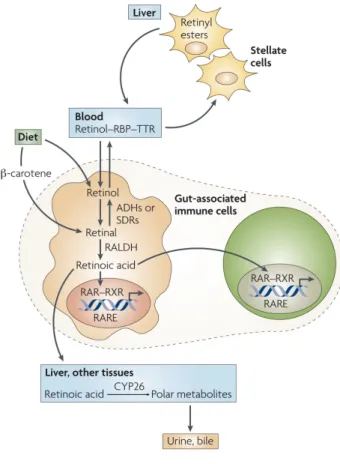
Figure 1: Vitamin A (retinol) is obtained from the
diet and is transported in the blood as a complex with retinol-binding protein (RBP) and trans-thyretin (TTR). In the liver, retinol is esterified to retinyl esters and stored in stellate cells. In other tissues, including gut-associated immune cells, retinol is oxidized to retinal by alcohol dehydrogenases (ADHs) or short chain dehydrogenase/reductases (SDRs). Retinal is then oxidized to all-trans-retinoic acid in an irreversible reaction that is catalysed by retinal dehydrogenases (RALDHs). Retinoic acid acts on immune cells by binding to the retinoic acid receptor (RAR). Retinoic acid is catabolized in the liver and in other tissues by the enzyme CYP26 and its metabolites are eliminated in the bile and urine. Nat Rev Immunol. 2008 September ; 8(9): 685–698.
2.1.2. The affects of vitamin A on adaptive immune cell subsets
ATRA is known to enhance T-cell proliferation and cytotoxicity. Consistent with its in vivo role in T-cell functions cells deficient in VA show defects in CD4+ T-cell activity that could
be due to the inavailability of ATRA to bind to RXR [16].ATRA interferes also with B-cell
proliferation and apoptosis, a process mediated by the binding of VA metabolites to the RAR receptors[17]. ATRA also modulates antigen presentation via exerting direct effects on DC activities leading to increased expression of matrix metalloproteinases able to increase the migration of tumour-infiltrating DCs to the draining lymph nodes thus boosting the potential of tumour-specific T-cell responses [18]. In the presence of inflammatory stimuli, such as TNF-α ATRA enhances DC maturation and antigen-presenting capacity, both mediated by RXR receptors. However, it should be noted that DCs pre-treated with ATRA can apparently store this metabolite, which when released could ultimately act directly on T-cells and other cells thus contributing to the final outcome of an immune response [19]. VA metabolites also modulate the Th1–Th2-cell balance and the differentiation of Treg and Th17 cells as shown in Figure 2. VA deficiency correlates with decreased Th2-cell responses and conversely, VA supplementation blocks the production of Th1-cell cytokines in vitro and in vivo [5, 20]. In fact ATRA promotes Th2-cell differentiation by inducing IL4 gene expression [21]. Moreover, ATRA blocks the expression of the Th1 master regulator T-bet and induces Th2-cell promoting transcription factors, such as GATA3 (GATA-binding protein 3), macrophage-activating factor (MAF) and signal transducer and activator of transcription 6 (STAT6). It has been proposed that ATRA exerts its Th2-cell-promoting effect indirectly through the modulation of APC as well as acts directly on T-cells to induce Th2-cell differentiation through RAR proteins[5].
Although transforming growth factor-β (TGFβ) drives the generation of induced Treg cells that express the transcription factor forkhead box protein 3 (FOXP3) in peripheral tissues, it was recently demonstrated that this process can significantly be enhanced by ATRA[2, 22] (Figure 2). DCs from the gut-associated lymphoid tissue (GALT) or in small intestinal lamina propria also enhance Treg-cell differentiation in a ATRA-dependent manner [23, 24]. A recent study indicated that Mf but not DCs are responsible for inducing Treg cells in the intestinal lamina propria[25], and that DCs are mainly involved in the induction of Th17 cells at this site[25, 26]. The reasons for these seemingly discrepant results are unclear. In addition to inducing FOXP3, ATRA also upregulates the gut-homing receptors on Treg cells thus targeting these cells to the gut mucosa [22, 24].
The differentiation of Treg and Th17 cells is regulated reciprocally by cytokine signals [27]. Exposure of activated CD4+ T-cells to TGFβ alone induces Treg cells, whereas the combination of TGFβ together with IL-6, IL-1β and IL-23 or IL-21 blocks FOXP3 induction and induces Th17-cell differentiation[28]. However, exposure of CD4+ T-cells to ATRA together with TGFβ and IL-6 negates the Th17-cell-promoting effect of IL-6 via enhancing the induction of Treg cells and blocking the induction of receptor-related orphan receptor-γt (RORγt), a key transcription factor for Th17-cell differentiation[2]. It should be noted that this effect is only observed when ATRA is used over a given concentration[26], while low concentrations of ATRA seem to be necessary for Th17-cell differentiation. Thus ATRA has a dual role in maintaining immunological tolerance: it favours the induction of Treg cells and simultaneously can block or enhance Th17-cell differentiation, depending on its concentration.
2.1.3. Vitamin A modulates lymphocyte homing
Although naive lymphocytes migrate preferentially through secondary lymphoid organs, effector and memory lymphocytes acquire ‘trafficing’ molecules that endow them with the capacity to migrate to selected extra lymphoid tissues and to sites of inflammation. The
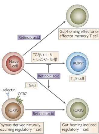
Figure 2: In addition to upregulating the
expression of gut-homing receptors, retinoic acid has also been reported to promote T-helper-2 (Th2) cell differentiation. Moreover, retinoic acid blocks the differentiation of T helper 17 (Th17) cells and induces forkhead box protein 3 (FOXP3)+ regulatory T (TReg)
cells in the presence of transforming growth factor-β (TGFβ) by reciprocally downregulating receptor-related orphan receptor-γt (RORγt) and inducing FOXP3 expression in T-cells, respectively. Retinoic acid also enhances the TGFβ-driven induction of TReg cells and induces gut-homing receptor
expression in both naturally occurring and induced TReg cells. Th17-cell differentiation
requires TGFβ, interleukin-6 (IL-6), IL-23 and, in humans, IL-1β. Cassani et al. Mol Aspects Med. 2012 Feb;33(1):63-76.
gastrointestinal mucosa is the main body surface exposed to environmental antigens and is also the paradigmatic tissues for which tissue-specific adhesion and chemoattractant receptors (also known as homing receptors) have been characterized in detail. For example, effector and memory lymphocytes migrating to the small bowel require expression of the α4β7-integrin and CC-chemokine receptor 9 (CCR9).
It has also been demonstrated that the lymphoid microenvironment in which the lymphocytes are activated determines the set of homing receptors they acquire, thus T-cells activated in the GALT acquire gut-homing receptors[29]. In the lymphoid microenvironment, DCs are essential contributors of efficient T-cell activation. Several groups have shown that DCs from Peyer’s patches and mesenteric lymph nodes are sufficient to induce the expression of α4β7-integrin and CCR9 supporting imprinting the gut-homing capacity of activated T- [30] and B-cells[26]. The means how GALT-resident DCs could imprint lymphocytes with a gut-homing phenotype was shown 25 years ago in rats suffering from both protein-caloric and VA deficiencies causing impaired migration of recently activated mesenteric lymphocytes to the small intestinal mucosa [31], while without VA deficiency it did not affect lymphocyte migration. Adoptive transfer experiments showed that impaired lymphocyte migration could be observed only when donor lymphocytes were from protein-caloric deficient and VA deficient rats and failed when wild-type cells were transferred to protein caloric-deficient and VA-deficient recipients. These results suggested that VA deficiency affected mostly the lymphocyte migratory capacity, but not target tissues[31]. The molecular basis for these observations was recently determined in a study showing that mice depleted of VA had decreased numbers of effector and memory T-cells in the gut mucosa, but not in other tissues [5]. The VA metabolite ATRA was sufficient to induce the expression of α4β7-integrin and the CCR9 chemokine by activated T-cells, even in the absence of DCs. Blocking ATRA receptors of the RAR family significantly decreased the induction of α4β7-integrin expression by T-cells in GALT-resident DCs, showing that ATRA is essential for the gut-imprinting capacity of the DCs. GALT-resident DCs, unlike DCs from other tissues, express RALDH enzymes, which are essential for ATRA biosynthesis. These results altogether indicate that ATRA plays a pivotal role for imprinting gut-homing T-cells [5].
Although VA deficiency decreases the number of T- and B-cells in the small bowel lamina propria[5] it does not affect lymphocyte migration to the colon[31]. Similarly, GALT resident DCs imprint T- and B-cells with homing capacity for the small bowel, but they do not induce homing T-cells. Therefore, ATRA is neither necessary nor sufficient to imprint colon-homing lymphocytes. The molecular signals that are responsible for lymphocyte colon-homing to
the colon and the reasons why T-cell migration to this compartment is controlled differently from homing to the small bowel are still to be determined.
2.2. The role of cytokines involved in the maintenance of gut homeostasis
Based on genetic and immunological studies cytokines have directly been implicated in the pathogenesis of IBD and their crucial role in controlling intestinal inflammation and the appearance of the associated clinical symptoms of IBD. The key role of cytokines in this process has also been highlighted by the blockade of TNF-α commonly used as a standard therapy for IBD and other inflammatory diseases[32].
A combination of IBD risk factors seems to initiate alterations in epithelial barrier function thereby allowing the translocation of luminal antigens including bacterial antigens from the commensal microbiota into the bowel wall. Excessive cytokine responses to such envi-ronmental triggers cause subclinical or acute mucosal inflammation in genetically susceptible hosts[32]. In patients that fail to resolve acute intestinal inflammation, chronic inflammation develops that is induced by the uncontrolled activation of the mucosal immune system. In mucosal immune cells such as Mf, DC and T-cells and the recently discovered subsets of innate lymphoid cells (ILCs) are able to respond to microbial products derived from the commensal microbiota by producing cytokines that promote chronic inflammation of the gastrointestinal tract.
Genome-wide association studies have identified several IBD susceptibility loci that contain genes encoding proteins involved in cytokine and chemokine receptor signalling, and T- helper cell responses. These involve various signalling molecules such as signal transducer and activator of transcription 1 (STAT1), STAT3, STAT4, CC-chemokine receptor 6 (CCR6), CC-chemokine ligand 2 (CCL2), CCL13, IL-12 receptor (IL-12R), IL-23R and Janus kinase 2 (JAK2). Further studies have identified IBD risk loci that contain genes encoding a plethora of cytokines: IL-2, IL-21, interferon-γ IL-10 and IL-27) highlighting a potentially major role for these cytokines in disease pathogenesis[33]. Loss-of-function mutations in the genes encoding IL-10 and IL-10R are associated with a very early-onset of IBD that is characterized by severe intractable enterocolitis in infants. Cytokine function in patients with IBD may be affected by the localization and type of inflammation, immune cell plasticity, different pathogenetic mechanisms or by changes in cytokine production patterns to occur in the course of the disease. The concerted action of these factors build up a complex mucosal cytokine networks. This mode of action suggests that anti-cytokine approaches targeting a single pro-inflammatory cytokine has major limitations in terms of effective therapy for the different
clinical subgroups of IBD[34]. In the following section we describe the major characteristics of the most potent proinflammatory cytokines involved in IBD i.e the IL-1 and TNF families.
2.2.1. The IL-1 family of cytokines and IBD
Lamina propria DCs and Mf are key APCs infiltrating the inflamed mucosa in IBD. In response to components of the commensal microbiota associated with the activation of signalling pathways triggered by various pattern recognition receptors (PRR), these cells produce large amounts of pro-inflammatory cytokines, such as IL-1β, IL-6, IL-18 and TNF-α [35]. A significant decrease in the ratio of IL-1 receptor antagonist of IL-1 could be detected in the intestinal mucosa in patients with Crohn’s disease and ulcerative colitis when compared to control subjects, indicating the activation of the IL-1 system in IBD. TNF-α but not IL-1 blockade was effective in the treatment of mice with chronic dextran sodium sulphate (DSS)-induced colitis. Collectively, these findings suggested that IL-1 has a prominent role in the initiation, rather than in the perpetuation of colonic inflammation. Importantly, blockade of the IL-1 family member IL-18, a cytokine preferentially induced in Mf and epithelial cells in patients with Crohn’s disease exerted beneficial effects in several murine models of acute and chronic colitis[36]. Furthermore, deficiency of the IL-1β-converting enzyme (ICE; also known as caspase 1), an enzyme that cleaves IL-1β and IL-18 into active cytokines, protected mice from DSS-induced colitis suggesting that the blockade of IL-1 family members may be relevant for the therapy of chronic intestinal inflammation[37].
2.2.2. Membrane-bound and soluble TNF in IBD
TNF is produced as a transmembrane protein and soluble TNF is released by proteolytic cleavage by the TNF-converting metalloproteinase enzyme (TACE; also known as ADAM17). The production of both membrane-bound and soluble TNF by mononuclear cells in the lamina propria is markedly augmented in patients with IBD. In particular, CD14+ macrophages, adipocytes, fibroblasts and T-cells from patients with IBD have been shown to produce large amounts of TNF-α [38]. TNF may exert various pro-inflammatory functions in colitis upon binding to its receptors TNFR1 and TNFR2, followed by the intracellular activation of the transcription factor nuclear factor-κB (NF-κB). TNFR1 signalling may also cause cell death via the activation of receptor-interacting protein kinase 1 (RIPK1) and caspase 3 proteins.
The treatment of IBD with neutralizing antibodies against soluble and membrane-bound TNF (such as infliximab and adalimumab) has been validated as highly effective treatment options
via inducing T-cell apoptosis in vivo, whereas agents that preferentially block soluble TNF (for example etanercept) haved no therapeutic effect[32]. Thus, the development of strategies that more specifically target the membrane-bound TNF–TNFR2 interaction is of potential interest for future therapy of IBD.
On the contrary, anti-cytokine therapies such as antibodies specific for TNF, IL-12 or IL-23 and the blockers of cytokine signalling such as tofacitinib seem to have beneficial clinical effects only in certain subgroups of patients[32]. This may reflect that cytokine networks in the inflamed mucosa are more complex than previously appreciated and are the subject of multiple layers in the regulation by microbial, genetic and immunological factors. A key feature of the mucosal cytokine network is its dynamic fluidity and ability to traverse spatial boundaries. Thus the blockade of a single cytokine in patients with IBD may lead to the development of alternative compensatory pro-inflammatory cytokine pathways. Furthermore, the pathological mechanisms driving mucosal inflammation may differ between patients and could explain why targeting of a single pro-inflammatory cytokine is not sufficient.
2.3. GM-CSF plays a significant role in the regulation of mucosal immunity
The cytokine granulocyte-macrophage colony stimulating factor (GM-CSF/Csf2) is a key determinant of myeloid lineage differentiation and is required for the optimal function of tissue resident mononuclear phagocytes (MNPs), including Mf and DCs thereby promoting host protection against environmental pathogens. Despite the key role of Csf2 in promoting MNP survival, differentiation and function, previous studies reported that mice lacking Csf2 or its receptor display only minor impairment in the development of spleen and lymph node resident DCs. Studies showing that Csf2 expression is increased in inflamed tissue and adoptively transferred monocytes can generate DCs in inflamed but not in steady-state spleen suggested that Csf2 acts as a major proinflammatory cytokine that controls the differentiation of inflammatory but not steady-state DCs in vivo. These results are consistent with the contribution of Csf2 to the pathophysiology of numerous inflammatory and autoimmune diseases. The role of microbial commensals that colonize the large bowel to promote the induction of Foxp3+ Treg cell differentiation has been established[39]. However the cellular cues that promote Treg accumulation in response to gut commensals have only recently started to be unraveled. The gut microbiota promotes intestinal homeostasis by supporting the crosstalk between IL-1β-secreting Mf and Csf2-producing ILC in the intestinal mucosa. Microbiota-driven IL-1β production by Mf promotes the release of Csf2 by ILC, which in turn acts on DCs and Mf, allowing the maintenance of colonic Treg cell homeostasis. Ablation
of Csf2 alters DC and Mf numbers and impaires the ability to produce regulatory factors such as ATRA and IL-10, which lead to disrupted Treg cell homeostasis in the large intestine. Conversely, administration of the Csf2 cytokine in mice increases Treg cell frequency in the gut. Most notably, the cell type-specific ablation of IL-1-dependent signaling in ILC abrogates oral tolerance to dietary antigens and compromises intestinal Treg homeostasis in vivo. Although the reduction in total Treg cell numbers is mostly observed in the large intestine, adoptive transfer studies in Csf2−/− mice revealed impaired Treg cell differentiation both in the small and large intestine. These results suggest that Csf2-dependent immunoregulatory functions control Treg cell induction in both tissues establishing intestinal tolerance being critical for the prevention of intestinal diseases such as IBD. A clinical study performed with more than 300 patients with Crohn’s disease (CD) revealed that the level of neutralizing antibodies against Csf2 in the serum correlated with ileal involvement and in the development of penetrating pathology, whereas a more recent study identified reduced levels of Csf2 receptor (Csf2R) and impaired receptor activity in a mixed group of IBD patients[40]. Previous clinical trials with recombinant Csf2 in IBD patients have established patient benefit in terms of reduced disease severity and lower burden of corticosteroid use. Unpublished results of a large Csf2-based clinical trial in IBD failed to achieve primary clinical end points, but it remains likely that a subset of IBD patients with defective Csf2 production or function still could benefit from this therapy. Thus the key role for Csf2 in the maintenance of intestinal tolerance is consistent with previous studies showing that the absence of Csf2 can also contribute to lupus-like disease, insulitis, and age-related glucose intolerance and emphasizes the critical role of tissue-resident phagocytes in the maintenance of tissue integrity. It is now well established that the gut commensal flora promotes immune homeostasis in the host. The commensal driven MNP-ILC-Csf2 axis acts as a key regulator of intestinal T-cell homeostasis in the mouse intestine. Disturbance of this regulation can radically alter MNP-associated effector functions resulting in impaired tolerance to dietary antigens (Figure 3).
2.4. Defensins are secreted by IEC as part of a first line of mucosal defence
Mammalian antimicrobial peptides (AMPs) are members of a diverse array of protein families, all having the potential to rapidly kill or inactivate microorganisms. The epithelial cells lining the gut, skin and respiratory tract produce a rich arsenal of AMPs, probably reflecting the complexity of microbial challenges these tissues are exposed to and the continuous threat of microbial invasion at these sites[41]. The AMPs of the gut encompass
representatives of several distinct protein families. These include defensins, cathelicidins, C-type lectins, ribonucleases and psoriasin[42].
Among the diverse, most abundant and highly expressed AMP families in the gut are the α-defensins. They are small peptides of ~2-3 kD with a conserved three-dimensional structure characterized by an amphipathic arrangement of cationic and hydrophobic residues[43], resulting in a positively charged surface that is spatially separated from a neighbouring hydrophobic region. This unique structure promotes the attraction of α-defensins to the negatively charged cell surface and insertion into lipid-rich membranes. In general, α-defensins have a broad spectrum of activity against both Gram-positive and Gram-negative bacteria, and in some cases are active against fungi, viruses and protozoa however, particular defensin species exhibit marked differences in their activity spectrum and expression patterns[44]. The human α-defensins include DEFA5/HD5 and DEFA6/HD6.
Under steady-state conditions epithelial cells produce the majority of AMPs on the body surface , although infiltrating immune cells during inflammation can also contribute to AMP production. The intestinal epithelial cell surface comprises distinct epithelial cell lineages each of which expresses a distinct group of AMPs. The enterocyte is the most abundant epithelial cell lineage of both the small and large intestine.
Colon enterocytes express β-defensins and cathelicidins[45]. Paneth cells are located at the base of crypts of Lieberkühn and are unique to the small intestine. Many AMPs are expressed
Figure 3: Innate Lymphoid Cells (ILC) translate microbial cues into immunoregulatory signals in the intestine.
Microbial structures sensed by intestinal
mononuclear phagocytes lead to IL-1β release. IL-1β engages IL-1receptor on ILC and promotes Csf2 release. ILC-derived Csf2 triggers DC and MNP production of regulatory molecules including RA and IL-10, which in turn promote the induction and expansion of Tregs. Csf2-primed DCs
and MNPs promote Treg homeostasis
locally and also in mesenteric lymph nodes. Mortha et al. Science. 2014 Mar 28;343(6178):1249288.
abundantly by Paneth cells including α-defensins[44] and RNase[46]. Goblet cells constitute a third epithelial cell lineage that is present in both the small and large intestine. A major function of goblet cells is to secrete mucin glycoproteins that assemble to form a thick gel-like mucus layer that overlies the epithelium and functions in part to concentrate secreted AMPs at or near to the epithelial surface[47].
2.4.1. Mechanism of AMP actions
Many AMPs target essential cell wall or cell membrane structures, which limits the ability of microorganisms to evolve resistance. Several AMPs act as enzymes that kill bacteria by carrying out an enzymatic attack on cell wall structures. Lysozyme and phospholipase A2 (PLA2), both highly expressed by Paneth cells, function through such a mechanism. Lysozyme hydrolyses the glycosidic bounds between the glucosamine and N-acetyl-muramic acid that constitute the carbohydrate backbone of cell wall peptidoglycan, whereas secretory PLA2 (sPLA2) kills bacteria by hydrolysing bacterial membrane phospholipids[48]. Many AMPs kill bacteria through non-enzymatic disruption of the bacterial membrane.
In vertebrates defensins comprise a major family of membrane-disrupting peptides. The clusters of cationic residues of most defensins interact with the surface of the bacterial membrane through electrostatic interactions with negatively charged phospholipid groups. This interaction is followed by the formation of defensin pores in the bacterial membrane that disrupts membrane integrity and promote the lysis of the targeted microorganism[49].
Certain AMPs such as RNase7[50] and dermcidin[51] do not act through membrane disruption but function by metal chelation thus regulating the availability of essential trace elements such as Zn2+ and Mn2+ to augment other host defence mechanisms for microbial killing[52]. The other proteins have very low direct antimicrobial potency but are abundantly expressed in epithelia arguing for their substantial role in host defence. These findings altogether suggest that different AMP families use distinct molecular mechanisms to kill microorganisms. The use of diverse killing strategies presumably helps to limit the evolution of microbial resistance to multiple AMPs.
2.4.2. Chemotactic activity of AMPs
The α- and β-defensins have been shown to have chemotactic activity and the capacity to recruit leukocytes by direct or indirect mechanisms, which can directly modify the outcome of inflammatory responses. Human α-defensins (DEFA1/neutrophil defensin 1 and 2 and β-defensins/DEFB103) and BD4/DEFB104 have been reported to be chemotactic for monocytes
and Mf, and BD2/DEFB4A[53]. The chemotactic activities of the different AMP groups differ from each other. For example, human α-defensins selectively induce the migration of naive CD4+CD45RA+ and CD8+ human cells, but not CD4+CD45RO+ memory T-cells[54]. By contrast, β-defensins are chemotactic for immature DCs and CD4+CD45RO+ memory T-cells. The chemotactic effect of human defensins can be inhibited by antibodies specific for CC-chemokine receptor 6 (CCR6)[54].
2.4.3. Expression and functions of antimicrobial peptides and proteins
The expression, secretion and activity of most AMPs are tightly controlled. This is necessary to prevent the potential toxic effects of many of these proteins on mammalian cell membranes. The chemokine-like activities and immune-modulatory effects of many AMPs presumably require tight control of expression in order to avoid triggering of unnecessary inflammatory responses. Studies performed in germ-free mice have revealed that some intestinal AMPs are expressed independently of the microbiota, whereas others require bacterial signals for their expression. For example, the expression of most intestinal α-defensins requires the transcription factor TCF4 belonging to the WNT pathway, but is independent on the microbiota[55]. Similarly, the expression of lysozyme and certain members of the β-defensin family do not require microbial signals[46].
Other intestinal AMPs depend on microbial cues for full expression. Members of α-defensins show increased levels of expression in conventionally raised mice in comparison to germ-free mice[55]. Similarly, key members of the human β-defensin family, including BD2, are expressed under the control of bacterial signals. For example, bacterial flagellin has been shown to be relevant for the induction of BD2 expression[56].
2.4.4. AMP secretion and IBD
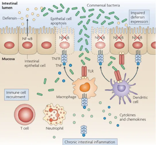
Gut epithelial AMPs not only protect against pathogen colonization but also determine microbiota composition and limit the access of the microbiota to host tissues (Figure 4). A key mechanism by which the mammalian intestine maintains homeostasis with its associated bacterial communities is to minimize contact between the bacteria and the intestinal epithelium[57]. This is accomplished in part by enhancing the physical barrier through the production and assembly of a thick mucus layer at the epithelial surface. Visualization of the spatial relationships between bacteria and the intestinal surface shows that the inner mucus layer remains relatively free of bacteria, whereas the outer mucus layer retains large numbers of bacteria[58]. Biochemical analysis of AMP localization has shown
that most AMP activity is confined to the mucus layer and is essentially absent from the luminal content[47]. Thus the mucus layer in addition to functioning as a physical barrier, limits bacterial penetration by forming a diffusion barrier that concentrates AMPs near to the epithelial cell surface. Given the crucial contributions of AMPs to the maintenance of homeostasis at body surfaces, dysregulation of AMP production and function can be associated with disease. Patients with IBD frequently exhibit increased numbers of epithelial cell surface-associated bacteria[59] suggesting a breakdown in immune mechanisms that normally limit direct contact between the microbiota and the intestinal mucosa. Several IBD risk alleles have been associated with altered intestinal epithelial AMP production. For example, genetic variants in the promoter region of the TCF4 and the NOD2 genes controlling α-defensin expression are associated with ileal Crohn's disease[60]. In these patients, severe intestinal inflammation is associated with decreased α-defensin expression[61]. These examples illustrate how genetic defects leading to decreased AMP production can be
associated with intestinal inflammation and disease. However, it is important to note that in mice the defects in AMP production alone generally do not result in intestinal inflammation [57] suggesting that in humans IBD may require multiple genetic defects that target other immune mechanisms in addition to AMPs[62].
Body surfaces continuously face complex microbial challenges that include the maintenance of homeostasis in close contact with indigenous microorganisms and limiting exposure to pathogens. The intestine confronts these challenges by producing a complex arsenal of AMPs that directly kill microorganisms and can modulate immune responses. It is also becoming clear that endogenous AMPs are essential not only for protecting these sites from pathogenic microbial evasion but also for shaping the composition and location of indigenous microbial communities. To get a better insight to the importance of AMPs in regulating innate immune activities we have developed a targeted mass spectrometry method for the identification and quentitation of AMPs. In the present study the selected reaction monitoring (SRM) method was used in a semi-quantitative manner for the relative quantification of defensin 1, β-defensin 2, β-β-defensin 3 and β-β-defensin 4 derived from human Caco2 colonic epithelial cell lysates and culture supernatants.
Figure 4: The intestinal epithelium has an essential role in the maintenance of immune homeostasis in the gut. By forming a mechanical barrier and expressing antimicrobial peptides (such as defensins), intestinal
epithelial cells (IECs) prevent commensal bacteria from invading the gut mucosa (left hand side). Nuclear factor- B (NF- B) inhibition results in increased death of IECs and decreased expression of antimicrobial peptides, thereby compromising the epithelial barrier and allowing commensal bacteria to invade the colonic mucosa, which triggers severe chronic colitis in a MYD88- and tumour necrosis factor receptor 1 (TNFR1)-dependent manner (right hand side). TLR, Toll-like receptor. Nature Reviews Immunology 9, 778-788 (November 2009)
3. AIMS OF THE STUDY
• Develop a human myeloid cell-based in vitro model system to mimic the response of colon epithelial cells (CEC) to inflammatory stimuli (IL-1β or TNF-α) and the effect of ATRA on this inflammation.
• Identify, how the molecular information, collected by activated CEC supernatant could be translated to T-lymphocyte polarization.
• To demonstrate the role of ATRA, a metabolite of VA, in modulating the outcome of both innate and adaptive immune responses.
• Dissect the characteristics of DC- and Mf-mediated responses in the presence and absence of ATRA.
• To show that properly ‘educated’ myeloid cells are able to induce tolerance when co-cultured with autologous CD4+ T-cells.
• To test, whether GM-CSF deficiency in the gut could alter the phenotypic properties of DC and Mf considering that GM-CSF is able to enhance IL-1β secretion of intestinal lymphoid cells and also has an effect on the number of Treg cells.
• To demonstrate the expression of β-defensins at the protein level in Caco2 cell lysates and in the supernatant of these cells.
• To introduce a new mass spectrometro based methodology for the semi quantitative determination of relative β-defensin expressions in Caco2 cells stimulated by IL-1β.
4. MATERIALS AND METHODS Cell Culture of Caco2 colon epithelial cells
The human colorectal adenocarcinoma cell lines Caco2 is from ATCC-number HTB-37™and HT-29 is from ATCC-number HTB-38™. The colorectal carcinoma cell line HCT116 was a generous gift from Dr. György Vereb, Department of Biophysics, University of Debrecen. Caco2 and HCT116 cells were cultured in RPMI-1640 medium supplemented with 1% antibiotic-antimycotic solution and 20% fetal bovine serum (GIBCO by Life Technologies, EU) in tissue culture flasks (Nunclon, Rochester, NY) at 37°C in 10% and 5% CO2, respectively. HT-29 cells were cultured in RPMI-1640 medium supplemented with 1% antibiotic-antimycotic solution and 10% fetal bovine serum in 5% CO2. Cell culture medium was replaced every 2-3 days and the cells were passaged when sub-confluent.
Protein Array for Chemokine analyses
CEC of 70-80% confluency were plated overnight in RPMI supplemented with 10% FCS-followed by stimulation with 10ng/ml pro-inflammatory cytokines (IL-1β or TNF-α) in combination with or without 10nmol/ml ATRA or left untreated for 1 hour. The cells were washed and replaced with fresh medium for 5 hr, when the supernatants were collected for chemokine analysis performed by a commercially available protein array (Proteome Profiler Arrays- ARY017, R&D Systems, Minneapolis, MN, USA) according to the manufacturer's instructions. Sample controls (transferrin R, gp130 and fibrinogen) included in the array allowed us the detection and quantitation of the secreted chemokines. Considering that ATRA dissolved in DMSO may have toxic effects on resting CEC, which could be further enhanced by activation with IL-1β or TNF-α, we performed preliminary titration experiments to optimize the cell culture conditions by using 24h 7AAD-based viability assays performed by FACS analysis. These results indicated 98% viability of Caco2 and HT-29 cells in both the presence and absence of 10nmol/ml ATRA that was similar to those measured for untreated CEC.
In vitro cell migration and the chemotaxis assay
Migration of three different groups of monocyte-derived cells, differentiated in GM-CSF+IL4, GM-CSF and M-CSF, were tested for cell migration to chemokines and cytokines secreted by Caco2, HT-29 and HCT116 cells. Monocytes (3x10⁵) differentiated in the presence of the 3 different growth factors were placed on the upper chamber of a 5micron Corning transwell plate and the CEC supernatants were added to the lower chamber of the
transwell. After 24 h the monocyte-derived cells that migrated to the lower chamber were collected. 10,000 polystyrene beads (15micron) were added to each sample (Fluka Analytical, Germany) and the number of migrating cells was counted by FACS Calibur (BD Biosciences, Franklin Lakes, NJ, USA). The data were analyzed by the FlowJo software (Tree Star, Ashland, OR, USA).
Peripheral blood monocyte-derived cells
Leukocyte enriched buffy coats were obtained from healthy blood donors drawn at the Regional Blood Center of the Hungarian National Blood Transfusion Service (Debrecen, Hungary) in accordance with the written approval of the Director of the National Blood Transfusion Service and the Regional and Institutional Ethics Committee of the University of Debrecen, Medical and Health Science Center (Hungary). PBMCs were separated by a standard density gradient centrifugation with Ficoll-Paque Plus (Amersham Biosciences, Uppsala, Sweden). Monocytes were purified from PBMCs by positive selection using immunomagnetic cell separation with anti-CD14 microbeads, according to the manufacturer's instruction (MiltenyiBiotec, BergischGladbach, Germany). After separation on a VarioMACS
magnet, 96–99% of the cells were CD14+ monocytes, as measured by flow cytometry.
Monocytes were divided and cultured in 12-well tissue culture plates at a density of 2 × 10⁶
cells/ml in 10% RPMI medium supplemented with three different growth factors: 80ng/ml GM-CSF (Gentaur Molecular Products, Brussels, Belgium), 100ng/ml IL-4 (PeproTech EC, London, UK), and M-CSF 50ng/ml (MACS, MiltenyiBiotec, Germany).
Pheripheral blood lymphocytes and CD4+ T-cells
Autologous naive T-cells were separated from human blood mononuclear cells using the
naive CD4+ T-cell isolation kit based on negative selection according to the manufacturer’s
instruction (MiltenyiBiotec).
Phenotypic characterization of myeloid cells by flow cytometry
Detection of the cell surface expression of monocyte-derived myeloid cells was performed by flow cytometry using CD1a-PE, CD209-PE, CD14-PE, CD83-PE, anti-CD103-PE, anti-CX3CR1-PE, anti-CCR7-PE (Beckman Coulter, Hialeah, FL, USA). The growth factor receptors were characterized by anti-GM-CSFRα-PE and anti-M-CSF R/CD115-PE (R&D Systems, USA) and isotype-matched control antibodies (BD PharMingen, San Diego, CA, USA). Fluorescence intensities were measured by FACS
Calibur (BD Biosciences, Franklin Lakes, NJ, USA), and data were analyzed by the FlowJo software (Tree Star, Ashland, OR, USA). The human chemokines Mk, CXCL7, CCL20 and CXCL16 were ordered from PeproTech, UK, CXCL8 and CXCL1 are from MiltenyiBiotec.
IL-17 and IFNγ ELISPOT assays
The monocyte-derived cells were cultured in GM-CSF+IL4, GM-CSF and M-CSF for 3 days along with the supernatant of unstimulated or cytokine activated Caco2 cells at 2×10⁵ cells/well density. The cells were washed to remove all growth factors and supernatants and
were co-cultured with naïve autologous CD4+ T-cells (106cells/well) in 10% RPMI medium
for 2 days at 37°C in a humidified atmosphere containing 5% CO2. PHA (phytohaemagglutinin) and Con A (concanavalin A) activated T-cells were used as positive controls, untreated monocyte-derived cells, CD4+ T-cell co-cultures and CD4+ T-cells served as negative controls. Detection of cytokine-secreting T-cells was performed by the avidin-HRP system (NatuTec GmbH, Germany). Plates were analyzed by an ImmunoScan plate reader (CTL, Shaker Heights, OH, USA).
RT-qPCR
Total RNA was isolated by Trizol reagent (Invitrogen, Carlsbad, CA, USA) in accordance with the manufacturer’s instructions and the total RNA content was quantitated by spectrophotometry (NanoDrop ND1000; Promega Biosciences, Madison, WI). cDNA was synthesized by using SuperScriptTM II Reverse Transcriptase (Invitrogen) following the manufacturer’s instructions. Quantitative PCR was performed using 0.125&U Taq DNA polymerase (Fermentas, St. Leon-Rot, Germany) to detect double-stranded cDNA synthesis and Rox Reference Dye (Invitrogen) was used for normalization of the fluorescent reporter signal. Taqman Gene Expression Assay Hs00175474_m1 (Applied Biosystems) was used for the detection of β-defensin 2. The PCR cycling conditions involved an initial polymerase activation step for 5&minutes at 95°C, followed by 40 cycles of 12&s at 95°C for denaturation, 45&s at 60°C for annealing, and 15&s at 72°C for elongation. Relative expression levels were determined with StepOne Software v2.1 for StepOne Plus Real-Time PCR Systems (Applied Biosystems). The expression levels were calculated by the ΔCt method using cyclophilin as a house keeping gene control.
Dot-blot analysis
40 µg protein of the Caco2 cell supernatants were spotted and dried to the PVDF membrane to perfom the dot-blot analysis. The membranes were blocked with 5% milk powder containing TBS solution for one hour at room temperature. Anti β-defensin 2 IgG antibodies (Abcam, ab66072) were used at 5µg/ml concentration, and incubated for 24 hours at room temperature. The primary antibodies were washed out by repeated washing (three times for 5 minutes) with TBS. HRP-conjugated anti-mouse IgG antibodies (Amersham Biosciences, NA931V) were used as a secondary antibody in 1 ng/ml concentration, and after one hour incubation at room temperature the membranes were washed with TBS three times for five minutes. Visualization of the bands and dots were carried out with ECL reagent (Thermo Scientific) developed on a radiographic film.
Densitometry
Films were scanned using PharosFX Plus Molecular Imager (Bio-Rad) and the densitometry was carried out with Quantity One 4.6.7 software (Bio-Rad). Individual background substraction was administrated in each case and the area under the intensity profile curve was calculated by the software.
ELISA
Determination of β-defensin 2 protein levels form 100 µl cell lysates and cell culture supernatants were performed by sandwich ELISA using the EK-072-37 kit (Phoenix Pharmaceuticals Inc) according to the provided protocol.
SRM-based targeted proteomic method development
In order to design specific SRM transitions for β-defensins we have utilized the amino acid sequences for β-defensins 1, 2, 3 and 4 available in the UniProt database (www.uniprot.org) followed by in silico trypsin digestion. Tryptic fragments with ≥95% cleavage probability were selected for BLASTp analysis and the NCBI non-redundant protein sequence database was searched to determine the protein-specific, unique tryptic peptide sequences. Stable-isotope-labeled synthetic (SIL) crude peptides were obtained from JPT Peptide Technologies GmbH, Germany, while purified hBD2 SIL was obtained from PepscanPresto, The Netherlands. The quality of the synthetic peptides was assessed by matrix-assisted laser desorption/ionization time-of-flight mass spectrometry (MALDI-TOFMS) conducted in our laboratory and the SRM spectra of singly charged y ions were recorded.
Sample preparation for mass spectrometry
The protein concentration of each sample was determined with Bradford method [30]. The proteins were denatured by the addition of 6 M urea and reduced using 10 mM dithiothreitol. The reduced samples were alkylated with 20 mM iodoacetamide and diluted with 25 mM ammonium bicarbonate to decrease the urea concentration to 1 M. Trypsin digestion was performed overnight at 37 oC by adding MS grade modified trypsin (ABSciex) in 1:25 enzyme: protein ratio. The digested peptides were lyophilized, dissolved in 1% formic acid. The samples were desalted with C18 ZipTip tips (Millipore) and the eluates were lyophilized and redissolved in 1% formic acid.
Mass spectrometry analysis
SRM experiments were carried out on a 4000 QTRAP (ABSciex) mass spectrometer using NanoSpray II MicroIon Source and controlled by the Analyst 1.4.2 software (ABSciex). Carried out in collaboration with Proteomics Core Facility of University of Debrecen.
Statistical analysis
Statistical analysis was performed by one way analysis of variance (ANOVA) for multiple comparisons. Results are expressed as mean ± SD. Two group differences were analyzed by Student's t-test. p value (two-tailed) less than 0.05 was considered statistically significant. The data for Mass spectrometry were statistically analyzed with the SigmaPlot 12.0 software (Systat Software Inc.) using student t-test. For the determination of correlation coefficient the linear regression method was used (Daniel W.W., Biostatistics: A foundation for analysis in the health sciences (Fifth edition), Wiley Series in probability and mathematical statistic-applied, 1991). The level of significance was set to 0.05.
5. RESULTS
5.1. The impact of ATRA on shaping human myeloid cell responses to epithelial cell-derived stimuli and on T-lymphocyte polarization
We designed in vitro experiments with resting human CEC and in an inflammatory milieu mimicked with TNF-α or IL-1β stimulation in the presence or absence of ATRA. This was performed by monitoring the levels of secreted chemokines measured at the protein level and by investigating their impact on the phenotype and functional attributes of myeloid cells generated by different growth/differentiation factors. Considering that DC have the potential to instruct T-cells for inflammatory or regulatory directions, our final goal was to identify the impact of stimulated CEC-induced and DC-mediated effects on CD4+ effector T-lymphocyte responses. !
5.1.1. Identification of chemokines secreted by resting and activated CEC
The single cell monolayer of CEC plays an essential role in the maintenance of gut homeostasis by supporting barrier function and defense against microbes preferentially through the secretion of chemokines. It is also well established that the pro-inflammatory cytokines IL-1β and TNF-α act as potent activators of CEC[63]. In this study we applied a high throughput approach for identifying the chemokines secreted by CEC (Caco2, HT-29 and HCT116) in response to IL-1β and TNF-α by using a commercially available Human Chemokine Array to quantify the relative levels of chemokines released by resting and activated CEC at the protein level. The sample controls provided (transferrin R, gp130 and fibrinogen) were used for calculating mean pixel densities of the respective dot blots. The results showed that resting Caco2 cells secrete detectable levels of CCL19, CCL21 and CCL22 constitutively (dots 2D, 9F, 8D in Figure 5A, summarized in Figure 5C). Similar results were obtained for HT-29 cells, but HCT116 secreted only CCL19 at detectable levels. Comparison of the relative cytokine levels secreted by the Caco2, HT-29 and HCT116 cell lines are summarized in Table 1. Considering that these CCL chemokines are known to attract myeloid cells, they may maintain a population of myeloid cells in the vicinity of CEC to support cellular interactions. It has previously been shown that MDC/CCL22 attracts TH2 cytokine producing cells and its mRNA and protein expression is upregulated against entero-invasive bacteria, but inhibition of the NF-κB pathway abolished CCL22 expression in response to pro-inflammatory stimuli[1]. The chemokines CXCL7, CXCL16 and Mk with different functional activities were also constitutively secreted by resting CEC (dot 9C, 7F, 7D in Figure 5A) suggesting their role in the maintenance of epithelial cell homeostasis.
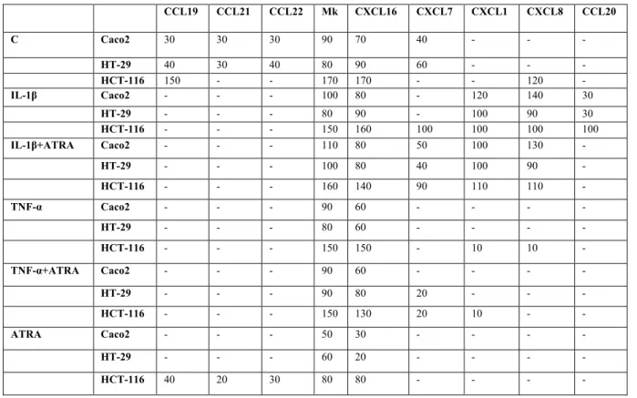
Table 1 : Relative expression of chemokines secreted by colonic epithelial cell lines
Caco2, HT-29 and HCT116 cells were treated with IL-1β or TNF-α in the presence or absence of ATRA for 60 min. After removing the cell culture supernatant the cells were washed and replaced with fresh medium and the supernatants were collected after 5 hours and used for the Chemokine Array analysis according to the manufacturer’s instruction to determine the variety of chemokines secreted by these cell lines. The values indicate the results of the densitometric analysis of dot blots and are normalized to the relevant sample control according to the manufacturer’s instructions. For simplicity the value of 1 obtained after dividing with the sample control is referred to as 100.
CCL19 CCL21 CCL22 Mk CXCL16 CXCL7 CXCL1 CXCL8 CCL20 C Caco2 30 30 30 90 70 40 - - - HT-29 40 30 40 80 90 60 - - - HCT-116 150 - - 170 170 - - 120 - IL-1β Caco2 - - - 100 80 - 120 140 30 HT-29 - - - 80 90 - 100 90 30 HCT-116 - - - 150 160 100 100 100 100 IL-1β+ATRA Caco2 - - - 110 80 50 100 130 - HT-29 - - - 100 80 40 100 90 - HCT-116 - - - 160 140 90 110 110 - TNF-α Caco2 - - - 90 60 - - - - HT-29 - - - 80 60 - - - - HCT-116 - - - 150 150 - 10 10 - TNF-α+ATRA Caco2 - - - 90 60 - - - - HT-29 - - - 90 80 20 - - - HCT-116 - - - 150 130 20 10 - - ATRA Caco2 - - - 50 30 - - - - HT-29 - - - 60 20 - - - - HCT-116 40 20 30 80 80 - - - -
Figure 5: Chemokine secretion of human epithelial cells activated by IL-1β or TNF-α in the presence or absence of ATRA: The relative levels of the Caco2 cell-derived chemokines were determined by calculating
mean pixel densities of the individual blots normalized to sample control. The mean+SD of 4 independent measurements are shown. A – Localization of the chemokine probes in the membranes related to the the positive controls. B – Representative dot blots showing the relative expression of chemokines produced by untreated Caco2 cells without or with ATRA (upper and lower panels 1) as compared to Caco2 cells stimulated by IL-1β (upper panel 2) and IL-1β in the presence of ATRA (lower panel 2). Upper and lower panels 3 correspond to Caco2 cells stimulated by TNF-α in the absence or presence of ATRA, respectively. C – Relative expression levels of CCL chemokines produced by unstimulated Caco2 cells.
A
B
C B
5.1.2. The effect of ATRA on the chemokine secretion by CEC pre-stimulated by IL-1β or TNF-α
In our model system CEC were left untreated or stimulated by IL-1β or TNF-α in combination with or without ATRA for 6 hrs and the cell culture supernatants were subjected to chemokine array analysis by calculating pixel densities using the relevant sample control (Figure 5). When Caco2 cells were activated by TNF-α or IL-1β, the secretion levels of the chemokines CXCL7, CXCL16 and Mk did not change significantly as compared to unstimulated cells (dots 9C, 7F, 7D in Figure 5A, summarized in Figure 6A, 6B). However, the expression of CXCL16 and Mk was down regulated in the presence of ATRA suggesting that the secretion of these chemokines may contribute to the maintenance of epithelial cell homeostasis. However, under inflammatory conditions they do not mediate positive signals for DC. Remarkably, the secretion of CXCL7 (dot 9C in Figure 5A, summarized in Figure 6A and 6B) could be induced only when IL-1β or TNF-α was combined with ATRA treatment demonstrating the dependence of its secretion on ATRA. DMSO, which is the standard solvent of ATRA also decreased CXCL7 secretion (Figure 6A and 6B). Depending on CEC sensitivity to DMSO, certain effects of this solvent have previously been reported, as inhibition of prostaglandin E2 production upon treatment of Caco2 cells with IL-1β and attenuation of mRNA levels of IL-6, IL-1α and IL-1β[64]. These findings might be in line with inhibition of CXCL7 observed in our system. In the IEC-18 cell line IL-1β induced increased mRNA levels of MCP-1/CCL2, MIP-1α/CCL3, inducible NO synthase and RANTES/CCL5, which are upregulated in a NF-κB dependent manner[65].
The secretion levels of CCL20 and CXCL1 was not affected by ATRA (dots 3D, 2F and 7E in Figure 5A, summarized in Figure 6C) and could be induced exclusively by IL-1β but not by TNF-α. Surprisingly, CXCL8 secretion was upregulated not only by IL-β with or without ATRA, but also by DMSO used as a vehicle for ATRA (Figure 6C). DMSO induced strain on the actin cytoskeleton and integrins expressed by Caco2 cells were suggested to be transferred to actin-associated molecules like α-actinin-1, acting as a scaffold protein and interacted directly with ERK1/2 leading to phosphorylation and increased secretion of CXCL8. The in vivo relevance of this effect is underscored by the physical deformation of CEC during peristalsis and villous motility[66]. Along with IL-8/CXCL8 and GROα/CXCL1, Caco2 cells also express MCP-1/CCL2 as a result of IL-1 stimulation, but our chemokine array-based method did not detect CCL2[67]. These results suggest that the expression of individual chemokines depends on the means of activation and also on ATRA, which can modulate the outcome of chemokine secretion, whereas the group of chemokines not affected by ATRA
indicates the complexity of chemokine-mediated regulation in the gut. Similar results were obtained for HT-29 and HCT116 CEC as summerised in Table1. [65, 68, 69].
Figure 6: Effects of ATRA on the expression of chemokines in cytokine-stimulated Caco2 cells: Relative
expression of chemokines in Caco2 cells, pre-stimulated by TNF-α or IL-1β in the presence or absence of ATRA, was determined as described in Figure 5 and was compared to unstimulated cells cultured with or without ATRA. The possible contribution of DMSO used as a solvent control of ATRA was tested in Caco2 cells cultured in the presence of TNF-α or IL-1β with or without DMSO. A – Expression of chemokines secreted by Caco2 cells pre-stimulated by TNF-α, in the presence or absence of ATRA. B – Expression of chemokines secreted by