EFFECTS OF POSTNATAL ENVIRONMENTAL TOBACCO SMOKE ON NON-NUTRITIVE SWALLOWING-BREATHING COORDINATION IN NEWBORN LAMBS
Charles Duvareille1, Marie St-Hilaire1, Nathalie Samson1, Parseh Bakirtzian1, Simon Brisebois1, Mathieu Boheimier1, Djamal-Dine Djeddi1, Alexandre A. Doueik2,
Jean-Paul Praud1
Neonatal Respiratory Research Unit; Departments of Pediatrics and Physiology1 and Department of pathology2, Université de Sherbrooke, Sherbrooke, QC - Canada
Running title: Cigarette smoke and swallowing in the newborn
Address for correspondence Jean-Paul Praud MD PhD
Departments of Pediatrics and Physiology Université de Sherbrooke
J1H 5N4 - QC – Canada
Email : [email protected] Fax: 1 (819) 564-5215
ABSTRACT
While prenatal environmental tobacco smoke (ETS) exposure is a well-known risk factor for sudden infant death syndrome, the effect of postnatal ETS exposure is less clear. The objective of this study was to investigate the effect of postnatal ETS exposure on non-nutritive swallowing (NNS) and NNS-breathing coordination. Twelve newborn lambs were exposed to either ten or twenty cigarettes per day for 15 days. Six controls were exposed to air. Lambs were instrumented for recording states of alertness, swallowing, electrocardiogram and breathing; recordings were performed in non-sedated lambs at the end of ETS exposure. Urinary cotinine/creatinine ratio confirmed relevant real-life exposure. Postnatal ETS exposure had no effect on NNS frequency but tended to decrease inspiratory NNS (p = 0.07) during quiet sleep. No effect on respiratory or heart rate (p > 0.6), apnea index (p = 0.2) or sleep states (p = 0.3) was observed. In conclusion, postnatal ETS exposure in lambs had only mild effects on NNS-breathing coordination.
1. INTRODUCTION
The early postnatal period is crucial for the newborn, due to the immaturity of a number of vital functions such as breathing and swallowing. Non-nutritive swallowing (NNS) is essential for clearing upper airway secretions and laryngopharyngeal refluxes, which are a frequent occurrence in newborns. In addition, a perfect NNS-breathing coordination is imperative to prevent cardiorespiratory events, due to laryngeal penetration and/or tracheal aspiration of saliva or gastric refluxate (Thach and Menon, 1985). Alterations in the perinatal maturation of NNS and NNS-breathing coordination may thus be involved in potentially dramatic pathologies, such as apneas of prematurity, apparent life-threatening events and sudden infant death syndrome (SIDS). However, studies on NNS and NNS-breathing coordination in the neonatal period are relatively rare (Kelly et al., 2006, 2008; Nixon et al., 2008; Wilson et al., 1981).
Following the worldwide campaign to promote supine sleep in infants, perinatal environmental tobacco smoke (ETS) appears as the main preventable risk factor for sudden infant death syndrome (SIDS) (Adgent, 2006; Liebrechts-Akkerman et al., 2011). Prenatal and postnatal ETS exposure is thought to increase SIDS by 2 to 5 times (Anderson and Cook, 1997; Liebrechts-Akkerman et al., 2011), and one third of SIDS victims could be prevented by avoidance of prenatal ETS (Mitchell and Milerad, 2006). In addition, the observation that 75% of the 40% of women who stop smoking before or during pregnancy relapse during postpartum (Bottorff et al., 2000) suggests the importance of investigating postnatal ETS exposure alone. The few studies on the effects of tobacco smoke exposure on swallowing function have been virtually restricted to chronic, adult smokers. Alterations of both swallowing and major protective
mechanisms against aspiration (pharyngo-glottal closure and upper esophageal sphincter closure reflexes) were observed in chronic smokers (Dua et al., 1998, 2002). Of note, nicotine alone applied by patch failed to reproduce these results (Dua et al., 2009), showing the importance of studying the effect of whole cigarette smoke. To our knowledge, the only assessment of the effects of postnatal ETS exposure on NNS was performed by our group in lambs and showed a reduction in NNS frequency immediately following laryngeal stimulation by liquids (St-Hilaire et al., 2010). However, the effects of early postnatal ETS exposure on NNS occurring spontaneously, outside of laryngeal chemoreflexes, are totally unknown.
The main objective of the present study was to test the hypothesis that postnatal ETS exposure in the first 15 days of life alters spontaneous NNS frequency and NNS-breathing coordination.
2. MATERIALS AND METHODS
2.1. Animals
Eighteen mixed-bred lambs were involved in the study. All lambs were born at term by spontaneous vaginal delivery at our local provider’s farm and housed in our animal quarters from day one of life until the end of the experiments. The study protocol was approved by the Ethics Committee for Animal Care and Experimentation of our institution.
2.2. Environmental tobacco smoke exposure
The lambs (6 per group) were exposed to secondary tobacco smoke and randomly exposed to either fresh air (C0 group) or 10 cigarettes/day (C10 group) or 20 cigarettes/day (C20 group) for the first 15 days of life. The 8-h daily exposure consisted of two periods of 4h separated by a 30-min pause at noon for bottle-feeding. Before and after exposure, the lambs were bottle-fed, body temperature and weight were measured, and a blood sample was collected for arterial blood gas measurement. A urine sample was also collected for cotinine and creatinine measurement on days 14 and 15 (U-Bag for newborn, Libertyville, IL, USA).
An automatic, custom-built cigarette-smoking machine (Duvareille et al., 2010) was used to expose the lambs to a mixture of mainstream, exhaled mainstream and side stream smoke. The machine was preset at 2 s puff duration, 35 ml puff volume, according to ISO 3308 norms, with a 30 s interval between 2 puffs. The most popular cigarette brand in Quebec at the time of the study (Peter Jackson King size) was used. Environmental tobacco smoke was delivered to a vented 1.2 X 1.2 m Plexiglas exposure chamber
where non-restrained lambs were housed by pair and stayed for the duration of the exposure (Duvareille et al., 2010). Following the 15-day exposure, a 6-h polysomnographic recording was performed in each lamb. Lambs were euthanized at the end of the recording.
2.3. Surgical preparation
At day one of life, a catheter was placed under local anesthesia in the brachial artery for arterial blood gas measurements. Aseptic surgery was performed at day 12 of life under general anesthesia, as previously described (St-Hilaire et al., 2010). Briefly, chronic instrumentation included placement of bipolar electrodes into the thyroarytenoid muscles (electromyographic TA activity; a glottal adductor) for recording swallowing activity, into the parietal cortex for electrocorticogram (ECog) recording and under the periosteum of the 5th rib for electrocardiogram (ECG) recording. Leads from all electrodes were
subcutaneously tunneled to exit on the back of the lamb. Correct electrode positioning was systematically verified at necropsy.
2.4. Recording equipment
The above surgical instrumentation was completed immediately prior to the polysomnographic recording performed on day 16 without sedation. Needle electrodes were inserted subcutaneously near the right eye socket for electrooculogram (EOG) recording. Respiratory inductance plethysmography was used to monitor respiratory thoraco-abdominal movements. In order to obtain data from prolonged recordings (with periods of wakefulness and sleep) in freely-moving, non-sedated lambs, we used our custom-built radiotelemetry system (Samson et al., 2011). The telemetry transmitter was
connected to the electrode leads and housed in the lamb’s jacket. The raw EMG signals were rectified, integrated and averaged (moving time average = 100 ms). Polysomnographic signals were recorded on a PC, using the MP100A data acquisition system and Acqknowledge software (version 3.7.3, Biopac Systems Inc., Santa Barbara, CA, USA).
2.5. Data analysis
2.5.1. Baseline data and laryngeal inflammation
Weight and arterial blood gases were averaged daily for each group of lambs throughout the study. Arterial blood gases were corrected for lamb temperature (Andritsch et al., 1981). Urinary cotinine was measured using an ELISA immunoassay kit (Bio-Quant COTININE Direct Elisa, San Diego, CA) and urinary creatinine was assayed in the Department of Biochemistry, Sherbrooke University Hospital, as previously described (Duvareille et al., 2010). Cotinine/creatinine ratio was calculated on days 14 and 15. Finally, laryngeal inflammation was graded for epithelial and subepithelial changes, as previously reported (Carreau et al., 2011).
2.5.2. States of alertness
Standard electrophysiological and behavioral criteria were used to define wakefulness (W), quiet sleep (QS), active sleep (AS) and arousals (Renolleau et al., 1999). Percentage of time spent in each state of alertness was calculated, as well as the arousal index (number by hour).
2.5.3. Cardiorespiratory parameters
Respiratory and heart rates were calculated for each state of alertness during 60 seconds of stable breathing and then averaged. Apneas were defined as two “missed” breaths, compared to the two preceding respiratory cycles, and sighs were defined as a twofold increase in amplitude of the respiratory inductance plethysmography signal. Apnea and sigh indexes per hour were calculated in the three states of alertness.
2.5.4. Non-nutritive swallowing and non-nutritive breathing coordination
Non-nutritive swallowing activity was recognized by a brief, high amplitude TA EMG burst with interruption of nasal airflow, as previously validated (Reix et al., 2003). Total NNS frequency and percentage were calculated for QS, AS and periods of quiet wakefulness. Non-nutritive swallows were then separated into isolated NNS and NNS occurring in bursts, defined as ≥ 2 NNS within 4 seconds (Duvareille et al., 2007). Thereafter, using the sum signal of the respiratory inductance plethysmography and nasal airflow traces, analysis of NNS-breathing coordination was performed on all isolated swallows preceded and followed by at least two quiet breathing cycles without NNS. In order to provide a complete assessment of the effect of ETS on NNS breathing coordination, two different but complementary analyses were performed.
2.5.6. Categorical assessment of NNS-breathing coordination
Isolated NNS were classified as e-type NNS (preceded by and followed by expiration), ei-type NNS (at the transition from expiration to inspiration), ie-type NNS (at the transition from inspiration to expiration) or i-type NNS (preceded by and followed by
inspiration) (Reix et al., 2003). NNS frequency per hour was then calculated for each type of NNS, in each state of alertness for each exposure condition. Furthermore, the percentage of each NNS type was calculated for each state of alertness and exposure condition.
2.5.7. NNS-breathing phase calculation
The goal of this analysis was to provide a quantitative assessment of the timing (phase) of NNS within the breathing cycle, hence attempting to provide a more thorough analysis of NNS-breathing coordination than the categorical assessment. In the present study, the non-calibrated sum signal provided by the respiratory inductive plethysmography was used for all measurements. This analysis has been described in detail elsewhere (McFarland and Lund, 1993). The duration from the beginning of the co-occurring breathing cycle to NNS onset was first measured. Because swallowing can perturb the co-occurring cycle, this duration measure is expressed as a percentage of the total duration of the immediately preceding control breathing cycle. In this manner, phase is normalized to total control cycle duration. NNS-breathing phase value can range from 0% to 100%, with 0% being the beginning of inspiration of the control cycle and 100% being the end of expiration of the control cycle. Swallows occurring during a prolongation of the NNS co-occurring cycle that extend beyond the control cycle duration, including during an expiratory pause, are classified as 100% +.
2.6. Statistical analyses
All results are expressed as mean (SD) in each lamb and for each state of alertness. Statistical analyses were performed on raw data for all variables. Normality was tested
using the Shapiro-Wilk test. The effect of ETS on baseline values (weight gain, blood gases, cotinine/creatinine ratio) was assessed by the Kruskal-Wallis test. All other variables were analyzed through either a general linear model 2-way or 3-way ANOVA for repeated measures using PROC MIXED of the SAS software (version 9.1.3, Cary, NC) with group exposure, states of alertness and NNS type as the independent variables. Differences were deemed significant if p < 0.05. In addition, given the relatively small number of studied lambs (related both to the complexity of the ovine model and ethical constraints), it was decided to give full consideration to the presence of a significant trend, defined as p < 0.1.
3. RESULTS
3.1. Baseline values and laryngeal inflammation
All lambs tolerated the 15 days of ETS exposure without any problems. In particular, no cough, nose secretion or fever was observed. Mean urinary cotinine/creatinine ratio on days 14 and 15 were significantly higher in ETS-exposed lambs, in relation to the number of cigarettes smoked per day (p = 0.005, see table 1 for details). Mean weight gain was identical in the three groups (p = 0.4, table 1). Results for arterial blood gas values and hemoglobin saturation in O2 are shown in table 1 and reveal no significant
differences for any of the variables between exposure groups. Finally, no significant laryngeal epithelial or subepithelial inflammation was observed in the exposure groups (C10: mean score = 1.8/15; C20: mean score = 0.5/15) compared to control lambs (mean score = 0/15).
3.2. Sleep architecture
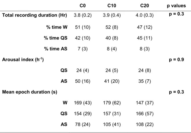
Sleep architecture is reported in table 2 for the 3 groups. Although not quantified, an increase in spontaneous activity during wakefulness was noted in C10 and especially C20 lambs. Overall, ETS had no significant influence on the % time spent in W, QS and AS, as well as on the arousal index and mean sleep state duration.
3.3. Cardiorespiratory variables
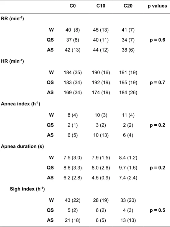
Data for cardiorespiratory variables are reported in Table 3. Regardless of the state of alertness, ETS had no influence on respiratory and heart rates, as well as on sigh index or apnea index and duration. Most apneas were post-sigh apneas.
3.4. Non-nutritive swallowing activity
NNS frequency data are reported in figure 1. Regardless of the exposure, NNS frequencies (isolated and bursts) were greater in AS than in W and QS. In addition, irrespective of the state of alertness, ETS had no effect on the frequency of total (p = 0.9) and isolated (p = 0.9) NNS, as well as NNS bursts (p = 0.6).
3.5. NNS-breathing coordination
Results on NNS breathing coordination are reported in figure 2. Regardless of the exposure, the same overall pattern of NNS-breathing coordination was observed, with i > ie > ei > e-NNS types. However, in QS i-type, NNS tended to decrease in C20 lambs compared to control (p = 0.07), whereas e and ei-type NNS were increased in C20 lambs when compared respectively to control (p = 0.07) and C10 lambs (p = 0.02). Furthermore, a decrease in e-type NNS in AS was noted between C10 and C0 lambs (p = 0.06). Finally, no differences were noted between exposure groups using the NNS-breathing phase calculation analysis (C0: 49% vs. C20: 52% p = 0.9).
Of note, identical overall statistical results were observed when C10 and C20 were pooled as the exposed group compared to the unexposed, control group.
4. DISCUSSION
Results from the present study show for the first time that postnatal ETS exposure for the first 15 days of life does not have any significant effects on baseline heart and respiratory rates, sleep architecture and non-nutritive swallowing frequency in newborn lambs. However, in support of our hypothesis, NNS-breathing coordination tends to be altered.
The effects of tobacco exposure on neonatal cardiorespiratory function have been previously assessed in a number of animal studies, including in newborn rats and lambs (Bamford et al., 1996; Hafstrom et al., 2002). While alterations in cardiorespiratory function and sleep have been reported, most studies used very high levels of nicotine exposure (> 1 mg/kg/day), which is likely irrelevant to human newborn exposure (Hussein et al., 2006). In addition, animal studies have been mostly focused on the effects of nicotine alone, whereas cigarette smoke contains approximately 4000 different chemical products, of which more than 100 are toxic and harmful for human health. In the present study, our custom-built smoking machine (Duvareille et al., 2010) was used to mimic postnatal ETS exposure and reproduce a urinary cotinine level at the upper limit of the range measured in newborn infants exposed to ETS at home (Blackburn et al., 2005; Joseph et al., 2007), thus ensuring real-life relevance of our results.
4.1. Baseline values and laryngeal inflammation
The normal weight gain observed throughout the 15 days of ETS exposure in the present study confirms similar observations in a previous study by our group (St-Hilaire
et al., 2010). Of note, longer ETS exposure (32 weeks) has been reported to reduce weight gain in mice (Talukder et al., 2011).
In the present study, 15 days of postnatal ETS exposure did not induce any observable laryngeal inflammation histologically. This is in agreement with a previous study in mice showing no increase in neutrophil accumulation or pro-inflammatory chemokine levels in the nasal turbinate tissue following a 24 weeks of ETS exposure (Huvenne et al., 2010). In contrast, several previous studies have reported that both ETS and active smoking (periods of 20 minutes to 24 weeks) induced laryngeal and supraglottic mucosa inflammation in adult humans and mice (Ben Gamra at al., 2007; Huvenne et al., 2010). The reason for the discrepant results between these studies is unknown.
4.2. Sleep architecture and arousal
In the present study, no effect of ETS was observed on sleep architecture. Sleep disturbances, including insufficient sleep, have been reported in active, chronic adult smokers (Davila et al., 2010). In human infants, while prenatal exposure to tobacco smoke did not modify sleep architecture (Dahlström et al., 2008; Horne et al., 2004), a decrease in both spontaneous arousals and arousal responses to obstructive apneas, hypoxia and trigeminal or auditory stimulation has been reported (Horne et al., 2004; Richardson et al., 2009; Tirosh and Libon, 1996). In addition, studies in lambs have shown that prenatal plus postnatal nicotine exposure blunts the arousal response to hypoxia (Hafstrom et al., 2000, 2002), while 15 days of postnatal ETS exposure blunts the arousal response observed during laryngeal chemoreflexes (St Hilaire et al., 2010). Overall, available results suggest that while perinatal ETS exposure does not alter
overall sleep architecture, prenatal and/or postnatal ETS exposure can blunt the arousal responses to various stimuli.
4.3. Cardiorespiratory effects
In agreement with the present observations, previous reports have shown that heart rate at rest is not affected by perinatal ETS exposure, including prenatal + postnatal nicotine exposure in mice (Bamford et al., 1996; Schuen et al., 1997), prenatal ETS exposure in human infants (Tirosh et al., 1996; Viskari-Lähdeoja et al., 2008) and postnatal nicotine exposure in lambs (Hafström et al., 2000).
Prenatal exposure to nicotine in animals (lambs or rats) and ETS exposure in humans has led to variable (no change or increase) effects on respiratory rate (RR) at rest (Bamford et al., 1996; Hafström et al., 2002; Huang et al., 2004; Schuen et al.,1997; Stéphan-Blanchard et al., 2010), possibly a reflection of variably-blunted prenatal lung development (Maritz and Harding, 2011). No effects on RR were observed after postnatal nicotine exposure in lambs (Hafström et al., 2000), in agreement with the present findings.
The absence of effect of 15 days postnatal ETS exposure on arterial blood gases and acid-base balance is in agreement with previous results obtained after prenatal plus postnatal nicotine exposure in lambs (Hafström et al., 2000, 2002), as well as after prenatal ETS exposure in term and preterm human infants for hemoglobin oxygen saturation (Stéphan-Blanchard et al., 2010; Tirosh et al., 1996).
Human studies on the effect of prenatal ETS exposure on apneas have yielded contradictory results, including no effect in term and preterm infants (Stéphan-Blanchard et al., 2010; Tirosh et al., 1996) or an increase in frequency and duration of obstructive
(Balaguer et al., 2009) or central (Toubas et al., 1986) apneas. Our present observations show for the first time an absence of effect of postnatal ETS exposure on spontaneous apneas.
Of note, we had previously reported that 15 days postnatal exposure to ETS enhances the cardiorespiratory inhibition observed as part of the laryngeal chemoreflexes in lambs (St-Hilaire et al., 2010), providing evidence of a deleterious cardiorespiratory effect of postnatal ETS exposure in lambs, which is not manifested in baseline conditions, as shown herein, but rather appears when the autonomic system of the lambs is challenged.
4.4 Non-nutritive swallowing frequency
As highlighted in the introduction, the reason for targeting NNS and NNS-breathing coordination in the present study was related to the crucial importance of this function in preventing lung aspiration of liquids secreted by the upper airways as well as part of the pharyngo-laryngeal refluxes, which are particularly frequent in the postnatal period. It has been previously proposed that anomalies of NNS may be involved in conditions such as apneas of prematurity, apparent life-threatening events of infants and sudden infant death syndrome (Praud, 2010). In this respect, the blunting effect of active smoking on various pharyngeal and laryngeal reflexes in adult humans, including swallowing, provided further support for the hypothesis tested in the present study (Dua et al., 1998, 2009, 2011). In addition, previous data suggest that active cigarette smoking could enhance gastro-esophageal and laryngopharyngeal reflux by altering closure of the upper (Dua et al., 1998) and lower (Kahrilas and Gupta, 1990) esophageal sphincter, together with inducing a delay in gastric emptying (Miller et al., 1989). The
present observation that NNS frequency is unaltered by 15 days of postnatal ETS exposure in newborn lambs is similar to results on NNS frequency in active smokers at rest (Dua et al., 1998) and suggests that early postnatal ETS exposure should not increase the risk of saliva aspiration. However, our previous results showing a decreased number of NNS elicited during laryngeal chemoreflexes (St-Hilaire et al., 2010) suggest that NNS alterations by early postnatal ETS exposure might predispose to lung aspiration during acute challenges such as pharyngolaryngeal reflux. In this respect, it would be of interest to study the effects of postnatal ETS exposure on nutritive swallowing, which constitutes another challenge for swallowing function (Bernier et al., 2012).
4.5. Non-nutritive swallowing-breathing coordination
Non-nutritive swallowing and breathing share common upper airways structures and their central pattern generator are located in the same brainstem nuclei (Jean, 2001). Conceivably, any condition which can alter the upper airways or the respiratory or swallowing centers may influence NNS frequency and the precise NNS-breathing coordination. We have previously shown that NNS beginning in inspiration are predominant over NNS beginning in expiration in lambs (Reix et al., 2003). In addition, we have reported that NNS-breathing coordination in lambs is unaltered by various neonatal conditions such as nasal application of continuous or intermittent positive airway pressure (Samson et al., 2005), hypoxia (Duvareille et al., 2007), preterm birth (Reix et al., 2004) and reflux laryngitis (Brisebois et al., 2010), suggesting that NNS-breathing coordination is established well before birth, in keeping with its importance for preventing both lung aspiration and respiratory inhibition. Until now, hypercapnia in
newborn lambs (Duvareille et al., 2007) and volition in adult humans (Kelly et al., 2007) were the only conditions known to alter NNS-breathing coordination. The present observation adds postnatal ETS to this list, which tends to alter NNS-breathing coordination, especially by increasing expiratory NNS at the expense of inspiratory NNS in the C20 lambs compared to control lambs. However, the potential physiological and/or clinical importance of this alteration is unknown. Similarly, it is also unknown whether prenatal tobacco exposure might lead to even more deleterious effects on NNS-breathing coordination.
The absence of any laryngeal inflammation in lambs suggests that the potential mechanism responsible for altering NNS-breathing coordination is of central origin. Previous studies have reported that perinatal ETS exposure leads to anomalies of neuronal network development in regions of the brainstem involved in respiratory and swallowing control. Examples include anomalies in nicotinergic, GABAergic and serotonergic receptors (Duncan et al., 2009) and microglial inflammation by NNK[4-methylnitrosamino-1-(3-pyridyl)-1-butanone] leading to neuronal destruction (Gosh et al., 2009).
5. CONCLUSION
In conclusion, postnatal ETS exposure in newborn lambs did not affect sleep, respiration, cardiac activity or NNS frequency, but tended to alter NNS-breathing coordination during quiet sleep. Though the potential consequences of this alteration are unknown, it adds to our previous observation of a decreased swallowing activity during
laryngeal chemoreflexes and supports an effect of postnatal ETS exposure on swallowing function.
ACKNOWLEDGEMENTS
The authors gratefully acknowledge the technical assistance of Jean-Philippe Gagné and the statistical assistance of Julie Hamon.
The experiments were supported by the Canadian Institutes of Health Research, the Canadian Foundation for Innovation and the Foundation of Stars. Jean-Paul Praud is the holder of the Tier 1 Canada Research Chair in Neonatal Respiratory Physiology and a member of the Centre de Recherche Clinique Etienne-Le Bel du Centre Hospitalier Universitaire de Sherbrooke.
REFERENCES
Adgent, M.A., 2006. Environmental tobacco smoke and sudden infant death syndrome: a review. Birth Defects Res. B. Dev. Reprod. Toxicol. 77, 69-85.
Anderson, H.R., Cook, D.G., 1997. Passive smoking and sudden infant death syndrome: review of the epidemiological evidence. Thorax 52, 1003-1009.
Bernier, A., Catelin, C., Hadj Ahmed, M.A., Samson, N., Bonneau, P., Praud, J.P., 2012. Effects of nasal continuous positive airway pressure on nutritive swallowing in lambs. J. Appl. Physiol. In press
Balaguer, C., Palou, A., Alonso-Fernandez, A., 2009. Smoking and sleep disorders. Arch. Bronconeumol. 45, 449-458.
Bamford, O.S., Schuen, J.N., Carroll, J.L., 1996. Effect of nicotine exposure on postnatal ventilatory responses to hypoxia and hypercapnia. Respir. Physiol. 106, 1-11. Ben Gamra, O., Mbarek, C., Charfi, S., Ouni, H., Hariga, I., Chedly, A., Zribi, S., El
Khedim, A., 2007. Chronic laryngitis in adults. Tunis. Med. 85, 641-643.
Blackburn, C.M., Bonas, S., Spencer, N.J., Coe, C.J., Dolan, A., Moy, R., 2005. Parental smoking and passive smoking in infants: fathers matter too. Health Educ. Res. 20, 185-194.
Bottorff, J.L., Johnson, J.L., Irwin, L.G., Ratner, P.A., 2000. Narratives of smoking relapse: the stories of postpartum women. Res. Nurs. Health 23, 126-134.
Brisebois, S., Samson, N., Fortier, P.H., Doueik, A.A., Carreau, A.M., Gagné, J.P., Praud, J.P., 2010. Effect of reflux laryngitis on non-nutritive swallowing in newborn lambs. Am. J. Respir. Crit. Care Med. 181, A2441.
Carreau, A.M., Patural, H., Samson, N., Doueik, A.A., Hamon, J., Fortier, P.H., Praud, J.P., 2011. Effects of simulated reflux laryngitis on laryngeal chemoreflexes in newborn lambs. J. Appl. Physiol. 111, 400-406.
Dahlstrom, A., Ebersjo, C., Lundell, B., 2008. Nicotine in breast milk influences heart rate variability in the infant. Acta Paediatr. 97, 1075-1079.
Davila, E.P., Lee, D.J., Fleming, L.E., LeBlanc, W.G., Arheart, K., Dietz, N., Lewis, J.E., McCollister, K., Caban-Martinez, A., Bandiera, F., 2010. Sleep disorders and secondhand smoke exposure in the U.S. population. Nicotine Tob. Res. 12, 294-299.
Dua, K., Bardan, E., Ren, J., Sui, Z., Shaker, R., 2002. Effect of chronic and acute cigarette smoking on the pharyngoglottal closure reflex. Gut 51, 771-775.
Dua, K., Bardan, E., Ren, J., Sui, Z., Shaker, R., 1998. Effect of chronic and acute cigarette smoking on the pharyngo-upper oesophageal sphincter contractile reflex and reflexive pharyngeal swallow. Gut 43, 537-541.
Dua, K., Surapaneni, S.N., Kuribayashi, S., Hafeezullah, M., Shaker, R., 2011. Protective role of aerodigestive reflexes against aspiration: study on subjects with impaired and preserved reflexes. Gastroenterology 140, 1927-1933.
Dua, K.S., Surapaneni, S.N., Santharam, R., Knuff, D., Hofmann, C., Shaker, R., 2009. Effect of systemic alcohol and nicotine on airway protective reflexes. Am. J. Gastroenterol. 104, 2431-2438.
Duncan, J.R., Garland, M., Myers, M.M., Fifer, W.P., Yang, M., Kinney, H.C., Stark, R.I., 2009. Prenatal nicotine-exposure alters fetal autonomic activity and medullary neurotransmitter receptors: implications for sudden infant death syndrome. J. Appl. Physiol. 107, 1579-1590.
Duvareille, C., Beaudry, B., St-Hilaire, M., Boheimier, M., Brunel, C., Micheau, P., Praud, J.P., 2010. Validation of a new automatic smoking machine to study the effects of cigarette smoke in newborn lambs. Lab. Anim. 44, 290-297.
Duvareille, C., Lafrance, M., Samson, N., St-Hilaire, M., Pladys, P., Micheau, P., Bournival, V., Langlois, C., Praud, J.P., 2007. Effects of hypoxia and hypercapnia on nonnutritive swallowing in newborn lambs. J. Appl. Physiol. 103, 1180-1188. Gosh, D., Mishra, M.K., Das, S., Kaushik, D.K., Basu, A., 2009. Tobacco carcinogen
induces microglial activation and subsequent neuronal damage. J. Neurochem. 110, 1070-1081.
Hafstrom, O., Milerad, J., Asokan, N., Poole, S.D., Sundell, H.W., 2000. Nicotine delays arousal during hypoxemia in lambs. Pediatr. Res. 47, 646-652.
Hafstrom, O., Milerad, J., Sundell, H.W., 2002. Altered breathing pattern after prenatal nicotine exposure in the young lamb. Am. J. Respir. Crit. Care Med. 166, 92-97. Hafstrom, O., Milerad, J., Sundell, H.W., 2002. Prenatal nicotine exposure blunts the
cardiorespiratory response to hypoxia in lambs. Am. J. Respir. Crit. Care Med. 166, 1544-1549.
Horne, R.S., Franco, P., Adamson, T.M., Groswasser, J., Kahn, A., 2004. Influences of maternal cigarette smoking on infant arousability. Early Hum. Dev. 79, 49-58.
Huang, Y.H., Brown, A.R., Cross, S.J., Cruz, J., Rice, A., Jaiswal, S., Fregosi, R.F., 2010. Influence of prenatal nicotine exposure on development of the ventilatory
response to hypoxia and hypercapnia in neonatal rats. J. Appl. Physiol. 109, 149-158.
Hussein, J., Farkas, S., MacKinnon, Y., Ariano, R.E., Sitar, D.S., Hasan, S.U., 2007. Nicotine dose-concentration relationship and pregnancy outcomes in rat: biologic plausibility and implications for future research. Toxicol. Appl. Pharmacol. 218, 1-10.
Huvenne, W., Perez-Novo, C.A., Derycke, L., De Ruyck, N., Krysko, O., Maes, T., Pauwels, N., Robays, L., Bracke, K.R., Joos, G., Brusselle, G., Bachert, C., 2010. Different regulation of cigarette smoke induced inflammation in upper versus lower airways. Respir. Res. 11, 100.
Jean, A., 2001. Brain stem control of swallowing: neuronal network and cellular mechanisms. Physiol. Rev. 81, 929-969.
Joseph, D.V., Jackson, J.A., Westaway, J., Taub, N.A., Petersen, S.A., Wailoo, M.P., 2007. Effect of parental smoking on cotinine levels in newborns. Arch. Dis. Child. Fetal Neonatal Ed. 92, F484-488.
Kahrilas, P.J., Gupta, R.R., 1990. Mechanisms of acid reflux associated with cigarette smoking. Gut 31, 4-10.
Kelly, B.N., Huckabee, M.L., Jones, R.D., Frampton, C.M., 2006. Nutritive and non-nutritive swallowing apnea duration in term infants: implications for neural control mechanisms. Respir. Physiol. Neurobiol. 154, 372-378.
Kelly, B.N., Huckabee, M.L., Jones, R.D., Carroll, G.J., 2007. The influence of volition on breathing-swallowing coordination in healthy adults. Behav. Neurosci. 121, 1174-1179.
Kelly, B.N., Huckabee, M.L., Frampton, C.M., Jones, R.D., 2008. Arousal has no effect on non-nutritive breathing-swallowing coordination during the first year of human life. Int. J. Dev. Neurosci. 26, 385-390.
Liebrechts-Akkerman, G., Lao, O., Liu, F., van Sleuwen, B.E., Engelberts, A.C., L'hoir, M.P., Tiemeier, H.W., Kayser, M., 2011. Postnatal parental smoking: an important risk factor for SIDS. Eur. J. Pediatr. 170, 1281-1291.
Maritz, G.S., Harding, R., 2011. Life-long programming implications of exposure to tobacco smoking and nicotine before and soon after birth: evidence for altered lung development. Int. J. Environ. Res. Public. Health. 8, 875-898.
McFarland, D.H., Lund, J.P., 1993. An investigation of the coupling between respiration, mastication, and swallowing in the awake rabbit. J. Neurophysiol. 69, 95-108.
Miller, G., Palmer, K.R., Smith, B., Ferrington, C., Merrick, M.V., 1989. Smoking delays gastric emptying of solids. Gut 30, 50-53.
Mitchell, E.A., Milerad, J., 2006. Smoking and the sudden infant death syndrome. Rev. Environ. Health 21, 81-103.
Nixon, G.M., Charbonneau, I., Kermack, A.S., Brouillette, R.T., McFarland, D.H., 2008. Respiratory-swallowing interactions during sleep in premature infants at term. Respir. Physiol. Neurobiol. 160, 76-82.
Praud, J.P., 2010. Upper airway reflexes in response to gastric reflux. Paediatr. Respir. Rev. 11, 208-212.
Reix, P., Fortier, P.H., Niyonsenga, T., Arsenault, J., Letourneau, P., Praud, J.P., 2003. Non-nutritive swallowing and respiration coordination in full-term newborn lambs. Respir. Physiol. Neurobiol. 134, 209-218.
Reix, P., Arsenault, J., Langlois, C., Niyonsenga, T., Praud, J.P., 2004. Nonnutritive swallowing and respiration relationships in preterm lambs. J. Appl. Physiol. 97, 1283-1290.
Renolleau, S., Letourneau, P., Niyonsenga, T., Praud, J.P., 1999. Thyroarytenoid muscle electrical activity during spontaneous apneas in preterm lambs. Am. J. Respir. Crit. Care Med. 159, 1396-1404.
Richardson, H.L., Walker, A.M., Horne, R.S., 2009. Maternal smoking impairs arousal patterns in sleeping infants. Sleep 32, 515-521.
Samson, N., St-Hilaire, M., Nsegbe, E., Reix, P., Moreau-Bussiere, F., Praud, J.P., 2005. Effect of nasal continuous or intermittent positive airway pressure on nonnutritive swallowing in the newborn lamb. J. Appl. Physiol. 99, 1636-1642.
Samson, N., Dumont, S., Specq, M.L, Praud, J.P., 2011. Radio telemetry devices to monitor breathing in non-sedated animals. Respir. Physiol. Neurobiol. 179, 111-118.
Schuen, J.N., Bamford, O.S., Carroll, J.L., 1997. The cardiorespiratory response to anoxia: normal development and the effect of nicotine. Respir. Physiol. 109, 231-239.
Stephan-Blanchard, E., Chardon, K., Leke, A., Delanaud, S., Djeddi, D., Libert, J.P., Bach, V., Telliez, F., 2010. In utero exposure to smoking and peripheral chemoreceptor function in preterm neonates. Pediatrics 125, e592-e599.
St-Hilaire, M., Duvareille, C., Avoine, O., Carreau, A.M., Samson, N., Micheau, P., Doueik, A., Praud, J.P., 2010. Effects of postnatal smoke exposure on laryngeal chemoreflexes in newborn lambs. J. Appl. Physiol. 109, 1820-1826.
Talukder, M.A., Johnson, W.M., Varadharaj, S., Lian, J., Kearns, P.N., El-Mahdy, M.A., Liu, X., Zweier, J.L., 2011. Chronic cigarette smoking causes hypertension, increased oxidative stress, impaired NO bioavailability, endothelial dysfunction, and cardiac remodeling in mice. Am. J. Physiol. Heart Circ. Physiol. 300, H388-H396.
Thach, B.T., Menon, A., 1985. Pulmonary protective mechanisms in human infants. Am. Rev. Respir. Dis. 131, S55-S58.
Tirosh, E., Libon, D., Bader, D., 1996. The effect of maternal smoking during pregnancy on sleep respiratory and arousal patterns in neonates. J. Perinatol. 16, 435-438. Toubas, P.L., Duke, J.C., McCaffree, M.A., Mattice, C.D., Bendell, D., Orr, W.C., 1986.
Effects of maternal smoking and caffeine habits on infantile apnea: a retrospective study. Pediatrics 78, 159-163.
Viskari-Lahdeoja, S., Hytinantti, T., Andersson, S., Kirjavainen, T., 2008. Heart rate and blood pressure control in infants exposed to maternal cigarette smoking. Acta Paediatr. 97, 1535-1541.
Wilson, S.L., Thach, B.T., Brouillette, R.T., Abu-Osba, Y.K., 1981. Coordination of breathing and swallowing in human infants. J. Appl. Physiol. 50, 851-858.
Table 1: Baseline data following postnatal exposure to environmental tobacco smoke for 15 days.
C0 C10 C20 p values
Cotinine/creatinine ratio
(ng.mg-1) 12 (7) 932 (346) 1535 (368) p = 0.005
Weight gain (g.day-1) 134 (50) 161 (40) 128 (54) p = 0.4
PaO2 (mmHg) 89 (8) 92 (10) 94 (80) p = 0.6 PaCO2 (mmHg) 45 (5) 44 (4) 41 (10) p = 0.5 pH 7.37 (0.05) 7.40 (0.04) 7.39 (0.07) p = 0.4 HCO3- (mmole.l-1) 24 (2) 26 (3) 24 (2) p = 0.4 Hemoglobin saturation in O2 (%) 96 (3) 97 (1) 97 (1) p = 0.9
C0: control lambs exposed to air, n = 6; C10: lambs exposed to 10 cigarettes per day, n= 6; C20: lambs exposed to 20 cigarettes per day, n = 6.
Table 2: Sleep architecture following postnatal exposure to environmental tobacco smoke for 15 days
C0 C10 C20 p values
Total recording duration (Hr) 3.8 (0.2) 3.9 (0.4) 4.0 (0.3) p = 0.3
% time W 51 (10) 52 (8) 47 (12) % time QS 42 (10) 40 (8) 45 (11) % time AS 7 (3) 8 (4) 8 (3) Arousal index (h-1) p = 0.9 QS 24 (4) 24 (5) 24 (8) AS 50 (16) 41 (20) 35 (7)
Mean epoch duration (s) p = 0.3
W 169 (43) 179 (62) 147 (37)
QS 154 (29) 157 (31) 166 (57)
AS 78 (24) 105 (41) 108 (22)
W: wakefulness; QS: quiet sleep; AS: active sleep. %time W, QS, AS: percentage of recording time spent in wakefulness, quiet and active sleep.
Table 3: Cardiorespiratory data in lambs after postnatal exposure to environmental tobacco smoke for 15 days
C0 C10 C20 p values RR (min-1) W 40 (8) 45 (13) 41 (7) p = 0.6 QS 37 (8) 40 (11) 34 (7) AS 42 (13) 44 (12) 38 (6) HR (min-1) W 184 (35) 190 (16) 191 (19) p = 0.7 QS 183 (34) 192 (19) 195 (19) AS 169 (34) 174 (19) 184 (26) Apnea index (h-1) W 8 (4) 10 (3) 11 (4) p = 0.2 QS 2 (1) 3 (2) 2 (2) AS 6 (5) 10 (13) 6 (4) Apnea duration (s) W 7.5 (3.0) 7.9 (1.5) 8.4 (1.2) p = 0.2 QS 8.6 (3.3) 8.0 (2.6) 9.7 (1.6) AS 6.2 (2.8) 4.5 (0.9) 7.4 (2.4) Sigh index (h-1) W 43 (22) 28 (19) 33 (20) p = 0.5 QS 5 (2) 6 (2) 4 (3) AS 21 (18) 6 (5) 13 (13)
Figure 1: Effect of cigarette smoke exposure on the frequency of total non-nutritive swallowing (NNS), isolated NNS and NNS bursts in control lambs (C0, exposure to air), lambs exposed to 10 cigarettes (C10) or 20 cigarettes (C20) per day (n = 6 for each group) in each state of alertness.
W, wakefulness; QS, quiet sleep; AS, active sleep.
There were no statistically significant differences between groups or states of alertness.
Figure 2: Effects of cigarette smoke exposure on NNS-breathing coordination in each state of alertness in C0, C10 and C20 groups (n = 6 for each group). i-type NNS, NNS beginning and ending in inspiration; e-type NNS, NNS beginning and ending in expiration; ie-type NNS, NNS beginning in inspiration and ending in expiration; ei-type NNS, NNS beginning in expiration and ending in inspiration. Results are expressed in percentage of the total number of NNS. *: significant difference vs. C0; β: significant difference C10 vs. C20. Underlined symbols indicate p < 0.05, normal font symbols indicate p < 0.1.
Overall, the same pattern of NNS-breathing coordination is observed between groups exposed to cigarette smoke or not, with i > ie > ei > e-NNS types. However, postnatal ETS tends to increase e-NNS in QS at the expense of inspiratory NNS in the C20 lambs compared to control lambs. The other differences (decrease in e-NNS in AS in C10 only compared to controls, or decrease in ei-NNS in QS in C20 compared to C10, but not to controls) do not provide a coherent message in relation to postnatal ETS.