Bacterial adaptation to temperature
stress: molecular responses in two
Gram-positive species from distinct
ecological niches
Dong XUE
Promoteur: Prof. Marc ONGENA
Co-promoteur: Prof. Jin WANG
2020
COMMUNAUTÉ FRANÇAISE DE BELGIQUE UNIVERSITÉ DE LIÈGE – GEMBLOUX AGRO-BIO TECH
Bacterial adaptation to temperature
stress: molecular responses in two
Gram-positive species from distinct
ecological niches
Dong XUE
Dissertation originale présentée en vue de l’obtention du grade de docteur en sciences agronomiques et ingénierie biologique
Promoteurs: Prof. Marc Ongena & Prof. Jin Wang Année civile: 2020
Résumé
Dong XUE (2020) Adaptation bactérienne au stress thermique : réponses
moléculaires chez deux espèces Gram-positives de niches écologiques distinctes
(thèse de doctorat). Gembloux, Belgique, Liège Université, Gembloux Agro-Bio Tech, 133 p., 14 Tableaux, 44 Figures.
Résumé : Les micro-organismes sont souvent affectés par divers facteurs environnementaux. Ces facteurs environnementaux affectent leurs fonctions physiologiques et biochimiques. Parmi ces facteurs environnementaux, la température joue un rôle important dans les activités physiologiques normales des micro-organismes. Pour s'adapter à différents environnements de température, les bactéries ont développé de nombreux mécanismes adaptatifs pour coordonner une gamme de changements dans l'expression génique et l'activité protéines. Dans cette étude, nous avons étudié les mécanismes d'adaptation de deux bactéries Gram-positives à différentes températures. Les principaux résultats de cette thèse sont les suivants :
(1) Deinococcus radiodurans est une bactérie Gram-positive, pigmentée de rose et à haut G + C. La réponse thermique de D. radiodurans est considérée comme un système de régulation classique induit par le stress qui se caractérise par une reprogrammation transcriptionnelle étendue. Dans cette partie, nous avons étudié les gènes fonctionnels clés impliqués dans le stress thermique qui a été exprimé et accumulé dans les cellules après un traitement thermique à 48 °C pendant 2 heures (R48). Considérant que la dégradation des protéines est un bioprocessus chronophage, nous avons prédit que pour maintenir l'homéostasie cellulaire, l'expression des protéines fonctionnelles clés serait considérablement diminuée dans les cellules qui s'étaient partiellement rétablies du stress thermique (RH) par rapport à leur expression dans les cellules cultivées sous température optimale (R30). La transcriptomique comparative a identifié 15 gènes qui étaient significativement régulés à la baisse dans l'RH par rapport à R30, dont sept avaient été précédemment caractérisés comme étant des protéines de choc thermique. Parmi ces gènes, trois gènes hypothétiques (dr_0127, dr_1083 et
dr_1325) sont très susceptibles d'être impliqués en réponse au stress thermique.
L'analyse de survie des souches mutantes dépourvues de dr_0127, dr_1083 et
dr_1325 a montré une réduction de la tolérance à la chaleur par rapport à la
souche de type sauvage. Ces résultats suggèrent que dr_0127, dr_1083 et
dr_1325 pourraient jouer un rôle dans la réponse au stress thermique.
Sur la base de nos données RNA-seq et des rapports précédents, nous avons identifié deux nouveaux ARN non codants (ncRNA) inductibles par la chaleur chez D. radiodurans, nommés DnrH et dsr11. L'analyse de la tolérance à la chaleur a montré que la suppression de DnrH inhibait significativement la viabilité en réponse à des conditions de température élevée. Des analyses phénotypiques et qRT-PCR comparatives d'un mutant DnrH (∆DnrH) et de type sauvage (WT) ont suggéré que DnrH est potentiellement impliqué dans la
régulation de l'expression du gène Hsp20 lié au choc thermique. La thermophorèse à l'échelle microscopique et la complémentation génétique ont montré qu'une séquence de 28 nucléotides (nt) dans la structure tige-boucle des paires DnrH (143-170 nt) interagit avec son homologue dans la région codante de mRNA Hsp20 (91-117 nt) via une séquence de 22 nt Région. In vivo, la mutation de la région des 22 nt dans le génome de D. radiodurans a entraîné une réduction de la tolérance à la chaleur similaire à celle observée chez le mutant DnrH. Nos résultats montrent que la DnrH influence positivement la tolérance à la chaleur en augmentant la transcription de mRNA de Hsp20, démontrant, pour la première fois, un ncRNA qui contrôle directement l'expression d'un gène de résistance au stress thermique. De manière similaire à DnrH, nous avons caractérisé un autre ncRNA dsr11. Notre résultat a montré que le niveau de transcription de dsr11 était augmenté de 4,2 fois sous stress thermique par analyse qRT-PCR. Un essai de tolérance à la chaleur a montré que la suppression de dsr11 inhibait significativement la viabilité dans des conditions de température élevée. Pour évaluer l'influence de dsr11 sur le transcriptome de D. radioduans, 157 gènes ont été trouvés différentiellement exprimés dans le mutant knock-out par l'expérience RNA-Seq. En combinant RNA-Seq et analyse bioinformatique, nous avons constaté que dr_0457 (protéine de transport de biopolymère) était probablement la cible directe de dsr11. D'autres résultats de thermophorèse à l'échelle microscopique ont démontré que le dsr11 peut se lier directement au mRNA de
dr_0457. Nos résultats ont indiqué que dsr11 peut améliorer la tolérance au stress
thermique de D. radiodurans en se liant au mRNA de dr_0457.
(2) Bacillus velezensis GA1 est une bactérie modèle Gram-positive qui vit naturellement en association avec les plantes et possède des propriétés de biocontrôle permettant de protéger son hôte végétal contre les phytopathogènes. Dans ce travail, nous avons évalué l'impact d'une température basse qui correspond à celle des sols sur cette bactérie. Une température basse a un impact négatif sur le taux de croissance, reflétant la réduction générale de l'activité métabolique des cellules cultivées à basse température. Les cultures in vitro ont révélé que la productivité de certains métabolites liés au biocontrôle changeait sensiblement lorsque la température était abaissée. Nous avons observé qu’après plusieurs cycles de culture à 15 et 18 ° C, la croissance de GA1 est devenue plus rapide qu'auparavant. Nous avons testé GA1 à 18 ° C sur tomate, les résultats ont montré que la formation de biofilm sur les racines était un peu plus lente qu'à 22 ° C, réduisant ainsi la population de GA1 sur ces tissus.
En résumé, cette étude a analysé en profondeur les voies d'adaptation et les mécanismes développés par deux bactéries à Gram-positive en réponse à différentes conditions de stress températures. Notre étude fournira une base théorique pour des applications futures dans l'industrie et l'agriculture.
Mots-clés : Deinococcus radiodurans, stress thermique, non codant RNA, dnrH,
Abstract
Dong XUE (2020) Bacterial adaptation to temperature stress: molecular
responses in two Gram-positive species from distinct ecological niches (PhD
thesis). Gembloux, Belgium, University of Liège, Gembloux Agro-Bio Tech, 133 p., 14 Tables, 44 Figures.
Abstract: Microorganisms are often affected by various environmental factors. These environmental conditions have an effect on their physiological and biochemical functions. Among these environmental factors, temperature plays an important role in the normal physiological behaviors of microorganisms. To adapt to different temperature environments, bacteria have evolved a variety of adaptive mechanisms to coordinate a range of gene expression and protein activity changes. In this study, we investigated the adaptation mechanisms of two Gram-positive bacteria at different temperatures. The main results of this thesis are as follows:
(1) Deinococcus radiodurans is a gram-positive, pink-pigmented, and high G+C bacterium. The heat response of D. radiodurans is considered to be a classical stress-induced regulatory system characterized by extensive transcriptional reprogramming. In this part, we investigated the key functional genes involved in heat stress that were expressed and accumulated in cells following heat treatment at 48°C for 2 hours (R48). Considering that protein degradation is a time-consuming process, we predicted that in order to maintain cellular homeostasis, the expression of the key functional proteins would be significantly decreased in cells that had partly recovered from heat stress (RH) relative to their expression in cells grown under optimal temperature (R30). Comparative transcriptomics identified fifteen genes that were significantly downregulated in RH relative to R30, seven of which were previously characterized as heat shock proteins. Among these candidates, three genes (dr_0127, dr_1083, and dr_1325) are more likely involved in response to heat stress as survival analysis of mutant strains lacking dr_0127, dr_1325, and dr_1083 showed a reduction in heat tolerance compared to the wild-type strain.
Based on our RNA-seq results and previous studies, we identified two novel heat-inducible ncRNAs in D. radiodurans, named DnrH and dsr11. Heat tolerance analysis showed that deleting DnrH significantly inhibited viability in response to high temperature conditions. Comparative phenotypic and qRT-PCR analyses of a DnrH mutant (∆DnrH) and wild-type (WT) suggested that DnrH is potentially involved in regulating the expression of the heat shock-related gene
Hsp20. Microscale thermophoresis and genetic complementation showed that a
28-nucleotide (nt) sequence in the stem-loop structure of DnrH (143–170 nt) pairs with its counterpart in the coding region of Hsp20 mRNA (91–117 nt) via a 22 nt region. In vivo, mutation of the 22-nt region in the D. radiodurans genome led to a reduction in heat tolerance similar to that observed in the DnrH-mutant. Our
results show that DnrH positively influences heat tolerance by increasing the transcription of Hsp20 mRNA, demonstrating, for the first time that a ncRNA may directly controls the expression of a heat stress-resistance gene. Similar to
dnrH, we characterized another ncRNA, dsr11. Our result showed that the
transcription level of dsr11 was upregulated 4.2-fold under heat stress by qRT-PCR analysis. Heat tolerance assays showed that deleting dsr11 significantly inhibited the viability under high temperature stress conditions. To assess the influence of dsr11 on the D. radioduans transcriptome, 157 genes were found differentially expressed in the knock-out mutant by RNA-Seq experiment. Combined RNA-Seq and bioinformatic analysis, we found that dr_0457 (biopolymer transport protein) was likely to be the direct targets of dsr11. Further microscale thermophoresis results demonstrated that dsr11 can directly bind to the mRNA of dr_0457. Our results indicated that dsr11 can enhance the tolerance to heat stress of D. radiodurans by binding to dr_0457 mRNA.
(2) Bacillus velezensis GA1 is a Gram-positive bacterium living in association with plant roots and which may provide some protective effects against phytopathogens (biocontrol). In this work, we evaluated the impact on GA1 of temperatures typical of soils and lower than the optimal one commonly used under laboratory conditions. Cold temperature negatively impacted the cell growth rate of GA1, reflecting a general reduction in the metabolic activity. In
vitro cultures revealed that production of some metabolites involved in biocontrol
changed markedly when the temperature was lowered. We observed that after several rounds of culture on RE liquid medium at 15 and 18°C the growth of GA1 became faster than before suggesting that the bacterium may somehow adapt to cold conditions. We also tested the behavior of GA1 at 18°C on tomato plants, and showed that biofilm formation on root was a bit slower than at 22°C which correlated with reduced populations as revealed by flow cytometry measurements.
In summary, this study deeply analyzed the adaptation pathways and mechanisms of two gram-positive bacteria in response to different temperatures. Our work will provide a theoretical basis for future applications in industry and agriculture.
Keywords: Deinococcus radiodurans, heat stress, noncoding RNA, dnrH, dsr11,
Acknowledgments
This PhD dissertation is the results of much collaboration with both scientific colleagues but also with nonscientific actors. I would like to give my thanks to everyone who contributed and help to make this doctoral dissertation finally happen. I would also like to express my gratitude especially to:
Firstly, I would like to express my sincere gratitude to my supervisor Prof. Marc Ongena, who has a really good sense of humor and always gives me understandings for my life and study. You always shared new ideas and discussed with me about my research interests. You have helped me a lot in technical research and writing this thesis. Without you, I could not finish this fruitful study. The word thanks cannot express my gratitude feeling.
I would also like to thank my co-supervisor Prof. Jin Wang for her guidance and suggestion during my thesis. She always shared her busy schedule to meet and have a discussion with me for the method and data analysis.
I would also like thank to Prof. Min Lin, who is rich in precious ideas, kind guidance and give me constructional advices in my study and cultivating my personal way of working in scientific search.
My sincere thanks and appreciation to the rest of my thesis committee: Prof. Philippe Jacques, Prof. Frank Delvigne, Prof. Marc Hanikenne, Prof. Yuquan Xu, and Dr. Anthony Arias Arguelles for contributing their valuable time to read my manuscript, and their insightful comments, suggestions to make my thesis manuscript better and also for the hard questions which incented me to widen my research from various perspectives.
Thanks to every member in molecular microbiology research team for their concern and help during my PhD study, thanks to Prof. Ming Chen, Prof. Wei Zhang, Prof. Shuzhen Ping, Prof. Wei Lu, Prof. Yongliang Yan, Dr. Zhengfu Zhou, Dr. Yuhua Zhan, and Dr. Xiubing Ke.
I would also thank Prof. Marc Ongena, Prof. Frank Delvigne, Prof. Patrick Fickers and Prof. Philippe Jacque who are executors of the lab and MiPI team. Thank all the members from MiPI lab. Thank you all for welcoming me, for your support, for your kindness and your friendship.
Thank my classmates for taking care of me in Gembloux, especially to Tie Cheng Bai, Xuewei Zhou, Minmin Li, Lin Li, and Wei Fang, for taking care of me during my operation in Belgium.
I would like to give thanks to my family. Thank you, mam and dad, for giving me the rights to choose what I want in my life. During the past five years, there were some struggling times for me, thank you all for all the warmness and supports you have offered. You are the most important reasons that I could finish this dissertation and get my PhD degree. Thank you for being there for me all the time. I also would like to thank my girlfriend Feng Li, thanks her for understanding and supporting me during my PhD study.
Additionally, I would like thank to the Key Research and Development Projects, the Ministry of Agriculture Transgenic Program, and the National Natural Science Foundation of China support this research. I wish to thank China Scholarship Council (CSC) for the financial support to study in Belgium. Finally, I would like to thank sincerely all the people who helped me to carry it out successfully.
Tables of Contents Résumé ... I Abstract ... III Acknowledgments ... V Tables of Contents ... VII List of Tables ... XII List of Figures ... XIII List of Abbreviations ... XV
Chapter I: General Introduction ... 1
1.1. The effect of temperature on the physiology in prokaryotes ... 1
1.2. Research progress on heat stress response in bacteria ... 2
1.2.1. Regulated by the sigma factor ... 2
1.2.2. Regulated by the CIRCE/HrcA regulatory system ... 3
1.2.3. Regulated by RNA sensors ... 3
1.2.4. DNA topology affects the gene expression ... 4
1.3. Key technologies of transcriptomics and application in bacterial functional genomics research ... 5
1.3.1. RNA-Seq technology ... 5
1.3.2. Transcriptomics application in the global regulation of bacterial stress response ... 6
1.4. Research progress on Deinococcus radiodurans ... 6
1.4.1. Introduction on D. radiodurans ... 6
1.4.2. The extreme resistance of D. radiodurans ... 7
1.4.2.1. Ionizing radiation resistance ... 7
1.4.2.2. Oxidative resistance ... 8
1.4.2.3. Desiccation resistance ... 9
1.4.2.4. Heat stress resistance ... 9
1.5. Research progress on noncoding RNA in bacteria ... 10
1.5.1. Definition of noncoding RNA in bacteria ... 10
1.5.2. Molecular mechanism of bacterial ncRNA ... 12
1.5.2.1. Interaction with target mRNA ... 12
1.5.3. Study progress of ncRNA in D. radiodurans ... 14
1.6. Biocontrol effect of plant growth-promoting rhizobacteria for plant protection ... 14
1.6.1. Plant growth-promoting rhizobacteria ... 14
1.6.2. The role of PGPR in plants under abiotic stresses ... 14
1.6.3. Bacillus velezensis GA1 ... 15
1.7. Objectives ... 15
1.8. Overview of the chapters ... 16
References ... 17
Chapter II: RNA-Seq-based comparative transcriptome analysis highlights new features of the heat-stress response in the extremophilic bacterium Deinococcus radiodurans ... 29
2.1. Foreword ... 29
2.2. Abstract ... 29
2.3. Introduction ... 31
2.4. Materials and Methods ... 33
2.4.1. Strain and growth conditions ... 33
2.4.2. Heat stress treatment of D. radiodurans and recovery conditions ... 33
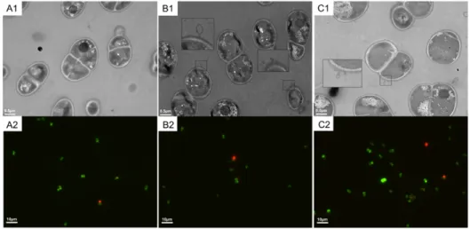
2.4.3. Transmission electron microscopy (TEM) and fluorescence assay ... 33
2.4.4. Total RNA extraction, complementary DNA (cDNA) library preparation, and sequencing ... 34
2.4.5. Assembly and functional enrichment analyses of differentially expressed genes (DEGs) ... 34
2.4.6. Gene Ontology (GO) and Kyoto Encyclopedia of Gene and Genomes (KEGG) enrichment analysis ... 35
2.4.7. Quantitative real-time PCR (qRT-PCR) validation ... 35
2.4.8. Construction of gene deletion mutant strains and heat stress phenotype assays ... 36
2.4.9. Statistical analysis ... 36
2.5. Results ... 37
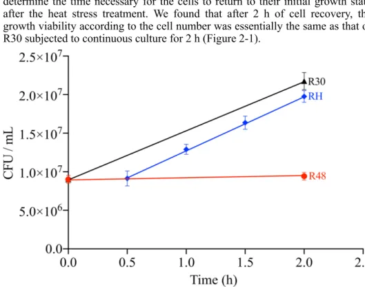
2.5.1. Cell growth state and viability of D. radiodurans under heat stress ... 37
2.5.2. Qualitative and quantitative analyses of the DEGs in the three groups under heat stress ... 38
2.5.3. GO and KEGG analyses of the DEGs ... 42 2.5.4. Analysis of heat-related genes with over eight-fold higher expressions in
response to heat stress ... 42
2.5.5. Function analysis of the novel, potentially heat-related genes ... 44
2.6. Discussion ... 46
2.7. Supplementary Materials ... 50
References ... 58
Chapter III: Targeting Hsp20 using the novel small noncoding RNA DnrH regulates heat tolerance in Deinococcus radiodurans ... 65
3.1. Foreword ... 65
3.2. Abstract ... 65
3.3. Introduction ... 66
3.4. Materials and Methods ... 67
3.4.1. Bacterial strains, plasmids, primers, and culture conditions ... 67
3.4.2. RNA isolation ... 68
3.4.3. 5′RACE ... 68
3.4.4. Construction of the mutant and complementary strains ... 69
3.4.5. Quantitative real-time PCR (qRT-PCR) ... 69
3.4.6. Bacterial growth curve and heat stress survival assays ... 70
3.4.7. Northern blot ... 70
3.4.8. Microscale thermophoresis (MST) measurements ... 70
3.5. Results ... 71
3.5.1. Experimental identification and transcriptional start sites of DnrH ... 71
3.5.2. DnrH is a novel factor involved in the heat stress response ... 74
3.5.3. Hsp20 as target of DnrH in response to heat stress ... 75
3.5.4. DnrH increases heat tolerance in D. radiodurans through regulation of Hsp20 mRNA ... 76
3.6. Discussion ... 79
3.7. Supplementary Materials ... 80
Reference ... 85
Chapter IV: A novel noncoding RNA dsr11 involved in heat stress tolerance in Deinococcus radiodurans ... 91
4.1. Foreword ... 91
4.2. Abstract ... 91
4.4. Materials and Methods ... 94
4.4.1. Strain and growth conditions ... 94
4.4.2. RNA extraction ... 94
4.4.3. Construction of gene deletion mutant strain ... 94
4.4.4. Quantitative real-time PCR (qRT-PCR) ... 94
4.4.5. Bacterial Growth curve and heat stress tolerance assays ... 95
4.4.6. RNA-seq and data analysis ... 95
4.4.7. Bioinformatics analysis ... 96
4.4.8. Microscale thermophoresis (MST) analysis ... 96
4.5. Results and discussion ... 96
4.5.1. Characterization of the novel ncRNA dsr11 ... 96
4.5.2. dsr11 is required for heat tolerance of D. radiodurans ... 97
4.5.3. RNA-Seq analysis sheds new light on the mechanisms of dsr11-mediated heat tolerance ... 98
4.5.4. Identification of the dsr11 targets ... 101
4.6. Conclusions ... 103
4.7. Supplementary Materials ... 103
Reference ... 105
Chapter V: Adaptation to cold stress in Bacillus velezensis GA1 and interaction with tomato at low temperature ... 111
5.1. Foreword ... 111
5.2. Introduction ... 111
5.3. Material and methods ... 112
5.3.1. Bacteria strain and plant used in this study ... 112
5.3.2. Time-frame study for cold-adaptive ... 112
5.3.3. Preparing tomato seeds for germination ... 113
5.3.4. Bacterial colonization on tomato rhizosphere ... 113
5.3.5. LC-MS analysis ... 114
5.3.6. Flow cytometry (FCM) analysis ... 114
5.4. Results and discussion ... 114
5.4.1 Bacterial growth and time-frame study in a solid medium under low temperature ... 114 5.4.2. Bacterial growth and time-frame study in liquid medium under low
temperature ... 115
5.4.3. Influence of cold temperature on colonization of tomato roots ... 117
References ... 120
Chapter VI: General discussion and perspectives ... 123
General discussion and perspectives ... 123
References ... 128
List of Tables
Table 1-1. The four types of prokaryotes according to OGT.
Table 2-1. The top 30 most upregulated and downregulated genes when exposed
to heat stress (R48 versus R30).
Table 2-2. Descriptions of the 16 genes with altered expression in cells recovered
from heat stress compared with non-stressed cells (RH versus R30).
Supplementary Table 2-1. The significantly different gene expression in three
groups.
Supplementary Table 2-2. GO annotation of the DEGs.
Supplementary Table 2-3. Summary of the control and heat stress transcriptome
sequencing data.
Supplementary Table 2-5. List of primers used for this study. Table 3-1. Strains and plasmids used in this study.
Supplementary Table 3-1. Premiers used in this study.
Supplementary Table 3-2. Synthesized ssRNA oligonucleotide derivatives for
MST.
Table 4-1. Selection of the most representative genes differentially expressed in D. radiodurans dsr11 deletion mutant with Log2 (FC) >1 or Log2 (FC) < 1. Supplementary Table 4-1. Premiers used in this study.
Supplementary Table 4-2. Synthesized ssRNA oligonucleotide derivatives for
MST.
Supplementary Table 4-3. List of genes differentially expressed in the dsr11
deletion mutant compared to the WT strain.
Supplementary Table 4-4. The possible targets of dsr11 predicted by
List of Figures
Figure 1-1. Wiring diagram of sig32 regulation.
Figure 1-2. Titration models for homeostatic control of chaperone expression. Figure 1-3. Schematic illustration of regulatory principles of riboswitches (A)
and RNA thermometers (B).
Figure 1-4. Effects of heat shock on plasmid topology in Escherichia coli and Sulfolobus islandicus.
Figure 1-5. Two-step of genome reconstitution in D. radiodurans shattered by
ionizing radiation.
Figure 1-6. Extreme resistance of D. radiodurans to desiccation. Figure 1-7. ncRNAs in global regulation cascades.
Figure 1-8. Overview of cis- and trans-encoded ncRNAs. Figure 1-9. Basic mechanisms of the ncRNA/mRNA interaction.
Figure 1-10. General properties of ncRNAs that modulate protein activity. Figure 2-1. Correlation analysis of the cell viability and the different treatments
using three biological replicates.
Figure 2-2. TEM and fluorescence images of the heat-induced lesions on D. radiodurans cells following exposure to 48 °C for 2 h.
Figure 2-3. Differential expression levels among the three treatment groups. Figure 2-4. Heat-related genes with over eight-fold higher expressions in
response to heat stress.
Figure 2-5. The effect of the dr_0127 deletion on the expression of its flanking
genes and survival phenotype plate assay upon heat stress.
Figure 2-6. Molecular response of D. radiodurans under high temperature
conditions.
Supplementary Figure 2-1. Validation of the RNA-Seq data using qRT-PCR. Supplementary Figure 2-2. Gene ontology (GO) enrichment analysis of
differentially expressed genes in D. radiodurans with and without heat stress.
Supplementary Figure 2-3. Top 20 KEGG biological pathway classification
histograms for annotated unigenes.
Supplementary Figure 2-4. Construction and verification of the three mutants. Supplementary Figure 2-5. Effect of the dr_1325 deletion on the expression of
its downstream genes (dr1326 and dr1327) under normal growth conditions.
Supplementary Figure 2-6. Pearson correlation analysis among 9 samples. Supplementary Figure 2-7. Cluster analysis of differentially expressed genes in
9 samples under heat stress.
Figure 3-1. Locus features and the expression of DnrH. Figure 3-2. Conservation of DnrH sequence and structure.
Figure 3-3. Transcriptional and functional analysis of DnrH ncRNA in Deinococcus radiodurans.
expression and phenotypic analysis of Hsp20 during heat stress.
Figure 3-5. Interaction between DnrH and Hsp20 mRNA in vitro and in vivo. Figure 3-6. A proposed working model for the DnrH-mediated regulatory
network in heat stress response in Deinococcus radiodurans.
Supplementary Figure 3-1. The original gels of 5’RACE results.
Supplementary Figure 3-2. Expression of heat regulatory genes in wildtype D. radiodurans under heat stress.
Supplementary Figure 3-3. Secondary structure of Hsp20 prediction performed
with RNAalifold.
Figure 4-1. The expression of dsr11 under heat stress and secondary structure of dsr11.
Figure 4-2. Transcriptional and functional analysis of dsr11 in Deinococcus radiodurans.
Figure 4-3. RNA-Seq analysis between WT and ∆Dsr11.
Figure 4-4. Network plots of dsr11 and targets interaction by RNA-Seq and in silico prediction.
Figure 4-5. Predicted interaction sites of dsr11 and target gene and determination
of the binding affinity of dsr11 to target mRNAs by microscale thermophoresis (MST).
Figure 5-1. The workflow diagram in solid RE medium (A) and liquid RE
medium (B).
Figure 5-2. Growth of GA1 on four rounds of continuous culture on RE solid
medium at three temperatures (15°C, 18°C, and 22°C).
Figure 5-3. The adaptation of GA1 after several rounds at lower temperatures
(15 °C and 18 °C).
Figure 5-4. Analysis the surfactin production of the supernatant of GA1 in RE
liquid medium at different temperatures and rounds.
Figure 5-5. B. velezensis GA1 colonization on tomato roots.
Figure 5-6. Root colonization by GA1 on tomato root exposed to different
temperatures by flow cytometry.
Figure 5-7. The different types of GA1 colonized on tomato root at different time
List of Abbreviations
Amp Ampicillin
cDNA Complementary DNA
Cm Chloromycetin
DNA Deoxyribonucleic
dNTP Deoxy-ribonucleoside-triphosphate EDTA Ethylene dinitrilotetracetic acid
IGR Intergenic region
Km Kamacycine
LB Luria-Bertani
mRNA Messenger RNA
MST Microscale thermophoresis
ncRNA Noncoding RNA
OD Optical density
ORF Opening read frame
PAGE Polyacrylamide gel electrophoresis
PCR Polymerase chain reaction
qRT-PCR Quantity real-time PCR
RNA Ribonucleic acid
RBS Ribosome binding site
rpm Rounds per minute
RT-PCR Reverse-transcription PCR
SD Shine-Dalgarno sequence
SDS Sodium dodecyl sulfate
TEMED N, N, N’, N’-Tetramethylethylenediamine Tris Tris (hytdroxymethyl) aminomethane
tRNA Transfer RNA
TSS Transcription start site
UTR Untranslated regions
UV ultraviolet
WT Wild type
X-gal 5-bromo-4-cholro-3-indoly-β-D-galactose
OGT Optimal growth temperature TEM Transmission electron microscopy
PI Propidium iodide
RSG Redox sensor green
RIN RNA integrity
DEGs Differentially expressed genes
FPKMs Fragments per kilobase of transcript per million mapped reads
GO Gene ontology
KEGG Kyoto Encyclopedia of gene and genomes
Spec spectinomycin
SD Standard deviations
PGPR Plant growth-promoting rhizobacteria
1
1.1. The effect of temperature on the physiology in
prokaryotes
Prokaryotes have high physiological diversity and wide ecological distribution (Whitman et al., 1998). Prokaryotes are the main components in the biosphere and play an important role in the biogeochemical cycle. The prokaryotes living environment often changes, such as temperature, pH, oxygen concentration, pressure, and nutrients (Knoll et al., 1989). Temperature is one of the key environmental factors in microbial life, it can determine whether organisms can survive. Most microorganisms have an optimal growth temperature (OGT) ranging from 24 °C to 50 °C, which called mesophile (Bell et al., 2002). Besides, there are also psychrophile with OGT<24°C, thermophile (50°C<OGT<80°C), and hyperthermophile (OGT>80°C) (Chandrayan et al., 2014; Galtier et al., 1997; Musto et al., 2006; Nakashima et al., 2003; Ohtani et al., 2010) (Table
1-1).
Table 1-1. The four types of prokaryotes according to OGT
Type OGT
Psychrophile OGT<24°C
Mesophile 24°C<OGT<50°C
Thermophile 50°C<OGT<80°C
Hyperthermophile OGT>80°C
The low temperature will cause a decrease in enzyme activity in microbial cells. The speed of various biochemical reactions, cell metabolism, and the fluidity of biomass membranes will be slow down (Hébraud et al., 1999; Shivaji et al., 2010). However, when the temperature is further lowered below the freezing point, the water in the cells will condense into ice crystals, causing irreversible mechanical damage to the biomass membrane such as the cell membrane, eventually leading to the death of the cells (Wilson et al., 2010). The high temperature will cause irreversible denaturation of protein in microorganisms. In particular, it affects normal cell physiological activities, such as affecting the enzymes involved in the tricarboxylic acid cycle (Ventura et al., 2006). Besides, high temperatures can lead to the destruction of the plasma membrane structure and to the damage of nucleic acid molecules, which leads to the death of the cells (Russell, 2003; Teixeira et al., 1997). When microorganisms in a non-fatal high-temperature environment, their growth will slow down or stagnate for a short period, and the level of cellular metabolism will decrease. The cells exhibit a heat shock response to cope with the effects of high temperature pressure on the cells. In the long-term heat stress process, bacteria can finally get the adaptability to heat stress through a series of stress responses (Gayán et al., 2016; Van der Veen et al., 2007).
distinct ecological niches
1.2. Research progress on heat stress response in
bacteria
Severe high-temperature stress can cause cell damage and cell death, while sub-lethal high-temperature stress can induce various cellular heat shock response (Suo et al., 2012). Heat shock response can protect cells from heat damage, help cells restore to normal physiological activities, and enhance the heat tolerance of bacteria. The thermal denaturation of proteins and enzymes is critical for cell viability. To adapt to high-temperature, microorganisms have developed a variety of adaptive mechanisms to protect cells from high-temperature stress, such as heat shock gene regulation mechanisms, and the stringent response at the global regulatory level (J.-s. Li et al., 2011; Murata et al., 2011). These components are discussed below:
1.2.1. Regulated by the sigma factor
In E. coli, sig32 is an RNA polymerase subunit that is encoded by the rpoH gene. In general, the expression level of the rpoH gene is very low, mainly because the molecular chaperone system (like DnaK and GroE-type) accelerates the degradation of sig32 by protease (FtsH) (Arsène et al., 2000; Guisbert et al., 2004; Yura et al., 1999). As the external temperature rises, the expression of
rpoH increases dramatically over time, and enhances the synthesis of sig32. The
increase in the level of sig32 promotes the synthesis of molecular chaperone (DnaK/J; GroEL/S) and protease (FtsH). Chaperones and proteases further bind the misfolded proteins to help them fold correctly or degrade the misfolded proteins. In the adaptation phase that follows the induction phase, sig32 is inactivated when excess heat shock proteins (HSPs) are present (Guisbert et al., 2008) (Figure 1-1).
Figure 1-1. Wiring diagram of sig32 regulation.
There are three primary modes of regulation as follows: (1) excess free DnaK/J and GroEL/S chaperones directly bind to and inactivate sig32; (2) the FtsH protease degrades
sig32, with chaperones participating in this process; and (3) temperature directly controls the rate of sig32 translation. Misfolded proteins titrate chaperones from these regulatory roles, allowing active sig32 to increase the synthesis of chaperones and proteases during
conditions where they are required (Guisbert et al., 2008).
1.2.2. Regulated by the CIRCE/HrcA regulatory system
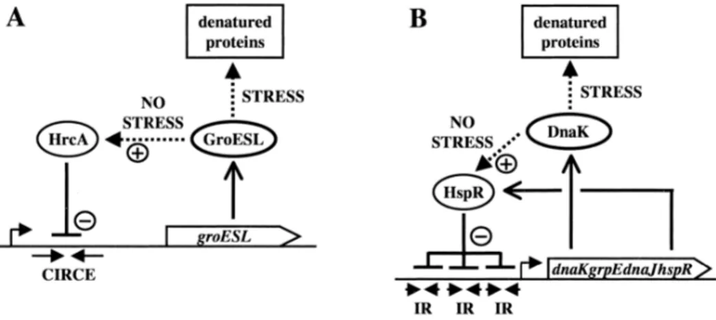
In the Gram-positive bacterium Bacillus subtilis, the expression of some chaperone genes (dnaK, groE) is regulated by the HrcA gene (Zuber et al., 1994). The HrcA gene-encoded protein products affect the transcription of these genes by binding to a conserved sequence (CIRCE). A related regulatory mechanism, HAIR/HspR, is also present in Streptomyces albus, and genes such as the dnaK operon and clpB are regulated by that regulatory system (Narberhaus, 1999) (Figure 1-2).
Figure 1-2. Titration models for homeostatic control of chaperone expression. A. GroEL as a cellular thermometer in HrcA-controlled systems. Dotted arrows indicate
protein-protein interactions; (+) symbolizes activation and (-) repression. B. DnaK titration in an HspR-controlled system (Narberhaus, 1999).
1.2.3. Regulated by RNA sensors
Bacteria use complex strategies to coordinate the expression of temperature-dependent genes. For these genes, riboswitches monitor the metabolic state of a cell by binding metabolites with high specificity and affinity. Riboswitches are located in the 5’ -UTR of genes involved in the biosynthesis, uptake or degradation of small metabolites and provide feedback control to these pathways. Their complex architecture consists in a receptor region (aptamer) and an output region (expression platform). The receptor region is characterized by the consensus sequence specifying the substrate molecule. A conformational switch is triggered by the binding of small molecules, which changes gene expression through one of three possible mechanisms: premature transcription
distinct ecological niches
termination; translation initiation, or mRNA processing (Mironov et al., 2002; Nahvi et al., 2002; Winkler et al., 2004) (Figure 1-3A). In comparison to highly specialized metabolic binding riboswitches, RNA thermometers respond to a fairly global physical signal (i.e. the intracellular temperature), which is a significant parameter under constant vigilance. A well-known feature of structured nucleic acid molecules is that they melt as the temperature rises. All currently known cis- and trans-acting molecular thermometers achieve translational regulation by sequestration of the ribosome-binding region (Narberhaus et al., 2006) (Figure 1-3B).
Figure 1-3. Schematic illustration of regulatory principles of riboswitches (A) and RNA thermometers (B).
The processes that are controlled are indicated in each case. Red circles represent metabolites. RNAP, RNA polymerase; SD, Shine–Dalgarno sequence; 30S and 50S,
ribosomal subunits, ∆T, temperature change. AUG and UUUUUU; ribonucleotide sequences (Narberhaus et al., 2006).
1.2.4. DNA topology affects the gene expression
In thermophilic and ambient species, temperature causes change in the topology of DNA, which primarily affects supercoiling (López‐García et al., 2000). The transcriptional efficiency of genes is very sensitive to changes in the topology of DNA (Pruss et al., 1989). Therefore, an increase of the environment temperature in which the bacteria is exposed can lead to changes in the topology of the DNA and thus influence the gene expression (Figure 1-4).
Figure 1-4. Effects of heat shock on plasmid topology in Escherichia coli and Sulfolobus
islandicus.
On the left of each panel is shown the variation of plasmid specific linking difference (σ=∆Lk/Lk0) with the time of exposure to the shock temperature. The right part of each
panel corresponds to the schematic interpretation of the respective topological changes and the proteins involved in their regulation. Hypothetical regulators are followed by (?). SC, supercoiling; Topo, topoisomerase; Gyr, gyrase; RG, reverse gyrase (López‐García et
al., 2000).
1.3. Key technologies of transcriptomics and
application in bacterial functional genomics
research
1.3.1. RNA-Seq technology
The effects of temperature on bacteria and the response to temperature changes are reflected in multiple levels from gene to phenotypic. The transcriptional level is an important part of this. A transcriptome is a set of transcript data in a cell at a particular developmental stage or physiological condition. In recent years, a new generation of high-throughput sequencing technology has developed rapidly. This is followed by high-resolution deep sequencing of transcripts across the genome, using high-throughput quantitative assays to sequence various types of transcripts called RNA-seq (Z. Wang et al., 2009). RNA-seq does not require the gene probes compared to the traditional chip hybridization platforms. RNA samples from any source can be tested, while high-throughput RNA sequencing can complete gene annotation and quantify genes across all subtypes. It has currently become the most powerful tool for a comprehensive analysis of the complexity of the transcriptome (Marioni et al., 2008).
RNA-seq has been successfully applied to the detection of gene expression levels, as well as the definition of gene boundaries, the study of transcriptional differences, the functional studies of noncoding small RNAs, and the discovery
of new transcripts. As one of the most valuable high-technologies in the 21st
century, transcriptomics can discover new genes that may cause disease or disease-related genes by comparing intracellular expression differences, in addition to making breakthroughs in basic research. It has been successfully
distinct ecological niches
applied to cell regulatory networks and biochemical metabolic pathway studies (Licatalosi et al., 2010), as well as gene function and environmental signaling responses (Livny et al., 2014).
1.3.2. Transcriptomics application in the global regulation
of bacterial stress response
Gene expression levels can be compared across the genome, and differentially expressed genes can be mapped to metabolic pathways to elucidate the regulatory mechanisms under specific bacterial environments. The bacteria’s living environment is constantly changing. To adapt to the changing environment, bacteria use their complex regulatory networks to respond to their physiology and phenotype (De Groot et al., 2014). Transcriptomics has been the necessary method to study the gene regulation of bacteria. Its characteristics are more in-depth than the traditional bacterial stress reaction research methods and can reveal the physiological mechanism of bacteria adapting to diverse environments.
Jozefczuk et al. used transcriptomics to analyze the response mechanisms of E.
coli under high pressure, low temperature, high temperature, salt, and acid stress,
indicating that the adaptation of bacteria to stress conditions may have a similar response mechanism. These transcriptomics studies are important for revealing the physiological mechanisms by which bacteria adapt to stressful environments (Dahlsten et al., 2014; Jozefczuk et al., 2010).
1.4. Research progress on Deinococcus radiodurans
1.4.1. Introduction on D. radiodurans
D. radiodurans is a gram-positive, nonsporulating, nonmotile, nonpathogenic
high G+C, pink-pigmented, and aerobic bacterium existing in the form of diads or tetrads. D. radiodurans was first isolated from gamma-radiated canned meat.
D. radiodurans has attracted more attention because of its unprecedented
resistance to ionizing radiation (Daly, 2009; Hua et al., 2016; Omelchenko et al., 2005). D. radiodurans was originally classified into Micrococcus based on morphology, and named M. radiodurans (Moseley, 1967). However, 16S rRNA analysis indicated that it forms a unique phylogenetic group of bacteria; hence, it was included in a new genus, the Deinococcus (Brooks et al., 1981), which is closely related to the genus Thermus of heat-resistant bacteria (Hensel et al., 1986; Omelchenko et al., 2005; Weisburg et al., 1989). D. radiodurans can often be observed in the form of a double or quadruplex under a microscope and the individual cells have an average diameter of 0.5 - 3.5 μm, an average of about 1 μm (Work et al., 1968). It is red due to carotenoid production, and gram-stain is positive but has a double-layered cell membrane (Tian et al., 2009). The optimal growth temperature was 30 °C, and cell doubling takes approximately 100 min in TGY liquid medium (0.5% tryptone, 0.1% glucose, 0.15% yeast extract) (He, 2009).
In 1999, White et al. have completed the whole genome sequencing of D.
radiodurans (White et al., 1999). The genome of D. radiodurans is 3.28M in
size, with an average GC content of 66.6% and protein-coding region accounted for 90.0%. D. radiodurans genome is composed of four replicons: two chromosomes (chromosome I: 2,648,638bp, and chromosome II: 412,348bp), a megaplasmid (177,466bp), and a small plasmid (45,704bp). The genome of D.
radiodurans encodes 3,187 proteins. About 75% of the D. radiodurans proteins
have homologs in other prokaryotes, and 24% of the proteins are unique. The function of excavating these genes has great significance for revealing the mechanism of extreme resistance in D. radiodurans.
1.4.2. The extreme resistance of D. radiodurans
1.4.2.1. Ionizing radiation resistance
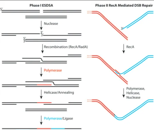
The most striking thing is that D. radiodurans can tolerate high doses of ionizing radiation, the exponentially grown cells can endure gamma-ray irradiation over 15 kGy , which is equivalent to more than 250 times of E. coli radiation resistance, more than 3,000 times the dose of human tolerance (Daly, 2009). D. radiodurans can still grow normally under 60 Gy/h continuous irradiation without affecting its growth rate and expression of the exogenous genes (Lange et al., 1998). The effects of irradiation on cells include genomic damage that can cause more than 200 DNA double-strand breaks (DSBs) into the genome of the bacterium. However, it can repair 100 to 200 DSBs per chromosome within 12 to 24 hours and accurately reconstruct the genome but under the same conditions, other bacteria can generally only repair several DSBs (Slade et al., 2011). Its superior radiation resistance is thus mainly due to the efficient and accurate DNA repair ability. As initially stated by Daly et al. (Daly et al., 1996), and more recently reinforced by Radman et al. (Slade et al., 2009; Zahradka et al., 2006), the recombinant repair of DSBs in D. radiodurans proceeds via two homologous recombination processes (Slade et al., 2009; Zahradka et al., 2006) (Figure 1-5). The first step is a mechanism known as extended synthesis-dependent single-strand DNA annealing (ESDSA). The second phase involves RecA protein-mediated double-strand break repair. The fragmented DNA is recessed in a 5’- to -3’ direction, releasing single-stranded 3’overhangs, which, via RecA- and RadA-mediated strand invasion, prime synthesis on overlapping fragments through a migrating D loop. DNA synthesis is initiated and elongated by polymerase, with polymerase filling up gaps arising from the excision repair of damaged bases. Two non-contiguous fragments are linked by the converging elongations on a third “bridging” fragment. Newly synthesized single strands dissociate from the template and anneal to complementary single-stranded extensions, forming dsDNA intermediates. The flaps are removed, and the gaps are filled. Long linear intermediates are joined into circular chromosomes by RecA-dependent crossovers.
distinct ecological niches
Figure 1-5. Two-step of genome reconstitution in D. radiodurans shattered by ionizing radiation.
The first step, ESDSA is dominated by nuclease and DNA polymerase functions. The second step is a more conventional RecA-mediated double-strand break repair process
focused on the final splicing of large chromosomal segments (Cox et al., 2010).
1.4.2.2. Oxidative resistance
D. radiodurans is also highly tolerant to a wide range of oxidative stresses.
Reactive oxygen species (ROS) induced oxidative stress is deleterious for all species. Oxidative stress results from the formation of ROS, the three primary groups of which are hydroxyl radicals, superoxide radicals, and hydrogen peroxide (D'Autréaux et al., 2007). It is well known that ROS can be generated endogenously by metabolism or formed by environmental factors such as exposure to ionizing radiation (Battista et al., 1999), ultraviolet (Battista, 1997), desiccation (Mattimore et al., 1996), and hydrogen peroxide (Slade et al., 2011). To adapt the oxygen-rich environment of earth and remove the dangerous ROS, aerobic species have evolved a range of ROS scavenging antioxidants to cope with oxidative stress. Noticeably, D. radiodurans provide an unprecedented antioxidant system to defend itself from oxidative damage relative to other species. It has been reported that the catalase activity of D. radiodurans was 127
times and 32 times greater than that of E. coli during the exponential and stationary phases, respectively (P. Wang et al., 1995). D. radiodurans genome encodes two KatE-type catalases, DR1998 (KatE1), and DRA0259 (KatE2), and one eukaryotic-type catalase, DRA0146 (Slade et al., 2011).
1.4.2.3. Desiccation resistance
D. radiodurans is also highly resistant to prolonged desiccation stress (Figure
1-6). Desiccation is defined as water content below 0.1 g H2O g-1 dry mass. D.
radiodurans can survive for 6 weeks in a dry environment with a relative
humidity of less than 5% and do not affect the growth ability (Mattimore et al., 1996). This exceptionally high survival efficiency is powerful for the bacteria that cannot produce spores. The dehydration process causes DNA damage and fragmentation. Both radiation- and desiccation-resistant bacteria have a high Mn/Fe ratio, and their proteins are less susceptible to protein oxidation than are those of sensitive bacteria (Fredrickson et al., 2008). Recently identified manganese complexes acting as the most efficient D. radiodurans ROS scavengers are expected to protect proteins against desiccation-induced damage (Daly et al., 2010). D. radiodurans is unique among bacteria in possessing as many as four homologs of plant desiccation resistance-associated proteins (Makarova et al., 2001), which may help the cells against the desiccation stress.
Figure 1-6. Extreme resistance of D. radiodurans to desiccation (Slade et al., 2011).
1.4.2.4. Heat stress resistance
distinct ecological niches
D. radiodurans cells treated with 100 °C for 10 min under dry heat conditions
still has more than 90% survival (Bauermeister et al., 2012). Under heat shock conditions, more than 130 genes were induced, including the common molecular chaperones (groESL, dnaKJ, and clpB) (Schmid et al., 2005a, 2005b). Recent studies have shown that the presence of the small heat shock protein two-component system HSP17.7/HSP20.2 in D. radiodurans plays an important role in preventing protein aggregation under heat stress (Bepperling et al., 2012). More recently, Meyer et al. show that ddrI is also involved in heat stress (Meyer et al., 2018).
1.5. Research progress on noncoding RNA in
bacteria
1.5.1. Definition of noncoding RNA in bacteria
Noncoding RNAs (ncRNAs) cannot be translated into proteins but are capable of functioning at the transcriptional level, and 10 - 30% of genes in the whole genome are regulated by ncRNA in bacteria (Wassarman et al., 1999). Normally, their size is 40 - 500 nucleotides (nt). The first ncRNA was found in E.
coli 40 years ago (Griffin, 1971), but its function was not determined at that time.
Recent studies have shown that ncRNAs control different cellular activities in bacteria, such as acid resistance (Opdyke et al., 2004), sugar metabolism (Vanderpool et al., 2004), environmental stress response (Eyraud et al., 2014; Zhang et al., 2019), and bacterial virulence (Mann et al., 2012). Most of the ncRNAs are influenced by environmental factors and have independent transcriptional targets (Gottesman, 2005; Storz et al., 2005) (Figure 1-7). ncRNAs exert their function by binding to the target mRNA through complementary base pairing, thereby affecting the translation and/or stability of the target mRNA (Valverde et al., 2008). An ncRNA may have multiple mRNA targets, and one target mRNA may be regulated by multiple ncRNAs, such as
oxyS and dsrA affects rpoS expression in E. coli (Majdalani et al., 2001). In
recent years, the research on the function and regulation mechanism of bacterial ncRNAs has become one of the research hotspots.
Figure 1-7. ncRNAs in global regulation cascades.
Various stress conditions and changes in growth lead to induction of global responses, generally by affecting the activity and/or synthesis of a transcriptional regulator. Depicted
here are several of the conditions, regulators, and resulting small RNAs and their targets that have been studied in Escherichia coli (Gottesman, 2005).
With the development of biotechnology detection methods for finding ncRNAs, which includes RNA labeling and staining, functional gene screening, protein co-purification, shotgun cloning, microarray detection, and bioinformatics (Ahmed et al., 2018). By these identification methods, more and more ncRNAs have been found in different bacteria in the past decade, and researchers have begun to study the functions of these ncRNAs. By studying how these ncRNAs exert their regulatory functions at the molecular level, it will help to understand the relationship between their structure and function and improve the prediction of ncRNA.
Normally, ncRNAs have been divided into two groups (cis-encoded ncRNAs and trans-encoded ncRNAs) according to functional base-pairing with their target (Wagner et al., 2015). The cis-encoded ncRNA is fully complementary to a single target mRNA to form a complete complex, while trans-encoded ncRNAs exert their regulatory roles through imperfect base-pairing with target mRNA to modulate their stability and/or translation (Figure 1-8) (W. Li et al., 2012). Trans-encoded ncRNAs are located in the intergenic regions of the genome and the genetic manipulation and functional studies are relatively easier than cis-encoded ncRNA, which has been the best characterized and most extensively studied. Therefore, the current research in bacteria is more in-depth on the function and regulation mechanism of trans-encoded ncRNA. The following figure provides a description of the regulatory mechanism of
distinct ecological niches
Figure 1-8. Overview of cis- and trans-encoded ncRNAs (W. Li et al., 2012).
1.5.2. Molecular mechanism of bacterial ncRNA
1.5.2.1. Interaction with target mRNA
Typically, the majority of bacterial trans-encoded ncRNAs act by imperfect base-pairing with target mRNA (Vogel et al., 2005), resulting in inhibition or activation of translation of the target mRNA. If the binding site covers or is close to the region adjacent to the SD (Shine-Dalgarno) sequence, or if the translation initiation codon is in the translation initiation region (TIR) of the target mRNA, it may result in blocking the ribosome binding site and inhibit the translation. If the inhibited secondary structure is melted, it leads to the exposure of the ribosome binding site and of the translation initiation site of mRNA and active the translation (Repoila et al., 2009) (Figure 1-9).
Figure 1-9. Basic mechanisms of the ncRNA/mRNA interaction.
A. mRNA regions targeted by a ncRNA; B. general mechanisms of translation regulation (Repoila et al., 2009).
1.5.2.2. Interaction with target proteins
mRNA is not the only target for ncRNAs. Previous studies have shown that three ncRNAs in E. coli and homologous genes in other bacteria can interact with cellular proteins to regulate their activity (Brantl, 2009). For example, 6S RNA (a highly abundant noncoding RNA) is highly conserved in prokaryotes and accumulates during the stationary phase. 6S RNA regulates transcription through interaction with σ70 polymerase in E. coli (Trotochaud et al., 2004, 2005). csrB and csrC RNAs have a binding site for the global post-transcriptional regulatory protein CsrA, which binds to regulatory proteins, attenuates the regulation of CsrA protein, and firmly controls the activity of this protein. This promotes or inhibits the expression of certain genes regulated by the CsrA protein, which in turn affects cell-related metabolism, such as cell membrane formation, movement, and acetate metabolism (Romeo, 1998; Weilbacher et al., 2003) (Figure 1-10).
Figure 1-10. General properties of ncRNAs that modulate protein activity. Bacterial ncRNA binding to proteins has been demonstrated to inhibit and/or modify
distinct ecological niches
protein activities (Storz et al., 2011).
1.5.3. Study progress of ncRNA in D. radiodurans
ncRNAs also play an important physiological role in D. radiodurans. At present, there are few reports on ncRNAs in D. radiodurans. Only in 2015, Tsai et al reported that there were 199 ncRNA candidates (Tsai et al., 2015), but there is no in-depth study on their actual function. With the improvement of biotechnology, more and more unknown ncRNAs have been identified and verified. It has become a frontier and hot spot in the field to identify their target, mode of action and physiological significance.
1.6. Biocontrol effect of plant growth-promoting
rhizobacteria for plant protection
1.6.1. Plant growth-promoting rhizobacteria
Plant growth-promoting rhizobacteria (PGPR) colonize the rhizosphere of
many plant species and can promote plant growth by a wide range of mechanisms
(Bhattacharyya et al., 2012). The main functions are: (1) to provide nutrition for crops (2) to stimulate plant growth (3) to inhibit the activity of phytopathogenic bacteria (4) to improve soil structure (5) to accumulate inorganic matter. PGPR often fixed around the root system of the plant to promote plant growth. At present, it is more popular to use biological methods to increase plant nutrition and crop yield (Dey et al., 2004; Shoebitz et al., 2009). There are several pathways by which PGPR promotes plant growth. Generally, PGPR can affect plants through two ways, both direct and indirect. PGPR directly promotes plant growth refers to the fact that PGPR can synthesize certain substances (such as auxin) that have a direct impact on plant growth and development and/or change the form of certain ineffective elements in the soil to make it effective and facilitate plant absorption (such as nitrogen fixation, phosphorus solubilization, etc.) (Dobbelaere et al., 2003; Rodríguez et al., 2006). Indirect effects refer to inhibiting or minimizing the detrimental effects of certain plant pathogens on plant growth and yield.
1.6.2. The role of PGPR in plants under abiotic stresses
Plant‒microbes symbiosis is affected by environmental changes in the soil, which
mainly include water, soil temperature, salinity, nutrient deficiencies, alkalinity and
acidity (Hungria et al., 2000; Meena et al., 2014; Meena et al., 2015). Low soil temperature is an important abiotic stress factor, pose a major limitation on plant growth and development. It affects the symbiosis of rhizobacteria and plants by regulating the rate of physico-chemical reactions (Sardesai et al., 2001). Exposure to low temperatures is one of the most damaging environmental factors affecting plants (Nagarajan et al., 2009). Recently, the use of beneficial microorganisms such as PGPR has emerged as a potential new approach to reduce abiotic stress-induced
damage (Yang et al., 2009). As a result, intimate associations between bacteria and host plants can be formed without harming the plant (Compant et al., 2005; Gray et al., 2005). PGPR colonizes the plant rhizosphere and has a beneficial impact on their growth and responses to stress (Compant et al., 2008; Yang et al., 2009).
1.6.3. Bacillus velezensis GA1
Many PGPR strains have been described, the main species of which include
Bacillus and Pseudomonas. Indeed, nowadays, the market of PGPRs is
dominated by Bacilli due to their adaptability to the agro-industrial world (i.e. endospore-forming ability), some of which are proven to be effective PGPRs. The operational group ‘amyloliquefaciens’, along with the species B. subtilis and B. pumilus, gather most of the available strains (Borriss, 2015). The dominant position of Bacillus products underlines the importance of this genus in the PGPR sector.
Our study focuses on one specific PGPR: Bacillus velezensis GA1 (GA1), which has become a typical bacterium for our team to study plant-bacterial interactions. GA1 is a Gram-positive, aerobic, spore-forming, motile, rod-shaped bacteria. GA1 is often encountered as B. amyloliquefaciens GA1 (Arguelles-Arias et al., 2009). However, some controversy has occurred regarding the taxonomy of B. velezensis, B. amyloliquefaciens subsp plantarum,
B. oryzicola and B. methylotrophicus. As a result, the above-mentioned four
species constitute only one species, Bacillus velezensis, and that this species belongs to the ‘operational’ group ‘amyloliquefaciens’. Bacillus sp. FZB42 is a closely related strain that belongs to the same species as GA1, is now classified as velezensis rather than amyloliquefaciens subsp plantarum. The same logic should be applied to Bacillus sp. GA1 and it should be called B. velezensis GA1 (Dunlap et al., 2016; Fan et al., 2017). It is worth noting that this operational group gathers most of the best PGPR Bacilli (Fan et al., 2017) and that it has undergone an evolutionary adaptation to plant associated habitat (Belbahri et al., 2017). Thus, GA1 is a good representative of the bacillus PGPRs.
GA1 can produce large amounts of Bioactive Secondary Metabolites (BSMs) (Arguelles-Arias et al., 2009). The BSMs produced by GA1 include non-ribosomal peptides (NRPs), polyketides (PKs), Lipopeptides (CLPs), siderophores and ribosomally synthesized peptides. Because of the large number of BSM, GA1 has an antimicrobial activity toward a large group of pathogens and thus has great potential as a biocontrol agent.
1.7. Objectives
Temperature is one of the stressful environments that can give pressure to biological evolution. The research on the response process of prokaryotic to heat or cold can help us better understand its stress adaptation mechanism, find important biomacromolecules with thermal/cold stability, and find the physiological effects and response methods caused by thermal/cold adaptation
distinct ecological niches
evolution. The results can be applied to evolutionary research, genetic engineering, food engineering, fermentation engineering, enzyme engineering, and new biomaterials.
D. radiodurans is capable of living in a variety of extreme environments, with
ionizing radiation resistance and a variety of abiotic stress (such as cold, heat, etc.) resistance, is an ideal strain to study the adaptation mechanism of extreme microbial abiotic stress. In recent years, there are many studies on the molecular mechanism of D. radiodurans anti-radiation, but little research on the response mechanism to heat stress. We used the Illumina sequencing platform for transcriptome analysis of wild-type strains under normal growth, heat stress treatment, and recovery after heat stress. The heat stress reaction process of D.
radiodurans was initially explored, and some novel heat stress response proteins
were discovered. It provided theoretical support for further understanding of the adaptation and response mechanism of bacteria in extreme environments.
In recent years, the study of ncRNA in D. radiodurans has been limited only to the discovery and identification of ncRNA. However, little research has been done on its mechanisms involved in various activities in cells. In the complex regulatory network of the heat stress response, in addition to the important regulatory role of key proteins, whether there is a more direct regulatory ncRNA involved in the entire regulatory process still needs further research. Based on our and previous report, we found two ncRNAs that are highly expressed under heat stress and carried out in-depth research on their molecular mechanisms involved in response to heat stress, which laid a theoretical foundation for further understanding of the mechanism of ncRNA involvement in heat stress. This will help to fully understand the importance of ncRNA in microbial heat stress response mechanisms at a new level.
PGPR is a group of bacteria that can enhance plant growth and yield during agricultural production. The environment during crop production is complex. Temperature is a vital factor during plant growth. The impact of temperature on biological control has occasionally been addressed in previous works. However, the effect of low temperature on the expression of biocontrol traits by those beneficial rhizobacteria. Our objective is to investigate the effect of temperature on several B. velezensis GA1 (a PGPR) traits, such as growth rate, motility, biofilm formation, and root colonization ability. We also gave special emphasis to finding out how B. velezensis GA1 metabolite production can be modulated at low temperatures in vitro and planta.
1.8. Overview of the chapters
In this chapter, we present a review of the bibliography on temperature adaptation in bacteria. Information is presented on the mechanisms of temperature tolerance in bacteria and the scope for further research in this area. We also provided a brief description of the context and the aims of this work.