RNA BIOCHEMISTRY
Transcriptome-wide distribution
and function of RNA
hydroxymethylcytosine
Benjamin Delatte,1*† Fei Wang,2* Long Vo Ngoc,3*‡ Evelyne Collignon,1
Elise Bonvin,1 Rachel Deplus,1Emilie Calonne,1Bouchra Hassabi,1Pascale Putmans,1Stephan Awe,4 Collin Wetzel,5§ Judith Kreher,4‖ Romuald Soin,3Catherine Creppe,1
Patrick A. Limbach,5Cyril Gueydan,3Véronique Kruys,3Alexander Brehm,4 Svetlana Minakhina,2¶ Matthieu Defrance,1Ruth Steward,2François Fuks1# Hydroxymethylcytosine, well described in DNA, occurs also in RNA. Here, we show that hydroxymethylcytosine preferentially marks polyadenylated RNAs and is deposited by Tet in Drosophila. We map the transcriptome-wide hydroxymethylation landscape, revealing hydroxymethylcytosine in the transcripts of many genes, notably in coding sequences, and identify consensus sites for hydroxymethylation. We found that RNA hydroxymethylation can favor mRNA translation. Tet and hydroxymethylated RNA are found to be most abundant in theDrosophila brain, and Tet-deficient fruitflies suffer impaired brain development, accompanied by decreased RNA hydroxymethylation. This study highlights the distribution, localization, and function of cytosine hydroxymethylation and identifies central roles for this modification inDrosophila.
I
n DNA, vertebrate Tet methyldioxygenases (Tet1, Tet2, and Tet3) catalyze hydroxylation of 5-methylcytosine to 5-hydroxymethylcytosine (1–3). Tet also catalyzes the formation of hy-droxymethylcytosine in RNA (referred to here as hmrC) (4, 5). To date, however, the distribution, localization, and functional relevance of hmrC re-main unknown.In the present study, we have sought to pro-vide a better understanding of hmrC. For ease of interpreting results, we analyzed hmrC in Dro-sophila melanogaster because (i) cytosine meth-ylation in Drosophila DNA is either absent or very low, being restricted to specific cellular contexts (6, 7), and (ii) we have found no evidence of DNA hydroxymethylation in this organism (fig. S1). To detect 5hmC RNA modification, we used an antibody
raised against 5-hydroxymethylcytosine (8, 9). To confirm that it will bind to hmrC, we performed dot blot experiments using in vitro transcribed templates containing either unmethylated, methy-lated, or hydroxymethylated cytosines (table S1). The antibody to 5hmC specifically detected 5hmC-containing RNA. In addition, detection of 5hmC was abolished after ribonuclease (RNase) A treat-ment (fig. S2A).
We detected hmrC in dot blot experiments on total RNA extracted from Drosophila S2 cells (Fig. 1A and fig. S2, B and C). Isolation of polyadenylated RNA from S2 cells followed by immunoblotting showed strong enrichment in hmrC signal as compared with that of total cellular RNA (Fig. 1B and fig. S2, D and E). No signal was de-tected in fractions enriched in small RNAs or
ribosomal RNAs (Fig. 1C and fig. S3, A and B). Drosophila possesses only one conserved Tet ortholog, CG43444 (dTet) (10, 11). Depletion of dTet in S2 cells, by using RNA interference for dTet (dTet KD), revealed a 54% decrease in dTet transcripts, as compared with control cells (Fig. 1D). Dot blotting with antibody to 5hmC showed a similar decrease in hmrC—44%—upon dTet knockdown (Fig. 1D and fig. S3, C and D).
To map the hmrC modification landscape in a transcriptome-wide manner, we adapted a re-cently used method [methylated RNA immuno-precipitation followed by sequencing (MeRIP-seq)] (12, 13), which we call hMeRIP-seq. This method involves immunoprecipitation of hmrC-containing RNA with the antibody to 5hmC followed by next-generation sequencing. hMeRIP-seq in S2 cells yielded 3058 significantly enriched regions (“hmrC peaks,” P < 10−10) within 1597 coding gene tran-scripts (fig. S4, A to C, and table S2). Examples of enrichment profiles are shown in Fig. 2A. Several key controls were performed so as to en-sure the validity and stringency of our experimental approach: (i) Further bioinformatic analyses dem-onstrated that our hMeRIP-seq experiments did not merely coprecipitate abundant RNA fragments
282 15 JANUARY 2016• VOL 351 ISSUE 6270 sciencemag.org SCIENCE
1Laboratory of Cancer Epigenetics, Faculty of Medicine, ULB Cancer Research Center (U-CRC), Université Libre de Bruxelles (ULB), Brussels, Belgium.2Waksman Institute, Department of Molecular Biology and Biochemistry, Cancer Institute of New Jersey, Rutgers University, Piscataway, NJ, USA.3Laboratory of Molecular Biology of the Gene, Faculty of Sciences, Université Libre de Bruxelles, Gosselies, Belgium.4Institut für Molekularbiologie und Tumorforschung, Philipps-Universität Marburg, Marburg, Germany. 5Department of Chemistry, University of Cincinnati, Cincinnati, OH, USA.
*These authors contributed equally to this work.†Present address: Division of Signaling and Gene Expression, La Jolla Institute for Allergy and Immunology, La Jolla, CA 92037, USA.‡Present address: Section of Molecular Biology, University of California, San Diego, La Jolla, CA 92093, USA. §Present address: Department of Cancer Biology, University of Cincinnati, Cincinnati, OH, USA. ||Present address: Roche Diagnostics, 68305 Mannheim, Germany. ¶Present address: Robert Wood Johnson Medical School, Depart-ment of Medicine, Rutgers University, Piscataway, NJ, USA. #Corresponding author. E-mail: [email protected]
0 5 10 15 20 25 30 Total Poly A RNA: 0 0.2 0.4 0.6 0.8 1 Total small RNA RNA: rRNA 5 Relativ e hmrC amount Relativ e hmrC amount 0 0.2 0.4 0.6 0.8 1 - RNase RNA: Relativ e hmrC amount Relativ e hmrC amount Relativ e dT et e xpression Ctrl dTet KD qRT- PCR Dot Blot hmrC Ctrl dTet KD 0 0.2 0.4 0.6 0.8 1 0 0.2 0.4 0.6 0.8 1
Fig. 1. RNA hydroxymethylation by dTet inDrosophila S2 cells. (A) Dot blotting on total RNA from Drosophila S2 cells with antibody to 5hmC, treated or not with RNase A (serially halved amounts of RNA, starting at 1mg). Data are mean ± SD (n = 4 experiments run) with a representative blot shown. (B) Immunoblotting with anti-5hmC antibody was performed on polyadenyl-ated and total RNA from S2 cells. Data are mean ± SD (n = 3 experiments
run). (C) hmrC content of total RNA as well as fractions enriched in small RNA or rRNA was assessed by dot blotting. Data are mean ± SD (n = 3 experiments run). A vertical line indicates juxtaposition of lanes within the same blot, exposed for the same time. (D) dTet knockdown leads to reduced hmrC levels. (Left) Quantitative RT-PCR analysis. (Right) Dot blotting. Data are mean ± SD (n = 4 experiments run).
nonspecifically (Fig. 2B and fig. S5); (ii) up to 79.4% of hmrC sites showed significant reduction levels in hMeRIP-seq upon dTet depletion as compared with that of control S2 cells, with 85.5%
of the sites showing a more than fourfold reduc-tion in hmrC levels (Fig. 2C and fig. S6); and (iii) replicate hMeRIP-seq experiments by using an additional antibody to 5hmC showed strong
agree-ment between experiagree-ments performed with the two 5hmC antibodies (fig. S7 and table S3).
The distribution of hmrC peaks revealed by hMeRIP-seq analyses was significantly nonrandom
SCIENCE sciencemag.org 15 JANUARY 2016• VOL 351 ISSUE 6270 283
Expression Level (log2)
Number of mRNAs 0 5 10 15 0 200 400 600
All expressed mRNAs Expressed mRNAs enriched for hmrC
bits
Down (49.6%) Up (50.4%) 574 differentially expressed 150 hMeRIP-Seq (1597) RNA-Seq (574) 26% Expected Observed * Number of hMeRIP peaks 0 375 750 1125 1500 19% 48% 16% 17%*
*
5'UTR CDS Intronic 3'UTR
RNA-Seq dTet depletion Ctrl 0 5 10 15
Normalized hmrC reads (log
2 ) 0 20 40 60 80 100 Reduced hmrC peaks (%) Reduced > 4 fold Reduced < 4 fold 79.4% 20.6% Not Reduced Reduced Ctrl Ctrl hMeRIP hMeRIP 0.0 1.0 2.0 5 10 15
Fig. 2. Transcriptome-wide distribution of hmrC inDrosophila cells. (A) Representative UCSC Genome Browser plot from hMeRIP-seq data. (B) Distribution of all expressed (gray) or hmrC-enriched (green) transcripts, showing the number of mRNAs as a function of their expression levels. In seq, enrichment in both abundant and less abundant fragments was observed. (C) hMeRIP-seq in cells depleted of dTet shows reduced hmrC levels at the majority of target regions. (Left) Box plot of the normalized number of hmrC reads in dTet-depleted cells versus control cells. (Center) Pie chart showing the percentage of reduced hmrC peaks, with (right) a more than fourfold reduction at most
targets. (D) Distribution of hmrC peaks according to the type of structural element within the transcript. *P < 10−5). (E) Sequence motif identified within hmrC peaks. (F) RNA-seq performed on dTet-knockdown and control S2 cells. (G) Partial overlap between hMeRIP-seq and RNA-seq data sets.
F-Luc Loading control F-luc nor maliz ed CPM (%) SDS PAGE/ Salicilate staining Ctrl rC mrC hmrC 0 20 40 60 80 100 120 hmrC Free RNP 40S 60S Polysomes P ercent of hmrC signal (%) 80S 0 20 40 60 80 100 0 10 20 30 40 50 OD 254 nm ( % sa tur a tion) (Sucrose gradient) 0 5 10 15 20 25 30 10 20 30 40 0 50
Fig. 3. RNA hydroxymethylation can favor mRNA translation. (A) Sucrose gradient fractionation followed by dot-blot quantification shows that active translation is associated with a high hmrC content. Data are mean ± SD (n = 3 experiments run). (B) In vitro translation of C-, mC-, and hmC-containing RNAs, as measured by incorporation of35S-radiolabeled methionine, shows that methylation decreases translation and hydroxymethylation restores it. Shown are the normalized scintillation counts (top) and the results of SDS–polyacrylamide gel electrophoresis (SDS-PAGE) followed by fluorography (bottom). Data are mean ± SD (n = 3 experiments run). RESEARCH | R E P O R T S
(P < 10–5), with many of these peaks found in coding sequences (48%) (Fig. 2D). Further analyses identified an overrepresented motif with oc-currences in a large proportion of peaks (64%) and which tends to be highly UC-rich, contain-ing UCCUC repeats (Fig. 2E and fig. S8A). Gene Ontology analysis of the hmrC targets showed enrichment for genes involved in basic cellular processes and notably in the regulation of em-bryogenesis and development (fig. S8B and table S6). In addition, exons were more enriched in hmrC than introns (fig. S9D). Thus, the above data present the hmrC-enriched transcriptome, revealing the presence of this modification in specific mRNA regions and in specific sequence contexts.
Next, we knocked down dTet expression in S2 cells and performed RNA-sequencing (RNA-seq) experiments in order to assess how many mRNAs might be regulated by dTet. We found the expres-sion of 574 mRNAs to change after dTet depletion
(50.4% were more abundant, whereas 49.6% were less abundant) (Fig. 2F; fig. S9, A to C; and table S4). Examples of mRNAs whose levels either increased or decreased upon dTet knockdown are shown in fig. S9, A and B. A positive correlation was observed between transcript abundance and the presence of hmrC peaks (Fig. 2B and fig. S5). We then compared these dTet-regulated mRNAs with the targets identified by hMeRIP-seq and found a slight but significant percentage (26%, P < 10−43) of the dTet-regulated mRNAs to con-tain at least one hmrC peak (Fig. 2G, figs. S9C and S11, and table S5). It is worth mentioning that dTet contains an N-terminal CXXC Zn-finger domain, which likely explains the reported ability of mammalian Tets to regulate gene expression independently of their catalytic activity (14, 15). It thus remains possible that dTet might affect gene expression via its CXXC domain, indepen-dently of its ability to hydroxylate methylated RNA (fig. S10, A and B). This domain, however,
is likely not required for the role of dTet in Drosophila brain development (see below) be-cause flies where dTet CXXC is deleted are still viable and show no specific phenotype (11).
To examine how cytosine hydroxymethylation might affect mRNA function, we examined the distribution of hmrC as a function of the mRNA translational status in Drosophila S2 cells. For this, we performed standard sucrose-gradient frac-tionation followed by dot blotting. A correlation between hmrC abundance and active mRNA trans-lation was observed; fractions with low transtrans-lation activity (free mRNAs and monosomes) were found to be poor in hmrC, whereas mRNAs heavily loaded with ribosomes (polysome fractions) showed a high hmrC content (Fig. 3A). We also assessed how mrC distributes across polysome fractions and found it to be high in monosomes and low in polysomes (fig. S12). Next, we examined whether mRNA hydroxymethylation might af-fect mRNA translation. In vitro translation of
284 15 JANUARY 2016• VOL 351 ISSUE 6270 sciencemag.org SCIENCE
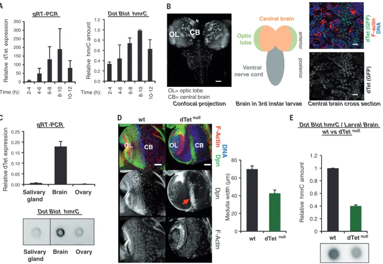
Dot Blot hmrC qRT-PCR Relativ e hm rC am ount Relativ e dTet ex pres s ion Time (h): 2-4 4-6 6-8 8-10 10-12 Time (h): 2-4 4-6 6-8 8-10 10-12 Central brain
Central brain cross section Confocal projection Optic lobe anter ior poster ior dT et (GFP) F-act in DNA Ventral nerve cord
OL
CB
OL= optic lobe
CB= central brain dT
et (GFP)
Brain in 3rd instar larvae RT-qPCR CB Brain Ovary Salivary gland Dot Blot hmrC qRT-PCR Relativ e dT et e xpression 0 20 40 60 80 wt dTet wt null OL CB Dpn F-A c tin DNA Dp n F -Actin OL CB null Medulla width (µm) wt dTetnull Relativ e hm rC amount
Dot Blot hmrC / Larval Brain wt vs dTet null Brain Ovary Salivary gland 0 50 100 150 200 250 300 350 0.00 0.05 0.10 0.15 0.20 0.25 0.0 0.2 0.4 0.6 0.8 1.0 1.2 0 0.2 0.4 0.6 0.8 1 1.2 dTet
Fig. 4. dTet-deficient fruitflies show impaired brain development, ac-companied by decreased RNA hydroxymethylation. (A) dTet expression and hmrC levels during Drosophila embryogenesis. Data are mean ± SD (n = 4 experiments run). (B) (Left) Pattern of endogenous GFP-tagged dTet in the larval brain. Scale bar, 50mm. (Center) Scheme of the larval brain. (Right) Confocal brain section showing the expression of endogenous dTet (green) and F-actin (red). DNA, blue. Scale bar, 10mm. (C) (Top) dTet expression in the salivary gland, brain, and ovary. Data are mean ± SD (n = 3 experiments run).
(Bottom) Immunoblotting with 5hmC antibody in RNA from salivary gland, brain, and ovary. Vertical line indicates juxtaposition of lanes within the same blot, exposed for the same time. (D) (Left) dTetnullbrains are smaller than wild-type brains. Dpn (neuroblasts), green; F-actin, red; DNA, blue. Scale bar, 50mm. (Right) Average width of the medulla region containing the neuro-blasts (left, red arrow). Error bars represent 95% confidence intervals (p < 2.4 10−9) for 20 brain lobes. (E) Brains of dTet-deficient larvae show a decrease in hmrC. Data are mean ± SD (n = 3 experiments run).
unmodified, methylated, and hydroxymethyl-ated Firefly Luciferase–encoding RNA templates in rabbit reticulocyte lysate showed a decrease in translation of methylated RNAs. In contrast, hmrC-modified templates gave rise to near-control protein levels (Fig. 3B), suggesting that hydroxy-methylation can restore the translation efficiency of previously methylated substrates.
We next sought to assess hmrC in vivo in fruit-flies. To determine the timing of dTet expression and of the appearance of hmrC during early embryo-genesis of D. melanogaster, we performed quantita-tive reverse transcription polymerase chain reaction (RT-PCR) to measure dTet transcript levels, and immunoblotting to estimate levels of hmrC. We found that dTet levels correlate positively with hmrC levels during fruitfly embryogenesis (Fig. 4A and fig. S13A). We also analyzed a publicly available database of RNA-seq results from different stages of D. melanogaster development and found that in third-instar larvae, dTet expression is highest in the central nervous system (fig. S13B). These findings suggest that dTet-mediated hydroxymethylation of RNA could play a role in the fruitfly brain. To confirm and extend these observations, we generated transgenic Drosophila flies expressing a GFP-dTet fusion construct under the control of the endog-enous dTet promoter (called dTet-Mi). The green fluorescent protein (GFP)–tagged dTet protein was detected throughout the larval brain, the highest levels being detected in the optic lobe and central brain (Fig. 4B).
It was of interest to assess whether the fly brain contains high levels of hmrC. To this end, in ad-dition to the brain, we used ovary [because this was the organ chosen to show the role of dTet in DNA m6A demethylation (11)]. We also used an-other organ, the salivary gland, from which we could extract enough RNA to measure hmrC levels. Quantitative RT-PCR and dot blotting showed higher dTet expression and hmrC content in the brain than in the ovary or in the salivary gland of the fruitfly (Fig. 4C). Hence, by revealing that the hmrC signal is highest in the brain, these data support the argument for the importance of hmrC in this organ.
We wished to evaluate RNA hydroxymethyl-ation levels in a complete loss-of-function mu-tant of dTet (fig. S13, C and D). In agreement with recent observations (11), dTet-deficient ani-mals survived through the larval stages but died at the pupal stage (no adult animals survived; n > 5000 dTet-null animals analyzed) (fig. S13E). Morphological defects were observed at larval stages: The brains of mutant larvae were smaller than those of normal larvae and showed abnor-mal organization of neuroblasts in their central part (Fig. 4D). To quantify the observed changes, we measured the width of the medulla region (based on 20 examined brains). We found it to be significantly reduced in dTetnullanimals (P <
2.4 × 10⁻⁹), likely reflecting a lower number of neuroblasts (Fig. 4D). To measure the effects of dTet loss on the RNA hydroxymethylation level, we performed immunoblotting analyses with antibody to 5hmC on brains from dTetnulland wild-type larvae. These experiments showed a
decreased hmrC content in the brains of dTet-deficient larvae (Fig. 4E and fig. S14).
Our understanding of the posttranscriptional modifications that decorate RNA, a regulatory layer positioned between DNA and proteins, is in its infancy. We have conducted a study addressing the distribution, localization, and function of cy-tosine hydroxymethylation in RNA, using Dro-sophila melanogaster as a model. Our work has yielded the following key findings: (i) It provides a picture of the hydroxymethylated transcriptome, (ii) reveals an unrecognized function for hmrC, and (iii) suggests a central role for this RNA modif-ication and dTet in the Drosophila brain. All in all, we expect this study to change the way we think about the roles played by cytosine hydroxy-methylation and the Tet proteins. Our findings open new research prospects in an emerging realm of biological regulation: epitranscriptomics.
R E F E R E N C ES A N D N OT ES
1. S. Kriaucionis, N. Heintz, Science 324, 929–930 (2009). 2. M. Tahiliani et al., Science 324, 930–935 (2009). 3. R. M. Kohli, Y. Zhang, Nature 502, 472–479 (2013). 4. I. Rácz, I. Király, D. Lásztily, Planta 142, 263–267 (1978). 5. L. Fu et al., J. Am. Chem. Soc. 136, 11582–11585 (2014). 6. G. Raddatz et al., Proc. Natl. Acad. Sci. U.S.A. 110, 8627–8631
(2013).
7. S. Takayama et al., Genome Res. 24, 821–830 (2014). 8. E. M. Kallin et al., Mol. Cell. 48, 266–276 (2012). 9. H. Wu et al., Genes Dev. 25, 679–684 (2011).
10. T. L. Dunwell, L. J. McGuffin, J. M. Dunwell, G. P. Pfeifer, Cell Cycle 12, 3357–3365 (2013).
11. G. Zhang et al., Cell 161, 893–906 (2015).
12. K. D. Meyer et al., Cell 149, 1635–1646 (2012). 13. D. Dominissini et al., Nature 485, 201–206 (2012). 14. R. Deplus et al., EMBO J. 32, 645–655 (2013). 15. K. Williams et al., Nature 473, 343–348 (2011). AC K N OW L E D G M E N TS
We thank L. Droogmans for advice, as well as A. Rao and J. Kadonaga for continued support. We also thank Y. N. Jan for providing the antibody to Dpn. R.D., B.H., C.C., and E.B. were supported by the Belgian Fonds de la Recherche Scientifique (FNRS). B.D. was supported by the FNRS (« Aspirant ») and a Belgian American Educational Foundation (BAEF) fellowship. L.V.N. was supported by the Belgian Fund for Research Training in Industry and Agriculture and a BAEF fellowship. E.Co. was supported by a“l’Oréal” fellowship, and M.D. was supported by the WBHealth grant (CanDx) from the Walloon Region. F.F. is a ULB Professor. F.F.’s lab was funded by grants from the FNRS and Télévie, the Interuniversity Attraction Poles (P7/03) program, the Action de Recherche Concertée (ARC) (AUWB-2010-2015 ULB-No 7), the WB Health program, the Belgian Fondation contre le Cancer, and the Fonds Gaston Ithier. F.W. was supported by a Charles and Johanna Busch graduate fellowship and the work at Rutgers by a NIH grant (RO1 GM089992) to R.S. V.K. was supported by the ARC (AV.12/17) as well as Brachet and Van Buuren Funds. P.A.L. received financial support from the National Science Foundation (NSF CHE 1212625 and 1507357). The authors declare no conflicts of interest. All sequencing data have been deposited to the Gene Expression Omnibus as series GSE66090.
SUPPLEMENTARY MATERIALS
www.sciencemag.org/content/351/6270/282/suppl/DC1 Materials and Methods
Figs. S1 to S14 Tables S1 to S6 References (16–35)
8 May 2015; accepted 8 December 2015 10.1126/science.aac5253
GENE REGULATION
Transcription factors LRF and BCL11A
independently repress expression of
fetal hemoglobin
Takeshi Masuda,1Xin Wang,2Manami Maeda,1Matthew C. Canver,3Falak Sher,3 Alister P. W. Funnell,4Chris Fisher,5Maria Suciu,5Gabriella E. Martyn,4 Laura J. Norton,4Catherine Zhu,1Ryo Kurita,6Yukio Nakamura,6,7Jian Xu,3,8 Douglas R. Higgs,5Merlin Crossley,4Daniel E. Bauer,3Stuart H. Orkin,3,9 Peter V. Kharchenko,2* Takahiro Maeda1*
Genes encoding human b-type globin undergo a developmental switch from embryonic to fetal to adult-type expression. Mutations in the adult form cause inherited
hemoglobinopathies or globin disorders, including sickle cell disease and thalassemia. Some experimental results have suggested that these diseases could be treated by induction of fetal-type hemoglobin (HbF). However, the mechanisms that repress HbF in adults remain unclear. We found that the LRF/ZBTB7A transcription factor occupies fetal g-globin genes and maintains the nucleosome density necessary for g-globin gene silencing in adults, and that LRF confers its repressive activity through a NuRD repressor complex independent of the fetal globin repressor BCL11A. Our study may provide additional opportunities for therapeutic targeting in the treatment of hemoglobinopathies.
D
uring human development, the site of eryth-ropoiesis changes from the embryonic yolk sac to the fetal liver and then, in newborns, to the bone marrow, where it persists through adulthood. Coincidentally, there is a“globinswitch” from embryonic to fetal globin genes in utero, and then a second switch from fetal to adult globin expression soon after birth. This pro-cess has been studied for more than 60 years (1). The latter transition from fetal to adult
SCIENCE sciencemag.org 15 JANUARY 2016• VOL 351 ISSUE 6270 285