Clustering algorithms and shape factor methods to discriminate
among small GTPase phenotypes using DIC image analysis
par
Arturo Papaluca
Département de Biochimie Faculté de Médecine
Mémoire présenté à la Faculté de Médecine
en vue de l’obtention du grade de Maître ès science (M.Sc.) En Biochimie
Option Génétique Moléculaire
Juillet, 2012
Faculté des études supérieures et postdoctorales
Ce mémoire intitulé:
Clustering algorithms and shape factor methods to discriminate among small GTPase phenotypes using DIC image analysis
Présenté par : Arturo Papaluca
A été évalué par un jury composé des personnes suivantes :
Pascal Chartrand, président-rapporteur Stephen Michnick, directeur de recherche
Résumé
Naïvement perçu, le processus d’évolution est une succession d’événements de duplication et de mutations graduelles dans le génome qui mènent à des changements dans les fonctions et les interactions du protéome. La famille des hydrolases de guanosine triphosphate (GTPases) similaire à Ras constitue un bon modèle de travail afin de comprendre ce phénomène fondamental, car cette famille de protéines contient un nombre limité d’éléments qui diffèrent en fonctionnalité et en interactions. Globalement, nous désirons comprendre comment les mutations singulières au niveau des GTPases affectent la morphologie des cellules ainsi que leur degré d’impact sur les populations asynchrones.
Mon travail de maîtrise vise à classifier de manière significative différents phénotypes de la levure Saccaromyces cerevisiae via l’analyse de plusieurs critères morphologiques de souches exprimant des GTPases mutées et natives. Notre approche à base de microscopie et d’analyses bioinformatique des images DIC (microscopie d’interférence différentielle de contraste) permet de distinguer les phénotypes propres aux cellules natives et aux mutants. L’emploi de cette méthode a permis une détection automatisée et une caractérisation des phénotypes mutants associés à la sur-expression de GTPases constitutivement actives. Les mutants de GTPases constitutivement actifs Cdc42 Q61L, Rho5 Q91H, Ras1 Q68L et Rsr1 G12V ont été analysés avec succès.
En effet, l’implémentation de différents algorithmes de partitionnement, permet d’analyser des données qui combinent les mesures morphologiques de population native et mutantes. Nos résultats démontrent que l’algorithme Fuzzy C-Means performe un partitionnement efficace des cellules natives ou mutantes, où les différents types de cellules sont classifiés en fonction de plusieurs facteurs de formes cellulaires obtenus à partir des images DIC. Cette analyse démontre que les mutations Cdc42 Q61L, Rho5 Q91H, Ras1 Q68L et Rsr1 G12V induisent respectivement des phénotypes amorphe, allongé, rond et large qui sont représentés par des vecteurs de facteurs de forme distincts. Ces distinctions
sont observées avec différentes proportions (morphologie mutante / morphologie native) dans les populations de mutants.
Le développement de nouvelles méthodes automatisées d’analyse morphologique des cellules natives et mutantes s’avère extrêmement utile pour l’étude de la famille des GTPases ainsi que des résidus spécifiques qui dictent leurs fonctions et réseau d’interaction. Nous pouvons maintenant envisager de produire des mutants de GTPases qui inversent leur fonction en ciblant des résidus divergents. La substitution fonctionnelle est ensuite détectée au niveau morphologique grâce à notre nouvelle stratégie quantitative. Ce type d’analyse peut également être transposé à d’autres familles de protéines et contribuer de manière significative au domaine de la biologie évolutive.
Mots-clés : évolution, petite GTPases Ras, morphologie cellulaire, fonction et structure des protéines, réseaux d'interaction protéine-protéine, facteurs de forme cellulaire, algorithmes de partitionnement.
Abstract
Evolution is a gradual process that gives rise to changes in the form of mutations that are reflected at the protein level. We propose that evolution of new pathways occurs by switching binding partners, hence creating new functions. The different functions encountered in a given family of related proteins have emerged from a common ancestor that has been duplicated and mutated to become implicated in new interactions and to gain new functions. In this study, we will use native and constitutive active mutant variants of the Ras-like family of small GTPases as working model, to explore such gene duplications, followed by neo / sub-functionalization. The reason for choosing this family resides in the fact that it is a defined set of proteins with well known functions that are mediated through multiple protein-protein interactions.
The aim of this master is to perform a classification of budding yeast phenotypes using different approaches in order to statistically determine at which level of the population these constitutively active mutations are capable to affect cell morphology. Working with a subset of the Ras-like small GTPases family, we recently developed an approach to catalogue and classify these proteins based on multiple physical and chemical criteria. Using microscopic and bioinformatics methods, we characterized phenotypes associated with over-expression of the native small GTPases of the budding yeast
Saccharomyces cerevisiae, showing that an established classification is not very clear.
We are interested to investigate how point mutations in small GTPases can affect the cell morphology and their level of impact on asynchronous population. We want to establish a method to determine and quantify mutant and wild type-like phenotypes on these populations using Differential interference contrast microscopy (DIC) images only. As for the first aim of this study, we hypothesize that clustering algorithms can partition mutant cells from wild type cells based on cell shape factor measurements. To prove this hypothesis, we proposed to implement different clustering algorithms to analyze datasets which combines measurements from wild type and respective mutant populations.
We created constitutively active forms of these small GTPases and used Cdc42, Rho5, Ras1 and Rsr1 to validate our results. We observed that Cdc42 Q61L, Rho5 Q91H, Ras1 Q68L and Rsr1 G12V mutations induced characteristic amorphous, clumped/elongated, rounded and discrete large phenotypes respectively. This classification allowed us to define a phenotypical classification related to functions. Phenotype classification of the small GTPases has been confirmed using shape factor formulas accompanied with bioinformatics approaches. These approaches which involved different clustering methods allowed an automated quantitative characterization of the phenotypes of up to 7293 mutant cells.
Sequence alignment of Cdc42 and Rho5 showed 46.1% identity as well as 62.6% for Ras1 and Rsr1 allowing the identification of diverged residues potentially involved in specific functions and protein-protein interactions. Directed mutagenesis and substitution of these sites from one gene to another have been performed in some positions to test for specificity and involvement in morphology changes. In parallel, interactions observed for native and constitutively active mutants Cdc42 and Rho5 will be assayed with protein-fragment complementation assay (PCA). This will enable us to determine whether a high correlation exists between functions switches and binding partner’s switches.
We propose to expand this approach to the whole Ras-like small GTPases family and monitor protein-protein interactions and functions at a network scale. This research will confirm whether enrichment or depletion of residues in specific sites induces a switch of function due to switching binding partners. Understanding the mechanism underlying such correlation is important to gain insight in the biological mechanisms underlying the Ras-like small GTPases and other proteins evolution. Such knowledge is of fundamental importance in biomedical and pharmaceutical fields, since Ras-like small GTPases represent important targets for therapeutic interventions and for the evolutionary biology field.
This thesis aims to:
1. Explore the evolution of the Ras-like small GTPases by looking at constitutively active mutants, switched mutants and their impact on the protein-protein interactions network.
2. Solve the issue of quantitative and statistically significant phenotypic analysis using only DIC (Differential interference contrast microscopy) images. We propose a measurement to quantitatively characterize different phenotypes of the budding yeast cells.
3. Apply different clustering methods on the proposed measurement to classify the budding yeast cells according to their phenotypes and choose the best approach to continue future research on DIC images quantification. These analyses are accomplished using the R statistical language.
4. Using the above approach to discriminate between wild type cells and those containing point mutations.
Keywords: Evolution, Ras-like small GTPases, cell morphology, protein function and structure, protein-protein interaction networks, shape factors, clustering algorithms.
Table of Content:
Résumé ... i Abstract ... iii Table of Content:... vi List of Tables: ... x List of Figures: ... xi Dedication ... xiii Acknowledgments ... xiv 1. Introduction ... 11.1. Preamble: Subject Situation ... 2
1.2. Meaning of Evolution ... 4
1.3. Gene duplication & protein evolution ... 5
1.3.1. Molecular Mechanisms of Gene Duplication ... 6
1.3.2. Models of Gene Duplication ... 9
1.3.2.1. Ohno’s Model ... 9
1.3.2.2. Divergence prior to duplication model ... 11
1.3.2.3. The Duplication-Degeneration-Complementation Model ... 12
1.3.2.4. Sub/Neo/Non-functionalization ... 14
1.3.2.5. Understanding how new pathways evolved ... 16
1.4. Budding yeast as a biological model ... 17
1.5. Ras-like small GTPases super family ... 19
1.5.1. How Ras-like small GTPases are regulated? ... 19
1.5.2. The Ras-like small GTPases are grouped in five major branches ... 20
1.5.3. Cell morphology: Regulation by Rho and Ras small GTPases ... 21
1.5.4. Ras-like small GTPases... 21
1.5.5. GDP-GTP cycle of Ras-like small GTPases ... 23
1.5.7. How do vesicles and tubular structures generate cellular transport? ... 31
1.5.8. Rab-like small GTPases ... 31
1.5.9. Arf-like small GTPases ... 32
1.5.10. Ran-like small GTPases ... 33
1.5.11. Ras-like small GTPases and their influence in cancer ... 34
1.6. Divergent amino acid positions involved in cell morphology ... 35
1.7. Shape Factors and morphologic properties ... 35
1.8. Cluster analysis algorithms ... 36
1.8.1. Hierarchical clustering algorithm ... 37
1.8.2. K-Means clustering algorithm ... 39
1.8.3. Fuzzy C-Means clustering algorithm ... 41
1.8.4. Clustering Quality Measure ... 43
1.9. Hypothesis ... 43
1.10. Specific aims ... 43
2. Materials and methods ... 45
2.1. Budding yeast media ... 46
2.2. E. coli media... 46
2.3. Strain and plasmids ... 46
2.4. Transformation procedures ... 49
2.5. Over expression of wild type Ras-like small GTPases ... 50
2.6. Sequence alignment to create constitutively active mutants ... 51
2.7. Jalview 2: Sequence alignment software ... 51
2.8. Perform site-directed mutagenesis to induce constitutively active mutants ... 51
2.9. Differential interference contrast (DIC) and fluorescence staining microscopic imagery...56
2.10. Use of a budding yeast expression tool to observe changes in phenotypes ... 59
2.11. Budding yeast cells measurements... 59
2.11.1.1. Aspect ratio ( ): ... 60
2.11.1.2. Circularity shape factor ( ): ... 60
2.11.1.3. Elongation shape factor ( ):... 61
2.11.1.4. Elliptical shape factor ( ): ... 61
2.11.2. Measurement parameters ... 63
2.12. Clustering methods ... 64
2.12.1. Clustering analysis packages and parameters using R ... 64
2.12.1.1. Data processing ... 64
2.12.1.2. Perform Hierarchical clustering ... 64
2.12.1.3. Perform K-Means clustering ... 65
2.12.1.4. Perform Fuzzy C-Means clustering ... 66
2.12.1.5. Determination of the number of clusters for K-Means and Fuzzy C-Means ... 67
2.13. Identify residues that are involved in specific functions ... 68
3. Results ... 69
3.1. Assembling a set of small GTPases ... 70
3.2. Creation of constitutively active mutants and morphological profiling... 72
3.2.1. Identify residues that are to be mutated using multiple sequence alignment of small GTPases protein sequences in budding yeast with other species ... 72
3.2.2. Create constitutively active mutants and express these mutant small GTPases ... 75
3.3. Shape factor of cells allows a benchmarking of phenotypes ... 82
3.3.1. 1st analysis: Manual quantification... 82
3.3.2. 2nd analysis: Simultaneous use of shape factors and macro design ... 86
3.3.3. 3rd Analysis: Clustering methods applied using R statistical language ... 91
3.3.3.1. Hierarchical clustering algorithm ... 91
3.3.3.2. K-Means clustering algorithm ... 95
3.3.3.3. Fuzzy C-Means clustering algorithm ... 99
3.4. Fuzzy C-Means outperforms Hierarchical and K-Means clustering and demonstrate the unique value of mutant phenotype ... 102
3.5. Data randomization ... 107
3.6. Divergent mutations do not result in pronounced phenotypic changes ... 108
4. Discussion and conclusion ... 109
4.1. Small GTPases and their influence in cell morphology ... 110
4.2. Use of the shape factor formulas along with clustering methods ... 113
4.3. Evolution of cell morphology ... 118
4.4. Concluding remarks ... 119
4.5. Perspectives and future approach ... 120
List of Tables:
Table 1: Primers used to create constitutively active mutants ... 52
Table 2: Primers used to create switch-of-function mutants... ... 54
Table 3 : Set of total number of measured cellular phenotypes corresponding with the number of pictures acquired... ... 57
Table 4: Description of parameters used in shape factor formulas. ... 62
Table 5: Range of characteristic shape factor measures... ... 83
Table 6: Values for phenotype classification using macro. ... 85
Table 7: Randomization results from each clustering method with their respective mean and standard deviation... 105
List of Figures:
Figure 1: Common mechanisms of gene duplication... 8
Figure 2: Ohno’s model ... 10
Figure 3: Divergence prior to duplication model (DPD) ... 11
Figure 4: Duplication-Degeneration-Complementation Model (DDC) ... 12
Figure 5: Gene fate after duplication ... 15
Figure 6 : Saccharomyces cerevisiae, a biological model ... 18
Figure 7: The GDP-GTP cycle of Ras-like small GTPases ... 23
Figure 8: Phylogenetic tree of 34 known Ras-like small GTPases of Saccharomyces cerevisiae... 24
Figure 9: The GDP-GTP cycle of Rho small GTPases ... 29
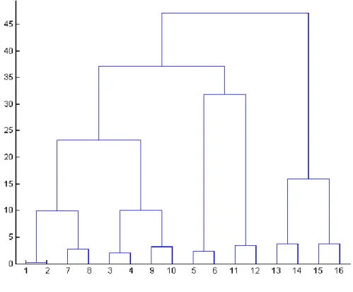
Figure 10: Hierarchical binary tree structure or dendrogram ... 37
Figure 11: K-Means dotplot representation ... 39
Figure 12: Fuzzy C-Means dotplot ... 41
Figure 13: BG1805 vector construction scheme ... 47
Figure 14: Illustration of Shape factor descriptor ... 61
Figure 15 : Sequence alignment comparison between Cdc42 and Rho5 ... 67
Figure 16: Over-expression of native small GTPases in S. cerevisiae BY4741 strain ... 70
Figure 17: G1/G3 sequence alignment of small GTPases ... 73
Figure 18: Images of constitutively active Cdc42 (amorphous), Rho5 (elongated/clumped) and wild type cells ... 76
Figure 19 : Morphologies of cells expressing small GTPases with point mutations in comparison with cells expressing native small GTPases ... 79
Figure 20: Comparison of tendency curves of shape factors used with 4 different mutants ... 82
Figure 21: Simultaneous use of different shape factors in a pool of wild type and mutants of 7293 cells ... 87
Figure 23: Hierarchical clustering results represented in histograms ... 92
Figure 24: Calculation of the between cluster sum of squares (blue) and the within cluster sum of squares (red) ... 94
Figure 25: K-Means clustering results represented in histograms ... 96
Figure 26 : Identification of clusters of phenotypes using Fuzzy C-Means algorithm ... 99
Figure 27: Average CQm for each mutant population ... 103
Figure 28: Percentage of unique cellular phenotypes ... 104
Dedication
A Papá y Mamá, quienes amo y me
aman sin dependencia y a mis
queridos hermanos
Acknowledgments
For those in Science and those about to Rock... I salute you!
To my beloved family, without your support, nothing would be possible. I am indebted in every possible way. Mamá & Papá for your undying love, support and care since I came to Canada (this one is for you and there is more to come in the future). To my brothers Oscar & Ulyses, I would like to thank you both for being excellent brothers, Abuela & Tía for whose continued support and prayers I am extremely grateful. My dogs Popi & Tita for being special and unique in the way they were, Ringo, Rocco, Uchy, Harri and Flopy for jumping in front of the camera while skyping! Martina the new member of the Papaluca family for your sweet and cheerful smile and punching the computer when I scream your name, Marcia Brun, thank you for all your love, patience and support (I will remember every day). To my dear friends, Fede Vega and Victor Gaete for long distance support and metallic brotherhood. To all, May the Force be with you.
With regards to my scientific career, the first person I want to thank is Professor Stephen Michnick, for accepting me into his lab and opening the door of science for me (un maestro, una causa, un efecto). You have influenced me tremendously to further continue the exploration and to understand life at the molecular level. My dear colleagues, from whom I learnt everything from scratch in order to perform my experiments and ask big questions! Relax guys, do the experiment! Specifically, I would like to thank Louis-Philippe “Calm down APZ” Bergeron-Sandoval for all the mentorship during this process and try to convert people to get free passports; Abdellali Kelil for the bioinformatics lessons; Benjamin Dubreuil for useful discussions and sharing ideas; Mohan “The mating expert” Malleshaiah for sharing so many good times until the end of his PhD; Durga Sivanesan for useful discussions and taking all my morning heavy jokes; The rest of the team; Diala Abd Rabbo, Jacqueline Kowarzyk, Emmanuelle Tchekanda, Luz Carrillo,
Jean-François Paradis, Emmanuel Levy, Po Hien Ear, Vincent Messier, Eugene Kanshin, Benjamin Ilunga Matala, Sinan Isik, Marcos Rodrigues and Bram Stynen.
Special thanks goes to Louis-Philippe, Abdellali, and Benjamin D. for all the bioinformatics support and useful comments, Durga, Jacqueline, and Diala for feedbacks and again Louis-Philippe for correcting the overall and for proving me with great feedback regarding this project, just relax mate! Cheers guys!!!
In addition, I would like to extend my appreciation and gratitude to Monique Vasseur for guidance and constant support with all my microscopy work. Also, I would like to acknowledge the members of my master thesis jury Dr. Pascal Chartrand and Dr. Gerardo Ferbeyre for accepting to review my work. Prof. Luc DesGroseillers and Sylvie Beauchemin for faculty procedural and administrative guidance to complete this research project.
1.1. Preamble: Subject Situation
Pathways evolution and new gene functions emerging from genes duplication is of biological and medical importance. Understanding these phenomena could be interesting in certain domains such as drugs development and certain types of medical therapy. Also to target the wanted pathway or engineer a certain cell type in order to accomplish a specific function.
The Ras-like small GTPases family represent important targets for pathway activation and development, since they can regulate a wide variety of cellular functions. These proteins have been the focus of cancer research since the discovery of the isoform
H-Ras p21 mutant in human tumour cells [1]. This discovery highlighted the importance
of the implication of proteins from the Ras-like small GTPases family in different types of diseases and different pathways.
Previous work accomplished by Heo et al. [2] showed that mutants with effective switch-of-phenotypes is related to functions. The authors developed an algorithm that predicts positions in a pair of protein classes that if exchanged will create mutants with switched functionality. They proved that specificity in a given protein family can be explored by combining genome-wide experimental functional classification with the creation of switch-of-function mutants. However, Heo et al. didn’t explain how the change in morphology arises as a result of switch-of-function mutants, this observation raises several questions, such as: (i) Will the GTPase activity of the mutated proteins be conserved? (ii) If not how do these mutations affect their targets? (iii) Do these mutations affect the interaction network of the implicated proteins? (iv) How their phenotypes have been quantified in order to statistically differentiate between the various phenotypes?
Proposed questions regarding such observations: (i) Which method can be used or developed to quantify cell phenotypes? (ii) How many mutations are necessary following a gene duplication to exchange functions between two Ras-like small GTPases? (iii) Which residues are relevant for functional specificity in the same small GTPase sub-family? (iv)
What are the possible effects of mutations at the divergent positions? (v) Would the divergent positions give rise to new protein interaction partner connections in order to gain new function and develop a new phenotype?
The aim of this master thesis is to perform a classification of budding yeast phenotypes using different approaches in order to statistically determine at which level of the cellular population, these constitutively active mutations are capable to affect cell morphology. As a starting point, native and constitutively active mutants of small GTPases were over-expressed to see whether there is a change or not in cellular morphology. Using the Ras-like small GTPases family as a working model, an extensive sequence alignment was elaborated in order to create constitutively active mutants. The known human H-Ras
Q61L constitutively active mutant has been used as a model to align with the rest of the
small GTPases of Homo sapiens, Mus musculus, Drosophila melanogaster, Oryzias
latipes, Rattus norvegicus, Gallus gallus, Schizosaccharomyces pombe, Saccharomyces cerevisiae, and other species. This showed conserved G1 and G3 domains among species.
Such domains are known to be involved in giving a constitutive activity by blocking the regulation by GTPase-activating proteins (GAP). In fact, creation of constitutively active mutants of proteins and their over-expression can induce different morphological cell changes which make their classification feasible.
Using microscopic techniques, Shape factor formulas have been used to measure coarse aggregate for concrete using digital image processing [3]. The adaptation and development of these formulas to cellular biology, helped us to define and describe quantitatively cellular shapes related to different phenotypes using various parameters. The main advantage of using the shape factors as a phenotype measurement is that it can be efficiently and easily applied on a large amount of data. All the analysis of this study was accomplished using the R statistical language. This phenotype classification is the first step to further understand if a neo or sub-functionalization process is taking place in order to induce different types of phenotypes. At the same time, this reflects various protein behaviours involved in important cellular events. This phenotype classification is
the first step to further understand if a neo or sub-functionalization process is taking place in order to induce different types of phenotypes. At the same time, this reflects various protein behaviours involved in important cellular events.
The second aim of this study is to explain the neo/sub-functionalization processes of the Ras-like small GTPases by looking at phenotypes linked with functions, signalling processes and protein-protein interaction networks. However, these presented goals are out of the scope of this thesis.
In summary, our phenotype classification method allowed us to observe how proteins from a defined sub-family can have two different roles, belong to different interaction networks and can induce two different phenotypes indicating a possible sub-functionalization process between these proteins. In addition, our applied method allow automating large-scale phenotypes quantification using shape factors and are addressing the issue of direct DIC quantification incognita. Moreover, this approach is opening doors to explore how protein-protein interaction networks can evolve and understand how pathways ends up developing important functions inside the cell.
1.2. Meaning of Evolution
Evolution is the only scientific theory that describes the diversification of life. Evolution theory explains the outstanding similarities among extremely great different forms of life, the development of new life forms, and the changes that occur within populations through inherited traits. Inherited traits are particular distinguishing aspects, including anatomical, biochemical or behavioural characteristics that are passed from one generation to another. The major sources of such variation are mutations, genetic recombination and gene flow (Adapted from Nature Education Knowledge Project). Darwin’s Theory “Evolution is a
vital process of life, and speciation is an evolutionary process by which new species comes to life” (C. Darwin. 1859) gave to evolution a strong, meaningful and controversial concept
that has been defying religious beliefs and point of views by putting in doubt creationism. This concept of evolution is critical point to biology since it helps understand important processes and is the only scientific explanation of life’s diversity among species. The theory of evolution has been called the cornerstone of modern biology since it provides a good basis for understanding science. As the prominent scientist Theodosius Dobzhansky stated “Nothing in biology makes sense except in the light of evolution” and without it, lots of biological events would remain unclear. Moreover understanding evolution is central for the advancement of biology and evolutionary medicine. In general words, by looking at the molecular point of view, evolution gives rise to changes and speciation which occurs in the form of mutation at the level of genetic sequences, also as a consequence often observed at the protein level. These concepts would lead scientist to develop tools in order to study at the genetic and molecular level, processes such as gene duplication, protein evolution, signalling and pathway development that has been the focus of research in the past decade. If we understand the evolution of a system we can target and engineer specific functions and we can dictate cells behaviour in one way or another and also understand the structure, function and mechanism of that system and we can make predictions about it.
1.3. Gene duplication & protein evolution
In protein evolution, gene duplication is considered to be the most important source of new DNA sequences which are in some cases devoid of selective pressure and other particular cases under selective pressure [4]. For instance, mutations and genomic modifications in these DNA sequence can lead to changes in sequences and switch domains among proteins respectively. These changes result in new protein structure, functions and abundance to just mention among others. It is interesting to know how two proteins from the same family can have totally different functions, how the same function can be conserve, and how they can share the burden of a determined function. Are these previous points affecting the interaction network? Are these previous observations pointing out on
how at the evolution of the protein-protein interaction network of system? These questions can be answered by first starting to understand mechanisms such as gene duplication, divergence and conservation which are the real phenomenon in charge of how eukaryotic signalling proteins can assemble a whole new set of functions and how this ends up implicating the whole network [5].
1.3.1. Molecular Mechanisms of Gene Duplication
The evolutionary biology field tried to address the following questions: (i) Can new genes emerge by duplication of existing ones and how do they acquire new functions after the duplication event? (ii) Can the new duplicate share functions with its ancestral form and which are the possible paths leading to this event? (iii) Which strategies can help us uncover and understand the consequences of gene duplications? It is worthwhile starting this chapter by briefly describing the different mechanisms causing genes duplication. We can cite mainly four major mechanisms orchestrating gene duplications in evolution (Figure 1) [6]:
A) Unequal Crossing-Over or Tandem Duplication: This phenomenon targets gene duplicates that are expected to have the same orientation but an unequal crossing-over. This first mechanism seems to be a common contributor of genetic material in duplicates.
B) Duplicative Transposition: Duplicative DNA Transposition is a mechanism accomplished by mainly two well known pathways: the non allelic homologous recombination (NAHR) and the non homologous end joining (NHEJ), where homologous sequences are used as templates during double-strand break repair.
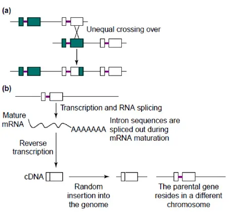
C) Retrotranposition: In 1991, Brosius et al. showed that retrotransposed genes result from the reverse transcription of mRNA into complementary DNA which is then inserted into a new genomic position giving birth to a new duplicate [7]. Some molecular features
about retrotransposition are loss of introns, presence of poly A tracts and presence of flanking short tandem sequences which deviate from the common pattern of this mechanism [8].
D) Polyploidy: Whole-genome duplication result in the retention of two copies of a gene. This process can be considered as duplication event of every single gene of the entire genome including flanking regulatory sequences [6]. It is known that after polyploidization the genes functions are maintained in a different manner from those duplicated by different mechanisms [9, 10].
Figure 1: Common mechanisms of gene duplication. a) Unequal crossing over, which results in a recombination event. b) Retrotransposition, which occurs when a mRNA is transcribed into a complementary DNA and inserted into a new genomic position resulting in a gene duplication event. Squares represent exons and bold lines represent introns (Adapted from Zhang, J.Z. [11]).
1.3.2. Models of Gene Duplication
Gene duplication is the principal event that orchestrates evolutionary novelty in eukaryotes, but what are the driving forces for gene duplication, and what are the selection pressures that model the evolving gene pair? Any aspect of genome evolution goes hand to hand with gene duplications among all kinds of species. This is a general way that genes evolve, acquire, develop or share new functions [12]. Understanding these processes is of pivotal interest, since after a gene undergoes duplication, this one can acquire a new truncated function which can segregate in the whole population causing a disease or can confer an adaptive advantage with a new function [13]. In the literature, many aspect and processes of gene duplication and evolution have been well described, but many of these theories have been disproved by experimental approaches [13]. First, we need to address the question of which model can be the most appropriate to understand evolution of gene duplication. Several important models have been proposed. Those models imply the known neo and sub-functionalization, gene conservation and gene loss, which give rise to protein evolution theory.
1.3.2.1. Ohno’s Model
Evolution by Gene Duplication, the widely cited theory of Susumu Ohno, was the
first to shed light on the evolution of new duplicates, explaining that “Adaptive mutations
accumulate in the new duplicate under no pressure of selection” [14]. This model is
characterized by maintaining a gene duplicate by simply increasing the number of genes coding for a protein (Figure 2) [11]. In an update of Ohno’s book “Evolution by Gene
Duplication”, the authors introduced the notion of “Gene Conservation” where both genes
maintain their original functions after duplication. The importance of this mechanism becomes relevant when the same function needs to be duplicated and conserved in order to fulfill certain needs inside the organism. This event is also necessary when there is deficiency in the original or ancestral function and need for higher protein concentrations. The Ohno Model introduced the sub-functionalization theory, where a gene undergoes
duplication accumulating mutations leading one copy to share a burden of the original function with the new copy. Moreover, Ohno’s model implies the neo-functionalization after gene duplication and introduces the sub-functionalization theory.
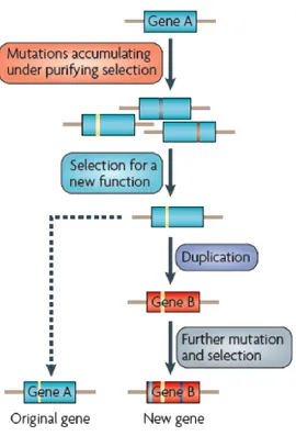
Figure 2: Ohno’s model. The new duplicate accumulates mutations under no pressure of selection. This model predicted that gene A undergoes duplication giving birth to gene B. Gene B accumulate mutations under no pressure and with no selection, therefore is selected for a new function by accumulating mutations. While gene A maintains in its original function. (Adapted from Soskine. M and D.F. Tawfik. [5]).
1.3.2.2. Divergence prior to duplication model
This model suggests that while initial levels of new evolving functions are acquired, the original function is maintained. The ancestor gene accumulates mutations under natural selection which leads to be selected for a new function. After duplication, the original gene is maintained (gene A) and new function is acquired by the new duplicate (gene B) (Figure 3). Here, duplication happens after the new duplicated gene is subjected to positive selection [5].
Figure 3: Divergence prior to duplication model (DPD). In the symmetric model the new function appears in the new duplicate and can also appear in the original copy. It proposes that gene A accumulates mutations under pressure of selection which leads to be selected for new function. Gene A undergoes duplication giving birth to gene B, the latter gene accumulates mutations resulting in the development of a new function, while gene A maintains the original function. (Adapted from Soskine. M and D.F. Tawfik. [5]).
1.3.2.3. The Duplication-Degeneration-Complementation Model
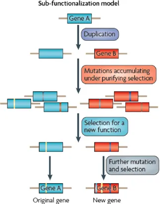
The Duplication-Degeneration-Complementation model also known as the sub-functionalization model can be described as a combination of Ohno’s and DPD models. This model is often considered as the only model of sub-functionalization in which the maintenance of duplicates does not required adaptive mutations [15]. The two copies may therefore acquire complementary loss-of-function mutations such that both genes are now required to maintain the function of a single ancestral gene (Figure 4).
Figure 4: Duplication-Degeneration-Complementation Model (DDC). In this model, one of the two loci results from duplication events, suffer a degenerating mutation and results in loss-of- function. Here gene A undergoes duplication giving birth to gene B. Gene A and B accumulate mutations under pressure of purifying selection, which makes a difference from
the Ohno’s model where the duplicates are under no pressure of selection. Gene A and B are selected for new functions where gene A maintains the original function and gene B accumulates mutations and is selected to share a new function (Adapted from Soskine. M and D.F. Tawfik. [5]).
1.3.2.4. Sub/Neo/Non-functionalization
This model explains how genes undergo duplication. Force et al. [15] suggested an improved theory of Ohno’s model. The authors were mainly interested in the division of expression domains among paralogs, which they claim to be the main genre of sub-functionalization.
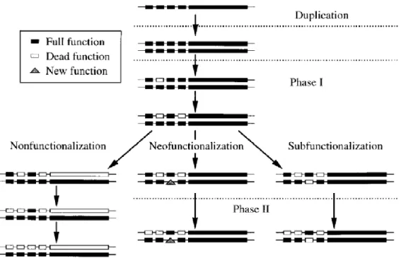
Gene’s fate after duplication depends on two major phases (Figure 5). Generally, after duplication, genes enter in phase 1 and experience one of the following three alternatives: neo-functionalization, sub-functionalization or non-functionalization (loss-of-function). The latter mechanism differs from the former ones, where the possibility of the loss of genes function was not considered. First, the new duplicate acquire a null mutation in the coding region which drifts to fixation that drive to gene loss (non-functionalization). This event also occurs whether the regulatory region of one duplicate is destroyed. Second, the duplicate may acquire a mutation which confers a totally new function and becomes fixed through Darwinian selection. This selection will lead the duplicate to acquire new mutations on regulatory regions causing a change while conferring a new function (neo-functionalization). Third, each duplicate may experience loss or reduction of expression for different sub-functions cause by degenerative mutations. In this process is known as neutral mutations (functionalization) where no adaptations are formed [14]). In the sub-functionalization process both duplicates can share the burden of a function by fulfilling the requirements of the ancestral function.
Figure 5: Gene fate after duplication. Notation: Small boxes denote regulatory elements with unique functions. Large boxes denote transcribed regions of a gene. Solid boxes denote intact regions of a gene. Open boxes denote full mutations. Triangles denote the evolution of a new function. (Adapted from Force, A. et al. [15]).
Studying these concepts is important to understand the mechanisms underlying the emergence of new phenotypes and how these mechanisms could be regulated in the heart of protein interactions network. Our goal is to show that in a family of proteins, those that were duplicated have evolved their original genetic code and developed new functions and novel structures which can result in phenotypic changes.
1.3.2.5. Understanding how new pathways evolved
The function of a protein is often determined by the identity of the amino acids composing its primary sequence and by various structural constraints. Conserved residues in a dispersed family of proteins are thought to have a direct role in protein structures and functions [4, 16]. In order to understand how proteins achieve specificity, we first need to identify which amino acids are relevant for a given functional class. Second, it’s important to determine the number of the proteins residues that need to be mutated in order to exchange functions from one protein into another. Many studies interested in gene duplication and evolution of protein functions were published [15, 17, 18], but none of them have focused on how to identify functional residues. Accordingly, we propose to study the functional differentiation and evolution of the Ras-like small GTPases super family and to examine how novel binding partners evolved and to investigate their influence on the creation of new pathways.
The Ras-like small GTPases protein networks provide an excellent model to demonstrate how new pathways have evolved by making random changes. Therefore, the structures found by evolution depend to some degree, on historical chance and are laden by biochemical details that require special description. The following section describes in details this wide family of proteins in order to understand their specificity and functions.
1.4. Budding yeast as a biological model
The eukaryotic organism Saccharomyces cerevisiae represents one of the most studied biological systems and constitutes an ideal model for understanding cellular processes. Used in biochemical and genetics studies, S. cerevisiae helped the development of a wide range of biochemical tools which have been designed and optimized to study any specific gene in this organism [19, 20]. Tools such as synthetic genetic array to study the relation between genotype and phenotype, yeast-two hybrid and protein-fragment complementation assay to study protein-protein interaction networks and astrobiology to study survival of different species in the outer space during the “Living interplanetary
flight experiment” (Phobos LIFE. www.interplanetary.org). In vivo studies of budding
yeast, revealed first evidence of small GTPases by discovering protein sequence similarity of certain proteins with human Ras small GTPases [21].
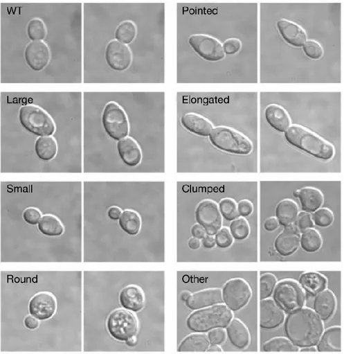
The approach followed in this research project consists of understanding the development of phenotypes linked to cellular functions involved in this process. This information brings us important clues of which approaches should be taken to start exploring evolution of protein-protein interaction networks using budding yeast as biological model. Furthermore, budding yeast displays a wide range of morphologies such as size and cellular division variants, which helps the development of this research (Figure 6). Moreover, this unicellular organism facilitates the study of cell cycle, cytoskeleton organization, sub cellular organelles, endoplasmic reticulum trafficking, secretion systems, metabolic regulation, signalling processes, chromosome recombination and receptor arrangements.
Figure 6 : Saccharomyces cerevisiae, a biological model, allowing the visualization of seven distinct phenotypes. These phenotypes serve as a model for classification. (Adapted from Giaever et al. [22]).
1.5. Ras-like small GTPases super family
Normal cellular development in multicellular organism is controlled by specific proteins that regulate a variety of complex signalling networks. The Ras-like family of small guanosine triphosphates (small GTPases) are known to be involved in various processes such as cell polarity, polarized growth, membrane trafficking, actin and septin organization and development, cell cycle regulation and cell survival. These proteins are of special interest because they regulate intracellular signal transduction pathways depending on external and internal stimuli. They act as molecular binary switches that are either turned on or off depending on the cell needs. This family of proteins has been well studied in both humans and Saccharomyces cerevisiae and showed to have a certain degree of conservation from yeast to humans [23]. Small GTPases serve as an excellent mechanism for the development of similar signalling processes established at levels of functions and structure [24].
1.5.1. How Ras-like small GTPases are regulated?
In Saccharomyces cerevisiae, the activity of proteins belonging to the small GTPases family is controlled by the ratio of bound GTP to GDP [25]. They exist in an inactive form (GDP-bound) or an active form (GTP-bound) that activates downstream effectors depending on the signal. They regulate several important biological cascades like the MAPK cascade, cAMP cascade, gene transcription, actin cytoskeleton organization, cell growth, cell cycle progression, bud emergence and pheromone response pathway among others [26]. The switch between active GTP-bound and inactive GDP-bound states is specifically controlled by Guanine nucleotide exchange factors (GEFs). These GEFs stimulate the exchange of GDP into GTP yielding an active form of the protein (Figure 7). This active form is inactivated by the hydrolysis of the GTP, which loses a phosphate group to become inactive GDP state. This inactivation cycle is controlled by the GTPases activating protein (GAPs). It is known that Ras-like small GTPases bind effectors in their
active state in order to activate important signalling pathways. In addition, Rho and Rab small GTPases subfamilies are also regulated by a third class of regulatory protein called Guanosine Nucleotide Dissociation Inhibitors (GDIs) These regulators not only prevent the GDP – GTP exchange cycle, but also maintain the protein in their GDP inactive state and from being localized at the membrane [27] that will be described later on. In addition, these GEFs and GAPs have proved to be important targets for drug design for cancer therapy [28] and rewiring of cellular morphology [29].
1.5.2. The Ras-like small GTPases are grouped in five major branches
Most of the available genomic studies on budding yeast Saccharomyces cerevisiae have allowed the classification, distribution and functional diversification of the Ras-like small GTPases. The available genome sequences of many eukaryotes allowed us to analyze this family from an evolutionary perspective. This super family is composed of more than 700 sequences from different species which contributes the understanding of origin, evolution, function and structure [30]. The Ras-like small GTPases is divided into five major branches (Figure 8). These branches are divided into five subfamilies which are Ras, Rho, Rab, Arf and Ran. Previous studies revealed that expansion and evolution of these genes have been related to unique eukaryotic cellular features such as cell division, cell cycle regulation, phagocytosis, apoptosis and signalling regulation. [31]. Each subfamily is involved in several important intracellular tasks. In addition, the comparison of these subfamilies using sequence alignment approaches showed that G domains are conserved among the five subfamilies, an observation that served for creation of constitutively active mutants [32].
1.5.3. Cell morphology: Regulation by Rho and Ras small GTPases
Maintaining the appropriate cell shape and morphology is essential for homeostasis and dynamic processes. Saccharomyces cerevisiae undergoes several morphological changes during the cell cycle [27], therefore this family of proteins participates actively in regulating actin cytoskeleton, cell growth, survival and differentiation, which implies regulation of cell cycle. These are fundamental processes that are essential for cellular development [33].
The Rho and Ras small GTPases are the most important subfamilies that particularly regulate cell shape. This regulation happens via the asymmetrical regulation of the actin cytoskeleton which leads to actin localization at sites of growth during budding and bud site selection, ending in normal budding process. Moreover, these subfamilies perform their tasks during the cell cycle through important signalling processes which are mediated by protein-protein interactions [34]. The cytoskeleton plays an important role in determining cell morphology, this process is known to be mainly regulated by Rho and Ras proteins which are known to be mutated in cancerous cells [35]. This chapter describes such processes and the involvement of Rho and Ras small GTPases and how these proteins orchestrate cell morphology behaviour.
1.5.4. Ras-like small GTPases
Ras subfamily was the first discovered proteins of the Ras-like small GTPases superfamily. These proteins are the key regulators of extracellular and cytoplasmic signalling networks that control cell growth, survival and differentiation [28]. An additional information is that in mammalian systems, the Ras subfamily is comprised of 36 proteins that has been studied extensively over the past two decades because of their roles in human cancers [36]. In contrast to mammals, Saccharomyces cerevisiae features a Ras subfamily comprised of only 4 members Ras1, Ras2, Rsr1 and Rhb1 which are involved in different
cellular roles. These proteins contain N-terminal portions with a significant homology to mammalian Ras and others subfamilies. As well they feature a C-terminus which includes at the terminal 4 amino acids that constituted the CAAX motif (C is cysteine, A is aliphatic amino acid And X is the C-terminal amino acid). This motif is important for posttranslational modifications that facilitate the association with the membrane. The conserved regions contain boxes known as G1, G2, G3, G4 and G5 which are short sequences of amino acids involved in the recognition of GEFs and GAPs [37].
Briefly, Ras1 is involved in G-protein signalling in the adenylate cycline activating pathway (cAMP) that plays a role in cell proliferation. It is localized at the plasma membrane and is a homolog of mammalian Ras proto-oncogenes [38]. His homolog Ras2 is in charge of the regulating the nitrogen starvation response, sporulation and filamentous growth, farnesyaltion and palmitoylation. Such functions are required for activity and localization to plasma membrane [38]. Following these descriptions, Rsr1 is required for bud site selection, response to mating pheromone and cell fusion. It is localized at the plasma membrane [39]. It has significant similarity to mammalian Rap small GTPases. Finally, Rhb1 which is related to mammalian Rheb and is involved in regulating canavanine resistance and arginine uptake [40]. These are just some functions that Ras proteins are capable to regulate.
1.5.5. GDP-GTP cycle of Ras-like small GTPases
The Ras subfamily of small GTPases feature a GDP-GTP cycle which is similar in other subfamilies such as Ran and Arf with the exception of Rho and Rab which are described in their own sections.
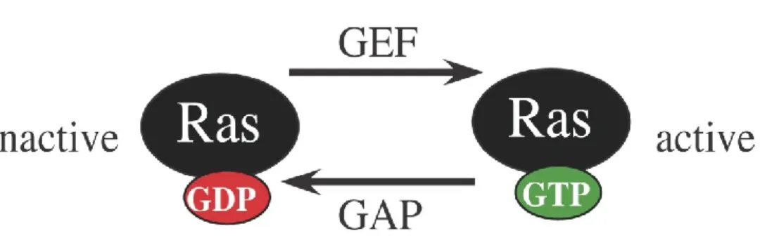
Figure 7: The GDP-GTP cycle of Ras-like small GTPases. Ras proteins are in their active state when bound to a GTP molecule and are inactive when bound to a GDP molecule. Ras activation is controlled by GEFs that stimulates the exchange of GDP into a GTP and Ras inactivation is controlled by GAPs that hydrolyse the GTP into GDP (Adapted from Downward, J. [41]).
Figure 8: Phylogenetic tree of 34 known Ras-like small GTPases of Saccharomyces
cerevisiae. Proteins are clustered into subfamilies of Rab, Ran, Ras, Rho and Arf
respectively. Clusters appear to be grouped according to cellular functions (Adapted from Garcia-Ranea et al. [30]).
1.5.6. Rho-like small GTPases
Through a series of complex biochemical networks, Rho small GTPases controls some of the most important processes of cell morphology. Rho small GTPases are also regulated by GEFs, GAPs and a third class of regulatory protein called Guanosine Nucleotide Dissociation Inhibitors (GDIs). These regulators not only prevent the GDP – GTP exchange cycle, but also maintain the protein in their GDP inactive state and from being localized at the membrane (Figure 9) [27]. This subfamily shares similar roles in signal transduction to Ras small GTPases subfamily and is best characterized for the regulation of actin cytoskeleton organization, morphogenesis and cell shape, movement and polarity and cell cycle progression [42, 43]. Hence, a particular cell shape is adopted and controlled via the asymmetry and organization of signalling pathways which involves the Rho subfamily of small GTPases [9].
The Rho subfamily of small GTPases are present in all eukaryotic systems being conserved from yeast to humans [44]. Such conservation suggests that basic mechanisms involved in cell morphology were conserved during evolution. Such mechanisms have defined the finest jobs in development and maintenance of cell morphology. The dynamics of the actin cytoskeleton are mainly controlled by Rho, which have significant roles during budding process [27, 45]. In addition, Rho subfamily is divided again into two subfamilies which are Rho and Rac and they apparently regulate overlapping pathways [30].
About Rho in mammals, recently Tybulewicz and Henderson reviewed Rho proteins describing their role in the immune response acting as regulators in lymphocytes development, differentiation, activation and migration. Rac1 and Rac2 have demonstrated important roles in B cell development, where loss of Rac1/Rac2 has for consequence a block of such process. This leads to an arrest of B cell development stage in the spleen, showing that Rac1 and Rac2 are required at the earliest stage of transitional B cells [46, 47]. Moreover, Rac1 is known to be mammalian homolog of budding yeast Rho5p, which
have similar roles in a protein kinase signal transduction pathway (Pkc1p) [48], being one of the reason to work with Rho5 in budding yeast.
A comparison of mammalian and budding yeast Rho small GTPases reveals that in mammals it comprises 23 members which are related in primary structure. Phylogenetic analysis shows in mammals that these proteins cluster into different subfamilies based on sequence similarity [47]. Whereas, in budding yeast is comprised of only 6 proteins; Rho1, Rho2, Rho3, Rho4, Rho5 and Cdc42 (Cell division cycle 42).
A wide range of studies have been made from mammals to yeast regarding Rho behaviour demonstrating important roles in cell integrity. To begin, Rho1 a small GTPase, involved in establishment of cell polarity, regulates protein kinase C (Pkc1p) and cell wall synthesizing 1, 3-beta-glucan synthase (Fks1p and Gscp) for fungal wall biosynthesis, which makes the most important enzyme responsible of the synthesis of cell wall polymer in budding yeast. [49]. Rho1 is essential and required for bud formation and development [50]. It is localized to the area of polarized growth independently of the actin cytoskeleton and is activated by a series of GEFs Rom1, Rom2 and Tus1 [51]. Rho1 GDP/GTP cycle is regulated by GAPs Bem2 and Sac7 which also have a role in negative regulation of the MAPK cell-integrity pathway [52, 53].
Rho1 has 67% identity with human RhoA, and both have similar roles and localized to places of active growth [54]. Recent work by Lee et al. showed that Rho1 confers resistance to oxidative responses in budding yeast providing protection. Yeast two hybrid assay showed that Rho1 associates with Ycf1, a vacuolar glutathione S-conjugate transporter, showing biomolecular fluorescent complex on the vacuolar membrane [55]. As well, several works revealed that Rho1 has the most notorious potential implications in control and coordination of three distinct biochemical pathways, which each of them contributes to growth and budding during the cell division cycle.
Rho subfamily also features four non essential small GTPases. Rho2 is involved in the establishment of cell polarity and microtubule assembly. Its disruption does not produce
a detectable phenotype, however, it may regulate aspects of the budding process [49]. Rho3 is involved in the establishment of cell polarity along with Rho2. Its GTPase activity is potentially regulated by the GAP Rgd1p, which is a GAP involved in control of actin cytoskeleton [56]. Rho4 is implicated in the establishment of cell polarity [56].
Moreover, the Rho3 and Rho4 proteins have a slightly difference. They show only 57% similarity in primary structure, but they are functionally related. Evidence about Rho3 and Rho4 tells that they seem to be required after bud formation to maintain cell polarity during the maturation of daughter cells [57]. They are also involved in the regulation of exocytosis and actin polarization [58]. Matsui et al. also showed that disruption of Rho3 gene produces viable cells with very poor growth, suggesting to be little essential after all, whereas Rho4 disruption has no significant effect on cell growth, but a double mutant yield cell with growth effects and over-expression of Rho4 is able to rescue Rho3 mutants.
Rho5, one of the main focus for this work, has been originally revealed in silico analysis of the entire yeast genome [30]. It is involved in protein kinase C (Pkc1p)-dependent signal transduction pathway that controls cell integrity. During functional characterization of Rho GAPs Bag7, Lrg1 and Rgd2, Roumanie et al., showed the first evidence of Rho5 in budding yeast using a systematic yeast two hybrid approach. It has been observed that Rho5 showed 46% sequence identity with Cdc42, an essential member of the Rho subfamily in Schizosaccharomyces pombe, as well with human homolog Rac1. In addition, Rho5 also shows 45% identity with members of the Rac subfamily, making Rho5 appears as a unique Rho/Rac-like protein in S. cerevisiae genome. Another feature that makes Rho5 interesting is that it is the only protein among the six belonging to the Rho subfamily that contains a PEST motif which plays a role in protein stability through ubiquitination process.
Despite that Rho5 was characterized as a non-essential protein in the S. cerevisiae genome, several research works have been done demonstrating its importance in development and regulation of cell integrity. In 2002 Schmitz et al. showed that Rho5 plays
an important in the protein kinase C (PKC1) dependent signal transduction pathway, where deletion of rho5 increased its activity. This deletion showed an increased resistance to drugs such as caffeine, calcofluor white and congo red, which indicates activation of the PKC pathway. In contrast, an overlapping activity has risen by over expressing a constitutively active mutant of Rho5 Q91H which renders cells more sensitive to these drugs, suggesting that Rho5 acts as an off-switch for the MAP-kinase cascade, which differentiates between MAPK-dependent and –independent functions of Pkc1 [48]. Other roles that have been assigned for Rho5 are; is necessary for H2O2-induced cell death, which goes with reactive oxygen species (ROS) accumulation and DNA fragmentation by over expressing a constitutively active mutant of Rho5p G12V which leads to a ROS accumulation followed by cell death upon H2O2 treatment [59]. Also Rho5 binds to Ste50 which is a protein involved in mating response and osmotolerance [60]. The expression of Rho5 Q91H allele in a ste50 deletion strain is lethal under osmotic stress. These data suggest a role in mediating the osmotic stress response via phosphorylation and ubiquitination [61].
Cell division control 42 or simply Cdc42 is a highly conserved small GTPase essential for establishment and maintenance of cell polarity acting as a molecular switch modulating a wide range of signalling processes. Mutant versions show defects in the organization of actin cytoskeleton and septins which have subtle roles for progression of the cell cycle. Essentially, Cdc42 is required to promote the assembly of bud components at the bud site [62].
This main regulator is known to be involved in processes like actin patch polarization, pheromone response pathway, actin cable nucleation and septin organization which are mostly related with the maintenance of cell morphology [27]. In S. cerevisiae Cdc42 is found at the plasma membrane being localized at specific domains coordinating polarized organization of actin cytoskeleton during cell growth [24, 63]. Cdc42 is known to induce specific phenotypes after activation to the GTP state. The Cdc42 Q61L has been reported to induce amorphous/misshapen/elongated phenotype giving his importance in the
regulation and maintenance of cell morphology. In addition, mammalian Cdc42 is known to control the formation of actin bundle containing filopodia at the cellular periphery [64].
These are the major reasons why we choose Rho small GTPases in order to study their phenotypic and protein network evolution. They are very efficient at inducing phenotypes and also inducing malignant cells in humans [26].
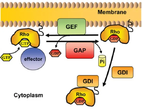
Figure 9: The GDP-GTP cycle of Rho small GTPases. As we can see in the cycle of small GTPases, the footstep from active state to inactive state is managed by GAPs and GEFs. In the case of Rho and Rab subfamilies, these have a third regulator GDIs. These important steps lead these proteins to regulate important intracellular responses like cell polarity, cell shape, cell migration, cell survival, cell proliferation among others which are controlled by different pivotal pathways. (Adapted from Perez et al. [27]).
1.5.7. How do vesicles and tubular structures generate cellular transport?
Vesicle trafficking is the movement of molecules inside the cell, from one compartment to another. These processes are known to be involved in novel functions at golgi complex and cilia assembly which implicates cell morphology [65]. Rab and Arf subfamilies of the Ras-like small GTPases are known to be membrane associated proteins. They are molecular switches modulated by GEFs and GAPs effectors. In their active state they promote the traffic and movement of vesicles and organelles in vesicle budding to specific cell compartments [66].
1.5.8. Rab-like small GTPases
This subfamily of Ras GTPases regulates membrane trafficking and intracellular transport. Rab small GTPases constitute the largest family of the known membrane trafficking processes [66]. It is composed of eleven proteins involved in several different functions. These small GTPases have the same activation mechanism but with different roles in their active state. They are molecular switches which in the active GTP bound state promotes trafficking of vesicle budding, vesicle motility, vesicle docking to specific membranes and vesicle fusion across the cell [66].
Rab small GTPases homologs, Ypt31 and Ypt32, are involved in the exocytic pathway. This protein mediates intra-golgi traffic and budding of the post-golgi from the trans-golgi [67]. The Ypt1 protein was one of the first discovered from Rab small GTPases subfamily, was found lying on actin and β-tubulin in the yeast genome [68]. Ypt1 is required for vesicle docking, and in the ER to golgi step of the secretory pathway [69]. The essential Sec4 small GTPase is required during vesicular transport and fusion from golgi to the plasma membrane and at the last steps of exocytic secretion. Sec4 also is located at bud tips and exocytic sites of plasma membrane and post golgi secretory vesicles. Its involvement in cell morphology can be observed at the bud tip in which is capable of
accumulating vesicles at bud tip. Its required for polarized transport of secretory vesicles thereby helping to regulate cell shape [70].
Ypt6p is required for ER to golgi and in cis- to medial-golgi transport. It has been proved to be involved in secretory pathway at elevated temperatures which makes it essential in such processes only at elevated temperatures in budding yeast [71]. Ypt7 is involved in endocytosis. It is known to be localized at the vacuole and is required during vacuole fusion reaction [72]. Protein Ypt10 is the only one of this subfamily that contains a PEST motif which is specific for control of proteolytic enzymes for degradation. Previous studies regarding over-expression of this protein showed an accumulation of golgi like cisternae with budding vesicles proving its involvement in cell shape control [73].
The homologues Ypt51, Ypt52 and Ypt53 small GTPases are required for endocytic transport, for sorting hydrolases and vacuolar proteins. They are detected at the mitochondria level and are known to be mammalian Rab5 homologs [74]. Last, Ypt11 mediates distribution of mitochondria and endoplasmic reticuli to daughter cells at the moment of budding [75]. These are known roles regarding this subfamily, showing its importance in vesicle trafficking and cellular morphology behaviour.
1.5.9. Arf-like small GTPases
ADP-ribosylation factor (ARF) is a subfamily best characterized to be regulators of membrane trafficking, organelle structure and intracellular transport. Functions of these proteins are GDP-GTP cycle dependant. An interesting observation regarding Arf’s GEFs and GAPs is that these GEFs contain a conserved Sec7 domain that catalyses hydrolysis of the GTP-bound state having intrinsic GTP hydrolysis activity, while the GAPs contain a conserved zinc-finger GAP catalytic domain [65]. These features facilitated the identification of Arf regulators from yeast to human.
Chromosome instability is mainly controlled by the Arf-like small GTPase Cin4, where its major function is observed at the folding of beta-tubulin [76]. Sar1 component of COPII coat of vesicles is required for transport vesicle formation between ER to golgi. The main function of Sar1 is to recruit COPII to the membrane mediated by its GEF Sec12 [77]. Related to previous roles, Arl3 is required for recruitment of Arl1 to the golgi [78]. Arl1 soluble small GTPase involved in regulation of membrane traffic and intracellular control of potassium influx. This role has been shown by creating a null Arl1 strain [79].
Homologs Arf1 and Arf2 are involved in regulation of coated vesicles formation in intracellular trafficking inside the golgi [80]. While, Arf2 gene encodes one of the three ADP ribosylation factors in the S. cerevisiae. These two proteins share a 96% identity in sequences. Mutant versions of Arf2 show no morphological changes whereas deletion of both Arf1 and Arf2 show no viability for the cell [80]. Not being a homolog but with similar name, Arf3 is involved in development of cell polarity, making it important for morphogenesis processes [81].
1.5.10. Ran-like small GTPases
This Ras-related nuclear small GTPases regulates nucleocytoplasmic transport of macromolecules, RNA, proteins and the organization of the spindle apparatus during mitosis [82]. Comprise only two proteins Gsp1 and Gsp2. It has also been shown that Ran small GTPases GDP/GTP cycling controls DNA replication and nuclear envelope gathering being important during cell cycle development [83]. In budding yeast have been characterized two major small GTPases which are homolog in sequences and involved in similar cellular tasks. Gsp1 represents an essential protein involved in the maintenance of nuclear organization and RNA transport and processing [84]. His homolog Gsp2 is not required for viability and is also involved in the maintenance of nuclear organization and RNA processing [84].