Effect of cigarette smoke on Candida albicans growth and
its interaction with human gingival fibroblasts
Mémoire
Humidah Alanazi
Maîtrise en biologie cellulaire et moléculaire
Maître ès sciences (M. Sc.)
Québec, Canada
iii
Résumé
L’objectif principal de cette étude est d’étudier l’effet de la fumée de cigarette (FC) sur C. albicans et sur son interaction avec les fibroblastes gingivaux humains. Nous avons démontré que Candida se multiplie pré-sence FC, en adoptant la forme hyphe. La FC rende C. albicans sensible au H2O2, mais résistant aux la
chaleur et au NaCl. La FC augmente la production de chitine par C. albicans. Préincubé avec le FC, C. albicans adhère beaucoup plus aux cellules gingivales en monocouche, prolifère plus et adopte la forme hyphe plus facilement. Les fibroblastes quant à eux ils montrent une réduction de leur croissance, mais produisent plus de l’IL-1b. En conclusion: le CFC module d’un côté C. albicans et de l’autre côté les cellules de l’hôte. Ceci suggère un négatif important de la fumée de cigarette favorisant les candidoses orales chez les fumeurs.
v
Abstract
The main objective of this study was to investigate the effect of cigarette smoke (CS) on C. albicans and its interaction with human gingival fibroblasts. We have shown that Candida multiplies presence CS, by adopting the hyphae form. CS makes C. albicans sensitive H2O2, but resistant to heat and NaCl. CS increases chitin
production by C. albicans. Preincubated with CS, C. albicans adheres more to gingival cells in monolayer, proliferating more and adopts the hyphae form more easily. Fibroblasts show a reduction of their growth, but produce more IL-1b. CSC modulates C. albicans and on the other side also CS modulates the host cells. This suggests a significant negative cigarette smoke promotes oral candidiasis in smokers.
vii
Table of content
Résumé ……….……….. iii
Abstract ……… v
Table of content ………..………... vii
List of tables ……..………... xi
List of Figures………….………... xiii
List of abbreviation ……..………... xv
Acknowledgement ………... xviii
Foreword ……….. xix
Chapter Ι………... 1
1.1 INTRODUCTION……….………. 1
1.1.1 The oral cavity and microbial niches……….……… 1
1.1.2 C. albicans... 2
1.1.2.1 C. albicans cell wall……… 3
1.1.2.2 C. albicans life cycle………... 5
1.1.3 C. albicans biofilms………... 7
1.1.3.1 C.albicans biofilm formation (phenotype switch)…..……... 8
1.1.3.1.1 Adhesion factors………. 9
a) Eap1 protein………... 9
b) Hwp1 protein………... 9
c) Chitin protein……… 10
1.1.3.1.2 Transition factors and regulatory pathways……. 10
1.1.3.2 Gene expression of biofilms formation………... 12
1.1.3.3 Factors contributing to biofilm distribution………..… 13
1.1.3.4 Biofilm resistance………... 14
1.1.4 C. albicans infection……….... 14
1.1.4.1 C. albicans and the host………... 16
1.1.4.2 C. albicans and different host tissues………. 16
1.1.4.3 C. albicans adaptation………... 17
1.1.4.4 C. albicans and host immune response………...….. 19
1.1.5 C. albicans and oral cavity………...…….. 20
1.1.5.1 Oral candidiasis……….. 21
1.1.6 C. albicans and risk factors………...………. 23
1.1.6.1 Saliva and oral candidiasis………...……… 23
1.1.6.2 Systematic diseases and oral candidiasis……….. 24
1.1.6.3 Antibiotic treatment and oral candidiasis……… 24
1.1.6.4 Tobacco smoke and oral candidiasis……….. 25
1.1.7 Cigarette smoke (CS)………. 26
1.1.7.1 Effect of cigarette smoke on human health……… 26
1.1.7.1.1 Cigarette smoke and cancer………. 28
1.1.7.1.2 Cigarette smoke and heart diseases…………...… 29
1.1.7.1.3 Cigarette smoke and respiratory diseases..……... 29
1.1.7.1.4 Cigarette smoke and oral diseases…………...….. 29
Chapter ΙΙ…Scientific article
Title: Cigarette smoke-exposed Candida albicans increased chitin production and modulated human
fibroblast cell responses (published article)……….. 33
Abstract………...………… 34
2.1 Introduction………... 35
2.2 Experimental protocol……… 37
2.2.1 Candida albicans………...……… 37
2.2.2 Gingival fibroblast isolation and culture………. 37
2.2.3 Preparation of the cigarette smoke condensate 37 2.2.4 Effect of CSC on C. albicans transition from blastospore to hyphal form……… 38
2.2.5 Effect of CSC on C. albicans response to stressful agents… 38 2.2.6 Effect of CSC on C. albicans cell wall chitin content………… 39
2.2.6.1 Cell wall isolation and purification……… 39
2.2.6.2 Chitin quantification……… 40
2.2.7 Evaluation of CSC-pretreated C. albicans adherence to a human gingival fibroblast monolayer 40 2.2.8 Effect of CSC on the interaction of C. albicans with gingival.. fibroblasts 41 2.2.8.1 Evaluation of CSC-pretreated C. albicans transition following contact with human gingival fibroblasts……….. 41
2.2.8.2 Evaluation of CSC-pretreated C. albicans and gingival fibroblasts growth……….. 41
2.2.9 Effect of CSC-pretreated C. albicans on IL-1β secretion by gingival fibroblasts…………..………... 42
2.2.10 Statistical analyses……….. 42
2.3 Results………... 43
2.3.1 CSC promoted C. albicans transition from blastospore to hyphae form……….. 43
2.3.2 CSC decreased C. albicans resistance to oxidative stress and increased its resistance to osmotic and heat stress….... 45
2.3.3 Exposure of C. albicans to CSC increased chitin content….. 49
2.3.4 CSC-pretreated C. albicans adhered better and changed form when in contact with the gingival fibroblast monolayer. 51 2.3.5 CSC-pretreated C. albicans proliferated better when inter-acting with the gingival fibroblasts………. 53
2.3.6 CSC-pretreated C. albicans decreased gingival fibroblast growth and increased IL-1ß secretion……….….. 54
2.4 Discussion………..….…… 57 2.5 Conclusion………..….…... 60 2.6 References……….. 61 Chapter ΙΙΙ………..……… 65 3.1 General discussion……….………. 65 3.2 Conclusion………...……….……… 67
ix
3.3 References………..………..……… 69
xi
LIST OF TABLES
Table number Title of the Table Page
Table 1-1 The environmental cues and pathways involved in filamentous growth regulation in C. albicans
11
Table 1-2 Candida-host recognition system: RGD, the amino acid sequence ar-ginine-glycine-aspartic acid, EMP, extracellular matrix protein of en-dothelial
17
Table 1-3 Pattern recognition receptor that sense fungal –associated PAMPs 20
xiii
LIST OF FIGURES
Figure number Title of the figure Page
Figure 1-1 C. albicans cell wall components 4
Figure 1-2 C. albicans morphogenesis 6
Figure 1-3 C. albicans life cycle 6
Figure 1-4 The biofilm formation mechanism 8
Figure 1-5 Roles of Efg1, Hgc1, and Sun41 in UME6-enhanced C. albicans
bio-films formation 12
Figure 1-6 C. albicans invasion through epithelial cells 15
Figure 1-7 Changes in carbon source program major changes in cell wall
archi-tecture 18
Figure 1-8 Changes in carbon source impact on immune surveillance by
alter-ing the recognition of C. albicans cells 18
Figure 1-9 The harmful components of the cigarette 27
Figure 1-10 Cigarette smoke modulates inflammation and promotes chronic
in-flammation in the conducting airways by a variety of mechanisms 28 Figure 2-1 C. albicans transition from blastospore to hyphal form following
con-tact with CSC 44
Figure 2-2 Susceptibility of CSC-treated C. albicans to a cell surface-disrupting
oxidative, osmotic and thermal stress agents 46
Figure 2-3 Oxidative stress reduced the growth of the CSC-pretreated C.
albi-cans 47
Figure 2-4 Osmotic and heat stresses increased the growth of CSC-pretreated
C. albicans 48
Figure 2-5 Chitin content in the non-exposed and CSC-exposed C. albicans 50 Figure 2-6 CSC-pretreated C. albicans adhered more to gingival fibroblasts 52 Figure 2-7 Transition of CSC-pretreated C. albicans following contact with
gingi-val fibroblasts 53
Figure 2-8 Growth of CSC-pretreated C. albicans following contact with gingival
fibroblasts 55
xv
List of Abbreviations
Als3 Agglutinin-Like Sequence 3
ANUG Acute Necrotizing Ulcerative Gingivitis AP-1 Activatory Protein-1
BAL Broncho Alveolar Lavage
cAMP Cyclic Adenosine MonoPhosphate
cdk Cyclin-Dependent Kinase
C. albicans Candida albicans
CHD Coronary Heart Disease
Chs Chitins
COPD Chronic Obstructive Pulmonary Disease
CS Cigarette Smoke
CSC Cigarette Smoke Condensate CVD Cardio Vascular Disease CWPs Cell Wall Proteins
Eap Enhanced Adherence to Polystyrene ECM ExtraCellular Matrix
GPI GlycosylPhosphatidyIinositol GPM1 PhosphoGlycerate Mutase 1 Hwp1 Hyphal Wall Protein 1 Hyr1 Hypha Regulated Protein 1
IL-1β Interleukin-1β
MAPK Mitogen-activated protein kinases MLST Multi-Locus Sequence Typing NF-κB Nuclear Factor Kappa-B
NNK Nicotine-derived Nitrosamine Ketone PAF Platelet-Activating Factor
PAMPs Pathogen Associated Molecular Patterns
PKA Protein Kinase A
PRRs Pattern Recognition Receptors
PVC PolyVinyl Chloride
Saps Secreted Aspartic Proteases
STAT Signal Transducer and Activator of Transcription
xvii
Acknowledgement
Here I would like to thank Dr. Mahmoud Rouabhia for assisting me to the master degree, he was and still supporting me to right way in the life and work. All the best I wish for a such great professor.
The most amazing post doctor I have ever met Hyun Jin Park, she is always beside me either in the life or work. Thanks a lot for you Hyun Jin and the coming is better.
Another wonderful dentist Neftaha Tazi, who always beside me and helped me to overcome hard times in my life.
There is a great couple Fatima Redjemi and Nader Maroc, the kid care provider (home care). They are my family here in Quebec City. This couple helped me a lot to pass difficult situations with my children even with me. I wish all the best for them and for their children.
I will never forget my mother and father, how they always pray and calling me to ask and support with their beautiful words.
Thanks for my husband Shadaid Alanezi, who always does not let me disappointment and give me the energy to keep going.
Finally, thanks for my sister Sara, the second mother, she is my inspiration to be better in the thinking and the life.
Thank you for my siblings and I hope one day my children Zaid and Mayar will proud what I have done for them.
xix
Foreword
Mahmoud Rouabhia, Witold Chmielewski, and Andrew Zakrzewski conceived the study. Humidah Alanazi, Abdelhabib Semlali, and Laura Perraud conducted the experiments. Humidah Alanazi, Abdelhabib Semlali, Laura Perraud, and Mahmoud Rouabhia analyzed and interpreted the data. Humidah Alanazi, Laura Perraud, and Abdelhabib Semlali drafted the Materials and Methods section. Mahmoud Rouabhia completed the paper with the help of Witold Chmielewski and Andrew Zakrzewski. All of the authors read and approved the final paper.
1
CHAPTER Ι
1.1
INTRODUCTION
1.1.1 The oral cavity and microbial niches
The human mouth is an appropriate environment for numerous microbes [1]. Microbial diversity in the human oral cavity was discovered long ago, thanks to culture methods and molecular-based approaches. The oral microbiome is heterogeneous showing different physicochemical properties [2]. The presence of shedding and solid surfaces in the oral cavity provides an opportunity for numerous microbial species to adhere and to grow, compared to other microbial habitats in the human body [3]. Multiple other factors such as oxygen content, saliva, gingival crevicular fluid, and diet play major roles in the microbial composition of these oral ecological niches [4]. Humans are able to modify the microbial load through daily hygiene habits such as cleaning the teeth and tongue [5].
The major constituents of the oral microbiome are bacteria and fungi. Many oral microbes, such as Firmicutes (genus Streptococcus, family Veillonellaceae, genus Granulicatella), Proteobacteria (genus Neisseria, Hae-mophilus), Actinobacteria (genus Corynebacterium, Rothia, Actinomyces), Bacteroidetes (genus Prevotella, Capnocytophaga, Porphyromonas), and Fusobacteria (genus Fusobacterium) are normal microbial com-plexes residing in the oral cavity in various intraoral niches (dental surfaces, cheeks, hard palate, tongue, and saliva) [3]. In contrast, the presence of fungi is associated with some diseases and infections, although this does not eliminate the role of fungi and fungi-bacteria interactions in such infections [6]. One of the most studied and most common fungi in the oral cavity is Candida albicans (C. al-bicans).
2
1.1.2 C. albicans
The yeast C. albicans is frequently found on the skin and in mucous membranes such as the mouth, rectum, etc. [7]. C. albicans is a mucosal commensal yeast capable of colonizing and proliferating within every organ of the body, sometimes causing serious infections. The immune system, can reduce C. albicans virulence and pathogenesis [8,9]. This highlights the importance of Candida both medically and biologically [10]. Of the 17 known Candida species, five (C. albicans, C. glabrata, C. tropicalis, C. parapsilosis, and C. krusei) are invasive and are the cause of candidiasis. Interestingly, C. albicans is frequently identified in humans (42.1%) compared to C. glabrata (26.7%), C. parapsilosis (15.9%), C. tropicalis (8.7%), and C. krusei (3.4%) [11].
C. albicans is associated with clinical materials in 90 to 100% of mucosal infections, while 40 to 70% of candidemia is caused by C. albicans [11]. C. albicans has the capacity to modulate its growth according to environmental changes, leading to the expression of various genes in an appropriate manner. These genes involve specific signaling pathways (MAP kinases and RLM1) which play an important role in the virulence and morphological transitions of C. albicans [12].
C. albicans takes advantage of inappropriate physiological changes (such as impaired saliva or immune de-fense dysregulation) to proliferate and to invade, causing pathogenesis [9]. For example, C. albicans adheres better to denture materials, and forms a biofilm to maintain chronic infection [9]. Furthermore, the elderly are more prone to C. albicans infections because of an immune system dysregulation [13].
It is important to note that Candida species have some differences:
Phylogenetically, C. glubrata is more similar to baker’s yeast (Saccharomyces cerevisiae) than to
3
Among all Candida infections, C.albicans is the most frequently involved in human infections; C. dubiliniesis evades the azole’s effects faster than C. albicans does; this explains the association
of C. dubiliniesis in HIV patients. Candidiasis has been found to be mainly caused by C. albicans [9].
C. albicans includes four major and eight minor clades typed among more than 400 C. albicans isolates identified by multi-locus sequence typing (MLST) techniques. This enabled the identification of flucytosine resistance associated with the major clades [14]. Pathogenesis of C. albicans involves the early adhesion steps involving cell wall proteins.
1.1.2.1 C. albicans cell wall
The C. albicans cell wall consists of 90% carbohydrate and 10% protein, each with an active role in the virulence of the yeast. Antigens and pathogen-associated molecular patterns (PAMPs) contribute to the con-version of C. albicans from commensalism to virulence. These PAMPs form two layers:
The inner layer consists of polysaccharides which contribute to the immunological signature of C.
al-bicans [15];
The outer layer consists of glycoproteins which are formed by covalent links between the O-and
N-linked mannose polymers (mannans) and proteins. These O-linked and N-linked mannans attract different Toll-like receptors (TLR) and C-type lectin pattern recognition receptors (PRRs) during C. al-bicans pathogenesis [16].
4
The outer layer mannoprotein contains β-glucan/chitin which give a strong shape to the cell wall; most of these glucans/chitins are glycosylphosphatidylinositol (GPI)-proteinscontributing to the attachment of both the inner and outer layers of the yeast membrane (Figure 1-1). Moreover, many proteins expressed during the morphological changes from blastospore to hyphal form can contribute to C. albicans infection. Cell wall proteins (CWPs) are major players at various levels, from fungus adhesion and growth to invasion of the host. These CWPs are related to specific genes expressed during hyphal switching, such as hyphal wall protein 1 (Hwp1), hyphally regulated protein 1 (Hyr1), and agglutinin-like sequence 3 (Als3). C. albicans most often proliferates as a budding yeast under neutral or alkaline conditions; increasing the temperature triggers the morphogenesis [8,9,17,18].
Figure 1-1: C. albican cell wall components. This figure is adapted from Gow and Hube. Curr Opin Micro-biol. 2012 Aug;15(4):406-12. The figure shows transmission electron micrograph of the C. albicans cell wall. This was supported by showing key components this cell well along with their role interacting with the host [7].
5
1.1.2.2 C. albicans life cycle
C. albicans is capable of changing its morphogenesis under certain circumstances and morphing into many different forms (yeast, pseudohyphae, hyphae) which is considered the main virulence factor and pathogenic feature of this fungus [19], (figure1-2). These morphological changes lead to different life cycles (figure 1-3).
1. C. albicans proliferation occurs when the cells go from blastospore form, which is spherical (figure 1-3), to a septum ring formation, after which time the DNA separation takes place across the mother-bud neck. When the cell cycle progresses from G1 to S at the same time as the DNA is replicated, the spindle
pole body is duplicated and the cell separation takes place following cytokinesis. The new cells can subsequently enter their own cell cycle when they reach the appropriate size/maturation.
2. In pseudohyphal cells, similar to the budding yeast, the septum ring development also occurs prior to DNA separation which takes place across the mother-bud neck; however, following cytokinesis, the new cells remain linked to the mother cell to form elongated branched colonies. It has been reported that pseudohyphal cells require more time to polarize and remain more in G2,compared to the yeast. The
daughter cells then enter the life cycle along with the mother cells until the daughter cells reach the same size as the mother.
3. In hyphal cells, spetins are major proteins of the cell wall. Septins are filament-forming GTP-binding proteins and were first identified for their roles in cell-cycle progression in S. cerevisiae. With C. albi-cans, septins localize at the sites of germ tube emergence at the early stage of cell division, and later a fraction of the molecules appear to persist at the growing tips over extended. Interestingly, other cell cycles begin during hyphal development. The DNA moves and separates in the germ tubes (hyphal form is tighter than that of pseudohyphal cells) [19].
6
Figure1-2: C. albicans morphogenesis: a) Yeast cell, b) pseudohyphal, c) hyphe. This figures is adapted from our laboratory work.
Figure 1-3: C. albicans cell cycle. Cell cycle of yeast and the first cell cycle of hyphae and pseu-dohyphae induced from unbudded yeast cells. Hyphal germ tubes emerge before the G1/S transition. This figure is adapted from Sudbery P and Gow N, Berman J. (Trends in microbiology. 2004;12:317-324.) [104].
7
1.1.3 C. albicans biofilms
Typical biofilm formation occurs when the microorganism attaches to a surface and/or to another microor-ganism and produces a protective extracellular matrix against the oral immune defense. This biofilm organi-zation consists of survival mechanisms [20]. The ability to adhere to different abiotic and biotic structures is the first step of biofilm formation. This is supported by C. albicans morphological transition from blastospore to hyphal form, along with extra-polymeric materials [21]. It has been reported that microorganisms growing into biofilms are well protected against antimicrobial agents and that they are different than their planktonic (floating) counterparts [21]. Generally, infection by C. albicans biofilms on different surfaces (medical devices, human tissues) is modulated by several factors including different yeast species [22]. C. albicans has a ten-dency to grow more into biofilms than do other Candida species (C. parapsilosis, C. pseudotropicalis, and Candida glabrata). Furthermore, C. albicans forms biofilms more on silicone elastomer than on polyvinylchlo-ride (PVC) [23]. In addition, the presence of conditioning film consisting of host saliva proteins was shown to facilitate biofilm formation on denture acrylic [24].
C. albicans biofilms are a mixture of extracellular matrix engulfing C. albicans cells in the form of blastospores and hyphae. Biofilm formation takes place through different yet sequentially organized stages (Figure 1-4). Microorganisms initially attach to available surfaces and after a certain time, the planktonic microorganisms undergo the morphogenesis process with significant surface adsorption through a thin extracellular polymeric material (Figure 1-1). The microorganisms will then proliferate and interact with each other to ensure their sustainability in the imposed environment. Through this proliferation, a thick extracellular matrix (mature bio-film) is formed to provide chemical as well as physical protection to the microorganisms. The formed biofilm then undergoes focal dispersion through specific signals, resulting in microbial release (Fig.1-1). The re-leased microorganisms are then free to spread to other locations and form new biofilm [4-7].
8
Figure 1-4: The different steps leading to biofilm formation. This figure is adapted from Rouabhia and Chmielewski (The Open Mycology Journal, 2012, 6, 27-32); showing the sequential formation of C. albicans biofilm.
1.1.3.1 C. albicans biofilm formation (phenotype switching)
The survival mechanism of C. albicans is the morphogenesis from blastospore to hyphal form and vice versa. This mechanism, known as ‘phenotypic switching’ from round to elongated form, confers to C. albicans the capacity to invade the host. In hyphal form, C. albicans is more virulent [9,25,26], adheres better to human tissue, and penetrates up to the basal layer. This is regulated by specific factors including fibrinogen-like and fibronectin-like receptors and non-specific factors such as hydrophobic or electrostatic interactions [15]. Phe-notypic switching of C. albicans also involves different proteolytic enzymes (Saps) and specific signaling molecules [27].
9
1.1.3.1.1 Adhesion factors
Biochemical examination of the adhesion stage of C. albicans reveals the involvement of two key proteins, namely, enhanced adherence to polystyrene (Eap) and hyphal wall protein (Hwp) [9].
a) The Eap1 protein
Eap1, an adhesion protein, was first discovered in S. cerevisiae then in Candida [28]. Both Eap1 and Als proteins have a structure similar to that of the substrate-binding domain located in the N-terminus. In addition, there are tandem repeats in the central region and the C-terminus contains a GPI anchorage sequence. Studies on eap1Δ/Δ null mutants of C. albicans and the expression of C. albicans EAP1 in S. cerevisiae revealed that Eap1 mediates adherence to epithelial cells and polystyrene [28,29]. Eap1 is also involved in biofilm formation both in vitro and in vivo. However, Eap1p is not known to be involved in C. albicans virulence during candidiasis.
b) Hwp1
Hwp1 is a GPI protein expressed on the surface of hyphae. This 9dhesion mediates the interaction of C. al-bicans with human oral epithelial cells through a specific adhesion mechanism [30,31]. Indeed, the N-terminal region of Hwp1 acts as a specific substrate for epithelial cells through small proline-rich proteins. Hwp1 re-portedly mediates epithelial cell adherence by functionally mimicking host cell proteins [30,31]. The knockout of Hwp1 was shown to lead to reduced C. albicans virulence in vivo, suggesting the involvement of Hwp1 in C. albicans pathogenesis [32]. Hwp1 can interact with other adhesions, such as Als1 and Als3, and can mediate the adherence of one C. albicans hypha to another. This interaction contributes to biofilm formation [33].
10
c) Chitin
Adhesion of C. albicans to different biotic and abiotic structures is also mediated by chitin [34]. It has four synthase iso-enzymes (Chs1, Chs2, Chs3, and Chs8) controlling chitin synthesis through protein phosphor-ylation. For instance, the phosphorylation/de-phosphorylation of Chs3 at the S139 site (on the tips of growing buds and hyphae) regulates the position of Chs3 in C. albicans cell walls during cell cycle. On the other hand, the inactivation of Chs1and Chs2 can trigger C. albicans cell death [35, 36]. Chitin synthase can be affected by the BNI4 gene, which promotes C. albicans cell wall shape to hyphal form by increasing chitin expression. The deletion or mutation in the BNI4 gene leads to a significant change in C. albicans cell shape and reduces the hyphal form [36]. Chitins have been found to play a role in C. albicans adhesion to corneocytes. This adhesion involves chitin receptors known as lectin-recognizing chitins [34,38].
1.1.3.1.2 Transition factors and regulatory pathways
Multiple factors such as serum, high temperature, pH, N-acetylglucosamine, and CO2 contribute to C.
albi-cans phenotypic changes through various signaling pathways, including cAMP/PKA [38,39] [table 1-1]. In-deed, transition from blastospore to hyphal form is regulated by two major temporal pathways: the MAPK signal transduction pathway involving CPH1; and the Efg1 pathway which requires cAMP modulation [41].
These two pathways are also inter-regulated via positive and/or negative interactions that modulate hyphal development. For example, tup1 was found to repress hyphal formation through Rbf1p and Int1p (41). Changes in cell polarity, cell cycle, and cell wall architecture are virulence factors that appear throughout hyphal development [41]. Many studies confirmed that pseudohyphals were linked to an extension of the G2 phase. They may also be linked to a delay related to the synthesis of active cyclin-dependent kinase (cdk) complex coupled with Cdc28p phosphorylation [41]. Ste20-like protein and Cla4p are involved in merging
11 transduction signals which are needed for hyphal development and polarity modifications [41]. Morphological changes from blastospore to hyphal form are also regulated by Efg1 and Efh1 [39]. Hyphae facilitate C. albi-cans adhesion to tissue through α5β1-like fibronectin receptors [40, 42].
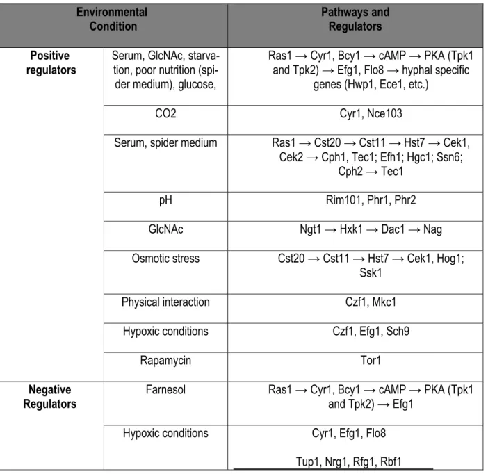
Table 1-1: The environmental condition and pathways involved in filamentous growth regulation in C. albi-cans (adapted from Huang et al., [105])
Environmental
Condition Pathways and Regulators Positive
regulators Serum, GlcNAc, starva-tion, poor nutrition (spi-der medium), glucose,
Ras1 → Cyr1, Bcy1 → cAMP → PKA (Tpk1 and Tpk2) → Efg1, Flo8 → hyphal specific
genes (Hwp1, Ece1, etc.)
CO2 Cyr1, Nce103
Serum, spider medium Ras1 → Cst20 → Cst11 → Hst7 → Cek1, Cek2 → Cph1, Tec1; Efh1; Hgc1; Ssn6;
Cph2 → Tec1
pH Rim101, Phr1, Phr2
GlcNAc Ngt1 → Hxk1 → Dac1 → Nag
Osmotic stress Cst20 → Cst11 → Hst7 → Cek1, Hog1; Ssk1
Physical interaction Czf1, Mkc1
Hypoxic conditions Czf1, Efg1, Sch9
Rapamycin Tor1
Negative
Regulators Farnesol Ras1 → Cyr1, Bcy1 → cAMP → PKA (Tpk1 and Tpk2) → Efg1
Hypoxic conditions Cyr1, Efg1, Flo8
12
1.1.3.2 Gene expression of biofilm formation
C. albicans growth and biofilm formation are controlled by various genes [43], such as HSP90, TEF3, ADH1, PYK1, RP10, and GFA, along with other genes that are expressed as a response to morphological changes. One of these, HYR1, is expressed when C. albicans changes from yeast to hyphal form through a modulation of the hyphal cell wall protein [43]. Moreover, UME6 is found to be specifically related to the hyphal form, contributing to biofilm formation through two signaling partners, namely, Hgc1 and Sun41. UME6 is respon-sible for the expression of 5 filament-specific transcripts (HYR1, HWP1, RBT4, ECE1, and ALS3). UME6 and these transcripts are affected by Efg1 and Sun4 (figure 1-5) [44].
Figure 1-5: Roles of Efg1, Hgc1 and Sun41 in UME6-enhanced C. albicans biofilms formation. This figure is adapted from Banerjee et al.; [44].
Efg1
Ume6
Sun41
Hgc1
Hyphae
de-velopment
Cell wall integrity
preservation
13
1.1.3.3. Factors contributing to biofilm dispersion
Multiple factors contribute to biofilm dispersion. Among these is nutrition. Nutrition limitation mediates mature biofilm dispersion and reduces the capacity of detached candida cells to adhere and to form biofilm elsewhere [45]. Biofilm dispersion is the opposite of biofilm formation and means the detachment of C. albicans cells from the biofilms. Medium pH is another factor contributing to biofilm dispersion. It was reported that C. albi-cans biofilms tend to grow at acidic rather than alkaline pH which triggers the lateral yeast proliferation out of the upper-most hyphal layers of the C. albicans biofilms [45]. This leads to the overexpression of CaPES1 which supports the dispersion of biofilms [45]. Of interest is that the presence of bacteria as a co-culture with C. albicans reduces C. albicans biofilm formation [46]. A decreased C. albicans biofilm formation refers to the inhibition of the activities of certain genes involved in C. albicans adherence and morphology transition. These genes include Sap5, Als3, Ece1, and Hwp1. However, co-culture of two C. albicans strains (such as C. albicans 53 and 163) leads to increased biofilm formation [46,47]. Heat shock proteins play a critical role in biofilm formation. Exposure of C. albicans to high temperatures leads to an upregulation of HSP104 ex-pression. This was suggested as a therapeutic target contributing to C. albicans biofilm inhibition [48].
RLM1 transcription factor also plays also a key role in C. albicans adhesion and biofilm formation because it manages and deals with different environmental stresses. Indeed, RLM1 has been found to participate in the recognition of C. albicans cell wall damage and to maintain cell integrity through the MAP kinase pathway by
balancing the C. albicans polysaccharide cell wall;
modifying the link between β-glucan, mannoproteins, and chitin; and producing β-,3-glucan synthase complex [49].
14
1.1.3.4. Biofilm resistance
Compared to growth as individual cells (planktonic form), growth as a biofilm is always more responsible for microbial resistance against antimicrobial agents [50,51]. Candida grown in biofilms escapes/resists anti-fungal drugs better compared to single Candida cells. This is linked to the capacity of C. albicans to adapt and to acquire many anti-fungal resistance mechanisms to maintain or increase its virulence. The presence of a thick extracellular matrix contributes to preventing antifungal agents from reaching the yeast cell. For example, β-1,3-glucans work as a barrier against anti-fungi drugs and thus impede attacks on biofilms [52]. This anti-fungi resistance of C. albicans biofilm is also demonstrated by the activation of certain genes. For example, FKS1 is essential for drug resistance, as it produces matrix β-1,3-glucans, thereby increasing bio-film formation and protecting C. albicans cells in this biobio-film [53].
Biofilm may contain one or more species. It has been confirmed that mixed-species biofilm (such as fungus plus bacteria) is more resistant to antimicrobial activity compared to single-species biofilm. This may be linked to the various extracellular matrix compounds secreted by the fungi and the bacteria [54].
1.1.4 C. albicans infection
C. albicans infection begins with the yeast’s adhesion, growth, and invasion of the host immune system. Once it overcomes this immune system, C. albicans adopts hyphal forms to facilitate the invasion [55]. In this way, C. albicans penetrates the host cells and reaches the blood to ultimately disseminate throughout the patient’s body (figure 1-6) [17,56]. There are several causes leading to C. albicans infections. These causes include antibiotic treatment (which disrupts the normal flora population), immunosuppression, etc. [57,58]. The most prominent diseases promoting C. albicans infection are AIDS and diabetes. With AIDS, the disorder
15 is related to the immune system, causing flaws in immune cells, while in the case of diabetes, the carbon source is increased, contributing to C. albicans nutrition and survival [57,58].
Figure 1-6: C. albicans invasion through epithelial cells. This figure is adapted from Gow and van de Veer-donk . Nature reviews. Microbiology, 2012; 10: 112-122. [106].
16
1.1.4.1 C. albicans and the host
In certain situations, C. albicans has a strong capacity to target the host innate immune system and to take advantage of it, converting from a commensal condition to an infection with an adaptation system that enables it to render the host suitable for its virulence purposes and ultimately reaching subendothelial matrix, endo-thelial cells, and keratinocytes [59,57].
1.1.4.2 C. albicans and different host tissues
In pathological conditions, C. albicans penetrates the tissue through its hyphae or can be endocytotic by epithelial cells. C. albicans can also secrete proteolytic enzymes, leading to cell/tissue destruction and yeast invasion [60]. It has been shown that C. albicans endocytosis by epithelial cells takes place through an en-docytotic process. In blood, C. albicans can be phagocytized by leukocytes then with blood circulation which lead to the infection of various body sites [60]. To penetrate cells, C. albicans first interacts with them through different molecules, including N-acetylglycosamine glycoside, which is expressed by human cells such as human buccal or vaginal epithelial cells (protein-sugar interaction) [61]. Integrin is also a key protein interact-ing with C. albicans proteins CR2 transmembrane protein (CR2) or heterodimeric transmembrane protein (CR3) [61]. C. albicans also interacts with arginine-glycine-aspartic acid (RGD) peptides that present at the surface of epithelial and endothelial cells [Table1-2] [61].
17 Table 1-2: Candida-host recognition system. RGD is the amino acid sequence arginine
glycine-aspartic acid. EMP is an extracellular matrix protein. This table is adapted from Calderone R. Trends in microbiology. 1993;1:55-58. [61].
Recognition
sys-tem Candida Host cell Type of host cell
I Lectin Fucose or
Glyc-NAC glycosides
Epithelial
II CR2 or CR3 RGD of EMP Endothelial
III Factor6 ? Epithelial
IV Chitin ? Epithelial
1.1.4.3 C. albicans adaptation
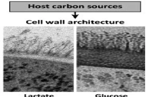
C. albicans displays unique properties in its adaptation process. It is able to take advantage of its interaction with the host to use all available host niches [57]. This adaptation system provides key factors to assist C. albicans during the transition (from commensal to pathogenic) through fungus cell wall modification, me-tabolism, and shape conversion [57]. When infectious conditions are available, C. albicans uses the host nutrition to produce the most virulent behaviours. For example, changing the sugar (carbon) source has been found to modulate C. albicans resistance by modifying the cell wall architecture through specific remodeling enzymes (figure 1-7) [62]. Moreover, C. albicans acquires many virulence factors during the metabolic adap-tation in the host. Indeed, the change from yeast to hyphal form is affected by glucose. The production of aspartic proteinase (SAP) is also affected by glucose/nitrogen sources [63,64]. C. albicans can also bypass the host immune system surveillance. This may involve cell wall alteration, which can influence host immune sensing of PAMPs (figure 1-8) [57,65].
18
Figure 1-7: Changes in carbon source lead to major changes in cell wall architecture. This figure is adapted from Brown A and Brown G. Trends in microbiology. 2014 pii: S0966-842X(14)00139-5.[57]
Figure1- 8: Changes in carbon source impact on immune surveillance by altering the recog-nition of C. albicans cells. This figure is adapted from Brown A and Brown G. Trends in microbiology. 2014pii:S0966-842X(14)00139-5.[57]
19
1.1.4.4 C. albicans and host immune response
C. albicans can dominate the different host tissues to reinforce its presence as an infection [59]. C. albicans escapes from the innate immune system by modulating and binding to different regulators such as H and H-like protein 1 factors [59]. C4BP and host extracellular matrix (ECM) proteins undergo lysis via plasmin stim-ulation through the binding of Gpm1 to plasminogen which later binds to C. albicans surface protein GPM1 (phosphoglycerate mutase1, which has glycolysis and plays a glyconeogenic role), factor H, and FHL-1 [59]. Furthermore, C. albicans also takes advantage of vitronectin (a part of ECM) to adhere and to grow [59].
Neutrophils are the most active cells against C. albicans. This involves different recognition receptors such as TLR2, TLR4, and mannose receptor (N-mannan) [66]. The binding of PRRs by their PAMPs leads to fungus recognition through various pathways that trigger protein profile changing (table 1-3). Interestingly, a discriminatory recognition pathway of C. albicans was discovered during infection and linked to the presence of a hyphal cell threshold promoting epithelial MAPKinase pathway activation (figure 9) [66].
Following C. albicans infection, epithelial cells secrete high levels of cytokines and chemokines, including IL-1α/β, IL-6, G-CSF, GM-CSF, TNF, RANTES, IL-8, and CCL20, and produce IL-12, IL-1α/β and TNF-α [67]. Of course, many other cells (neutrophils, dendritic cells, and T cells) contribute to the immune defense against C. albicans infection by increasing their anti-Candida properties [67].
20
Table 1-3: pattern recognition receptor that sense fungal –associated PAMPs. This figure is adapted from Moyes D and Naglik J. Clinical & developmental immunology. 2011;2011:1-9. [66]
1.1.5 C. albicans and oral cavity
In healthy conditions, the presence of C. albicans in the oral cavity is part of normal flora alongside many bacteria to maintain a microbial balance [68]. However, in certain conditions, this balance can be deregulated, resulting in infection in the oral cavity [69]. Many oral infections are caused by C. albicans. For example, one oral disorder caused by C. albicans is denture-related stomatitis which is an adhesion of C. albicans to den-tures and the formation of biofilms leading to chronic inflammation [69]. Denture-related stomatitis is affected by patient age, smoking, oral hygiene, and the presence of yeast [69,70]. It is identified as an oral disorder known as oral candidiasis.
Family
Receptor
PAMP
TLRs TRL2 Phosoholipomannan
TRL3 Double-stranded RNA
Mannan
TRL4 O-linked MAnnan residues
TRL9 CPG DNA
CLRs Dectin-1 Β-1,3-glucan
Dectin-2 High-mannose structures
Α-mannan
Mannose receptor Mannan
MINCLE Unknown
Galectin-3 Β-1,2-Mannosides
DC-SIGN High-mannose structures
NLRs NLRP3 Unknown
21
1.1.5.1 Oral candidiasis
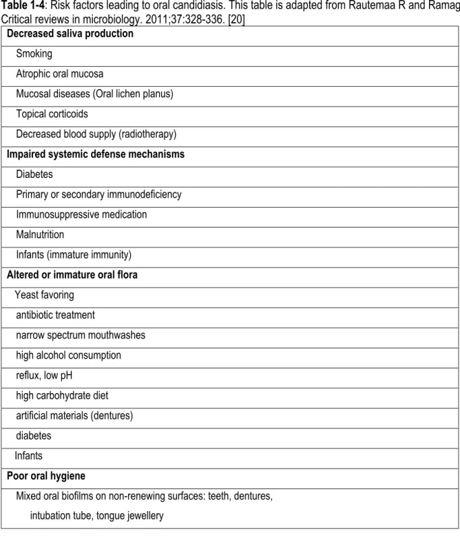
Oral candidiasis is an oral infection most often caused by C. albicans which forms white patches and inflam-mation on the oral mucosa and can cover the tongue and other oral regions. This is due to the presence of large quantities of C. albicans in the biofilm. C. albicans overgrowth and biofilm formation is also the conse-quence of a reduced immune response (Table 1-4) [20]. For example; the compromised immune system in HIV patients is the primary reason for frequent C. albicans colonization causing oral candidiasis through the combination of fungi and bacteria [71]. Oral candidiasis groups pseudomembranous candidiasis or thrush, erythematous candidiasis, characterized as a red patch on the upper posterior part of the tongue, and hyper-plastic candidiasis, identified as small, smooth, white areas changing to larger, dense, rough, and opaque plaques [71].
22
Table 1-4: Risk factors leading to oral candidiasis. This table is adapted from Rautemaa R and Ramage G. Critical reviews in microbiology. 2011;37:328-336. [20]
Decreased saliva production
Smoking
Atrophic oral mucosa
Mucosal diseases (Oral lichen planus) Topical corticoids
Decreased blood supply (radiotherapy)
Impaired systemic defense mechanisms
Diabetes
Primary or secondary immunodeficiency Immunosuppressive medication Malnutrition
Infants (immature immunity)
Altered or immature oral flora
Yeast favoring antibiotic treatment
narrow spectrum mouthwashes high alcohol consumption reflux, low pH
high carbohydrate diet artificial materials (dentures) diabetes
Infants
Poor oral hygiene
Mixed oral biofilms on non-renewing surfaces: teeth, dentures, intubation tube, tongue jewellery
23
1.1.6 C. albicans and risk factors
Oral candidiasis can be caused by a disruption in the oral microflora that affects oral C. albicans carriage and impairs local defense mechanisms. This disruption occurs following a decrease in saliva production, as well as by systematic diseases, antibiotic treatment, and tobacco smoke [21].
1.1.6.1 Saliva and oral candidiasis
Saliva has a role in fighting C. albicans infection. Because saliva contains many proteins, it can fight the presence of C. albicans infection with an anti-fungi composition [72]. The presence of C. albicans in the oral cavity for a long time due to decreased saliva flow can make oral candidiasis infection easier [73]. It has been demonstrated that placing epithelial cells with saliva inhibits C. albicans adhesion to these cells [74]. Thanks to specific adhesion proteins, C. albicans cannot adhere to free saliva [34]. Saliva thus has at least two major roles:
1- Continuous saliva flow contributes to eliminating non-adherent fungus and promotes the clearance activity [42]. Saliva composition also has an effect on C. albicans colonization; for example, the quan-tity of glucose present can affect the tendency by C. albicans to grow better, which explains the high probability of oral candidiasis in diabetic patients.
2- Saliva contains several non-specific defense factors capable of modulating the presence of C. albi-cans [75]. Both low salivation and low saliva quality contribute to C. albialbi-cans binding between cells and denture acrylic surfaces and increase the proliferation of the fungus [42]. Thus any decrease in saliva flow or volume will promote C. albicans adhesion and colonization in the oral cavity. Further-more, a decrease in saliva quantity and quality alters the oral microflora which play a role in Candida
24
resistance to antifungal treatments [72,74]
1.1.6.2 Systematic diseases and oral candidiasis
Another factor contributing to C. albicans overgrowth is systematic disease. It has been demonstrated by many studies that 90% of HIV patients suffer from oral candidiasis [76]. Connections were found between oral candidiasis and immunologic mechanisms (such as T cells) in AIDS patients [77]. Another systematic disease that promotes oral candidiasis is diabetes mellitus due to the reduced capacity of neutrophils to phagocytose and kill C. albicans [78,79]. Moreover, oral candidiasis in persons with diabetes mellitus is the result of an increased glucose level in the blood and saliva which renders the oral cavity more attractive to C. albicans colonization, in addition to the host’s suppressed immune system [80]. Children who have leuke-mia reportedly suffer from oral Candida infections, which may be due to the leukeleuke-mia itself or to the chemo-therapy and/or radiochemo-therapy treatments which have an impact on immune dysfunction leading to C. albicans colonization and infection [81].
1.1.6.3 Antibiotic treatment and oral candidiasis
Another cause of oral candidiasis is the inadequate use of antibiotic treatments. It has been found that anti-biotic treatment (wide-spectrum antianti-biotics) leads to C. albicans infection which takes advantage of a dis-rupted oral microflora [82,73]. This may be due to an inhibition of the anti-fungal immunity, including de-creased phagocytosis which disrupts the equilibrium between C. albicans carriage and normal flora [83,84]. Dysregulation leading to oral candidiasis can also be promoted by external agents such as smoking.
25
1.1.6.4 Tobacco smoke and oral candidiasis
Cigarette smoke (CS) has been found to have a strong impact on oral cavity microflora leading to microbiome deregulation which may promote the growth of pathogenic microorganisms [85]. For example, CS has been shown to increase biofilm formation [86]. Several studies have suggested a link between oral Candida car-riage and CS, as a strong percentage of smokers carry Candida, in contrast to non-smokers. It was suggested that CS provides an appropriate environment for Candida to grow and colonize the host. Furthermore, CS constituents such as aromatic hydrocarbon contents may serve to feed C. albicans [87]. CS-induced oral candidiasis may thus occur through a decrease in gingival cell immune activities. Indeed, CS reduces the level of oral antimicrobial peptide activity [87,88] which in turn leads to C. albicans growth and biofilm for-mation [87,86]. CS also appears to promote the production of lysis enzymes (such as phospholipase) by C. albicans, thereby contributing to tissue degradation as well as C. albicans invasion [89]. C. albicans viru-lence following exposure to CS can occur through gene activation. Indeed, HWP1, EAP1, and SAP2 gene expression was shown to increase in the presence of CS [90]. Thus CS can trigger oral candidiasis by acti-vating various pathways that not only induce C. albicans virulence but alter the host’s defense response. CS may also affect the interaction between C. albicans and host/cells, resulting in C. albicans overgrowth and infection [90,87].
26
1.1.7 Cigarette smoke (CS)
CS contains approximately 4000 chemicals (including chemical toxicants) which are found in the cigarette itself and include cigarette burning. Moreover, two types of smoke are achieved through the burning phase: the main stream (filtered) that the smoker produces by inhaling the cigarette and the side stream (unfiltered) which is the smoke emanating from the end of the cigarette bar (second hand smoke). Nicotine (another cigarette component) is responsible for the addiction. Other chemicals (N-nitrosamines, carbon monoxide, aldehydes hydrogen cyanide, polyaromatic hydrocarbons, and nitrogen oxides) are also present in cigarette smoke and are proven harmful [91,92].
1.1.7.1
Effect of cigarette smoke on human health
The different chemicals present in cigarettes are reported to be cytotoxic, mutagenic, carcinogenic, or anti-genic (innate and adaptive immunity). Entering the human body in different ways, including the oral cavity, nasal passages, and airway mucosa, these chemicals are reported to be responsible for genetic alterations in cell cycle, DNA damage, and tumor suppressor gene dysregulation (figure 1-9) [93,88]. Smoking is re-sponsible for 5 million human deaths each year [94].
27 Figure 1-9: The harmful components of the cigarette. This figure is adapted from Journal of dental
research. 2012;91:142-149. [93]
Burning one cigarette produces thousands of reactive oxygen species (ROS) which can destroy airway epi-thelial cell linings through the activation of intracellular signaling pathways (figure 1-10), induce the inflam-matory response by increasing IL-8 and TNFα secretion, and finally, deregulate neutrophil function and fibro-blast activity. CS also contributes to the activation of the inflammatory response through Toll-like receptors (TLRs), mitogen-activated protein kinases (MAPK), nuclear factor kappa-B (NF-κB), signal transducer and activator of transcription (STAT), and activator protein-1 (AP-1) [93,88]. Immune system dysregulation may thus explain cancer development in smokers [88].
28
Figure1-10: Cigarette smoke modulates inflammation and promotes chronic inflammation in the con-ducting airways by a variety of mechanisms. This figure is adapted from Journal of dental research. 2012;91:142-149. [93]
1.1.7.1.1 Cigarette smoke and cancer
Research has shown that smoking is responsible for 25% of deaths in men and 4% of deaths in women by cancer. Tobacco-related cancers can be found in the kidneys, liver, stomach, uterine cervix, and breasts [95]. For example, several studies showed that breast cancer develops more in long-term female smokers than in non-smokers. Indeed, tobacco smoke can contribute to metastasis in the lung, as breast cancer cells are capable of adhering to the lung endothelium via endothelial PAF (platelet-activating factor) [96]. Multiple studies also report a link between tobacco and other cancers, such as esophageal cancer, laryngeal cancer and oral cancer [98].
29
1.1.7.1.2 Cigarette smoke and heart diseases
In the United States, cigarette smoke is responsible for 30% of deaths by coronary heart disease (CHD) [99] and incriminated in cardiovascular disease (CVD). Exposure to cigarette smoke in the form of second hand smoke also raises the incidence of cardiovascular disease [100]. Heart surgery failures, myocardial re-infarc-tions, and death are also exacerbated by smoking [100]. CS also causes coronary thrombosis, as it alters the role of endothelial cells, platelets, fibrinogen, and coagulation factors and leads to an altered equilibrium of the antithrombotic/prothrombotic and profibrinolytic/antifibrinolytic factors involved in heart attacks [101].
1.1.7.1.3 Cigarette smoke and respiratory diseases
Smoking is highly involved in respiratory diseases and causes significantly more health problems than does cancer [95]. The entire lung organ is affected by cigarette smoke, which promotes chronic obstructive pul-monary disease (COPD). Smokers run a greater risk of infection and lung dysfunction. Indeed, smokers have been shown to be more sensitive to respiratory tuberculosis, influenza, and pneumonia, with a greater per-centage of deaths compared to non-smokers [95]. This is probably due to a reduced immune defense caused by smoking [98].
1.1.7.1.4 Cigarette smoke and oral diseases
The oral cavity is the first organ in contact with CS which is responsible for many oral diseases, including cancer. Oral cancers were found to increase 3 to 5 times more among smokers than in non-smokers and also in a dose-depending manner (number of smoked cigarettes/day and smoking years) [94]. CS promotes
30
periodontal disease (half caused by smoking). Furthermore, CS decreases tissue healing following oral sur-gery [102]. Moreover, much evidence suggests a link between smoking and tooth loss, inadequate dental restorations, and dentures changing color. Smoking also affects taste, promotes tooth decay [94], and pro-motes chronic gingivitis (inflammation and gingival bleeding) and acute necrotizing ulcerative gingivitis [94,103]. CS also alters the oral microflora, converting non-pathogenic C. albicans to pathogenic, resulting in oral candidiasis [94].
31
1.1.8 Hypotheses and objectives
Hypotheses
1. C. albicans may take advantage of the various products present in cigarette smoke to adhere, grow, and increase its infective capacity.
2. Cigarette smoke can reduce gingival cell defense reponses against C. albicans infection.
Objectives
Although some groups have reported a link between cigarette smoke and oral candidiasis, multiple questions remain as to the direct effect of cigarette smoke on C. albicans and human cells, and human cell interactions with C. albicans when present in the oral cavity. Our specific objectives were thus to investigate
1. The effect of cigarette smoke condensate (CSC) on C. albicans adhesion/growth and transition from blastospore to hyphal form;
2. The effect of CSC on C. albicans chitin production;
3. The effect of cigarette smoke-pretreated C. albicans on adhesion and growth following contact with gingival fibroblasts; and
4. The effect of cigarette smoke-pretreated C. albicans on gingival fibroblast proliferation and cytokine
secretion.33
Chapter ΙΙ
Publication:
Cigarette smoke-exposed Candida albicans increased chitin production and
modu-lated human fibroblast cell responses
Humidah Alanazi, Abdelhabib Semlali, Laura Perraud, Witold Chmielewski, Andrew
Zakrzewski and Mahmoud Rouabhia
Biomed Res Int; 2014; 2014: 963156. doi: 10.1155/2014/963156.
Epub 2014 Sep 11.
PMID: 25302312
34
Abstract
The predisposition of cigarette smokers for development of respiratory and oral bacterial infections is well documented. Cigarette smoke can also contribute to yeast infection. The aim of this study was to investigate the effect of cigarette smoke condensate (CSC) on C. albicans transition, chitin content, and response to environmental stress, and to examine the interaction between CSC-pretreated C. albicans and normal human gingival fibroblasts. Following exposure to CSC, C. albicans transition from blastospore to hyphal form in-creased. CSC-pretreated yeast cells became significantly (p<0.01) sensitive to oxidation, but significantly (p<0.01) resistant to both osmotic and heat stress. CSC-pretreated C. albicans expressed high levels of chitin, with 2 to 8 folds recorded under hyphal conditions. CSC-pretreated C. albicans adhered better to the gingival fibroblasts, proliferated almost three times more, and adapted into hyphae, while the gingival fibro-blasts recorded a significantly (p<0.01) slow growth rate but a significantly higher level of IL-1 when in contact with CSC-pretreated C. albicans. CSC was thus able to modulate both C. albicans transition through the cell wall chitin content as well as the interaction between C. albicans and normal human gingival fibro-blasts. These findings may be relevant to fungal infections in the oral cavity in smokers.
35
2.1 Introduction
Mucosal candidiasis, especially the oropharingeal type (OPC), is a common opportunistic infection in both immunocompromised and immunocompetent persons [1]. The leading cause of candidiasis is Candida albi-cans, a commensal dimorphic yeast that colonizes up to 60% of normally healthy individuals [2]. Symptomatic OPC appears under a number of predisposing conditions [3, 4]. From these, tobacco smoking was consid-ered predisposing to oral candidiasis [5]. Indeed, epidemiological studies in immunocompromised patients have identified tobacco as a major risk factor for symptomatic infection [6, 7]. Furthermore, the rate of oral candida carriage in tobacco smokers was reported to be higher in smokers than in non-smokers [8, 9]. This may explain why 98% of Indian villager’s smokers suffer candidal leukoplakia that can be resolved after cessation of tobacco [10, 11].
The exact mechanism by which candidal carriage may be affected by tobacco is still to be discovered. Pre-vious studies have demonstrated that smoking lead to innate immune reduction facilitating candida coloniza-tion and host infeccoloniza-tion [12]. This may suggest the use of tobacco compounds by C. albicans as nutricoloniza-tional factors, since aromatic hydrocarbons contained in cigarette smoke can be converted by Candida species to carcinogen end products [13]. It has also been reported that C. albicans can catalyze the formation of N-nitrosobenzylmethylamine supporting the high candidal leukoplakia level in smokers [14].
During the development of candidiasis, C. albicans adheres to and invades the tissue [15, 16]. Tis adhesion is promoted by yeast cell wall proteins [17]. Candida cell walls contain glucans, mannans, glycoproteins and chitins [18-20]. In C. albicans, three genes encoding different chitin synthases (CHS1, 2 and 3) were identi-fied. Chitin production is dependent on the experimental culture condition. Indeed, the CHS2 and CHS3 are preferentially expressed under hyphal culture condition [21, 22], whereas, CHS1 remained at low levels in
36
both yeast and hyphae [23]. Interestingly, Chs1p was found to be essential for cell integrity and virulence [23]. Mutants defective in chitin proteins are less virulent than the parental strain in a mouse model. Thus, with high level of chitin, C. albicans can overcome cigarette smoke effects, and escape the host immune defense [22].
Gingival fibroblasts are major actor in the host immune defense against Candida infection [24, 25]. Gingival fibroblasts play role in tissue structure and function [26]. They are active in the inflammatory response by secreting inflammatory cytokines, such as 6 and 8, as well as
IL-periodontopathic bacteria [27, 28]. The secretion of these cytokines may, thus be modulated when fibroblasts are exposed to cigarette smoke.
Exposure to smoke-derived toxicants has been shown to lead to immune dysfunction [29]. Smokers thus run a greater risk of contracting invasive diseases caused by various bacterial pathogens [30-32]. Exposure to cigarette smoke has also been shown to induce the formation of biofilm by various oral/respiratory pathogens in vitro, including Porphyromonas gingivalis, Staphylococcus aureus, Streptococcus. pneumoniae, Klebsiella pneumonia and P. aeruginosa, as well as Streptococcus mutans [30-32]. Cigarette smoke was also reported to increase C. albicans adhesion and growth, as well as biofilm formation [33, 34]. These effects were sup-ported by the overexpression of EAP1, HWP1, and Sap2 genes known to be active players in C. albicans virulence [34]. By modulating C. albicans adhesion, proliferation and biofilm formation in vitro, cigarette smoke may also modulate the yeast interaction with human cells such as fibroblasts. The aim of this study was thus to investigate the effect of cigarette smoke condensate on C. albicans transition from blastospore
37 to hyphal form, its response to environmental stress, its production of cell wall chitin, and its interaction with human gingival fibroblasts.
2.2 Experimental protocol
2.2.1 Candida albicans: C. albicans SC5314, known to be strongly invasive [35] was cultured for 24 h on Sabouraud dextrose agar plates (Becton Dickinson, Oakville, ON, Canada) at 30°C. For the C. albicans suspensions, one colony was used to inoculate 10 ml of Sabouraud liquid medium supplemented with 0.1% glucose, pH 5.6. The cultures were then grown in a shaking water bath for 18 h at 30°C, after which time the yeast cells were collected, washed with phosphate-buffered saline (PBS), counted by means of a haemocy-tometer, and adjusted to 106/ml prior to use.
2.2.2 Gingival fibroblast isolation and culture: Small biopsies of lamina propria tissue (gingival connective tissue) were collected from healthy non-smokers (18–25 years old) following their informed consent in ac-cordance with Laval University Ethics Committee guidelines. To isolate the gingival fibroblasts, the connec-tive tissue was placed in a collagenase P solution (0.125 U/ml; Boehringer Mannheim, Laval, QC, Canada) for 45 min at 37°C under gentle agitation. The isolated cells (2 × 10⁵) were seeded in 75-cm2 flasks (Falcon,
Becton-Dickinson, Cockeysville, MD, USA) and grown in Dulbecco’s modified Eagle’s (DME) medium (Invi-trogen) containing 10% fetal calf serum (Invitrogen Canada Inc., Burlington, ON, Canada). Upon 90% con-fluence, the gingival fibroblasts were used between passage two and four for the different experiments.
2.2.3 Preparation of the cigarette smoke condensate: 1R3F cigarettes were purchased from the Kentucky Tobacco Research & Development Center (Orlando, FL, USA) and used to prepare the cigarette smoke condensate solution. Each cigarette was placed into one end of a silicone tube linked to an Erlenmeyer flask
38
containing 200 ml of 0.09% sodium chloride. On the other end, a second silicone tube linked to the Erlen-meyer was connected to a standard vacuum. The cigarette was attached to the cigarette holder and lit and the smoke was extracted by applying vacuum, pulling the smoke directly into the 0.09% sodium chloride solution. The procedure was repeated with a total of ten whole cigarettes. The resulting cigarette smoke condensate (CSC) solution was then sterilized by filtration through a 0.22-μm filter and subsequently stored at 4°C until use.
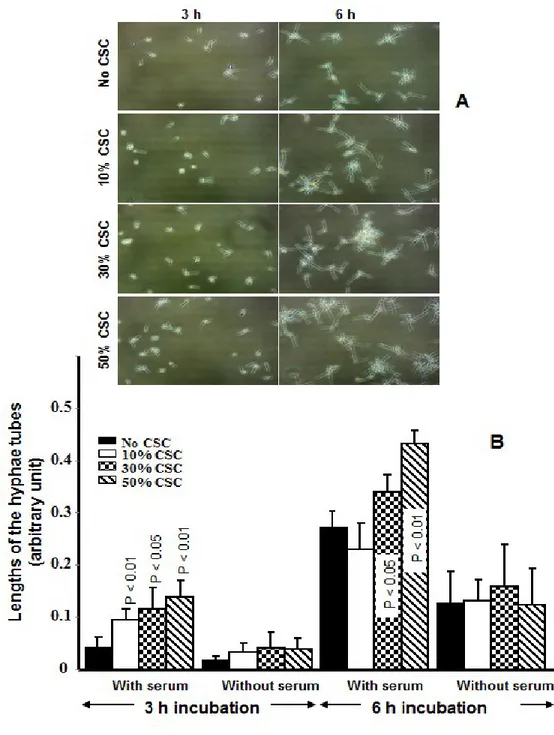
2.2.4 Effect of CSC on C. albicans transition from blastospore to hyphal form: To determine the effect of CSC on the yeast-to-hyphae transition we used qualitative and quantitative assays. C. albicans (105 cells)
was grown in 3 ml of Sabouraud dextrose broth supplemented with 0.1% glucose and 10% fetal bovine serum (FBS) with or without CSC at various concentrations (10, 30, and 50%). The negative controls refer to the C. albicans cultures without CSC. Different conditions were tested: (1) the hyphae-inducing conditions were previously reported [30], consisting of culture medium supplemented with 10% fetal calf serum followed by incubation at 37°C; (2) culture at 37°C, without serum; (3) culture in the presence of 10% serum at 30°C; and (4) culture at 30°C without serum. Following incubation for 3 or 6 h, the cultures were observed micro-scopically and photographed to record C. albicans morphology (n = 5). The density of the C. albicans transi-tion was also measured. Furthermore, the length of the hyphal forms in each conditransi-tion was measured by means of NIH-ImageJ software.
2.2.5 Effect of CSC on C. albicans response to stressful agents: To investigate the effect of CSC on C.
albicans sensitivity/resistivity, we exposed the yeast cells to oxidative, osmotic or heat stress. To do so, C. albicans cells were cultured in Sabouraud with or without CSC at various concentrations (10, 30, or 50%). The culture medium was adjusted to have the same level of nutriments as did the control (absence of CSC).
39 Cultures were incubated in a water bath at 30°C for 24 h under agitation. Following the incubation, the cells were counted with a haemocytometer, after which time 106 cells/ml were treated with hydrogen peroxide (5,
10 or 50 mM) or NaCl (1.2 M) or heat treated at 45°C. Incubation under oxidative, osmotic or heat stress took place for 60 min under agitation. The cells were then washed twice with sterile PBS and suspended thereafter in Sabouraud medium at 106cells/ml. The sample volume referring to 103 or 104 of each condition was spotted
on Sabouraud agar and incubated at 30°C and colony growth was monitored at 24 and 48 h. Culture on a PDA plate lacking the disrupting agents was also performed and used as a growth control. Cell viability following contact with oxidative, osmotic or heat stress was evaluated by means of the (3-(4,5-dimethylthia-zol-2-yl)-2,5-diphenyl tetrazolium bromide) (MTT) assay (Sigma-Aldrich, St. Louis, MO, USA) which measures viable cells as a function of mitochondrial activity as we previously reported [34]. Absorbance (optical density, OD) was subsequently measured at 550 nm by means of an xMark microplate spectropho-tometer (Bio-Rad, Mississauga, ON, Canada).
2.2.6 Effect of CSC on C. albicans cell wall chitin content.
2.2.6.1 Cell wall isolation and purification: C. albicans in Sabouraud medium was cultured overnight in the presence or absence of CSC at various concentrations (0, 10, 30, or 50%). Sabouraud medium was supple-mented or not with 10% fetal calf serum for hyphae culture conditions. Cells were collected by centrifugation at 1200 rpm for 10 min and were then used to extract cell wall proteins. The C. albicans pellets were sus-pended in 500 l of sterile PBS and supplemented with 200 l of glass beads (0.425-0.6 mm in diameter; Sigma-Aldrich, G9268). The samples were cooled at -80°C for 60 sec before being subjected to disruption by means of a MiniBead-beater (Biospec Products, Bartlesville, OK, USA) for 2 min at 5,000 rpm. The sam-ples were then cooled for 2 min at -80°C and the disruption cycle was repeated 15 times. The cells were then washed twice with 1 M NaCl and extracted thereafter in an SDS-MerOH extraction buffer (50 mM Tris, 2%