HAL Id: inserm-00679701
https://www.hal.inserm.fr/inserm-00679701
Submitted on 16 Mar 2012HAL is a multi-disciplinary open access
archive for the deposit and dissemination of sci-entific research documents, whether they are pub-lished or not. The documents may come from teaching and research institutions in France or abroad, or from public or private research centers.
L’archive ouverte pluridisciplinaire HAL, est destinée au dépôt et à la diffusion de documents scientifiques de niveau recherche, publiés ou non, émanant des établissements d’enseignement et de recherche français ou étrangers, des laboratoires publics ou privés.
permeabilization by the electric field and/or the
Pluronic® L64 in vitro and in vivo.
Michel Bureau, Luc Wasungu, Lauriane Jugé, Daniel Scherman, Marie-Pierre
Rols, Nathalie Mignet
To cite this version:
Michel Bureau, Luc Wasungu, Lauriane Jugé, Daniel Scherman, Marie-Pierre Rols, et al.. Investigating relationship between transfection and permeabilization by the electric field and/or the Pluronic® L64 in vitro and in vivo.. The Journal of Gene Medicine, Wiley, 2012, 14 (3), pp.204-15. �10.1002/jgm.2610�. �inserm-00679701�
Investigating relationship between transfection and permeabilization by the
electric field and /or the pluronic
®L64, in vitro and in vivo
Short title : Transfection vs permeabilization relationship
Michel F. Bureau*abcd, Luc Wasunguef, Lauriane Jugégd, Daniel Schermanabcd, Marie-Pierre
Rolsef, Nathalie Mignetabcd
a Unité de Pharmacologie chimique et Génétique et d’Imagerie : CNRS UMR 8151, Paris, F-75270. France
b INSERM U 1022, Paris, F-75270. France
c Université Paris Descartes, Faculté des Sciences Pharmaceutiques et Biologiques, Paris, F-75270. France
d Paristech, Paris, F-75270. France
e CNRS, IPBS (Institut de Pharmacologie et de Biologie Structurale), Toulouse, F-31077, France f Université de Toulouse. UPS, IPBS Toulouse, F-31077, France
g Laboratoire de RMN, ENSCP, Paris, F-75270. France
*
Corresponding author: Unité de Pharmacologie Chimique et Génétique & d’Imagerie,INSERM U1022, CNRS UMR8151, Faculté de Pharmacie 4 av de l’Observatoire, 75006, Paris, France.
Tel :+33 1 53739581 ; fax : +33 1 43266918
Abstract
Background
Electrotransfer can be obtained by the successive delivery of a high voltage short duration pulse (HV) inducing membrane destabilization then a low voltage long duration pulse (LV) allowing DNA electrophoresis (HVLV mode). Pluronic L64 (L64) has permeabilizing properties and amplifies expression of DNA. We wondered whether L64 could have an adjuvant effect on transfection by electrotransfer and whether the sequence L64 injection then application of a LV pulse could induce transfection comparable to that observed with the HVLV mode.
Methods
In vitro, we used fluorescence-activated cell sorting to evaluate the CHO cell transfection by a
plasmid coding GFP, and permeabilization to propidium iodide. In vivo, the transfection efficiency of mice tibial cranial muscle was evaluated by optical imaging using a plasmid DNA encoding luciferase. For the same animals, permeabilization indices were evaluated by magnetic resonance imaging from the uptake of a T1 contrast agent.
Results
Using the HVLV mode, transfection efficiency was low in vitro on CHO cells but high for muscles in vivo. Pre-treatment by L64 increased transfection efficiency of electrotransfer for CHO cells but not for the muscle. In mice muscles the L64 amplified the expression of DNA. Nevertheless, neither transgene expression nor permeability indices were further amplified by subsequent delivery of one LV pulse.
Conclusion
A major finding is that the nature of the membrane modification induced by electric pulses is not comparable to the one mediated by L64. The electrophoretic LV pulse does not induce additive effects to that of L64 for transfection improvement.
1 Introduction
Transfection of muscle using electric field application was developed in 1998 [1, 2] and is now a well-known, efficient procedure [3-6] but whose mechanisms remains largely unknown [7]. In addition, electrotransfer safety needs improvement, studies having shown that it induces an inflammation for 2 weeks after treatment [8] and can induce pain when high current intensities are applied [9, 10]. It has been proposed that electric field efficiency for DNA transfer through cell membranes results from two effects: (i) membrane modification as evidenced by its
permeabilization to small molecules and (ii) DNA electrophoresis [11]. On the muscle model in
vivo we have shown that the two effects can be combined by using the successive delivery of one
high-voltage (HV) short-duration pulse, which is exclusively permeabilizing, and a low-voltage (LV) pulse of long duration, which is not permeabilizing but can induce DNA electrophoresis (HVLV mode) [12, 13]. Such a procedure is now also practically used for the transfection of other tissues than muscle [14-16]. More recently various copolymers of polyethylene oxide and polypropylene oxide have been shown to be efficient agents for improving muscle transfection [17-19] to a level comparable to that of electrotransfer [20]. These copolymers are chemically neutral and do not interact with DNA [21]. Physical studies with membrane models or liposomes showed that these copolymers interact with lipids with their more hydrophobic part,
polypropylene oxide [21-23]. However, observation of labeled copolymers and DNA incubated with cells did not allow evidencing the copolymer at the level of the cell membrane, but rather inside the cells as macromolecular clusters [24, 25]. Moreover, copolymers were shown to improve transfection by acting on DNA nuclear import and its transcription through NFkB activation [26]. Among these copolymers, we chose the pluronic L64, which exhibits cell
permeabilizing properties on artificial membranes [21, 27], on cells in vitro [28], and in vivo [20]. In a previous study, we had compared muscle transfection and permeabilization, either mediated by the pluronic L64 or electrotransfer [20]. In this study, we combined both pluronic L64 and electrotransfer. To obtain further insights, the present study aimed to determine whether pluronic L64, by its effects on nuclear import of DNA and its transcription, could have an adjuvant effect on transfection by electrotransfer. This had been shown on CHO cells in vitro [24], but had not been studied in vivo on a muscle model. This effect of pluronic L64 has also been demonstrated for in vitro transfection by a cationic polymer, polyethyleneimine [29].
Another question was does permeabilizing properties of the pluronic L64 can be compared to that of the electric field ?
It had been shown in previous studies on the muscle model that the order of the sequence HVLV was critical to amplify DNA transfection. Under conditions of these studies one HV or one LV pulse alone have no effect on DNA transfection as compared to simple DNA injection. For an efficient transfection amplification, DNA may be injected either before or after the HV pulse, and before the LV pulse, which is consistent with a direct effect of the LV pulse on DNA, i.e.
electrophoresis [13]. It was also shown that the LV pulse was efficient only after the HV pulse had been applied, indicating that DNA electrophoresis is useful only when the cell membrane is in a modified state [12, 13]. Therefore, we investigated wether the same behaviour could be observed when the membrane modified state was induced by the pluronic L64. For that, we compared the HVLV mode to the delivery of pluronic L64 followed by a LV pulse (L64LV mode).
In summary we investigated, whether the pluronic L64 combined with electric pulses could improve gene transfection efficiency, and whether its permeabilizing property could be compared to permeabilization induced by electric field application. For this purpose we performed
experiments in vitro on CHO cells and in vivo on muscle, in which transfection and
permeabilization to small molecules were evaluated. In both cases, for the HVLV procedure HV was a brief high-voltage pulse, permeabilizing but not transfecting, and LV a long-duration pulse, not permeabilizing but allowing DNA electrophoresis.
2 Materials and Methods
2.1 Plasmid DNA
The pVAX2 construct was a pVAX1 plasmid (Invitrogen, Carlsbad, CA) in which the promoter was replaced by the pCMVβ plasmid promoter (Clontech, Palo Alto, CA). The pVAX2-luc plas-mid was a 4.6 kb vector encoding a cytosolic firefly luciferase plus protein (luc+) subcloned in the pVAX2 backbone. The expression vector pEGFP-C1 was a 4.7 kb plasmid containing the gene coding the Green Fluorescent Protein (GFP) under the control of the CMV promoter. It was obtained from Clontech. The plasmid DNA was purified from E. coli transformed cells using en-do-free Qiagen Gigaprep kits (Qiagen, Courtaboeuf, France), and then diluted in saline (0.9% NaCl). The quality of the plasmid was assessed by calculating the ratio of light absorption (260 nm/280 nm) and by visualization on ethidium bromide-stained 1% agarose gel. Plasmid DNA was supercoiled for at least 80% of the preparation. Light absorption at 260 nm was used to de-termine DNA concentration.
2.2 Polymer
Pluronic L64 (Fluka, Sigma-Aldrich, L'Isle-d'Abeau Chesnes, Saint-Quentin Fallavier, France) is a copolymer of propylene oxide (PO) and ethylene oxide (EO). Its formula is (EO)13(PO)30(EO)13.
For in vivo experiments, pluronic L64 solutions were prepared on the day of the experiment as described previously [20]. The final injected solution (30 µL) contained: 4.8 µg of DNA, 3.33 µmoles of Gd-DOTA and 0 or 0.25% pluronic L64, according to the experiment. We chose the concentration of 0.25% in pluronic L64 because we previously showed that this concentration was optimal for transfection amplification in the mice muscle [20].
2.3 In vitro experiments 2.3.1 Cell culture
Chinese hamster ovary (CHO) cells were grown in Eagle’s minimum essential medium (EMEM; Gibco-Invitrogen, Carlsbad, CA, USA) supplemented with 8% fetal calf serum (Gibco),
100 units/ml of penicillin and 1 mg/ml of streptomycin (Gibco-Invitrogen), 0.58 mg/ml of L
-glutamine (Eurobio, France) and vitamins (vitamin solutions from Ginco-Invitrogen, see manufacturer for composition). The cells were maintained in a 5% CO2 humidified incubator at
37 °C (Jouan, Saint-Herblain, France).
2.3.2 Electric pulse delivery in vitro
Electrotransfection was performed using a CNRS cell electropulsator (Jouan, Saint-Herblain, France). Alternatively, for high-voltage and low-voltage electrical conditions a Cliniporator generator (IGEA, Carpi, Italy) was used. Stainless steel flat parallel electrodes 2 cm in length set 4 mm apart were used. For all experiments in vitro (as in vivo) electric field intensity was estimated as the voltage divided by the distance between the plate electrodes. Electropulsation was applied on 500,000 CHO cells suspended in 100 µl of pulsation buffer (10 mM K2HPO4/
KH2PO4 buffer, 1 mM MgCl2, 250 mM sucrose, pH 7.4, conductivity 1.6 mS/cm) to which was
added 1 µl of plasmid DNA pEGFP-C1 solution (1µg/µl). Several electrical parameters were tested as described in table 1. After pulsation the cells were transferred to a 35 mm Petri dish with 1.5 ml of growth medium and kept at 37 °C in a 5% CO2 atmosphere.
For experiments with Pluronic L64 0.25%, cell pre-treatment, electropulsation was carried out on adherent CHO cells plated on a 24-well plate at a density of 50,000 cells per well 16 h before experiments, according to a procedure described previously [24]. Briefly, electropulsation was carried out directly in the well on adherent cells with stainless steel flat parallel electrodes (length 1 cm, set 1 cm apart). The electric field intensity used for optimal transfection was lower than for cell suspension because of the larger size of adherent cells. Conditions tested are described in table 1. For all experiments transfection efficiency was given as the percentage of fluorescent cells expressing the GFP and the average fluorescence level of positive cells was determined with a BD FACSCAN flow cytometer (BD Biosciences) 24 h after transfection.
2.3.3 Electropermeabilization of cell suspension in vitro
Electropermeabilization was studied with pulse conditions identical to the one used for
electrotransfection of cells in suspension. The pulsation buffer contained 100 µM of propidium iodide (PI, Sigma Chemical Co. St. Louis, USA) and no plasmid DNA. Directly after
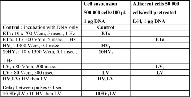
electropulsation, cells were transferred to 300 µL of PBS and analyzed by flow cytometry with a BD FACSCAN flow cytometer (BD Biosciences). The efficiency of permeabilization was reported as the percentage of PI positive fluorescent cells and the average fluorescence level of these cells. Cell suspension 500 000 cells/100 µl, 1 µg DNA Adherent cells 50 000 cells/well pretreated L64, 1 µg DNA Control : incubation with DNA only Control
ETs: 10 x 700 V/cm, 5 msec., 1 Hz ETs
ETa: 10 x 500 V/cm, 5 msec., 1 Hz ETa
HV1 : 1300 V/cm, 0.1 msec. HV1 10HV1 : 10 x 1300 V/cm, 0.1 msec., 1 Hz 10HV1 LV0 : 80 V/cm, 200 msec. LV0 LV : 80 V/cm, 500 msec. LV LV HV1LV: HV then LV
Delay between pulses 0.1 sec
HV1LV
10 HV1LV : 10 HV then LV 10HV1LV
Table 1: Different modalities of electric pulses delivery used in vitro. For all experiments,
temperature was set at 37°C.
2.3.4 Cell viability
Cell viability was measured by monitoring cell growth through coloration with crystal violet [30]. After staining with crystal violet and cells lysis the absorbance at 590 nm was measured with a spectrophotometer (Pharmacia Biotech). The viability was expressed by the percentage of coloration obtained as compared to cells treated in the same way but without application of an electric field.
2.4 In vivo experiments 2.4.1 Animals
In vivo studies were carried out on 6–8-week-old female BalbC/J mice (Janvier, Le
Genest-Saint-Isle, France). Before all procedures (treatment and imaging), animals were anesthetized by intra-peritoneal injection of ketamine and xylazine (Bayer Pharma, Puteaux, France) (100 mg/kg and 10 mg/kg, respectively). The studies were conducted following the recommendations of the European Convention for the Protection of Vertebrate Animals used for Experimentation, and the local Ethics Committee on Animal Care and Experimentation.
2.4.2 General experimental procedure and electric pulse delivery in vivo
For each experiment, the tibial cranial muscle was injected longitudinally with 30 µl of a solution containing DNA coding the firefly luciferase (4.8 µg) and the Gd-DOTA (3.33 µmoles) with or without pluronic L64 (as previously described) by means of an insulin syringe (MYINJECTOR 29Gx1/2, Terumo, Leuven, Belgium). MRI measurements were performed at day 3 and optical imaging at day 7 after injection. In a few experiments only DNA was injected and consequently only measurement of transfection by optical imaging was performed. When required electric pulses were delivered 20 s after plasmid DNA injection through two stainless steel plate
electrodes (10 × 20 mm), placed 4 mm apart at each side of the mouse leg. Electrical contact with the shaved leg skin was ensured by means of a conductive gel (Aquasonic 100, Parker
Laboratories, Fairfield, New Jersey, USA). Electric pulses were generated by a Cliniporator electropulsator (IGEA, Carpi, Italy). Several electrical parameters were tested as described in the table 2. Our procedure of electrotransfer ET that we named standard was used as an efficient condition in many of our previous studies studies [12, 13, 20]. The HVLV condition was derived from our study of 2002 [13] but with a LV of 500 msec. duration in order to accentuate
electrophoretic effect. These optimal conditions for the muscle are different from those used on CHO cells in vitro because of a different threshold for permeabilization. Indeed we have shown in a previous study that 8 pulses of 600 V/cm (1 msec.) at 1 Hz were lesional for the muscle. Thus 10 pulses of 700 V/cm 5 msec. at 1 Hz are not adapted to muscle transfection whether they are optimal for CHO cells [31].
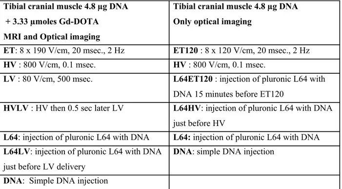
Tibial cranial muscle 4.8 µg DNA + 3.33 µmoles Gd-DOTA
MRI and Optical imaging
Tibial cranial muscle 4.8 µg DNA Only optical imaging
ET: 8 x 190 V/cm, 20 msec., 2 Hz ET120 : 8 x 120 V/cm, 20 msec., 2 Hz
HV : 800 V/cm, 0.1 msec. HV : 800 V/cm, 0.1 msec.
LV : 80 V/cm, 500 msec. L64ET120 : injection of pluronic L64 with
DNA 15 minutes before ET120
HVLV : HV then 0.5 sec later LV L64HV: injection of pluronic L64 with DNA
just before HV
L64: injection of pluronic L64 with DNA L64: injection of pluronic L64 with DNA
L64LV: injection of pluronic L64 with DNA
just before LV delivery
DNA: simple DNA injection DNA: Simple DNA injection
Table 2: Different modalities of electric pulses delivery used in vivo. 2.4.4 MRI acquisition
MRI examinations were performed with a 7 T vertical wide-bore magnet (Bruker) equipped with a standard proton micro-imaging probe and ParaVision 2.1.1 software. Mice were housed and kept upright using a custom-made support of diameter 38 mm. The two hind limbs were
positioned symmetrically and examined simultaneously in transversal planes using a T1-weighted
spin-echo sequence with the following parameters: repetition time (TR) 600 msec., echo time (TE)
10 msec., 12 contiguous slices of thickness 1 mm, field of view 30 mm, spatial resolution
117 × 117 µm² in-plane and acquisition time 20.3 min. The slice centered on the largest signal of interest was chosen for T1 and T2 measurements, using a single slice spin-echo sequence with the
following parameters: TR 3000, 2000, 1200, 800, 600, 500, 400 and 300 msec., eight echoes, first
echo at TE 9 msec., echo spacing 8 msec., field of view 30 mm, in-plane spatial resolution 117 ×
2.4.5 MRI processing
The assay Gd-DOTA trapped in tibialis muscle was obtained from T1-weighted images.
We used two parameters to characterize this uptake: the volume of enhanced T1 MRI signal (permeabilized volume) and the concentration of the contrast agent Gd-DOTA in this volume, calculated from T1 values. In brief, the permeabilized volume is correlated to the number of
muscle fibers that are permeabilized, and the concentration of contrast agent to the amount of compound taken up by those fibers.
The volume labeled by the Gd-DOTA and the concentration of Gd-DOTA in this volume were evaluated as described previously [20].
2.4.6 In vivo optical imaging of luciferase activity
Luciferin potassium salt (SYNCHEM, Felsberg/Altenburg, Germany) diluted in PBS was injected locally into the tibial cranial muscle at a dose of 100 μg/40 μl, which is in large excess relative to the amount of luciferase [32]. Optical imaging was performed with a cooled intensified charge-coupled device (CCD) camera (Biospace, Photo Imager, Paris, France) placed in a black box. Luminescence level was measured in ROI corresponding to the tibial cranial muscle as described previously [20]. ROIs were very similar from one experiment to another. We opted to take the mean values in cpm of all the measurements for 10 min after the start of acquisition [32].
2.4.7 Statistical analysis of in vivo results
Because of the non-Gaussian distribution of the results, we used nonparametric tests: Kruskal-Wallis nonparametric variance analysis of the measured parameters and pairwise Wilcoxon test for comparisons between treatments. In addition, we used the Spearman correlation test to check for correlations between some parameters. Statistical program used were Statview (SAS Institute inc) and R (the R foundation for Statistical Computing).
3 Results
First transfection and permeabilization experiments were performed in vitro on CHO cells then in
vivo on the mice tibial cranial muscle.
3.1 In vitro CHO model
3.1.1 Transfection and cell viability
As expected, electrotransfer applied in appropriate conditions for CHO cells in suspension (ETs: 10x700 V/cm, 5 msec.) allowed for a significant transfection. However, the pulse combination HVLV mode, which was shown to be very efficient for muscle transfection, had a low efficacy on CHO cells in vitro (Fig. 1A) even though HV1 (1300 V/cm, 0.1 msec.) electric field intensity
was well above the threshold to permeabilize cells [33] and LV (80 V/cm, 500 msec.) was at a level and duration sufficient to induce DNA electrophoresis [34].
Transfection Figure 1 A B 0 10 20 30 40 Control ETs HV1LV 10HV1LV 10HV1 0 50 100 150 200 250 ** NS *** *** F lu o re s ce n c e le ve l (A U ) G F P p o s it iv e ce ll s (% ) 0 20 40 60 80 100 120 ETs HV1LV 10HV1LV 10HV1 0 20 40 60 80 100 120 ETa LV0 LV V ia b ili ty V ia b ili ty Viability C D 0 10 20 30 ETa LV0 LV 0 1 2 3 4 5 G F P p o si ti ve c el ls (% ) Not Treated Pretreated with L64 F lu o re s ce n c e le ve l( A U x1 0 3) **
However using 10 HV1 pulses allowed a significant transfection which was not obtained with one
HV1 (data not shown). The addition of a LV pulse to the 10 HV1 pulses did not improve the
transfection. The patterns for the percent of cell transfected or the mean fluorescence intensity per cell were similar. Co-incubation of DNA and pluronic L64 with CHO cells did not result in cell
Permeability Figure 2 0 20 40 60 80 100 120 Control ETs HV1LV 10HV1LV 10HV1 0 100 200 300 400 500 *** *** *** *** NS F lu o re s ce n c e le ve l (A U ) P I p o s it iv e c e lls (% )
transfection (data not shown), as observed by other authors with COS7 cells [18]. However, pre-treatment of adherent CHO cells with pluronic L64 at 0.25 % improved transfection by
electrotransfer (Eta: 10x500 V/cm, 5 msec., 1 Hz) (Fig. 1 B). But, this pre-treatment followed by delivery of a low voltage pulse of long duration (80 V/cm, 200 or 500 msec.) did not induce a detectable transfection. Also, as expected, LV alone did not induce transfection.
For all these experiments, cell viability was of about 60 % after electrotransfer (ETa or ETs) and more than 80 % for all other conditions (Fig. 1C and 1D).
3.1.3 Permeabilization
The percentage of cells permeabilized in vitro (Fig. 2) increased after ETs or pulse combination HV1LV. As expected, LV did not increase the percentage of cells permeabilized to propidium
iodide (data not shown).
The mean propidium iodide intensity in cells was much lower after HV1LV than after ETs for
both series of experiments (Fig. 2).
We did not study the permeabilizing effect of pluronic L64 in vitro, since it had no effect on transfection when co-incubated with DNA and CHO cells (data not shown). However, such a permeabilising effect in vitro had been shown previously [35]. Also, we have not studied the permeabilization to propidium iodide under the conditions of figure 1B, since we had previously shown that pre-treatment with 0.05 % pluronic L64 was not related to an increased
3.2 In vivo muscle model
3.2.1 In vivo muscle transfection
Previous studies had shown that in vivo luminescence was a reliable index of the luciferase reporter gene transfection level [32, 36]. As shown in Fig. 3A, with an injected amount of 4.8 µg DNA, the luciferase transgene expression (luminescence) was significantly increased by either standard electrotransfer (ET, 8 × 190 V/cm, 20 msec., 2 Hz) or pulse combination (HVLV: HV 800 V/cm, 0.1 msec. then LV 80 V/cm, 500 msec.) at a comparable level, relative to the simple DNA injection (DNA). As expected, electrotransfer with HV or LV alone had no significant effect on transfection level (Fig. 3A HV, LV). Pulse combination with a LV of 300 msec. was less efficient than with an LV of 500 msec. (data not shown). Luminescence after DNA co-injection with pluronic L64 at 0.25% was also significantly increased relative to luminescence after simple DNA injection (Fig. 3A, L64). However, in this group of experiments this
luminescence value was significantly lower than after pulse combination (HVLV). Delivery of one LV pulse after co-injection of DNA and pluronic L64 had no effect on the transfection level (Fig. 3A, L64LV). We hypothesised that pluronic L64 muscle fiber internalisation and
consequently its amplification effect on DNA expression could be favored by a permeabilizing HV pulse. However, delivering an HV pulse before or after co-injection of DNA and
pluronic L64 had no detectable transfecting effect. Similarly, combining LV or HV pulse with a lower concentration of 0.1% pluronic L64 had no transfecting effect (data not shown).
3 4 5 6 7 8 lo g (L u m i) Fig 3 B 2 3 4 5 6 7 lo g (l u m i) ** * DNA HV L64ET120 ET120 L64 L64HV A DNA HV LV ET HVLV L64 L64LV **** **** * # 230 646 260 212 326 287 2 697 992 4 013 343 1 356 432 1 027 538 49 125 247 533 96 240 285 509 189 435 127 589
In order to know if as in vitro, in vivo pre-treatment of muscle with pluronic L64 can improve transfection level we performed another series of experiments (Fig 3B). We assumed that with a mild electrotransfer condition (8x120 V/cm, 20 msec., 2 Hz) amplification effect of the pluronic L64 on DNA expression could be more easily detected. However, pluronic L64 was unable to improve transfection when injected 15 minutes before DNA and a mild electrotransfer condition ((Fig. 3 B, L64ET120) or an HV pulse (Fig. 3 B, L64HV).
3.2.2in vivo muscle permeabilization
3.2.2.1 - Distribution of the Gd-DOTA in the tibial cranial muscle
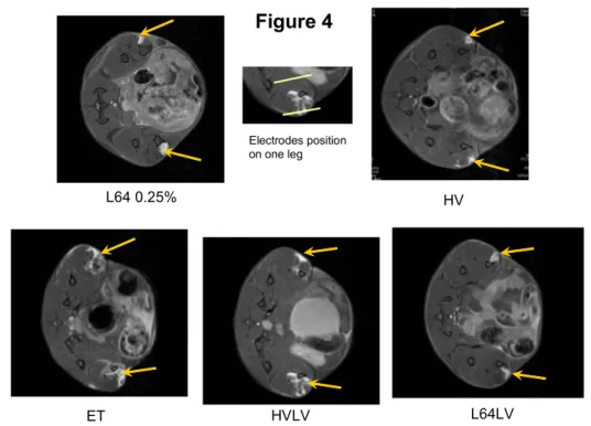
Figure 4
L64 0.25% HV
ET HVLV L64LV
Electrodes position on one leg
Figure 4
shows characteristic T1-weighted images. Gd-DOTA as DNA and pluronic L64 wereinjected locally into muscle (see Materials and Methods). We see that the distribution of the contrast agent in the tibial cranial muscle zone is localized and relatively homogenous after pluronic L64 (0.25 %) or HV treatment. By contrast, this distribution of the Gd-DOTA uptake is heterogeneous and enlarged after standard ET as well as after HVLV delivery. For L64 (0.25 %) injection then LV application distribution of Gd-DOTA seems similar to the one obtained with L64 alone.
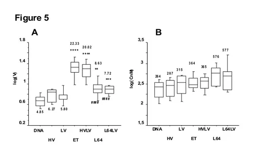
3.2.2.2 - Permeabilized volume (VGd)
As shown in Figure 5A
,
relative to the simple DNA injection, pulse combination (HV 800 V/cm, 0.1 msec. then LV 80 V/cm, 500 msec.: HVLV), standard electrotransfer (8 ×190 V/cm, 20 m, 2 Hz: ET) and DNA co-injection with pluronic L64 at 0.25% (L64) induced a significant increase in the volume permeabilized to Gd-DOTA. This increase in the permeabilized volume was higher for standard electrotransfer or HVLV relative to that induced by DNA co-injection withpluronic L64. Electrotransfer with HV alone induced a slight, non significant increase in the permeabilized volume. Just as for transgene expression (luminescence), the use of LV alone had no significant effect, nor did the delivery of one LV pulse after co-injection of DNA with
pluronic L64 0.25%. Similarly, we did not observe any effect on the permeabilized volume of HV or LV combination with pluronic L64 0.1% (data not shown).
3.2.2.3 - Concentration of the Gd-DOTA in the permeabilized volume (CGd)
Effects of the different treatments on the concentration of the contrast agent (CGd) in the
permeabilized volume were less obvious. Mean values for ET, HVLV and pluronic L64 were higher than for simple DNA injection, but not significantly. Just as for the permeabilized volume and transfection level, the CGd after DNA coinjection with pluronic L64 0.25 % was not modified
by the delivery of an LV pulse (Fig. 5 B).
Figure 5 A B 0.2 0.6 1 1.4 1.8 lo g( V) DNA HV ET HVLV L64 L64LV LV *** ** **** **** # ## # ## # # 4 .8 5 6 . 27 5 .8 0 2 2 .3 3 2 0 .0 2 8. 6 3 7 .7 2 1,5 2 2,5 3 3,5 lo g (C n M ) DNA HV ET HVLV L64 L64LV LV 26 4 2 8 7 3 1 5 3 6 4 36 5 5 7 6 5 7 7
3.2.3 Relations between “permeabilization” and transfection
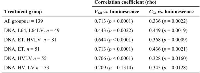
With the muscle model in vivo it was possible to study statistically the relations between transfection and permeabilization parameters measured on the same mice [20].
The strength of the correlation between the permeabilzed volume to Gd-DOTA (VGd) or the
concentration of Gd-DOTA in this volume (CGd) and transgene expression (luminescence) was
established using the nonparametric Spearman test. From this test, considering all studied groups, we found a correlation coefficient (rho) between luminescence and VGd of 0.718, which was
statistically very significant (p < 0.0001), as shown in Table 3. To improve the analysis, we treated different sub-groups separately. These results show that with three modalities of DNA transfection inducing muscle fibre permeabilization, i.e L64, ET, HVLV, the level of transfection was correlated to the volume permeabilized to Gd-DOTA. The correlation with the concentration of Gd-DOTA in the permeabilized volume was lower but also significant.
Table 3. Correlations between luminescence and volume permeabilized to Gd-DOTA (VGd) or concentration of Gd-DOTA in the permeabilized volume (CGd). V2
Correlation coefficient (rho)
Treatment group VGd vs. luminescence CGd vs. luminescence
All groups n = 139 0.713 (p < 0.0001) 0.336 (p = 0.0022) DNA, L64, L64LV. n = 49 0.443 (p = 0.0022) 0.449 (p = 0.0019) DNA, ET, HVLV n = 81 0.644 (p < 0.0001) 0.368 (p = 0.0009) DNA, ET. n = 51 0.713 (p < 0.0001) 0.436 (p = 0.0021) DNA, HVLV n = 55 0.706 (p < 0.0001) 0.328 (p = 0.0160) DNA, HV, LV n = 53 0.209 (p = 0.1314) 0.345 (p = 0.0128) Rho: strength of the link between the paired values (0 no relation, 1 and −1 perfect correlation; 1: direct, −1: inverse); n: number of paired values; p: statistical significance.
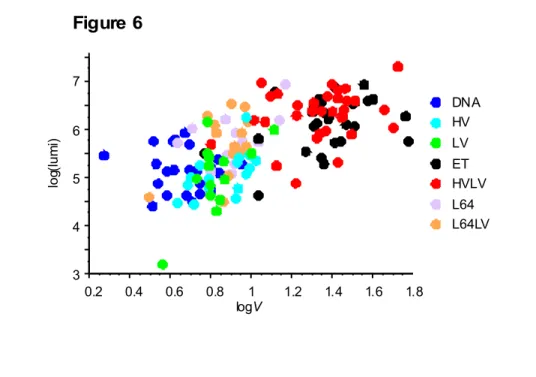
Fig 6 providesa graphic depiction of the relation between luminescence, our index of muscle transfection and the muscle volume permeabilized to Gd-DOTA (VGd). Top right: scatter plot for
electrotransfer by the standard (ET) or pulse combination (HVLV) procedure. Bottom left: scatter plot for DNA injection or DNA injection followed by an HV or LV pulse. Center: scatter plot for DNA coinjection with pluronic L64 followed or not followed by an LV pulse.
3 4 5 6 7 lo g( lu m i) 0.2 0.4 0.6 0.8 1 1.2 1.4 1.6 1.8 logV L64LV L64 HVLV ET LV HV DNA Figure 6
4 Discussion
In this study we compared transfection and permeabilization in vitro and in vivo with different modalities of electric pulse delivery with or without association of pluronic L64 (L64). Our in
vitro model was CHO cells and the in vivo model was the mouse tibial cranial muscle. In both
models we confirmed a high level of transfection associated with permeabilization to small mo-lecules with a series of electric pulses in conditions previously defined [11, 33], which we term standard electrotransfer (ET). However, using a combination of one high-voltage short-duration (HV) and one low-voltage long-duration (LV) pulses, the HVLV mode [12, 37], we found that transfection efficiency was low in vitro on CHO cells but high for muscles in vivo, at a level sim-ilar to standard electrotransfer ET.
We will first discuss the combination of HV and LV pulses and then go on to analyze results for the association of pluronic L64 and the electric field.
4.1 HVLV in vitro
In vitro, HV1LV did not induce many transfected cells even though a great number of cells were
permeabilized to propidium iodide (PI). However, the mean PI fluorescence level per cell was much lower than with the ETs condition. In these conditions DNA electrophoresis by LV is inef-fective and thus pointless. Other studies showed an effect of HVLV mode in vitro. Sukharev et al [37] who proposed the pulse combination (HVLV) approach showed electrotransfection effi-ciency on a different cell type (simian cos cell) and with very different parameters (HV : 4-7 kV/cm, 10-20 µsec; LV 0.2-0.5 kV/cm, 1-10 msec.). Cepurniene et al with conditions closer to ours, found a low percent of GFP positive cells, which is similar to our results [38]. Using a more sensitive reporter gene such as luciferase they revealed a clear effect of HVLV on transfec-tion. Although the detection sensitivity of transfected cells could be improved with HVLV in the present study, the mean level of “permeabilization” per cell remained low compared to the stand-ard condition ETs (10 x 700 V/cm, 5 msec., 1 Hz) (see Fig 1). In summary, the high number of weakly permeabilized cells after one HV1 pulse (1300 V/cm, 0.1 msec.) was not increased by
adding one LV pulse (80 V/cm, 500 msec.) and nor was the permeabilization level. In a study of Pavlin et al. the high voltage treatment is given by four HV pulses of 200 µs duration with amp-litude of 1.0 kV/cm [39]. In our study increasing the number of HV1 pulses to 10 allowed
indu-cing a clear cell transfection associated to increased permeabilization. Application of one LV pulse after the 10 HV1 pulses had no significant effect either for permeability or for transfection.
This lack of effect could be related to the level of DNA concentration. Indeed, it was shown with CHO cells that transfection obtained with HV pulses could be increased by a subsequent LV pulse only at DNA concentration below 10 µg/ml [39, 40]. For higher DNA concentration, elec-trophoretic improvement might not be needed as a result of there being sufficient DNA interac-tion with the cell membrane.
4.2 HVLV in vivo
We had shown in previous studies on muscle, and confirm here, that one HV (800 V/cm, 0.1 msec.) or one LV pulse (80 V/cm, 500 msec.) alone does not greatly improve transfection relative to simple DNA injection [12, 13]. The high transfecting condition HVLV led to a significant in-crease in the volume permeabilized to the contrast T1 agent Gd-DOTA (VGd). The concentration
of Gd-DOTA (CGd) in this volume was increased but non significantly. Overall, the uptake of
electrophores-It is not possible here to determine whether the increased uptake of Gd-DOTA is related to an in-crease in the membrane permeability coefficient or to a longer duration of the permeabilized state. However, in one study based on the pore theory of electropermeabilization it was suggested that in the pulse combination procedure (HVLV) the long-duration low-voltage pulse LV could stabilize the pores created by the HV pulse [41].
4.3 HVLV in vivo vs . in vitro
A better transfection in muscle by HVLV compared to that obtained in vitro may results from the additive effect of LV on membrane permeabilization by HV. The required LV field intensity in
vitro on CHO cells to obtain synergy between HV and LV for increasing membrane
permeabiliz-ation may need to be higher. This seems possible since the threshold electric field intensity to permeabilize CHO cells is close to 300 V/cm [33] whether it would be close to 100 V/cm for muscle fibers [11]. To note that for muscle fibers a threshold of 200 V/cm was found with 8 pulses of 0.1 msec. at 1 Hz [42]. However, it was shown on CHO cells that the necessary electric field intensity for electroporation of a given fraction of cells was decreasing with pulses of longer duration [43]. Consistently, in the present study delivering 8 pulses of 190 V/cm and 20 msec. duration at 2 Hz was clearly above the threshold for permeabilization and transfection.
Another hypothesis is that injected DNA that is transfected is in a particular compartment [44] that may be close to fiber membrane (as in T tubule….) [45]. Also, cell nuclei are closer to the cell membrane than for CHO cells. Thus small electrophoretic movements of DNA induced by LV may be more efficient than in vitro. It was shown in vitro that LV pulse after HV pulse in-creased transfection efficiency only for low concentration [40]. It is assumed by the authors that there is low local concentration in vivo. However the plasmid solution injected into muscle was highly concentrated (160 µg/ml) and we do not know how DNA is distributed in this tissues.
4.4 Pluronic L64 in vivo vs. in vitro
The purpose of our study was also to compare electric field effects with those of pluronic L64 and their combination. From the literature, it is known that pluronic L64 is inefficient in vitro but allows muscle transfection to be improved in vivo when co-injected with DNA [18], as confirmed by our study. Thus, it was shown to improve gene expression in vivo in the muscle where a basal transfection occurs by simple DNA injection [46]. The proposed mechanism is that pluronic L64 favors nuclear import of cytoplamic DNA and its transcription via activation of the NFkB pathway [29]. For this to happen it is necessary that DNA crosses the plasma membrane. This
occurs in the muscle, since a simple DNA injection allows fiber transfection [46]. However,this apparently does not occur in vitro by coincubation of DNA and pluronic L64 with CHO cells. It is also necessary to have DNA with consensus sequences for NFkB, which was shown for the CMV promoter of plasmid DNA that we used [47].
4.5 Pre-treatment with Pluronic L64 vs . ET
One of our aims in undertaking this study was to assess the possibility that the pluronic L64, by its effects on nuclear import of DNA and its transcription, could amplify transfection by
electrotransfer in muscle in vivo as we have shown in vitro with CHO cells in a previous study [24]. In vitro pre-treatment of CHO cells with pluronic L64 0.25 % allowed improvement of transfection by standard electrotransfer (10 x 500 V/cm, 5 msec., 1 Hz) whereas in vivo, muscle pre-treatment with 0.25% before a mild electrotransfer (8 pulses 120 V/cm, 20 msec., 2 Hz) did not improve transfection. A first hypothesis is that interaction of pluronic L64 with muscle fiber membrane and its subsequent internalization is not the same as with CHO cells. Another
hypothesis is that electrotransfer alone is able to activate the NFkB pathway. Consequently, any additional effect of pluronic L64 is reduced or null. Such an activation of NFkB by ET is possible, since it is known that this procedure induces an inflammation [8, 20, 48], and thus an activation of the NFkB pathway [49]. In our previous in vitro study we observed that
electrotransfer was more efficient using a plasmid with NFkB consensus sequences [24] (see Fig 6 of this article). Lastly, a slight improvement in transfection by pluronic L64 as observed with CHO cells in vitro (less than twofold increase) is difficult to detect in vivo owing to a variation of about one log between data in the same group.
4.6 Comparison between membrane destabilization with Pluronic L64 and HV
Another question was whether membrane destabilization induced by pluronic L64 could be equivalent to that induced by HV, and consequently whether we could evidence any synergy between pluronic L64 and LV as we observed between HV and LV for transfection and
permeabilization of the muscle. We found that pre-treatment by pluronic L64 before an LV pulse was inefficient both in vitro and in vivo. In vitro it could be expected since LV or pluronic L64 alone does not lead to DNA transfection. In vivo, the delivery of one LV pulse after pluronic L64 administration did not further amplify the transfection level, the muscle volume permeabilized to Gd-DOTA and the concentration of Gd-DOTA relative to pluronic L64 alone, in contrast to what
membrane modifications similar to those obtained by the delivery of an HV electric pulse. Even though, pluronic L64 is able to create pores in phospholipid membranes, this destabilisation is more than likely not of the same nature as the one by an electric field. First pluronic L64 might cover the phospholipid heads, masking the charges at the surface of the membrane. Second, insertion of pluronic L64 might not induce charge distribution modification as opposed to electrotransfer.
We showed in our previous study [24] that on CHO cells in vitro, pluronic L64 induced
permeabilization to propidium iodide (PI), but at a lower level than with ETa (10 pulses of 500 V/cm, 5msec. at 1 Hz). In pre-treatment experiments, i.e CHO cells incubated with pluronic L64 followed by ETa, the permeabilization to PI was similar to that obtained with ETa alone. This confirms that the nature of membrane modification induced by electric pulses is therefore not comparable to that obtained by pluronic L64, and these effects are not additives. Alimi-Guez et
al. also showed that gene transfection was not related to membrane permeabilization using
non-permeabilizing copolymer [28]. In addition, other authors have shown that permeabilization induced by cationic polymer or cationic lipids is a side effect not related to transfection [50]. DNA electrophoresis by LV could improve transfection by favoring DNA interaction with the membrane or its movement through the membrane. As shown in vitro it seems that DNA interaction with the membrane is favored by electrophoresis for a given state induced by the electric field [51]. Our experiment shows that such a state cannot be induced by the pluronic L64. Another effect of DNA electrophoresis on transfection could be to modify its biodistribution in such a way that it would reach a higher concentration near the membrane. We have seen that this effect was non significant in vitro with the DNA concentration used. In vivo, in the muscle such an effect was also non detectable. Indeed LV did not improve significantly DNA transfection.
4.7 Correlations between transfection and permeabilization indices
In vivo, we observed a positive correlation between the permeabilized volume VGd and transgene
expression (luminescence) for pluronic L64 and for ET (8 pulses of 190 V/cm and 20 msec. at 2 Hz) confirming our previous results [20]. The same significant correlation was evidenced with HVLV. Permeabilization to small molecules is an index of membrane modification associated with transfection. But DNA transfer probably does not occur by passive diffusion as it does for small molecules. In vitro we had no such possibility of statistical analysis. However, we observed that a minimal level of membrane modification, as evidenced by its permeabilization to
knowledge on in vitro gene transfer it is also probable that electroporation does not allow DNA transfer by passive diffusion [7].
In conclusion, the main finding of the present study is that the nature of membrane modification induced by permeabilizing electric pulses differs from that induced by pluronicL64. The
electrophoretic LV pulse does not induce additive effects to the transfection mediated by pluronic L64. This indicates that DNA electrophoresis would only favour DNA interaction with
membranes modified by permeabilizing electric pulses but not with membranes permeabilized by pluronic L64. In addition, the possible effect of DNA electrophoresis on its tissue biodistribution had no significant effect.
Acknowledgements: This work was supported in part by a grant from Agence Nationale pour la Recherche (ANR PCV06 139888), CNRS and INSERM French National Research. We are grateful to Julie Orio and Laetitia Helaudais for their assistance with some in vitro experiments.
The authors declare no conflict of interest.
References
1. Aihara H, Miyazaki J Gene transfer into muscle by electroporation in vivo. Nat Biotechnol 1998; 16: 867-870
2. Mir LM, Bureau MF, Rangara R, et al. Long-term, high level in vivo gene expression after electric pulse-mediated gene transfer into skeletal muscle. C R Acad Sci III 1998;
321: 893-899
3. Bloquel C, Fabre E, Bureau MF, et al. Plasmid DNA electrotransfer for intracellular and secreted proteins expression: new methodological developments and applications. J Gene Med 2004; 6 Suppl 1: S11-23
4. Wells DJ Gene therapy progress and prospects: electroporation and other physical methods. Gene Ther 2004; 11: 1363-1369
5. Mir LM Nucleic acids electrotransfer-based gene therapy (electrogenetherapy): past, current, and future. Mol Biotechnol 2009; 43: 167-176
6. Hojman P Basic principles and clinical advancements of muscle electrotransfer. Curr Gene Ther 2010; 10: 128-138
7. Escoffre JM, Portet T, Wasungu L, et al. What is (still not) known of the mechanism by which electroporation mediates gene transfer and expression in cells and tissues. Mol Biotechnol 2009; 41: 286-295
8. Chiarella P, Massi E, De Robertis M, et al. Electroporation of skeletal muscle induces danger signal release and antigen-presenting cell recruitment independently of DNA vaccine administration. Expert Opin Biol Ther 2008; 8: 1645-1657
9. Zupanic A, Ribaric S, Miklavcic D Increasing the repetition frequency of electric pulse delivery reduces unpleasant sensations that occur in electrochemotherapy. Neoplasma
10. Zhang L, Rabussay DP Clinical evaluation of safety and human tolerance of electrical sensation induced by electric fields with non-invasive electrodes. Bioelectrochemistry 2002; 56: 233-236
11. Mir LM, Bureau MF, Gehl J, et al. High-efficiency gene transfer into skeletal muscle mediated by electric pulses. Proc Natl Acad Sci U S A 1999; 96: 4262-4267
12. Bureau MF, Gehl J, Deleuze V, et al. Importance of association between permeabilization and electrophoretic forces for intramuscular DNA electrotransfer. Biochim Biophys Acta 2000; 1474: 353-359
13. Satkauskas S, Bureau MF, Puc M, et al. Mechanisms of in vivo DNA electrotransfer: respective contributions of cell electropermeabilization and DNA electrophoresis. Mol Ther 2002; 5: 133-140
14. Andre F, Gehl J, Sersa G, et al. Efficiency of High and Low Voltage Pulse Combinations for Gene Electrotransfer in Muscle, Liver, Tumor and Skin. Hum Gene Ther 2008
15. Cemazar M, Golzio M, Sersa G, et al. Control by pulse parameters of DNA electrotransfer into solid tumors in mice. Gene Ther 2009; 16: 635-644
16. Pavselj N, Preat V DNA electrotransfer into the skin using a combination of one high- and one low-voltage pulse. J Control Release 2005; 106: 407-415
17. Lemieux P, Guerin N, Paradis G, et al. A combination of poloxamers increases gene expression of plasmid DNA in skeletal muscle. Gene Ther 2000; 7: 986-991
18. Pitard B, Pollard H, Agbulut O, et al. A nonionic amphiphile agent promotes gene delivery in vivo to skeletal and cardiac muscles. Hum Gene Ther 2002; 13: 1767-1775 19. Yang Z, Zhu J, Sriadibhatla S, et al. Promoter- and strain-selective enhancement of gene
expression in a mouse skeletal muscle by a polymer excipient Pluronic P85. J Control Release 2005; 108: 496-512
20. Bureau MF, Juge L, Seguin J, et al. Muscle transfection and permeabilization induced by electrotransfer or pluronic L64: Paired study by optical imaging and MRI. Biochim Biophys Acta 2010; 1800: 537-543
21. Pembouong G, Morellet N, Kral T, et al. A comprehensive study in triblock copolymer membrane interaction. J Control Release 2011; 151: 57-64
22. Firestone MA, Wolf AC, Seifert S Small-angle X-ray scattering study of the interaction of poly(ethylene oxide)-b-poly(propylene oxide)-b-poly(ethylene oxide) triblock copolymers with lipid bilayers. Biomacromolecules 2003; 4: 1539-1549
23. Kostarelos K, Kipps M, Tadros T, et al. Molecular structure and conformation in phospholipid vesicles sterically stabilized by (tri)-block copolymers investigated by multi-nuclear magnetic resonance techniques. Colloids and Surfaces A: Physicochemical and engineering aspects 1998; 136: 1-9
24. Wasungu L, Marty AL, Bureau MF, et al. Pre-treatment of cells with pluronic L64 increases DNA transfection mediated by electrotransfer. J Control Release 2011; 149: 117-125
25. Batrakova EV, Li S, Brynskikh AM, et al. Effects of pluronic and doxorubicin on drug uptake, cellular metabolism, apoptosis and tumor inhibition in animal models of MDR cancers. J Control Release 2010; 143: 290-301
26. Goncalves C, Ardourel MY, Decoville M, et al. An optimized extended DNA kappa B site that enhances plasmid DNA nuclear import and gene expression. J Gene Med 2009;
11: 401-411
27. Gau-Racine J, Lal J, Zeghal M, et al. PEO-PPO block copolymer vectors do not interact directly with DNA but with lipid membranes. J Phys Chem B 2007; 111: 9900-9907 28. Alimi-Guez D, Leborgne C, Pembouong G, et al. Evaluation of the muscle gene transfer
29. Yang Z, Sahay G, Sriadibhatla S, et al. Amphiphilic block copolymers enhance cellular uptake and nuclear entry of polyplex-delivered DNA. Bioconjug Chem 2008; 19: 1987-1994
30. Gabriel B, Teissie J Generation of reactive-oxygen species induced by
electropermeabilization of Chinese hamster ovary cells and their consequence on cell viability. Eur J Biochem 1994; 223: 25-33
31. Wolf H, Rols MP, Boldt E, et al. Control by pulse parameters of electric field-mediated gene transfer in mammalian cells. Biophys J 1994; 66: 524-531
32. Bloquel C, Trollet C, Pradines E, et al. Optical imaging of luminescence for in vivo quantification of gene electrotransfer in mouse muscle and knee. BMC Biotechnol 2006;
6: 16
33. Rols MP, Teissie J Electropermeabilization of mammalian cells to macromolecules: control by pulse duration. Biophys J 1998; 75: 1415-1423
34. Zaharoff DA, Yuan F Effects of pulse strength and pulse duration on in vitro DNA electromobility. Bioelectrochemistry 2004; 62: 37-45
35. Debborah Alimi-Guez CL, Gaelle Pembouong, Laetitia Van Wittenberghe, Nathalie Mignet, Daniel Scherman, Antoine Kichler Evaluation of the muscle gene transfer activity of a series of amphiphilic triblock copolymers. Journal of Gene Medicine 2009; in press 36. Wu JC, Sundaresan G, Iyer M, et al. Noninvasive optical imaging of firefly luciferase
reporter gene expression in skeletal muscles of living mice. Mol Ther 2001; 4: 297-306 37. Sukharev SI, Klenchin VA, Serov SM, et al. Electroporation and electrophoretic DNA transfer into cells. The effect of DNA interaction with electropores. Biophys J 1992; 63: 1320-1327
38. Cepurniene K, Ruzgys P, Treinys R, et al. Influence of plasmid concentration on DNA electrotransfer in vitro using high-voltage and low-voltage pulses. J Membr Biol 2010;
236: 81-85
39. Pavlin M, Flisar K, Kanduser M The role of electrophoresis in gene electrotransfer. J Membr Biol 2010; 236: 75-79
40. Kanduser M, Miklavcic D, Pavlin M Mechanisms involved in gene electrotransfer using high- and low-voltage pulses--an in vitro study. Bioelectrochemistry 2009; 74: 265-271 41. Smith KC, Neu JC, Krassowska W Model of creation and evolution of stable electropores
for DNA delivery. Biophys J 2004; 86: 2813-2826
42. Corovic S, Zupanic A, Kranjc S, et al. The influence of skeletal muscle anisotropy on electroporation: in vivo study and numerical modeling. Med Biol Eng Comput; 48: 637-648
43. Pucihar G, Krmelj J, Rebersek M, et al. Equivalent pulse parameters for electroporation. IEEE Trans Biomed Eng; 58: 3279-3288
44. Bureau MF, Naimi S, Torero Ibad R, et al. Intramuscular plasmid DNA electrotransfer: biodistribution and degradation. Biochim Biophys Acta 2004; 1676: 138-148
45. Wolff JA, Dowty ME, Jiao S, et al. Expression of naked plasmids by cultured myotubes and entry of plasmids into T tubules and caveolae of mammalian skeletal muscle. J Cell Sci 1992; 103 ( Pt 4): 1249-1259
46. Wolff JA, Ludtke JJ, Acsadi G, et al. Long-term persistence of plasmid DNA and foreign gene expression in mouse muscle. Hum Mol Genet 1992; 1: 363-369
47. Breuzard G, Tertil M, Goncalves C, et al. Nuclear delivery of NFkappaB-assisted DNA/polymer complexes: plasmid DNA quantitation by confocal laser scanning microscopy and evidence of nuclear polyplexes by FRET imaging. Nucleic Acids Res 2008; 36: e71
48. Durieux AC, Bonnefoy R, Busso T, et al. In vivo gene electrotransfer into skeletal muscle: effects of plasmid DNA on the occurrence and extent of muscle damage. J Gene Med 2004; 6: 809-816
49. Tak PP, Firestein GS NF-kappaB: a key role in inflammatory diseases. J Clin Invest 2001;
107: 7-11
50. Prevette LE, Mullen DG, Holl MM Polycation-induced cell membrane permeability does not enhance cellular uptake or expression efficiency of delivered DNA. Mol Pharm 2010;
7: 870-883
51. Golzio M, Teissie J, Rols MP Direct visualization at the single-cell level of electrically mediated gene delivery. Proc Natl Acad Sci U S A 2002; 99: 1292-1297
Figures Captions Figure 1
CHO cells in vitro. Effect of different electric pulse delivery modalities and of pre-treatment with pluronic L64 on Transfection and Viability.
CHO cells in suspension or adherent were electrotransfected with a plasmid coding the Green Fluorescent Protein (GFP).
Group designations on the abscissa are for ETs: 10 pulses of 700 V/cm , 5 msec. duration at 1 Hz. ETa: 10 pulses of 500 V/cm, 5 msec. at 1 Hz. HV1: 1 pulse of 1300 V/cm, 0.1 msec..
HV1LV: the combination of high voltage and low voltage electrical conditions with LV: 1 pulse
of 80V/cm, 500 msec.. LV: low voltage alone. 10 HV1LV: ten HV1 pulses then one LV pulse. 10
HV1: ten HV1 pulses. The control group corresponds to simple DNA incubation with CHO cells.
LV0: 1 pulse of 80 V/cm, 200 msec.. Transfection
Panel A cell suspension
The percentage of GFP positive cells is represented with histograms on the left axis. This is the percent of viable cells transfected. The level of fluorescence (AU: arbitrary units) in the
population of fluorescent cells is represented with squares on the right axis. Each experiment was performed three times in triplicate, and a minimum of 5000 cells were counted with cytometry. Six to nine experiments were performed and four for control. The error bars are the standard deviation. Statistical comparisons between the different treatments were made using a PLSD Fisher test. Significance of the difference relative to control for the percent of GFP positive cells and the mean fluorescence intensity per cell: ** p<0.01 *** p< 0.001
Panel B adherent cells
Before transfection adherent cells were treated (grey histograms) or not treated (white
histograms: control) with 0.25% L64. The percentage of GFP positive cells is represented with histograms on the left axis. The level of fluorescence (AU: arbitrary units) in the population of fluorescent cells is represented with squares on the right axis. Error bars represent the standard deviation. Each experiment was performed from two to four times in duplicate and a minimum of 5000 cells were counted by cytometry. The error bars are the standard deviation. Statistical comparisons between the different treatments were made using a PLSD Fisher test. For
electrotransfer we observed a significant difference between cells pretreated by the pluronic L64 0.25 % and cells not pretreated for the percent of GFP positive cells ** p<0.01.
Cell viability
Column levels represent the mean percent of viable cells relative to the total number of cells. The error bars are the standard deviation. N=6 to 9.
Panel C cells suspension
Panel D adherent cells: Gray column design experiment without pluronic. White column design
experiments with adherent cells pretreated by pluronic L64, 16 h before electric pulses delivery.
Figure 2
Permeabilization of CHO cells to propidium iodide
CHO cells in suspension were electropermeabilized in the presence of the fluorescent probe propidium iodide (PI). Several electrical conditions were tested: standard electropermeabilization (ETs: 10 pulses of 700 V/cm, 5 msec, 1 Hz), high voltage electrical parameters (HV1: 1 pulse of
1300 V/cm, 0.1 msec.), the combination of high voltage and low voltage electrical conditions (HV1LV, LV: 1 pulse of 80V/cm, 500 msec.) and finally low voltage alone (LV). Non treated
cells which are only incubated with PI are represented as control. The percentage of PI positive cells is represented with histograms on the left axis. The level of fluorescence (AU: arbitrary units) in the population of fluorescent cells is represented with squares on the right axis. Values are the mean of 3 to 9 experiment performed in triplicate and a minimum of 5000 cells were counted by cytometry. The error bars are the standard deviation. Significance of the difference relative to control for PI positive cells and the mean fluorescence intensity per cell: *** p<0.001. The level of fluorescence did not differ significantly from the control for the HV1LV group.
Fig 3
Effect of pluronic L64 and / or electric field on transfection levels in vivo.
The distribution of values for each treatment are represented by box plot i.e out of the box smallest and maximum values (10 % and 90 %) then in the box the lower quartile, median and upper quartile.
A Tibial cranial muscle in vivo. Effect of different electric pulse delivery modalities on
transfection . The mean value of the luminescence in cpm/min is given near each box. Group
designations on the abscissa are for injection of free DNA (DNA); DNA then HV (HV); DNA then LV (LV); DNA then ET (ET); DNA then HVLV (HVLV); DNA and L64 0.25% (L64); DNA and L64 0.25% then LV (L64LV). With HV 800 V/cm, 0.1 msec., LV 80 V/cm, 500 msec.; HVLV one HV pulse followed 0.5 s later by one LV pulse; ET 8 pulses of 190 V/cm, 20 msec. at 2 Hz. The number of muscle for each treatment is of 12 to 33. Statistical significance of the difference relative to injection of free DNA : **** p < 0.0001, * p < 0.05; relative to HVLV: # p < 0.05
B Tibial cranial muscle in vivo: Effect of muscle pre-treatment with L64 0.25% injection before a mild electrotransfer or an HV pulse. The mean value of the luminescence in cpm/min
is given near each box. Group designations on the abscissa are for injection of free DNA (DNA); DNA then mild electrotransfer (ET120); DNA then HV (HV); DNA and L64 0.25% (L64); Pre-treatment with L64 0.25% then mild electrotransfer (L64ET120); Pre-Pre-treatment with L64 0.25% then HV (L64HV). With HV 800 V/cm, 0.1 msec.; ET120 8 pulses of 120 V/cm, 20 msec. at 2 Hz. The number of muscle for each treatment was of 10. Statistical significance of the difference relative to injection of free DNA : ** p < 0.01, * p < 0.05
Fig 4.
Characteristic T1-weighted images of Gd-DOTA distribution in the tibial cranial muscle.
Mouse leg axial section as visualized by MRI at day 3 after injection in the tibial cranial muscle of Gd-DOTA with DNA and pluronic L64 0.25 % (L64 0.25 %), DNA followed by delivery of one pulse 800 V/cm, 0.1 msec. (HV), DNA followed by delivery of 8 pulses, 190 V/cm, 20 msec., 2 Hz (ET) , DNA followed by delivery of 1 pulse 800 V/cm, 0.1 msec. and 0.5 sec later 1 pulse 80 V/cm, 500 msec. (HVLV) and DNA co-injected with pluronic L64 0.25 % followed by delivery of 1 pulse 80 V/cm, 500 msec. (L64LV). The Gd-DOTA trapped in muscle fibers is
visualized by the bright zone near the tibia bone and is indicated by an arrow. On the leg section HVLV approximate position of the two electrodes is indicated by two white bars.
Fig 5.
Effect of pluronic L64 and / or electric field on permeabilization in vivo
The distribution of values for each treatment are represented by box plot i.e out of the box smallest and maximum values (10 % and 90 %) then in the box the lower quartile, median and upper quartile.
A Tibial cranial muscle volume permeabilized to the MRI contrast agent Gd-DOTA
The mean value of the volume in µl is given near each box. Group designations on the abscissa are for injection of free DNA (DNA); DNA then HV (HV); DNA then LV (LV); DNA then ET (ET); DNA then HVLV (HVLV); DNA and L64 0.25% (L64); DNA and L64 0.25% then LV (L64LV). With HV 800 V/cm; 0.1 msec., LV 80 V/cm, 500 msec.; HVLV one pulse HV followed 0.5 s later by one LV pulse; ET 8 pulses of 190 V/cm, 20 msec. at 2 Hz; Statistical significance of the difference relative to injection of free DNA : **** p < 0.0001, *** p < 0.001, ** p < 0.01; relative to HVLV: #### p < 0.0001
B Concentration of the MRI contrast agent Gd-DOTA in the tibial cranial muscle volume permeabilized
The mean value of the concentration in nanomoles is given near each box plot. Group
designations on the abscissa are for injection of free DNA (DNA); DNA then HV (HV); DNA then LV (LV); DNA then ET (ET); DNA then HVLV (HVLV); DNA and L64 0.25% (L64); DNA and L64 0.25% then LV (L64LV). With HV 800 V/cm; 0.1 msec., LV 80 V/cm, 500 msec.; HVLV one pulse HV followed 0.5 s later by one LV pulse; ET 8 pulses of 190 V/cm, 20 msec. at 2 Hz; There was no significant difference for any group relative to injection of free DNA.
Fig 6.
Correlation between luminescence and volume permeabilized to Gd-DOTA.
Log values of the luminescence and corresponding permeabilized volume to Gd-DOTA of muscle in paired optical imaging and MRI experiments for all the mice studied. Each color corresponds to one treatment. Group designation: for injection of free DNA (DNA); DNA then
HV (HV); DNA then LV (LV); DNA then ET (ET); DNA then HVLV (HVLV); DNA and L64 0.25% (L64); DNA and L64 0.25% then LV (L64LV). With HV 800 V/cm; 0.1 msec., LV 80 V/cm, 500 msec.; HVLV one pulse HV followed 0.5 s later by one LV pulse; ET 8 pulses of 190 V/cm, 20 msec. at 2 Hz.