Université de Montréal
La relation entre le stress vécu par les bébés et la suppression neuronale mesurée en EEG
par Florence Deguire
Département de psychologie Faculté des arts et des sciences
Mémoire présenté en vue de l’obtention du grade de maîtrise (M. Sc.) en psychologie
Septembre 2018
ii Résumé
L’apprentissage et la mémoire sont sensibles aux effets du stress, et il a été démontré que chez l’adulte la relation entre le stress et l’apprentissage suit une courbe en « U » inversé. Cependant, il y a peu de données dans la littérature démontrant l’existence d’une telle relation chez les nourrissons. Dans ce mémoire, nous utilisons le phénomène de suppression neuronale et le cortisol salivaire recueilli en situation de stress afin de mesurer la relation entre le stress et l’apprentissage chez le nourrisson. Nous posons l’hypothèse que le stress affectera la suppression neuronale en suivant aussi une relation en « U » inversé. Nous avons mesuré en électroencéphalographie la réponse cérébrale pendant une tâche d’apprentissage et recueillis des échantillons de salive durant l’expérimentation chez 35 bébés (18 mâles) âgés entre 6 et 26 mois. Nous avons utilisé un modèle linéaire mixte afin de mesurer l’effet du stress sur l’apprentissage en incluant le niveau de cortisol salivaire, l’âge et le sexe comme prédicteurs. Les résultats démontrent que chez les jeunes enfants, un niveau de stress plus élevé n’est pas associé à une diminution de la réponse de suppression neuronale mais est plutôt lié à une réponse cérébrale plus élevée au premier stimulus de chaque essai. Compte tenu de ces résultats, le stress peut être considéré comme de la vigilance, expliquant ainsi cet effet du stress sur la réponse d’apprentissage chez le nourrisson. Puisque nous en savons peu sur le stress et l’apprentissage chez les jeunes enfants et que l’enfance est une période critique du développement, les futures recherches devraient continuer d’étudier l’influence du stress sur les mécanismes d’apprentissage et de la mémoire.
iii Abstract
The over activation of the hypothalamo–pituitary–adrenal (HPA) axis in stress situations can influence learning and memory. Researchers have underlined an inverted-U shape relationship between stress level, and learning and memory, in adults. Whether this model fits learning performances in infants is not well documented. In this study, we use the repetition suppression phenomenon and salivary cortisol collected during a stressful situation to measure the relationship between stress and learning in infants. We hypothesized that the effect of stress on repetition suppression will also follow an inverted-U shape relationship. We measured brain activity using EEG during a repetition-based learning task and saliva samples were collected during the experiment in 35 infants (18 males) aged between 6 and 26 months. The effect of stress level on learning was modeled with a linear mixed model, using cortisol, age and sex as predictors. Results indicate that in healthy infants, higher stress level is not associated with poorer repetition suppression response but is rather linked to an increased response on the first stimulus of each trial. Considering these results, stress could be interpreted as vigilance, thus explaining this effect of stress on learning response in infants. Since little is known about stress and learning in infants and considering that early childhood is a critical period of development, future studies should keep investigating the influence of stress on learning and memory mechanisms.
iv Table des matières
Résumé ... ii
Abstract ... iii
Liste des figures ... v
Liste des abréviations ... vi
Remerciements ... vii
Introduction générale ... 1
Position du problème ... 1
Les effets du stress ... 1
Potentiels liés aux évènements ... 4
Objectifs et hypothèses ... 5 Contribution à l’article ... 6 Scientific Article ... 7 Abstract ... 8 Introduction ... 9 Method ... 12
Materials and procedure ... 12
Results ... 18 Discussion ... 21 Conclusion ... 25 Acknowledgments ... 25 References ... 26 Discussion générale ... 29 Contributions ... 30 Limites ... 31 Perspectives ... 32
v Liste des figures
Scientific article
Figure 1. Experimental design. Example of one trial. A trial consisted of four frames depicting a man or a woman articulating the vowel /a/………. 15
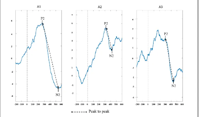
Figure 2. Averaged ERP of 10 participants for each /a/. This figure illustrates the auditory P2 and N2 components and the peak to peak measure. Peak to peak was assessed by subtracting the amplitude of the two components……….. 17
Figure 3. Effect of stress, measured by the area under the curve with respect to increase, on the intercept. The blue curve represents the mean repetition suppression response of all participants. The green curve represents the effect of high stress level on the intercept and the red curve represents the effect of low stress level on the intercept. The same effect is found with the difference between the second and the first saliva sample (S2-S1) and with the difference between the third and the first saliva sample (S3-S1)……… 20
vi Liste des abréviations
ACTH: hormone adrénocorticotrope; adrenocorticotropic hormone AUCI: Area under the curve with respect to increase
CRF: facteur de libération de corticotropine; corticotropin-releasing factor DLT: dépression à long-terme
EEG: électroencéphalographie; electroencephalography ERP: evoked-related potential
HPA: hypothalamo-pituitary-adrenal HPS: hypothalamo-pituito-surrénalien Hz: Hertz
ICA: independent component analysis ICC: inter-class correlation
kΩ: kiloohm
LMM: linear mixed model LTD: long-term depression LTP: long-term potentiation ML: maximum likelihood
PLT: potentialisation à long-terme PRE: potentiel lié aux événements ROI: region of interest
SE: standard error µV: microvolt
vii Remerciements
Je tiens d’abord à remercier Sarah Lippé de m’avoir accueillie dans le Laboratoire NED. Merci de m’offrir l’opportunité de réaliser des projets stimulants et de m’amener à constamment me dépasser. Merci également à l’équipe du Laboratoire NED d’avoir été là pour m’encourager chaque fois que je devais recommencer quelque chose. Merci pour votre aide et pour vos précieux conseils.
Je tiens à remercier tout spécialement mon amoureux, Raphaël, qui me soutient, qui m’encourage et qui m’endure depuis le tout début de mon parcours universitaire. Tu continues de croire en moi quand moi je n’y crois plus. Merci d’être toujours là quand j’en ai besoin. Merci de prendre soin de ma santé mentale, de me forcer à arrêter et à lâcher prise quand il le faut. Merci d’être toi!
Merci à ma famille pour tout le support et les encouragements. Merci de toujours me rappeler à quel point vous êtes fiers. Merci à mes parents, Manon et Jean, d’être les parents extraordinaires que vous êtes et de m’avoir inculqué la valeur du travail acharné et de la persévérance. Merci à mes grandes sœurs, Josée et Valérie, d’être mes modèles de femmes fortes et résilientes.
Merci aux « girls » de psycho. Vous avez été là quand ce n’était pas facile et vous avez été les premières à célébrer avec moi mes réussites. Merci de m’avoir inclus dans votre cohorte, même si je n’en faisais pas partie, vous avez rendu cette année de maîtrise beaucoup plus agréable.
Finalement, merci à tous ceux qui ont contribué, de loin ou de près, à la réalisation de ce mémoire de maîtrise!
Introduction générale Position du problème
Le stress affecte le fonctionnement du cerveau, notamment au niveau de l’apprentissage et de la mémoire. Il a été démontré que chez l’adulte, la relation entre le stress et l’apprentissage suit un modèle en « U » inversé (Lupien, Buss, Schramek, Maheu et Pruessner, 2005). Nous ignorons toutefois quels sont les effets du stress sur les fonctions cognitives chez le bébé puisqu’il y a peu d’informations dans la littérature à ce sujet. Pourtant, les premières années de vie sont critiques pour le développement du cerveau et particulièrement pour le développement des structures impliquées dans l’apprentissage et dans la mémoire. Il est donc nécessaire de mieux comprendre les facteurs influençant l’apprentissage et la mémoire chez les nourrissons.
Les effets du stress
La relation en « U » inversé entre le stress et l’apprentissage s’expliquerait par l’effet modulateur des glucocorticoïdes sur la potentialisation à long-terme (PLT). Lorsqu’une situation de stress est détectée par le cerveau, deux processus physiologiques sont activés ; le système sympathique médullosurrénalien et l’axe hypothalamo-pituito-surrénalien (HPS). L’activation du premier système provoque une libération rapide d’adrénaline et de noradrénaline par la glande médullosurrénale afin de permettre la réaction de fuite ou de combat (Gunnar et Quevedo, 2007). Parallèlement, l’axe HPS est un processus plus lent qui débute par l’activation de l’hypothalamus qui synthétise le facteur de libération de corticotropine (CRF), ce qui entraîne la sécrétion de l’hormone adrénocorticotrope (ACTH) par la glande pituitaire. Lorsque l’ACTH atteint la glande surrénale, cela provoque la libération de glucocorticoïdes, le cortisol majoritairement (Dedovic, D'Aguiar et Pruessner, 2009; Lupien et al., 2005; Seo et Lee, 2010).
2
Des boucles de rétroaction négative sont également mises en place, permettant de contrôler les activations subséquentes de l’axe HPS en agissant sur l’hypothalamus et la glande pituitaire. De plus, d’autres structures dotées d’un grand nombre de récepteurs de glucocorticoïdes, telles que l’hippocampe, le cortex préfrontal et l’amygdale, agissent comme régulateurs de l’axe HPS (Dedovic et al., 2009).
L’activation des récepteurs de minéralocorticoïdes augmente la force de la PLT dans l’hippocampe, tandis que l’activation des récepteurs de glucocorticoïdes réduit la PLT et facilite la dépression à long-terme (DLT) (Pavlides, Watanabe, Magarin et McEwen, 1995). Donc, un stress accru mène à une détérioration de la PLT et facilite l’induction de la DLT au niveau de l’hippocampe ce qui produit une réduction prolongée de l’efficacité synaptique impliquée dans les fonctions hippocampiques (Wong et al., 2007). Chez l’adulte, il a été démontré qu’à un faible niveau de stress, lorsque les récepteurs de glucocorticoïdes ne sont pas complètement saturés, l’apprentissage est amélioré. Toutefois, lorsque les récepteurs sont tous saturés sous des niveaux élevés de stress, l’apprentissage est altéré (Lupien et al., 2005; Zoladz et Diamond, 2009).
L’axe HPS semble également être impliqué dans les états d’éveils et de vigilance (Hancock, 1989). Selon les neuroscientifiques et les psychologues, la vigilance fait référence à la capacité de maintenir son attention sur une tâche et de rester alerte aux stimuli durant une certaine période de temps (Parasuraman, 1986). Il y aurait aussi une relation en « U » inversé entre les performances cognitives et les états d’activation corticale, de telle sorte que les performances sont optimales à des niveaux ni trop bas ni trop élevés de vigilance (Oken, Salinsky et Elsas, 2006). Lorsque la vigilance est réduite, l’amplitude des potentiels liés aux évènements est réduite et il y a une diminution de la réponse d’orientation (Oken et al., 2006).
3
Les recherches sur les effets du stress sur l’apprentissage ont été restreintes par la difficulté de mesurer l’apprentissage et la mémoire chez les nourrissons. Bien que les paradigmes de suivi oculaire soient possibles, l’électroencéphalographie (EEG) offre toutefois la possibilité d’examiner les réponses cérébrales d’apprentissage de façon plus précise. La suppression neuronale en particulier est un phénomène bien documenté qui reflète les mécanismes d’apprentissage chez le nourrisson (Bouchon, Nazzi et Gervain, 2015; Nordt, Hoehl et Weigelt, 2016; Snyder et Keil, 2008). De nombreuses études ont établi que lorsqu’un stimulus est présenté de façon répétée, il tend à y avoir une diminution de l’activité neuronale dans les régions impliquées dans le traitement du stimulus (Schacter et Buckner, 1998). Les régions activées lors de la première présentation du stimulus montrent une réponse atténuée lors de la deuxième présentation du stimulus (Henson, 2003; Schacter et Buckner, 1998). Ce phénomène survient à différents niveaux corticaux (Grill-Spector, Henson et Martin, 2006). Chez les bébés, les chercheurs font plus référence au phénomène d’habituation (Nordt et al., 2016; Sirois et Mareschal, 2002). Cette forme d’apprentissage joue un rôle important dans le développement perceptuel et cognitif du nourrisson (Sandman, Wadhwa, Hetrick, Porto et Peeke, 1997). Il a été mis de l’avant que l’habituation neuronale à un stimulus répété pourrait être un signe d’apprentissage dans le cerveau du nourrisson, de la même façon que l’effet d’habituation reflète un apprentissage au niveau comportemental (Aslin, 2007). Une étude de Snyder et Keil (2008) a démontré qu’une réduction des ondes gamma dans les régions postérieures du cerveau durant une phase d’encodage était associée à une préférence marquée pour le nouveau stimulus plutôt que vers celui qui était familier. Une étude de répétition en EEG conduit par Dehaene-Lambertz et Dehaene (1994) a établi un fort effet de suppression neuronale chez de très jeunes enfants. Ils ont présenté à des enfants âgés de deux à trois mois des séquences
4
de cinq syllabes. Certains essais contenaient que des syllabes identiques (essais répétés) et d’autres essais contenaient une cinquième syllabe différente (essais déviants). Ils ont trouvé qu’il y avait aux électrodes temporales postérieures un pic positif à 400 ms qui diminuait en amplitude dès la présentation de la deuxième syllabe. De plus, lorsque la cinquième syllabe présentée était déviante, cette composante augmentait d’amplitude, indiquant un relâchement de la suppression neuronale.
Potentiels liés aux évènements
Le phénomène de suppression neuronale peut se mesurer grâce aux potentiels liés aux évènements (PRE). Les PRE permettent d’extraire des réponses cognitives, sensorielles et motrices de l’ensemble du signal électroencéphalographique (Luck, 2014). Le signal d’un électroencéphalogramme (EEG) peut être décomposé en différentes composantes avec des polarités et des latences caractéristiques (Luck, 2014). Le terme « composante » peut soit faire référence aux déflexions positives et négatives qui surviennent dans des fenêtres de temps précises (Luck, 2014) où faire référence à des processus cérébraux sous-jacents dans le signal EEG, lesquels contribuent à la polarité des déflexions (Näätänen et Picton, 1987). Les PRE peuvent être enregistrés dès les premières secondes suivant la présentation du stimulus (Picton et Taylor, 2007). Les premières composantes sont reliées aux activités du tronc cérébral ou du cortex sensoriel primaire et donc varient d’une modalité sensorielle à une autre. Les composantes plus tardives (comme la N2) quant à elles sont générées par les régions pariétales, temporales et frontales associatives et représentent des phénomènes cérébraux de plus haut niveau (Knoth et Lippé, 2012; Picton et Taylor, 2007).
5
Les PRE corticaux auditifs sont largement utilisés chez les jeunes enfants et permettent d’étudier la réponse cérébrale d’apprentissage. Chez le nourrisson, la réponse auditive correspond principalement à une large composante positive (P2) dont l’amplitude maximale est enregistrée dans les régions fronto-centrales du scalp autour de 200-250 ms (Picton et Taylor, 2007). D’autres auteurs ont également démontré que dès les premiers mois de vie, il est possible d’identifier une large composante positive (P2) suivie d’une large composante négative (N2) dans le potentiel évoqué auditif (Lippé, Kovacevic et McIntosh, 2009; Lippé, Martinez-Montes, Arcand et Lassonde, 2009).
Objectifs et hypothèses
L’objectif de ce mémoire est de déterminer l’effet du stress sur la réponse cérébrale d’apprentissage chez les bébés. Nous posons l’hypothèse que le stress affectera la réponse de suppression neuronale et que cet effet suivra la même relation en « U » inversé que celle retrouvée chez les adultes. Nous prévoyons observer une réponse de suppression neuronale plus élevée sous un faible niveau de stress, jusqu’à un certain point où un taux de stress trop élevé réduira la réponse de suppression neuronale.
6 Contribution à l’article
L’article inclus dans ce mémoire a été réalisé dans le cadre de la maîtrise de Florence Deguire, selon une idée originale de sa directrice de recherche Sarah Lippé. Ce projet s’inscrit également dans le cadre du projet doctoral de Fanny Thebault-Dagher qui vise à étudier la relation entre la présence de convulsions fébriles atypiques et l’anatomie de l’hippocampe, la réaction au stress et le développement cognitif chez l’enfant. Comme première auteure, Florence Deguire a participé au recrutement et aux enregistrements EEG avec l’aide de Fanny Thebault-Dagher, Inga Sophia Knoth et Marc-Philippe Lafontaine. Elle a effectué la majeure partie du prétraitement des données d’électroencéphalographie et des analyses des potentiels liés aux évènements avec l’aide de Fanny Barlaam. Les analyses statistiques ont été entièrement réalisées par Florence Deguire. Sarah Lippé l’a guidé et aidé tout au long du processus. Cette première version a entièrement été écrite par Florence Deguire. L’article sera soumis au journal
7
Scientific Article
The relationship between stress and repetition suppression in infants, measured in EEG
Florence Deguire1,2,3,
Fanny Thébault-Dagher1,2,3,
Fanny Barlaam3,
Inga Sophia Knoth3,
Marc-Philippe Lafontaine1,2,3,
Sonia Lupien4,5,
Sarah Lippé1,2,3
1 Psychology department, University of Montréal
2 Centre de recherche en neuropsychologie et cognition, University of Montréal 3 Research Center of the Sainte-Justine Hospital, University of Montréal
4 Psychiatry department, University of Montréal
8 Abstract
The over activation of the hypothalamo–pituitary–adrenal (HPA) axis in stress situations can influence learning and memory. Researchers have underlined an inverted-U shape relationship between stress level, and learning and memory, in adults. Whether this model fits learning performances in infants is not well documented. In this study, we use the repetition suppression phenomenon and salivary cortisol collected during a stressful situation to measure the relationship between stress and learning in infants. We hypothesized that the effect of stress on repetition suppression will also follow an inverted-U shape relationship. We measured brain activity using EEG during a repetition-based learning task and saliva samples were collected during the experiment in 35 infants (18 males) aged between 6 and 26 months. The effect of stress levels on learning was modeled with a linear mixed model, using cortisol, age and sex as predictors. Results indicate that in healthy infants, higher stress level is not associated with poorer repetition suppression response but is rather linked to an increased response on the first stimulus of each trial. Considering these results, stress could be interpreted as vigilance, thus explaining this effect of stress on learning response in infants. Since little is known about stress and learning in infants and considering that early childhood is a critical period of development, future studies should keep investigating the influence of stress on learning and memory mechanisms.
9 Introduction Stress effects
Stress affects learning and memory. This relationship has been shown to follow an inverted-U shape model. The inverted-U shape relationship between stress and learning and memory has been well documented in adults (Lupien et al, 2005). However, we do not know how stress influences learning and memory in infants. Considering that infancy is a crucial learning period for later development, it is mandatory to have a better understanding of learning and memory modulators early in life.
The inverted-U shape relationship between stress and learning has been linked to the modulatory effect of glucocorticoids on long-term potentiation (LTP). When a stressful situation is encountered, two physiological systems are activated: the sympathetic-adrenomedullary system and the hypothalamic-pituitary-adrenal (HPA) axis. The activation of the first system induce a rapid release of epinephrine and norepinephrine by the adrenal medulla to allow the fight-or-flight response (Gunnar et Quevedo, 2007). In comparison, the HPA axis is a slower response that starts with the activation of the hypothalamus which synthesize the corticotropin-releasing factor (CRF) leading to the secretion of the adrenocorticotropic hormone (ACTH) by the pituitary gland. Glucocorticoids, cortisol predominantly, are released when the ACTH gets to the adrenal cortex (Lupien et al., 2005). Negative feedback inhibition is exerted on the hypothalamus and the pituitary gland, regulating the next HPA axis activation. Several structures with large numbers of glucocorticoids receptors, such as hippocampus, amygdala and prefrontal cortex, are important regulators of the HPA axis (Dedovic et al., 2009).
Activation of mineralocorticoids receptors increases LTP in the hippocampus while activation of glucocorticoids receptors reduces LTP and facilitates long-term depression (LTD)
10
(Pavlides et al., 1995). Thus, an acute stress leads to impaired LTP and favors LTD in the hippocampus, which results in a prolonged reduction in synaptic efficiency involved in hippocampus-dependant functions (Wong et al., 2007). Adult data demonstrate that learning is enhanced under low stress levels since both types of receptors are not fully saturated. However, when the receptors are all saturated under high stress levels, learning is impaired (Lupien et al., 2005; Zoladz et Diamond, 2009).
The HPA axis is also thought to play a role in arousal states and vigilance (Hancock, 1989). According to psychologists and neuroscientists, vigilance refers to the ability to maintain attention to a task and remain alert to stimuli for a period of time (Parasuraman, 1986). There is also an inverted-U shape relationship between cognitive performances and states of cortical activation, such that performances are enhanced at arousal states that are neither to high or to low (Oken et al., 2006). Hence, with decreased vigilance, there is a decreased amplitude of the event-related potentials and a decreased orientating response (Oken et al., 2006).
Researches on the effect of stress on learning have been hindered by the difficulty to measure learning and memory in infants. While eye-tracking paradigms are possible, EEG offers the possibility to examine more precisely neural learning responses in infants. Repetition suppression, in particular, is a well-documented phenomenon thought to reflect learning mechanisms in infants (Bouchon et al., 2015; Nordt et al., 2016; Snyder et Keil, 2008). When a stimulus is repeatedly presented, there is a reduction of the neural activity in the regions involved in stimulus processing (Schacter et Buckner, 1998). The regions activated during the first presentation of the stimulus tend to be less activated at the second presentation of the stimulus. This phenomenon is observable at many cortical levels of the brain (Grill-Spector et al., 2006). In infancy research, this phenomenon is referred to as neural habituation (Nordt et al., 2016;
11
Sirois et Mareschal, 2002). This form of learning plays a major role in perceptual and cognitive development in infants (Sandman et al., 1997). It has been advanced that neural habituation to repetitive stimuli could be a sign of learning in the infant brain, the same way habituation effects reflect learning at the behavioral level (Turk-Browne, Scholl et Chun, 2008). A study made by Snyder et Keil (2008) showed that a reduction of gamma activity in the posterior regions during an encoding phase was associated with a preference for the novel compared to the familiar stimulus. An early ERP repetition study by Dehaene-Lambertz et Dehaene (1994) has demonstrated strong repetition suppression effect in very young infants. They presented two-to-three-month-old children with sequences of five syllables. In some trials, all the syllables were the same (repeated trials) and in some trials, the fifth syllable was different (deviant trials). They found a positive peak around 400 ms at posterior temporal channels that decreased in amplitude at the second syllable. Moreover, for the fifth syllable in deviant trials, this component showed an increase in amplitude, thus indicating a release from repetition suppression.
Aim and Hypotheses
Infancy is critical for brain development and especially for the development of structures involved in learning and memory. In addition, the stress-learning relationship and the brain mechanisms affected by stress during the first year of life are still unclear. For these reasons, we aim to investigate how stress, experienced by young infants, affects learning and memory. The goal is to determine how stress affects repetition suppression in infants. We hypothesize that the stress-learning relationship will follow the same inverted-U shape relationship as the one found in adults. We expect to find a greater repetition suppression response under low to mild stress levels and a poorer repetition suppression response under higher stress levels.
12 Method Participants
The participants were 50 (27 males) healthy infants aged between 6 and 26 months (mean age ± SD: 15.77±6.03). Families were recruited in day cares and through social networks. Developmental information was gathered from developmental questionnaire completed by the parents. All infants were born at term with no pregnancy or delivery complications, had no significant health problem or developmental delay. Parents signed the consent form authorized by the ethics, administrative and scientific committees of the Ste-Justine’s University Hospital Research Center. Testing was stopped whenever infants reacted negatively. EEG data of five participants were rejected because of excessive artefacts, seven participants were excluded from the analyses because they had extreme or incomplete salivary cortisol data, two participants were excluded because of their age and one participant was excluded because of extreme scores (+3 Z score) on multiple variables. Therefore, the final sample included 35 infants (18 males; mean age ± SD: 14.81±5.58).
Materials and procedures Cortisol
To assess stress reactivity, salivary cortisol levels were measured by collecting saliva samples. Saliva samples were collected using Salivette device (SalivaBio Children’s Swab; Salimetrics LLC, Carlsbad, CA). A cotton dental roll was placed in the infant’s mouth for 30 seconds, or until it was sufficiently imbibed with saliva. It was then placed in a pierced tube, fitted in an external tube. To reduce bacterial growth, saliva samples were placed in a freezer until it was further analysed. Installation of the EEG net served as the stressor since it is new
13
and unknown for the infants and implies restriction of the infant’s arms by the parents. Saliva samples were collected before the stressor (M=7.58), 20 minutes after the stressor and 45 minutes after the stressor. In order to prevent any contaminations, we asked parents not to give food to their infant before taking the samples. If the infant had drunk or ate, the mouth was rinsed with water
Saliva samples were analysed at the Center for studies on Human Stress. Samples were centrifuged at 15000g x 15 minutes. Saliva was analysed with a high sensitivity enzyme immunoassay kit (Salimetric State College, PA, catalogue number 1-3102.) according to manufacturer’s instructions. The range of detection was from 0.0012 µg/dl to 3 µg/dl. Samples showing an out of curve reading were rerun after dilution. Samples were also rerun if the intra and interassay coefficients of variation were > 15%, unless the difference between the two values were below 0.03 µg/dl. All samples with enough volume were run in duplicate. All batch of saliva samples included samples from boys and girls and from infants of different ages.
To assess stress reactivity, area under the curve with respect to increase and differences between each saliva samples were used as stress measures. The area under the curve with respect to increase (AUCI) is calculated by using a derived of the trapezoid formula (Reinhardt et
Soeder, 2001). To calculate the formula, saliva measurements and time distance between the measurements are needed. The advantage of using AUCI is that the first measurement serves as
the baseline, thus assessing the change in reactivity over time rather than the total hormonal response. To calculate the difference between saliva samples (S2-S1, S3-S1, S3-S2), we made a subtraction between two saliva samples, then divided it by the time interval between the two. For all the stress measures, positive values indicate an increase in cortisol levels while negative values indicate a decrease in cortisol levels.
14 Stimuli
The task consisted of audio-visual stimuli featuring a woman and a man alternating in the articulation of the vowel /a/ or /i/. The audio-visual features served to maximize infants’ attention. Stimuli were generated by a Dell GX150 PC using E-Prime 2.0 software (Psychology Software Tools Inc., Pittsburgh, PA, USA) on a screen placed at a viewing distance of 60 cm. The sounds were delivered through two speakers located laterally at 30 cm from the infants’ ears. Infants were seated on their parents’ lap. The onset of the auditory vowel coincided with a visual clip lasting 200 ms of the mouth fully opened (frame 1). Following the end of the sound, two frames of 60 ms showing the mouth gradually closing were presented. During the next 280 ms, the face with a closed mouth was presented and then followed by the onset of the next vowel (Figure 1). The initial learning phase comprised 16 trials respecting a xxxY (aaaI) specific rule, called “standard”. During the test phase, out of 80 trials, 75% followed the xxxY(aaaI) specific rule. The other 25% followed a xxxx (aaaa) rule, called “deviant”. To facilitate learning, all deviant trials were followed by a standard trial. The sound-face combination was determined randomly. To assess repetition suppression, only the first three /a/ were used in the analyses.
15
Figure 1. Experimental design. Example of one trial. A trial consisted of four frames depicting a man or a woman articulating the vowel /a/.
EEG
The EEG recording took place in a dark soundproof experimental chamber. EEG was recorded continuously from a high-density EEG system containing 128 electrodes (Electrical Geodesics System Inc., Eugene, OR, USA). Signals were acquired and processed by a G4 Macintosh computer using NetStation EEG Software (Version 2.0). Data were acquired at a 1000 Hz sampling rate and analog bandpass filter of 0.1-100 Hz was applied. Impedances were kept bellow 40 kΩ (Tucker, 1993). Off-line signal processing and analyses were performed with MATLAB (Mathworks, Inc., Natick, MA) and with the EEGLAB toolbox (Delorme et Makeig, 2004). Data were digitally filtered with a lower-bound 0.5 filter and a 60 Hz notch filter. Twenty-eight electrodes containing muscular artifacts placed around the neck and face were excluded. Channels presenting voltage lower than 2 μV and higher than 200 μV were rejected. Data were re-referenced to an average reference. Eyes movement artefacts were rejected using semi-automatic independent component analysis (ICA) as implemented in EEGLAB. Visual
16
inspection of the segmented (-1000-2500 ms) data was also performed to reject manually the epochs with significant artefacts. Following epochs exclusion, an average of 57/80 (SD = 11.1) epochs were kept in the final analyses.
Event-related potentials. Auditory evoked potential analyses were conducted as they are widely used in infant’s studies. In infants, the most prominent wave in the auditive response is a large positive component (P2) with a maximum amplitude recorded at frontocentral scalp at around 200-250 ms (Lippé, Martinez-Montes, et al., 2009; Picton et Taylor, 2007). Other authors have demonstrated that in the first months of life, a large positive component (P2) is followed by a large negative component (N2) (Lippé, Kovacevic, et al., 2009; Lippé, Martinez-Montes, et al., 2009). Thereby, ERPs analyses were conducted on the auditory P2 and N2 components (Figure 2). Each epoch was segmented into three segments representing the time-window of each stimulus (A1: 0–600ms, A2: 600–1200m and A3: 1200–1800ms). We first
segmented and cleaned the data in 3500 ms time window to ensure that each A’s had the same numbers of trials. A region of interest (ROI) was created by selecting five electrodes in the FCz region (E5, E6, E12, E13, E112) since this region seemed to show the highest P2 amplitude. Amplitudes were defined using a peak to peak measure between the two auditory components (200 ms pre-stimulus baseline) and amplitudes were measured at the mean value extracted from the ROI. Peaks were obtained by averaging separately the first, the second and the third /a/ (A1
17
Figure 2. Averaged ERP of 10 participants for each /a/. This figure illustrates the auditory P2 and N2 components and the peak to peak measure. Peak to peak was assessed by subtracting the amplitude of the two components.
Statistics
Linear mixed model. Statistical analyses were performed using SPSS statistics, version 25 (IBM Corp., Armonk, NY, USA). Linear mixed models (LMM) were chosen to assess how stress levels predicted changes in cerebral activity across repetitions. To verify if there was an age effect, age was added as a predictor. We chose to use LMM because it can deal easily with missing data and with small sample size, in addition to enabling random intercepts and slopes, allowing for nonlinear modeling and selecting appropriate covariance structure (Field, 2013; West, 2009).
Linear trajectories were modeled individually to estimate the infant’s initial cerebral activity (intercept), the slope of infant’s cerebral activity trajectory and error, which is how well
18
the linear model fits the participant’s data. Each measure of stress levels was tested in a different model. Maximum likelihood (ML) was used to estimate all the models because both random and fixed effects were assessed (Singer et Willett, 2003) and heterogeneous first-order autoregressive covariance structure was used since it is most commonly found in longitudinal data (Field, 2013). Finally, sex was added sequentially to the models and a chi-square likelihood ratio was used to verify model fit improvement.
Results Preliminary analyses
Preliminary analyses showed no significant difference between boys and girls regarding age, timing of testing, time before the stressor, total time of testing and cortisol variables. T-tests were run to see if the stressor produced a significant increase in cortisol. The results show that there is rather a general decrease in cortisol levels over time, but the differences are not significant, thus seemingly infant’s stress level was stable throughout the experiment. We also tested age with the mean amplitude of each /a/ on the two components to ensure there were no correlations between age and amplitude and none of the correlations were significant.
Linear mixed models
Baseline models. We first tested the unconditional mean model to serve as a baseline to examine individual difference in amplitude without including time as a variable. This model gives the intraclass correlation coefficient (ICC) which indicate the amount of variance in the outcome that is related to interindividual differences. The interclass correlation coefficient is 0.25, which
19
justify the use of linear growth model (Heinrich et Lynn Jr, 2001). It indicates that 25% of the total variation in amplitude is due to interindividual differences.
Secondly, we tested the unconditional linear growth curve model. This model focuses on individual growth curve, more specifically individual change over time and serves as a baseline for comparing the subsequent models. Intercept and slope were allowed to vary randomly. Using the chi-square likelihood ratio test, best fit for the model was found using a quadratic model so the linear and the quadratic growth curve parameters were retained in the following models. The average (SE) initial peak to peak amplitude between the P2 and N2 components was 10.83 (1.76) (intercept) and the average (SE) slope was -6.10 (1.80). The quadratic model indicates that this trend slowed down after the second repetition and the average (SE) slope was then 1.31 (0.44).
Predictors. Predictors were added to the unconditional linear growth curve model to assess their effect on the repetition suppression response.
Age. When adding age to the unconditional model, there were no significant results and model fit was not significantly improved (χ2 (3) = 5, p > 0.1). Therefore, age was not retained in the subsequent models.
AUCI. AUCI was added as a time-varying predictor in the unconditional model as well as
interaction between repetitions and AUCI. Results indicate that stress level is associated with
change in the intercept (F (1, 54.4) = 6.2, p = 0.016) but was not a significant predictor of the linear and quadratic changes in amplitude across repetitions (Figure 3). Adding AUCI as a
20
Figure 3. Effect of stress, measured by the area under the curve with respect to increase, on the intercept. The blue curve represents the mean repetition suppression response of all participants. The green curve represents the effect of high stress level on the intercept and the red curve represents the effect of low stress level on the intercept. The same effect is found with the difference between the second and the first saliva sample (S2-S1) and with the difference between the third and the first saliva sample (S3-S1).
S2-S1. Difference between the second and the first saliva samples was also added as a time-varying predictor in the unconditional model as well as interaction between this stress measure and repetitions. A significant change in the intercept was found (F (1, 54.3) = 5.5, p = 0.023) but this stress measure was not a significant predictor of the linear and quadratic changes in amplitude across repetitions. Model fit was significantly improved when adding the difference between the second and the first saliva samples as a predictor in the model (χ2 (3) = 13.4, p < 0.005).
S3-S1. The difference between the third and the first saliva samples was added to the unconditional model as a time-varying predictor and interaction between this stress measure and
0 2 4 6 8 10 12 14 1 2 3 Ampli tude ( µ V) Stimulus presentation
Effect of stress on the intercept
21
repetitions. Results indicate this stress measure is significantly related to a change in the intercept (F (1, 56.9) = 4.5, p = 0.038). Again, this stress measure was not significantly related to linear and quadratic changes in amplitude across repetitions. There was again a significant improvement in model fit (χ2 (3) = 8.3, p < 0.05).
S3-S2. Finally, the difference between the third and the second saliva samples was added as a time-varying predictor to the unconditional model as well as interaction between this stress measure and repetitions. This stress measure was not significantly related to linear and quadratic changes in amplitude across repetitions nor to the intercept and did not improve model fit. Sex. Sex was added to the models as a time in-varying predictor and interaction between gender, stress measures and repetitions. Sex was not, in every case, a significant predictor of the linear and quadratic changes in amplitude across repetitions and was not associated with the intercept.
Discussion
The aim of this study was to investigate how stress, experience by infants, affects the learning response. Learning was measured by the repetition suppression phenomenon and stress was assessed by salivary cortisol. First, our results indicate that infants show a repetition suppression phenomenon that was not affected by age nor by sex. Second, results suggest that our experiment did not trigger elevated levels of stress in infants, suggesting that EEG is not a stressful procedure for infants. Nevertheless, our results indicate that normal elevation of stress is associated with amplest peak to peak measure between the P2 and the N2 auditory components on the first /a/ of each trial, but not to the repetition suppression phenomenon. Hence, more stress resulted in augmented orientation response, without affecting the learning response per see.
22 Repetition suppression
Results provide evidence that in healthy infants, repetition of the same stimulus generates a repetition suppression response, and that this response attenuates after the second presentation of the stimulus. This finding is in accordance with the existing literature positing that the second presentation of the same syllable results in a decreased component amplitude, whereas additional presentations of the syllable do not lead to further decrease (Dehaene-Lambertz et Dehaene, 1994). Mechanisms influencing the repetition effect are still unclear. However, there are some factors, such as attention, expectation, stimulus recognition, explicit memory and learning (Segaert, Weber, de Lange, Petersson et Hagoort, 2013) and the number of repetitions (Müller, Strumpf, Scholz, Baier et Melloni, 2012) that are known to influence repetition effect. The direction of the repetition effect can also vary between infants depending on their processing capacities (Nordt et al., 2016). Electrophysiological activity is known to change with age. In our study, adding age as a predictor did not improve model fit, suggesting that age does not influence the repetition suppression response.
Orientating response
Our results indicate that stress is generally associated with amplest peak to peak measure between the P2 and the N2 auditory components on the first /a/ of each trial. In a study of Dehaene-Lambertz et Dehaene (1994), amplitude was also higher in response to the first syllable of each trial. In our study, infants with higher stress levels showed a more prominent response to the first /a/ of each trial compared to infants with lower stress levels. This result could be explained by the role of the HPA axis in vigilance. Infants who were more stressed were also more vigilant to their environment and more alert to new stimulus whereas infants less stressed
23
were less vigilant and displayed the usual decreased amplitude associated with decreased vigilance (Oken et al., 2006). The first stimulus of each trial elicited a stronger orientating response in infants with higher stress levels, demonstrating that stress level is associated with the orientating response.
Moreover, results show that stress does not seem to influence the repetition suppression response in infants, which suggests that the relationship between stress and learning in infants differs from the inverted-U shape relationship found in adults. These results must be taken with a certain perspective since infants in the study showed no statistically significant cortisol increase. A review of Gunnar, Talge et Herrera (2009) has verified the effectiveness of stressor task in elevating cortisol levels in infants. For infants of four to nine months of age, stressor paradigms are barely successful in elevating cortisol levels and it is even worse for infants from twelve to twenty-four months of age. During the first year of life, many infants display no cortisol increase to stressors that typically cause important behavioral distress reactions. (Gunnar et Donzella, 2002). Neurodevelopmental changes affecting reactivity and regulation of the HPA system during infancy, such as improved negative feedback regulation of the axis and decrease sensitivity of the adrenal cortex to ACTH, could partially explain the diminution of the HPA responsiveness and the variability in effectiveness of stressor task (Lashansky et al., 1991). Since in our study parents could console their infant during the experimentation, it could explain why infants were not significantly stressed. Indeed, infant’s access to supportive adult care helps to buffer the activity of the HPA axis (Gunnar et Quevedo, 2007).
24 No sex effects
This study showed that sex was not a significant predictor of the linear and quadratic changes in amplitude across repetitions and was not associated with the initial peak to peak amplitude between the auditory P2 and N2 components. Few controlled stress studies have been conducted in children, but the few data available are contradictory. Few studies assessing stress in infants have reported significant main or interaction effects with sex (Gunnar et al., 2009). Some studies find no sex difference in stress response while other studies seem to find the same pattern of sex difference in infants as the one found in adults (Kudielka et Kirschbaum, 2005). In adults, stress in men is associated with activity in frontal regions and it is highly correlated with cortisol levels whereas stress in women activate principally the limbic system and it is less correlated with cortisol levels (Wang et al., 2007).
Study design and limitations
The use of repetition suppression paradigm as our learning measure provides multiple advantages. First, paradigms made for testing repetition suppression follow principle very similar to those used for testing habituation. In both cases, the aim is to measure the decrease in neural activity following the repeated presentation of the stimulus. Both paradigms show that infants have memory for the repeated stimulus and that they can discriminate the dimensions and features of the stimulus (Turk-Browne et al., 2008). Moreover, repetition suppression paradigms can be conducted without collecting overt behavioral response which makes it possible to use them in age groups, such as infants, where active task are not easy to realize (Nordt et al., 2016). In infants studies, salivary cortisol as a stress measure is widely used since it is a non-invasive method for assessing cortisol levels (Gunnar et White, 2001). Precautions
25
were systematically applied (i.e. sustained parental arm restraint, reduced parental support) to increase the stressor effectiveness and to seize individual differences in stress response. However, our stressor did not succeed in generating elevated stress. Future studies could try different types of stressors and different physiological stress measures.
Finally, personality or environmental factors may have led some families to decline to participate or to withdraw prior to participation, inducing selection bias.
Conclusion
In conclusion, this study as shown that normal variation of stress affects vigilance, but not the learning response in infants. Thereby, higher stress level is not associated with poorer repetition suppression response but is rather linked to an increased response on the first stimulus of each trial. Since little is known about stress and learning in infants, future studies should keep investigating this relationship in order to eventually translate experimental results into investigation tools and recommendation for the public.
Acknowledgments
The authors would like to thank the funding sources and the participating families. The authors would also like to thank the working team at the Neurosciences of Early Development Laboratory for their contribution in data collection and data processing as well as the team at the Center for Studies on Human Stress for their contribution with cortisol analyses.
26 References
Dedovic, K., D'Aguiar, C. et Pruessner, J. C. (2009). What stress does to your brain: a review of neuroimaging studies. The Canadian Journal of Psychiatry, 54(1), 6-15.
Dehaene-Lambertz, G. et Dehaene, S. (1994). Speed and cerebral correlates of syllable discrimination in infants. Nature, 370(6487), 292.
Delorme, A. et Makeig, S. (2004). EEGLAB: an open source toolbox for analysis of single-trial EEG dynamics including independent component analysis. Journal of neuroscience
methods, 134(1), 9-21.
Field, A. (2013). Discovering statistics using IBM SPSS statistics. sage.
Grill-Spector, K., Henson, R. et Martin, A. (2006). Repetition and the brain: neural models of stimulus-specific effects. Trends in Cognitive Sciences, 10(1), 14-23. doi: http://dx.doi.org/10.1016/j.tics.2005.11.006
Gunnar et Donzella. (2002). Social regulation of the cortisol levels in early human development.
Psychoneuroendocrinology, 27(1), 199-220.
Gunnar et Quevedo, K. (2007). The neurobiology of stress and development. Annual review of
psychology, 58, 145-173.
Gunnar, M. R., Talge, N. M. et Herrera, A. (2009). Stressor paradigms in developmental studies: What does and does not work to produce mean increases in salivary cortisol.
Psychoneuroendocrinology, 34(7), 953-967.
Gunnar, M. R. et White, B. P. (2001). Salivary cortisol measures in infant and child assessment. Hancock, P. A. (1989). A dynamic model of stress and sustained attention. Human factors,
31(5), 519-537.
Heinrich, C. J. et Lynn Jr, L. E. (2001). Means and ends: A comparative study of empirical methods for investigating governance and performance. Journal of Public
Administration Research and Theory, 11(1), 109-138.
Kudielka, B. M. et Kirschbaum, C. (2005). Sex differences in HPA axis responses to stress: a review. Biological psychology, 69(1), 113-132.
Lashansky, G., Saenger, P., Fishman, K., Gautier, T., Mayes, D., Berg, G., . . . Reiter, E. (1991). Normative data for adrenal steroidogenesis in a healthy pediatric population: age-and
27
sex-related changes after adrenocorticotropin stimulation. The Journal of Clinical
Endocrinology & Metabolism, 73(3), 674-686.
Lippé, S., Kovacevic, N. et McIntosh, A. R. (2009). Differential maturation of brain signal complexity in the human auditory and visual system. Frontiers in Human Neuroscience,
3.
Lippé, S., Martinez-Montes, E., Arcand, C. et Lassonde, M. (2009). Electrophysiological study of auditory development. Neuroscience, 164(3), 1108-1118.
Lupien, S. J., Buss, C., Schramek, T. E., Maheu, F. et Pruessner, J. (2005). Hormetic influence of glucocorticoids on human memory. Nonlinearity in biology, toxicology, medicine,
3(1), nonlin. 003.001. 003.
Müller, N. G., Strumpf, H., Scholz, M., Baier, B. et Melloni, L. (2012). Repetition suppression versus enhancement—it's quantity that matters. Cerebral cortex, 23(2), 315-322. Nordt, M., Hoehl, S. et Weigelt, S. (2016). The use of repetition suppression paradigms in
developmental cognitive neuroscience. Cortex, 80, 61-75.
Oken, B., Salinsky, M. et Elsas, S. (2006). Vigilance, alertness, or sustained attention: physiological basis and measurement. Clinical Neurophysiology, 117(9), 1885-1901. Parasuraman, R. (1986). Vigilance, monitoring, and search.
Pavlides, C., Watanabe, Y., Magarin, A. et McEwen, B. (1995). Opposing roles of type I and type II adrenal steroid receptors in hippocampal long-term potentiation. Neuroscience,
68(2), 387-394.
Picton, T. W. et Taylor, M. J. (2007). Electrophysiological evaluation of human brain development. Developmental neuropsychology, 31(3), 249-278.
Reinhardt, F. et Soeder, H. (2001). Atlas Mathematik: Deutscher Taschenbuch Verlag, Munich. Sandman, C. A., Wadhwa, P., Hetrick, W., Porto, M. et Peeke, H. V. (1997). Human Fetal Heart Rate Dishabituation between Thirty and Thirty‐Two Weeks Gestation. Child
development, 68(6), 1031-1040.
Schacter, D. L. et Buckner, R. L. (1998). Priming and the brain. Neuron, 20(2), 185-195. Segaert, K., Weber, K., de Lange, F. P., Petersson, K. M. et Hagoort, P. (2013). The suppression
of repetition enhancement: a review of fMRI studies. Neuropsychologia, 51(1), 59-66. Singer, J. D. et Willett, J. B. (2003). Applied longitudinal data analysis: Modeling change and
28
Sirois, S. et Mareschal, D. (2002). Models of habituation in infancy. Trends in Cognitive
Sciences, 6(7), 293-298.
Snyder, K. A. et Keil, A. (2008). Repetition suppression of induced gamma activity predicts enhanced orienting toward a novel stimulus in 6-month-old infants. Journal of cognitive
neuroscience, 20(12), 2137-2152.
Tucker, D. M. (1993). Spatial sampling of head electrical fields: the geodesic sensor net.
Electroencephalography and clinical neurophysiology, 87(3), 154-163.
Turk-Browne, N. B., Scholl, B. J. et Chun, M. M. (2008). Babies and brains: habituation in infant cognition and functional neuroimaging. Frontiers in Human Neuroscience, 2. Wang, J., Korczykowski, M., Rao, H., Fan, Y., Pluta, J., Gur, R. C., . . . Detre, J. A. (2007).
Gender difference in neural response to psychological stress. Social cognitive and
affective neuroscience, 2(3), 227-239.
West, B. T. (2009). Analyzing longitudinal data with the linear mixed models procedure in SPSS. Evaluation & the health professions, 32(3), 207-228.
Wong, T. P., Howland, J. G., Robillard, J. M., Ge, Y., Yu, W., Titterness, A. K., . . . Christie, B. R. (2007). Hippocampal long-term depression mediates acute stress-induced spatial memory retrieval impairment. Proceedings of the National Academy of Sciences,
104(27), 11471-11476.
Zoladz, P. R. et Diamond, D. M. (2009). Linear and non-linear dose-response functions reveal a hormetic relationship between stress and learning. Dose-Response, 7(2), dose-response. 08-015. Zoladz.
29
Discussion générale
L’objectif de l’article inclus dans ce mémoire était d’évaluer de quelle manière l’apprentissage et la mémoire sont affectés par le stress chez les nourrissons. Il est bien connu que chez l’adulte, la relation entre le stress et l’apprentissage et la mémoire suit une courbe en « U » inversé. Cependant, la relation stress-apprentissage et les mécanismes cérébraux affectés par le stress pendant les premières années de vie sont encore mal connus. En se basant sur la littérature existante, nous avons émis l’hypothèse que, chez le nourrisson, il y aurait également une relation en « U » inversé entre le stress et l’apprentissage. Les résultats obtenus ne nous permettent pas de confirmer ou d’infirmer cette hypothèse. Néanmoins, nos résultats indiquent qu’il y a une réponse de suppression neuronale chez les jeunes enfants et que cette dernière ne semble pas être influencée par l’âge ou par le sexe. De plus, il apparaît que l’EEG n’est pas suffisamment stressant pour générer une augmentation significative du niveau de stress. Nous remarquons plutôt qu’une élévation normale du niveau de stress engendre une augmentation de la réponse de vigilance, caractérisée par une mesure de pic à pic plus ample entre les composantes auditives P2 et N2 sur le premier /a/ de chaque essai. De ce fait, il semble que le niveau de stress soit associé à la réponse d’orientation chez le nourrisson. De façon générale, les tâches répétitives comme celle utilisée dans notre étude entrainent une diminution de la réponse d’orientation (Oken et al., 2006). Toutefois, nos résultats suggèrent qu’une augmentation normale du niveau de stress maintient la réponse d’orientation à chaque essai. Les jeunes enfants qui ont plus de cortisol salivaire sont plus vigilants et plus alertes au nouveau stimulus.
La faible augmentation du niveau de stress pourrait expliquer pourquoi le modèle adulte n’a pas été observé chez nos très jeunes participants. Le modèle en « U » inversé chez l’adulte
30
résulte d’une augmentation importante du taux d’hormones de stress dans le système, entrainant une modulation de la potentialisation à long-terme et de la dépression à long-terme au niveau de l’hippocampe (Pavlides et al., 1995). Dans notre étude, la réponse de stress induite par la pose du casque EEG était trop faible pour moduler ces mécanismes. Étant donné que la petite enfance est une période du développement durant laquelle la sensibilité de l’axe HPS est réduite, il est difficile d’élever le niveau de cortisol chez les nourrissons (Gunnar et Donzella, 2002; Gunnar et al., 2009). Cette faible réactivité est vue comme une façon de protéger leur cerveau en développement contre les effets délétères des hormones de stress (Sapolsky et Meaney, 1986).
Contributions
Ce mémoire de maîtrise s’intéresse à une période particulièrement importante dans le développement du cerveau. Durant l’enfance, le cerveau subit un grand nombre de changements qui serviront de base pour le bon développement des fonctions cognitives (Stiles et Jernigan, 2010) et une élévation soutenue du niveau d’hormones de stress peut perturber le développement de l’architecture du cerveau (Shonkoff et al., 2012). Notre étude n’a pu confirmer l’existence d’une relation en « U » inversé entre le stress et l’apprentissage chez les nourrissons. Toutefois, nous avons pu démontrer que l’EEG et le contexte autour de l’EEG ne déclenchent pas de grande réponse de stress chez le nourrisson et donc que l’EEG est une méthode non stressante pour mesurer la réponse cérébrale d’apprentissage. De plus, nous avons pu montrer que notre tâche d’apprentissage est efficace pour générer une réponse de suppression neuronale. D’autres études confirment également qu’avec les enfants il est mieux d’utiliser des tâches d’apprentissage où
31
les stimuli sont présentés rapidement. Ce type de tâche favorise l’apprentissage et est plus sensible aux effets de répétitions (Nordt et al., 2016; Snyder, Garza, Zolot et Kresse, 2010).
Limites
L’utilisation d’un paradigme de suppression neuronale comme mesure d’apprentissage présente une force de notre étude. D’abord, les paradigmes permettant de tester la suppression neuronale suivent des principes très similaires à ceux utilisés dans les paradigmes testant l’habituation. Dans les deux cas, l’objectif est de mettre en lumière la diminution de la réactivité neuronale à la suite de la présentation répétée d’un stimulus. Ceux-ci permettent également de démontrer l’intégration en mémoire du stimulus répété et la distinction des différents stimuli (Turk-Browne et al., 2008). De plus, les paradigmes de suppression neuronale ne nécessitent pas de recueillir une réponse comportementale, pouvant ainsi être utilisés avec des groupes d’âge où les tâches motrices sont plus difficiles à réaliser, comme avec les jeunes enfants par exemple (Nordt et al., 2016).
Le cortisol salivaire est fréquemment utilisé dans les études avec les enfants comme mesure de stress puisque c’est une méthode non invasive permettant d’évaluer le niveau de cortisol (Gunnar et White, 2001). Certaines précautions (c.-à-d. contrainte des bras par les parents, support parental réduit) ont été systématiquement appliquées à chaque participant afin d’augmenter l’efficacité du stresseur et permettre de capter les différences individuelles dans la réponse de stress. Toutefois, la pose du casque EEG n’a pas réussi à élever significativement le niveau de stress des nourrissons. Dans le contexte des recherches en neurodéveloppement, cela représente un avantage. En effet, cela démontre que les phénomènes cérébraux enregistrés en EEG chez les enfants ne sont pas tributaires de l’activation de la réponse physiologique de stress.
32
Cela démontre notamment la validité de l’EEG comme mesure d’apprentissage chez les nourrissons.
Finalement, des facteurs personnels ou environnementaux ont pu mener certaines familles à refuser de participer à l’étude ou de se retirer avant de participer, induisant un biais de sélection. Afin de minimiser ce biais, le recrutement a été fait dans plusieurs garderies différentes et via les médias sociaux pour s’assurer d’avoir un échantillon qui était représentatif de la population.
Perspectives
Dans de futures études, il serait pertinent d’utiliser d’autres types de stresseur afin de voir, d’abord, s’il est possible de générer une réponse de stress suffisamment grande pour moduler les mécanismes de l’hippocampe, et ensuite étudier si l’augmentation du niveau de cortisol a le même effet en « U » inversé sur l’apprentissage chez le nourrisson que chez l’adulte. Selon une revue de Gunnar et al. (2009), chez les enfants de 4 à 9 mois, les paradigmes de stress réussissent à peine à augmenter les niveaux de cortisol et cela serait encore plus vrai pour les enfants de 12 à 24 mois. Il semblerait que le soutien des adultes est un élément critique pour déterminer l’efficacité d’un stresseur puisque le soutien joue un grand rôle dans l’atténuation de l’activité de l’axe HPS chez les jeunes enfants (Gunnar et al., 2009). Les chercheurs considèrent qu’une menace au système de soutien parental est ce qui pourrait stresser le plus les nourrissons. Bien qu’éthiquement cela peut poser un défi, selon ces chercheurs, les futures études devraient se concentrer sur la conception de paradigme de stress qui implique une atteinte à la source principale de sécurité de l’enfant. Il pourrait être intéressant d’utiliser la situation étrangère comme stresseur dans de futures études d’EEG étant donné que cette dernière compromet le
33
soutien parental du nourrisson. La situation étrangère est fréquemment utilisée chez les enfants pour mesurer l’attachement et il a d’ailleurs été démontré que cette situation provoque une élévation du niveau de cortisol, particulièrement chez les plus jeunes enfants (Gunnar, Mangelsdorf, Larson et Hertsgaard, 1989; Gunnar et al., 2009; Larson, Gunnar et Hertsgaard, 1991; Van Bakel et Riksen‐Walraven, 2004).
34
Références citées dans l’introduction générale et la discussion générale Aslin, R. N. (2007). What's in a look? Developmental science, 10(1), 48-53.
Bouchon, C., Nazzi, T. et Gervain, J. (2015). Hemispheric asymmetries in repetition enhancement and suppression effects in the newborn brain. PloS one, 10(10), e0140160. Dedovic, K., D'Aguiar, C. et Pruessner, J. C. (2009). What stress does to your brain: a review
of neuroimaging studies. The Canadian Journal of Psychiatry, 54(1), 6-15.
Dehaene-Lambertz, G. et Dehaene, S. (1994). Speed and cerebral correlates of syllable discrimination in infants. Nature, 370(6487), 292.
Delorme, A. et Makeig, S. (2004). EEGLAB: an open source toolbox for analysis of single-trial EEG dynamics including independent component analysis. Journal of neuroscience
methods, 134(1), 9-21.
Field, A. (2013). Discovering statistics using IBM SPSS statistics. sage.
Grill-Spector, K., Henson, R. et Martin, A. (2006). Repetition and the brain: neural models of stimulus-specific effects. Trends in Cognitive Sciences, 10(1), 14-23. doi: http://dx.doi.org/10.1016/j.tics.2005.11.006
Gunnar et Donzella. (2002). Social regulation of the cortisol levels in early human development.
Psychoneuroendocrinology, 27(1), 199-220.
Gunnar et Quevedo, K. (2007). The neurobiology of stress and development. Annual review of
psychology, 58, 145-173.
Gunnar, M. R., Mangelsdorf, S., Larson, M. et Hertsgaard, L. (1989). Attachment, temperament, and adrenocortical activity in infancy: A study of psychoendocrine regulation.
Developmental Psychology, 25(3), 355.
Gunnar, M. R., Talge, N. M. et Herrera, A. (2009). Stressor paradigms in developmental studies: What does and does not work to produce mean increases in salivary cortisol.
Psychoneuroendocrinology, 34(7), 953-967.
Gunnar, M. R. et White, B. P. (2001). Salivary cortisol measures in infant and child assessment. Hancock, P. A. (1989). A dynamic model of stress and sustained attention. Human factors,
35
Heinrich, C. J. et Lynn Jr, L. E. (2001). Means and ends: A comparative study of empirical methods for investigating governance and performance. Journal of Public
Administration Research and Theory, 11(1), 109-138.
Henson, R. (2003). Neuroimaging studies of priming. Progress in neurobiology, 70(1), 53-81. Knoth, I. S. et Lippé, S. (2012). Event-related potential alterations in fragile X syndrome.
Frontiers in Human Neuroscience, 6, 264.
Kudielka, B. M. et Kirschbaum, C. (2005). Sex differences in HPA axis responses to stress: a review. Biological psychology, 69(1), 113-132.
Larson, M. C., Gunnar, M. R. et Hertsgaard, L. (1991). The effects of morning naps, car trips, and maternal separation on adrenocortical activity in human infants. Child development,
62(2), 362-372.
Lashansky, G., Saenger, P., Fishman, K., Gautier, T., Mayes, D., Berg, G., . . . Reiter, E. (1991). Normative data for adrenal steroidogenesis in a healthy pediatric population: age-and sex-related changes after adrenocorticotropin stimulation. The Journal of Clinical
Endocrinology & Metabolism, 73(3), 674-686.
Lippé, S., Kovacevic, N. et McIntosh, A. R. (2009). Differential maturation of brain signal complexity in the human auditory and visual system. Frontiers in Human Neuroscience,
3.
Lippé, S., Martinez-Montes, E., Arcand, C. et Lassonde, M. (2009). Electrophysiological study of auditory development. Neuroscience, 164(3), 1108-1118.
Luck, S. J. (2014). An introduction to the event-related potential technique. MIT press.
Lupien, S. J., Buss, C., Schramek, T. E., Maheu, F. et Pruessner, J. (2005). Hormetic influence of glucocorticoids on human memory. Nonlinearity in biology, toxicology, medicine,
3(1), nonlin. 003.001. 003.
Müller, N. G., Strumpf, H., Scholz, M., Baier, B. et Melloni, L. (2012). Repetition suppression versus enhancement—it's quantity that matters. Cerebral cortex, 23(2), 315-322. Näätänen, R. et Picton, T. (1987). The N1 wave of the human electric and magnetic response to
sound: a review and an analysis of the component structure. Psychophysiology, 24(4), 375-425.
Nordt, M., Hoehl, S. et Weigelt, S. (2016). The use of repetition suppression paradigms in developmental cognitive neuroscience. Cortex, 80, 61-75.
36
Oken, B., Salinsky, M. et Elsas, S. (2006). Vigilance, alertness, or sustained attention: physiological basis and measurement. Clinical Neurophysiology, 117(9), 1885-1901. Parasuraman, R. (1986). Vigilance, monitoring, and search.
Pavlides, C., Watanabe, Y., Magarin, A. et McEwen, B. (1995). Opposing roles of type I and type II adrenal steroid receptors in hippocampal long-term potentiation. Neuroscience,
68(2), 387-394.
Picton, T. W. et Taylor, M. J. (2007). Electrophysiological evaluation of human brain development. Developmental neuropsychology, 31(3), 249-278.
Reinhardt, F. et Soeder, H. (2001). Atlas Mathematik: Deutscher Taschenbuch Verlag, Munich. Sandman, C. A., Wadhwa, P., Hetrick, W., Porto, M. et Peeke, H. V. (1997). Human Fetal Heart Rate Dishabituation between Thirty and Thirty‐Two Weeks Gestation. Child
development, 68(6), 1031-1040.
Sapolsky, R. M. et Meaney, M. J. (1986). Maturation of the adrenocortical stress response: neuroendocrine control mechanisms and the stress hyporesponsive period. Brain
Research Reviews, 11(1), 65-76.
Schacter, D. L. et Buckner, R. L. (1998). Priming and the brain. Neuron, 20(2), 185-195. Segaert, K., Weber, K., de Lange, F. P., Petersson, K. M. et Hagoort, P. (2013). The suppression
of repetition enhancement: a review of fMRI studies. Neuropsychologia, 51(1), 59-66. Seo, S.-H. et Lee, J.-T. (2010). Stress and EEG (Convergence and hybrid information
technologies: InTech.
Shonkoff, J. P., Garner, A. S., Siegel, B. S., Dobbins, M. I., Earls, M. F., McGuinn, L., . . . Care, D. (2012). The lifelong effects of early childhood adversity and toxic stress. Pediatrics,
129(1), e232-e246.
Singer, J. D. et Willett, J. B. (2003). Applied longitudinal data analysis: Modeling change and
event occurrence. New York, NY, US: Oxford University Press.
Sirois, S. et Mareschal, D. (2002). Models of habituation in infancy. Trends in Cognitive
Sciences, 6(7), 293-298.
Snyder, K. A., Garza, J., Zolot, L. et Kresse, A. (2010). Electrophysiological signals of familiarity and recency in the infant brain. Infancy, 15(5), 487-516.