HAL Id: dumas-02305376
https://dumas.ccsd.cnrs.fr/dumas-02305376
Submitted on 4 Oct 2019HAL is a multi-disciplinary open access archive for the deposit and dissemination of sci-entific research documents, whether they are pub-lished or not. The documents may come from teaching and research institutions in France or abroad, or from public or private research centers.
L’archive ouverte pluridisciplinaire HAL, est destinée au dépôt et à la diffusion de documents scientifiques de niveau recherche, publiés ou non, émanant des établissements d’enseignement et de recherche français ou étrangers, des laboratoires publics ou privés.
Trente ans de chirurgie de l’épilepsie à Grenoble :
résultats et évolution des pratiques
Guillaume Martin
To cite this version:
Guillaume Martin. Trente ans de chirurgie de l’épilepsie à Grenoble : résultats et évolution des pratiques. Human health and pathology. 2019. �dumas-02305376�
AVERTISSEMENT
Ce document est le fruit d'un long travail approuvé par le
jury de soutenance et mis à disposition de l'ensemble de la
communauté universitaire élargie.
Il n’a pas été réévalué depuis la date de soutenance.
Il est soumis à la propriété intellectuelle de l'auteur. Ceci
implique une obligation de citation et de référencement
lors de l’utilisation de ce document.
D’autre part, toute contrefaçon, plagiat, reproduction illicite
encourt une poursuite pénale.
Contact au SID de Grenoble :
bump-theses@univ-grenoble-alpes.fr
LIENS
LIENS
Code de la Propriété Intellectuelle. articles L 122. 4
Code de la Propriété Intellectuelle. articles L 335.2- L 335.10
UNIVERSITÉ GRENOBLE ALPES
UFR DE MÉDECINE DE GRENOBLE
Année 2019
THIRTY YEARS OF EPILEPSY SURGERY IN GRENOBLE: RESULTS AND
CHANGES IN PRACTICES.
TRENTE ANS DE CHIRURGIE DE L’EPILEPSIE A GRENOBLE : RESULTATS ET EVOLUTION DES PRATIQUES
THÈSE PRÉSENTÉE POUR L’OBTENTION DU TITRE DE DOCTEUR EN MÉDECINE DIPLÔME D’ÉTAT
Guillaume MARTIN
THÈSE SOUTENUE PUBLIQUEMENT À LA FACULTÉ DE MÉDECINE DE GRENOBLE Le 30/09/2019
DEVANT LE JURY COMPOSÉ DE
Président du jury : Monsieur le Professeur Philippe KAHANE Membres
Monsieur le Professeur Stephan CHABARDES
Madame le Maitre de Conférence Universitaire Sylvie GRAND Madame le Docteur Lorella MINOTTI (directrice de thèse) Madame le Docteur Laurence MARTINEAU
L’UFR de Médecine de Grenoble n’entend donner aucune approbation ni improbation aux opinions émises dans les thèses ; ces opinions sont considérées comme propres à leurs auteurs.
Remerciements
A Monsieur le Professeur Philippe Kahane,
Merci Philippe de me faire l’honneur de présider cette thèse. Tu m’as fait confiance dans mon travail d’interne et de thèse, je t’en remercie. Apprendre à tes côtés a été riche en enseignements et bons moments. Tu sais nous transmettre avec passion et rigueur les connaissances en neurologie dans la bienveillance. Merci pour ton soutien dans mes projets d’avenir.
A Monsieur le Professeur Stephan Chabardes,
Je suis honoré que vous ayez accepté de juger ce travail de thèse, la lecture de vos compte rendus opératoires et de consultation de suivi m’a été d’une grande utilité dans le recueil des données. Veuillez recevoir l’expression de toute ma gratitude.
A Madame le Maitre de conférences Sylvie Grand,
Je vous remercie sincèrement de me faire l’honneur de votre présence dans ce jury de thèse. Vous avez été tout au long de mon internat d’une aide précieuse dans l’interprétation des imageries en vous montrant toujours disponible et pédagogue. Veuillez trouver ici l’expression de mes sincères remerciements.
A Madame le Docteur Laurence Martineau,
Merci Laurence d’avoir accepté de faire partie du jury, j’en suis très heureux. J’ai eu beaucoup de chance de travailler à tes côtés pendant ton fellowship, à la fois pour tes compétences mais aussi pour ces bons moments passés au « labo ». Merci pour ton aide dans les démarches administratives pour le Canada, j’aurai grand plaisir et j’ai déjà hâte de passer du temps avec Sébastien et toi dans votre cher Québec.
A Madame le Docteur Lorella Minotti,
Je suis honoré que tu ais accepté de diriger ce travail. Merci pour ta disponibilité et ton aide à la fois pour le recueil des données et l’écriture. Je suis très fier d’avoir lu ma première vidéo-EEG à tes côtés, et je te remercie pour la confiance que tu as su m’accorder dans la prise en charge des patients. L’ambiance et les repas à l’Italienne au laboratoire resterons un très bon souvenir, chaleureux et convivial.
Table of contents
Remerciements...6 Table of contents ...7 Glossary...8 Résumé...9 Abstract...10 1. Introduction ...11 2. Method ...14 2.1 Study design...14 2.2 Study population ...14 2.3 Data collection ...14 2.4 Data analysis ...17 2.5 Statistics...18 3. Results ...19 3.1 Population characteristics ...19 3.2 Surgery outcomes ...223.3 Patients and practices evolution...27
4. Discussion...33
4.1 Seizure outcome ...33
4.2 New surgical candidates...37
4.3 Study limitation...40
Conclusion ...41
Appendix ...44
References...45
Glossary
AED: anti-epileptic drugs DRE: drug-resistant epilepsy EEG: electroencephalography EZ: epileptogenic zone FCD: focal cortical dysplasia HS: hippocampal sclerosis
ILAE: international league against epilepsy LEAT: long-term epilepsy-associated tumors MCD: malformations of cortical development MEG: magnetoencephalography
(f-)MRI: (functional-) magnetic resonance imaging
FDG-PET: 18-fluorodesoxyglucose positron-emission tomography scan SD: standard deviation
SEEG: stereoelectroencephalography
SPECT: single photon emission computed tomography T : tesla
Résumé
Contexte : la chirurgie de l’épilepsie est reconnue comme le moyen le plus efficace pour traiter les
patients atteints d’une épilepsie partielle ne répondant pas, ou insuffisamment, aux traitements antiépileptiques. Elle est associée à une amélioration des capacités cognitives et de la qualité de vie et pourrait limiter la mortalité due aux morts soudaines et inattendues Elle a montré sa supériorité par rapport à un traitement pharmacologique pour la prise en charge des épilepsies temporales dans des études contrôlées. Une évaluation pré-chirurgicale est indispensable pour déterminer la zone épileptogène et les zones corticales responsables des fonctions essentielles et ainsi évaluer la faisabilité d’une intervention chirurgicale. Ces dernières décennies, les méthodes diagnostiques et leurs applications pour sélectionner les patients éligibles à la chirurgie ont progressées, parallèlement aux progrès scientifiques et aux connaissances en épilepsie.
Objectifs : notre objectif principal était d’évaluer les résultats postopératoires sur les crises d’épilepsie
chez les patients ayant subi une intervention pour une épilepsie focale pharmacorésistante et décrire les évolutions des techniques du bilan pré-chirurgical au CHU de Grenoble entre 1990 et 2017. Les résultats ont ensuite été analysés afin de comprendre l’impact de ces progrès dans le bilan préopératoire sur les résultats et sur le profil clinique des candidats à la chirurgie.
Méthode : nous avons réalisé une étude de cohorte rétrospective analytique unicentrique en incluant
704 patients, enfants et adultes, opérés entre 1990 et 2017 d’une chirurgie de résection ou déconnexion pour épilepsie partielle pharmacorésistante. Les données recueillies comprenaient les caractéristiques du patient et de la maladie, les étapes de l'évaluation pré-chirurgicale, le type de chirurgie, l'histopathologie et le résultat de la chirurgie de l’épilepsie (évalué par le score de Engel au dernier suivi). Les patients avec un suivi inférieur à deux ans ont été exclus de l’analyse des résultats. Nous avons réalisé des analyses en sous-groupes pour déterminer les facteurs de bons pronostics. Dans un second temps nous avons comparé les patients en fonction des changements de techniques utilisées dans le bilan préopératoire en s’intéressant à la transition de l’IRM 1.5 Tesla à 3 Tesla, à l’installation du PET scanner, et de l’IRM fonctionnelle en relai du test de Wada.
Résultats : parmi les patients opérés 479 avait un suivi supérieur à 2 ans et ont donc été inclus pour
l’analyse des résultats sur les crises d’épilepsie. Au dernier suivi 70.8% des patients étaient classés Engel I et 56.4% étaient libres de crise d’épilepsie et d’auras (Engel IA). Nous n’avons pas constaté d’amélioration des résultats au cours des vingt-sept dernières années, ce qui pourrait être expliqué par la complexité croissante des patients opérés. Au fil du temps, le nombre de cas opérésa diminué dans notre centre, alors que la durée moyenne de la maladie avant chirurgie et l'âge à la chirurgie n'ont pas évolués. Les résultats étaient meilleurs pour les patients avec une IRM positive, n’ayant pas subi de SEEG et opérés d’une épilepsie temporale (p<0.001). Il n’existait pas de différence de résultat en fonction du type de pathologie diagnostiquée. Les principales évolutions consistaient en une proportion plus importante de chirurgies extra-temporales, en un accroissement des dysplasies corticales focales au dépend des tumeurs associées à l’épilepsie (p<0.001), et en une diminution du recours à l’EEG intracrânien au fur et à mesure des années. Ces évolutions semblaient en lien avec l’utilisation combinée de l’IRM 3 Tesla et du PET scanner dans le bilan pré-chirurgical.
Conclusion : les résultats de la chirurgie de l’épilepsie à Grenoble sont similaires à ceux issus des
grandes études de cohortes Nos résultats suggèrent également que les évolutions technologiques récentes apportées au bilan préopératoire ces dernières années ont fait émerger de nouveaux candidats pour la chirurgie de l’épilepsie. Les défis du futur sont la poursuite du développement des techniques d’investigations pour les patients chez lesquels la chirurgie reste la moins efficace (IRM négative, histologie négative, épilepsie extra-temporale) et la réduction de la durée d'évolution de l'épilepsie et de ses conséquences en orientant rapidement les patients réfractaires aux traitements antiépileptiques vers un centre d’évaluation.
Mots clés : épilepsie pharmacorésistante, chirurgie de l’épilepsie, résultats, bilan pré-chirurgical. Filière: neurologie.
Abstract
Background: epilepsy surgery is recognized as the most effective way to treat patients presenting focal
epilepsy which does not, or insufficiently, respond to anti-epileptic drugs (AED). It is associated with cognition and quality of life improvements and could reduce mortality related to sudden unexpected death in epilepsy. Controlled trials have shown its superiority with respect to continued medical treatment for temporal lobe epilepsy. Presurgical assessment is essential to delineate the epileptogenic zone (EZ) and eloquent cortical areas and thus to evaluate the feasibility and extent of surgical resection if indicated. The last decades permitted, following progresses in the scientific knowledge on epilepsy, an improvement in diagnostic methods and their applications to identify patients eligible for epilepsy surgery.
Objectives: our main aim was to evaluate postoperative seizure outcomes for patients who underwent
surgery for focal drug-resistant epilepsy (DRE) and to describe presurgical assessment techniques evolution at Grenoble University Hospital between 1990 and 2017. Results were thereafter analyzed in order to understand the impact of these advances in preoperative assessment on seizure outcome and on the clinical profile of surgical candidates.
Method: we conducted a retrospective monocentric study on cohort of 704 children and adult patients
operated by resective or disconnective surgery for focal DRE between 1990 and 2017. Collected data included patient and disease characteristics, presurgical assessment steps, type of surgery, histopathology and seizure outcome (as assessed by the Engel’s score at last follow-up). Patients with less than two years of follow-up have been excluded for outcome evaluation. Subgroup analyses have been performed in order to determine good prognosis factors. In a second part we compared patients according to progresses made in techniques used in their preoperative assessment by evaluating the impact of the switch from 1.5 Tesla (T) magnetic resonance imaging (MRI) to the 3T-MRI, the use of 18-fluorodesoxyglucose positron-emission tomography scan (FDG-PET), and the switch from Wada test to functional MRI (f-MRI).
Results: of all operated patients 479 had a follow-up superior to 2 years and have thus been included
for seizure outcome analysis. Overall, at last post-operative follow-up 70.8% of patients were Engel class I and 56.4% were completely seizure-free without auras (Engel class IA). We did not observe outcome improvement over the last 27 years, which could be explained by the increasing complexity of surgical candidates. Over time the number of operated cases decreased in our center, whereas mean disease duration at surgery and age at surgery did not evolved. Results were better for patients with positive MRI, no stereoelectroencephalography (SEEG) and for temporal epilepsy surgery (p<0.001). No seizure outcome difference was observed between the main diagnosed pathologies. Main changes consisted in a greater proportion of extra-temporal surgeries, an increase rate of focal cortical dysplasia (FCD) as compared with long-term associated-epilepsy tumors (LEAT) (p<0.001), and a decrease in the use of intracranial electroencephalogram (EEG) over years (p<0.001). These evolutions seem to be related to the combined use of 3T-MRI and FDG-PET in presurgical assessment.
Conclusion: seizure outcome in our center was similar to those described in previous large cohort
studies showing effectiveness of epilepsy surgery. Our results also suggest that the recent technological developments in preoperative assessment have led to the emergence of new candidates for epilepsy surgery. Future challenges are the further development of investigative techniques to better evaluate patients for whom epilepsy surgery has the worst prognosis (negative MRI, non-informative histology, extra-temporal epilepsy) and to reduce epilepsy duration before surgery and its subsequent consequences by an early referral for evaluation of patients presenting refractory focal epilepsy.
1. Introduction
Epilepsy is a common neurological condition affecting at least 70 million of persons worldwide, as to say around one percent of world's population. Active epilepsy prevalence in Europe is estimated around 6 cases per 1000 inhabitants with an incidence of 50 new cases for 100 000 inhabitants/year. In children and adults, focal epilepsy seems to be more frequent than generalized forms.1
Thirty percent of epileptic patients will progress to drug resistance, regardless of epilepsy’s type.2
Drug-resistance is defined by the International League Against Epilepsy (ILAE) as a failure to achieve clinical objective, i.e. sustained seizure freedom for twelve months or three times the pretreatment interseizure time-lapse, with at least two tolerated and adequate AED (whether in monotherapy or combined).3
Drug-resistant epilepsy is associated with an increased morbidity and mortality compared to the general population. It has a significant negative impact on quality of life and is associated with social and professional difficulties as well as cognitive, behavioral and psychiatric disorders.3 The incidence of
sudden unexpected death in epilepsy in young adults is 27 times higher than sudden death in control populations.4 It is also interesting to note that 80% of health expenditure in epilepsy concerns patients
presenting DRE.5
Epilepsy surgery, thanks to substantial advances in both surgical techniques and presurgical diagnostic methods, is currently the only way to cure DRE, despite new drugs development. Three randomized trials established efficacy and safety of epilepsy surgery in both pediatric and adult population: of them, two reported the short term benefits of anterior temporal lobe epilepsy compared with medical treatment for refractory temporal lobe epilepsy, the third one showing similar results including extra-temporal lobe epilepsy in children.6–8 In all cases of DRE, it is now recommended to refer patients to an expert
epilepsy center for consider surgery.
Surgical techniques can be divided into curative and palliative procedures. Curative techniques include cortical resection and disconnection, stereotaxic radiosurgery, interstitial laser thermotherapy and
radiofrequency thermocoagulation. Palliative surgical procedures concern mainly patients inaccessible to curative surgery (bitemporal, multifocal or generalized epilepsy syndrome) or for whom surgery had failed, they include different neurostimulation treatments (intermittent vagus nerve stimulation (VNS), deep brain stimulation (DBS), closed loop cortical stimulation) and callosotomy. Respective goals of these curative and palliative approaches are to render the patients seizure free or to reduce the frequency and severity of seizures respectively.9
Figure 1 - Decision tree. Pre-surgical assessment. The minimum required assessment includes an optimal quality
anatomical MRI, repeated interictal EEGs, a detailed clinical evaluation and a neuropsychological assessment. In some lesional well-selected patients, this first evaluation is enough on the condition that good concordance is demonstrated between clinical data, EEGs and lesion localization. However, most of the time a video-EEG recording is necessary. This evaluation may be sufficient in a substantial number of patients, especially those with temporal and/or lesional epilepsy. This is particularly the case for patients with a so-called mesiotemporal epilepsy syndrome, for whom a standardized surgery can be performed in most cases without the use of invasive EEG. Other examinations may be useful to improve data from the video EEG monitoring, to evaluate the risk of post-operative deficit, or to help with invasive recordings planning. The latter are considered when non-invasive examinations are insufficiently consistent, prove to be inconsistent, do not allow for a conclusion, or when they suggest an early compromise of highly functional cortical regions. SISCOMSPECT: substraction ictal SPECT co-registered with MRI. From Kahane and Bartolomei, EMC.
Curative surgery on which this work is focused, aims at removing (disconnecting, destructing) the EZ (i.e. cortical area responsible for the initiation and development of the epileptic seizure),10 without
creating new unacceptable deficits. This implies a careful presurgical evaluation which aims at precisely identifying the brain tissue that generates seizures and evaluating its functional integrity. Currently, such a presurgical evaluation typically implies different procedures conducted in successive steps, the choice of which depends upon in individual characteristics of each patient [Figure 1].
This approach, however, has largely evolved with time and since the first surgical procedures reported in the late 19th century,11a number of advances have been made, both technically and conceptually,
which had progressively lead to the present place of epilepsy surgery as standard of care for many DRE patients.12
Recent series of patients with refractory temporal epilepsy, the most frequently focal epilepsy syndrome involved in epilepsy surgery, have estimated the proportion of patients free of seizures at 10 years from surgery at 49-83% in the case of a pre-surgical brain MRI showing a lesion and around 40-60% in the case of a negative brain MRI. This result is estimated around 14.7-55% at 5 years in extra-temporal surgery.13 These studies also suggest an improvement in quality of life and social integration,
associated with a low complication rate. In a recent review, the proportion of minor complications after surgery was estimated at 5.1% and major complications at 1.5%, the most frequent one being focal amputation of the visual field.14 Several studies compared mortality between patients undergoing
surgery and those who did not, showing a significantly lower seizure frequency and shorter epilepsy duration in the surgical group, which are both risk factor for sudden unexpected death in epilepsy.15
Overall, it appears that resective epilepsy surgery works, with large differences across studies16 and
one of the aim of our work was to evaluate our own results at Grenoble University Hospital (France), where epilepsy surgery has been developed over the past thirty years. Such a long activity of epilepsy surgery in our center also allowed us to evaluate how presurgical assessment evolved with time, and how such an evolution have had an impact on patient’s profiles and seizure outcome.
2. Method
2.1 Study design
We performed a retrospective monocentric study on a cohort of patients treated by surgery for their focal DRE between 1990 and 2017 at the Grenoble University Hospital. Our first aim was to evaluate the surgical results in term of seizure outcomes in a population of patients with a sufficient follow-up period, at least two years at last clinical evaluation. The secondary aim was to evaluate our practices evolution and patient profile evolution and their possible influence on surgical outcome.
2.2 Study population
From our database of 899 patients who underwent surgery and/or invasive recordings for focal DRE during the 1990-2017 period, we selected those patients, adults and children, who were treated by resection and/or disconnection whatever presurgical evaluation was. Patients with a sufficient follow-up period (at least 2 years) were considered for analysis of the results of the surgery. Patients with a less than two years follow-up were considered lost-to follow-up and included for patient and practice evolution analysis. We also included patients who had been previously investigated in another epilepsy centers and referred to discuss surgical management and then operated on in our center. In this cases, presurgical evaluation was sometimes limited but included at least video-EEG monitoring, brain anatomical MRI and neuropsychological evaluation. Exclusion criteria therefore included patients who did not benefit from any surgery or who were treated by palliative procedures only or treated with thermocoagulation only or with insufficient data.
2.3 Data collection
All patients had at least a presurgical evaluation including a detailed neurological assessment, neuropsychological evaluation, video-EEG monitoring and anatomical brain MRI. Further additional examinations were performed depending on the results of this first step.
The clinical evaluation was performed by an epileptologist trained in epilepsy surgery to collect information on disease and AED history, in order to confirm drug resistance, seizures frequency and their exact semiology. The first seizure was considered as the beginning of the disease, regardless of childhood febrile convulsions. Neuropsychological assessment was carried out by a neuropsychologist trained in epilepsy surgery, assessing both the cognitive impact of epilepsy and laterality of dysfunctions, including IQ, memory, language, executive functions and mood tests. Anatomical brain MRI was performed at the Grenoble University Hospital’s neuroradiology department. MRI sequences evolved between 1990 and 2017, adapting to advances in imaging and thus a systematic protocol could not be precisely defined for all the patients. Video-EEG monitoring was performed to collect ictal and interictal abnormalities, to record and characterize seizures, sometimes by reducing or stopping AED.
A B C
Figure 2 – Example of a patient operated on without invasive recordings. 35-year-old woman suffering from left
temporal lobe epilepsy associated with left hippocampal sclerosis clearly visualized on 3T-MRI (A). Ictal video-EEG discharges affected the left temporal region - arrows (B). Patient was seizure free after left anterior temporal lobe resection (C).
The analysis of the video-EEG interictal and ictal findings together with brain MRI and clinical data was the pivotal step of the presurgical work-up allowing us some hypothesis of the EZ localization and decide to perform or not invasive recordings [Figure 2]. Thus, additional tests, especially invasive recordings, were performed accordingly to each patient profile, depending on whether it was necessary to better locate the EZ and to define postoperative risk of neurological deficit. Intracranial EEG registration at Grenoble University Hospital was performed exclusively by SEEG [Figure 3]. We also collected data concerning the FDG-PET performed in Grenoble. We did not include in our study the exams performed in other hospitals for research purpose (i.e. single photon emission computed
tomography, SPECT; and magnetoencephalography, MEG). Hemispheric dominance evaluation for language and memory evolved in our center, the Wada test being performed until the prohibition of AMOBARBITAL in France, around 2003, and therefore replaced by f-MRI. All patients included in the analysis had cortical resection, disconnection or both. Almost all the histopathological examinations were carried out at the Grenoble University Hospital. More recently some histopathological specimens were sent to the neuropathology reference center for epilepsy surgery at the European Brain Bank (Prof Blümke, University Hospital Erlangen).
A B
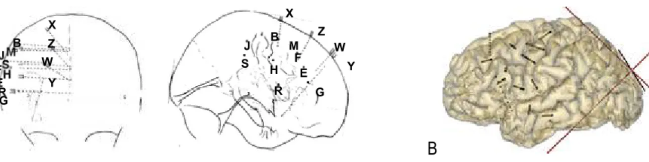
Figure 3 - SEEG scheme of a right frontal exploration as used during the first years in our center (face and profile) showing
the position of the intracerebral electrodes (A) or reconstruction of a SEEG implantation in left frontal lobe allowing the carefully visualization of the electrodes position in the patient 3D-MRI (B) as developed in the last years in our center.
Seizure outcome was considered at the last follow-up. Post-surgical outcomes were collected from the last surgery for patients who underwent multiple surgeries (at least twice). For those of patients who had a resective/disconnective surgery and were further treated by a palliative procedure, the last follow-up was set at the day before palliative intervention. For patients referred to our expert center by other neurologists, seizure outcome was assessed by direct phone call to the neurologist responsible for their clinical follow-up. When the clinical issue two years from surgery was missing, patient have been directly contacted by telephone if at least one postoperative evaluation in our center was available. Overall, the following historical data have been collected from medical files: patients’ general characteristics, disease and AED history, age at epilepsy onset, age at surgery, year of surgery, epilepsy duration, seizures frequency before surgery, data related to anatomical brain MRI, SEEG and FDG-PET if performed. Type of surgery, its location and side as well as the number of procedures for
MB J S H F E R G Y X Z W M B J S H F E R G Y W Z X
each patient, histopathology, follow-up duration and outcome at last follow-up according to the Engel score were carefully collected. We also specified palliative techniques after surgery if necessary. A data spreadsheet was completed by converting data for each variable into a numerical or alphabetical value (e.g., no MRI lesion = 0; MRI lesion =1). In case of contradictory information between database and the medical file, original medical letters or reports were used as reference document.
2.4 Data analysis
In our study, we analyzed the following variables: age at surgery by distinguishing two populations, pediatric (i.e. < 18 years) and adult population (in addition, for prognostic evaluation purposes, we also considered subgroups ranging from 0 to 3 years, 4 to 6 years, 7 to 13 years, 13 to 17 years, 18 to 40 years and over 40 years); epilepsy duration, calculated from the first epileptic seizure to the date of surgery; seizure frequency classified into three groups: rare to monthly, weekly and daily; and accordingly to brain MRI results we classified patients into two groups, lesional or non-lesional. Regarding the presurgical evaluation we considered if patients underwent or not invasive recordings (SEEG done or not).
Concerning surgical procedures, we considered the type of surgery, i.e. resection, disconnection or both, the side of surgery right or left and the site of the resection temporal lobe resection (lobar or partial including or not the mesiotemporal structures), or extra-temporal lobe resection (including frontal, parietal, occipital and insular). In our analysis, we included the multilobar resections in the extra-temporal group.
Histopathological data were divided into four groups: i)hippocampal sclerosis (HS); ii)LEAT (dysembryoplastic neuroepithelial tumor, gangioglioma, pleomorphic xanthoastrocytoma, pilocytic astrocytoma, angiocentric glioma, hamartoma...), iii)malformation of cortical development (MCD) (mainly FCD and some cases of heterotopia); iv)other pathology. The “other pathology” group included glial scars, vascular pathologies (cavernoma...), cortical tubers in patient with tuberous sclerosis complex,
Rasmussen encephalitis, dual pathology (i.e. HS association with another histopathological lesion), double pathologies (i.e. two types of lesions not including HS).17,18
The analysis regarding seizure outcome included only patients with a minimum postoperative follow-up of two years, as recommended in Engel’s classification (Appendix 1).19,20 We considered as free of
seizures and therefore with a good postoperative outcome, patients Engel class I at last follow-up. Seizures occurring within three months after surgery were considered symptomatic and not included in the analysis. In case of doubt about the patient's Engel class ranking, the case was discussed with neurologists’ team to rule on the assigned score. Usual post-surgery follow-up in our center was organized at 3, 6 and 12 months after the surgery and then on an annual basis.
To study the influence of presurgical assessment advances on patients’ clinical profiles and seizure outcome (i.e. the secondary objective of the study), for a given variable of interest, we compared the period before and after a relevant modification in our presurgical exams, regardless the duration of the follow-up. For MRI, we defined two groups: patients evaluated with or without 3T-MRI; for the FDG-PET, we compared patients who did or not benefit from this exam.
For functional tests performed to lateralize language and memory, we analyzed all patients, classing them in the Wada test or f-MRI subgroups, depending on the years of presurgical assessment, Wada test being switched to f-MRI in the local history.
2.5 Statistics
The main result studied the Engel’s score as a qualitative modality, given as a percentage of the patient population. For subgroup analysis, a Chi-squared test has been used for categorical variables and a Student-t independence test on the mean for quantitative variables. In case of abnormal data distribution for a quantitative variable highlighted by a normality test (Shapiro-Wilk) we performed a Mann-Whitney test. We did not have to use the exact Fisher test for the different categorical modalities. In case of continuous variable compared to categorical variable with more than two modalities an
ANOVA was performed. In the description of patient’s characteristics continuous variables are presented as mean, standard deviation and extreme values; categorical variables being presented as percentages with the corresponding number of subjects. Missing data on quantitative variables were ignored during calculation of means and considered as a category of their own for histopathology. Statistical analyses were done using JASP® software.
3. Results
3.1 Population characteristics
Seven hundred and four patients (350 men and 354 women) were operated for focal DRE between 1990 and 2017 and were included in the present study [figure 4]. One hundred and fifty-three (22.7%) were children. Mean age at epilepsy onset was 11 years (standard deviation (SD) +/-8.9) and epilepsy duration before surgery was 16.9 years (SD+/-10.6). Seizure’s frequency distribution in this population was homogeneous within each group (i.e. rare to monthly, weekly and daily), respectively around one-third of the overall population.
Figure 4 - Study flow chart.
Five hundred and fifty-three patients (78.6%) had a positive MRI while 151 patients (21.4%) had non-lesional epilepsy. Four hundred and forty-seven patients (63.5%) had intracranial EEG exploration by SEEG before surgery compared to the 257 patients (36.5%) for whom this exploration was not considered necessary after the initial assessment or could not be performed.
Resection was the main surgical procedure for 90.5% of the patients, whereas 9% underwent disconnection, this practice having been used mainly between 1999 and 2002. Temporal lobe resection was the most common surgical treatment (428 patients, 60.8%) compared to extra-temporal surgical procedures (276 patients, 39.2%). Among this latter group, one hundred and twenty-nine patients (18.3%) had a multilobar resection and 147 patients (20.9%) had a unilobar resection, mainly frontal for 114 patients (16.2%). Surgery lateralization was right for 380 patients (54%) and left for 321 patients (45.6%).
Total patients n = 899
Included patients n=704
More than or equal to 2 years follow-up n=479
Less than two years follow-up n=225 i.e. lost to follow-up Excluded patients : 195 - VNS 80 - DBS 16 - callosotomy 2 - radiosurgery 2 - only thermocoagulation 19 - no intervention 49 - insufficient data 27
Variable Total cohort n=704 ≥ 2 yrs follow-up n=479 < 2 yrs lost to-follow-up n=225 P-value Sexe (%) Male 49.7 (350) 48.9 (234) 51.6 (116) 0.5 Female 50.3 (354) 51.1 (245) 48.4 (109)
Age at surgery (years) 27.8 +-12.2
(1.5-63.9) 28.4+-12.8 (1.5-63.9) 26.7+-10.8 (2-61.3) 0.1 Children (%) 21.7 (153) 23 (110) 19.1 (43) 0.2
Age at epilepsy onset (years) 11.0+-8.9 (0-50) 11.2+-9.2 (0-50) 10.6+-8.4 (0-40) 0.5
Epilepsy duration (years) 16.9+-10.6 (0.5-56) 17.1+-11.3 (0.5-56) 16.1+-8.8 (1-49.3) 0.7
Seizures frequency (%) Monthly/rare 32.5 (229) 38.4 (184) 20.0 (45) <0.001 Weekly 36.9 (260) 34.0 (163) 43.1 (97) Daily 30.4 (214) 27.3 (131) 36.9 (83) Missing data 1 1 0 Febrile seizure (%) 21.9% (154) 24% (115) 17.3% (39) <0.05 MRI outcome (%) Lesion 78.6 (553) 78.3 (375) 79.1 (178) 0.5 No lesion 21.4 (151) 21.7 (104) 20.9 (47) SEEG (%) Yes 63.5 (447) 63.5 (304) 63.6 (143) 0.8 No 36.5 (257) 36.5 (175) 36.4 (82) Surgery type (%) Resection 90.5 (637) 90.6 (434) 90.2 (203) 0.7 Disconection 8.9 (63) 9.0 (43) 8.9 (20) Mixed 0.6 (4) 0.4 (2) 0 .9(2) Surgery location (%) Temporal 60.8 (428) 61.0 (292) 60.4 (136) 0.3 Multilobar 18.3 (129) 16.9 (81) 21.3 (48) Extra-temporal 20.9 (147) 22.4 (107) 18.1 (41) Frontal 16.2 (114) 17.1 (82) 14.2 (32) Parietal 1.7 (12) 1.5 (7) 2.2 (5) Occipital 1.6 (11) 1.7 (8) 1.3 (3) Insular 1.1 (8) 1.7 (8) 0 Other 0.3 (2) 0.2 (1) 0.4 (1) Multiple surgeries (%) 7.4 (52) 7.3 (35) 7.6 (17) 0.9 Surgery side (%) Left 45.6 (321) 48.2 (231) 40.0 (90) 0.1 Right 54.0 (380) 51.4 (246) 59.6 (134) Other 0.4 (3) 0.4 (2) 0.4 (1)
Palliative care after surgery (%) 3.0 (21) 3.1 (15) 2.7 (6) 0.7
Histopathological findings (%) HS 18.0 (127) 19.2 (92) 15.6 (35) <0.001 LEAT 20.2 (142) 12.7 (61) 35.1 (79) MCD 18.3 (130) 22.3 (107) 9.8 (22) Other 30.8 (217) 28.8 (138) 34.1 (77) Gliar scare 5.5 (39) 6.3 (30) 4.0 (9) Vascular 3.4 (24) 2.1 (10) 6.2 (14) Cortical tubers 1.1 (8) 1.0 (5) 1.3 (3) Encephalitis 0.9 (6) 0.4 (2) 1.8 (4) Cryptogenic 9.9 (71) 7.9 (38) 14.2 (32) Dual pathology 7.7 (54) 8.4 (40) 6.2 (14) Double pathology 2.0 (14) 2.7 (13) 0.4 (1) Missing data 12.6 (89) 16.9 (81) 5.3 (12)
Table 1 – Clinical characteristics and data from the presurgical assessment. Data are means +- standard deviation
(minimum/maximum) or % (number of patients). P-value compares patients with less or more than 2 years follow-up calculated by the chi-square or the Student t-test (age at epilepsy onset) or with the Mann-Whitney test (age at surgery and epilepsy duration), Dual pathology: HS associated with another principal pathology. Double pathology refers to two etiologically independent pathologies (HS not included).
Multiple surgeries were indicated for 52 patients (7.4%) because of an incomplete resection of the EZ. Some patients classified Engel class IV after surgery, as to say no surgery effect or even epilepsy worsening, were treated with palliative method, which represented 21 patients (3%) of the cohort. Histopathological data was missing for 89 patients (12.6%), especially related to the disconnection practice. The available data shown 18% (127 patients) of HS, 20.2% of LEAT, 18.3% of MCD, the remaining 30.8% representing the "other" group, including 5% of glial scars and 3% of vascular lesions. The majority of histopathological findings in the MCD group were FCD, type I or II, nodular heterotopias was the only other malformation subtype in this cohort (n=11). Ten percent of the histopathological analyses did not reveal any lesion [Table 1].
Among the 704 patients included into our study, 479 had more than 2 years follow-up and thus considered for surgery outcome evaluation. The other 225 patients had less than 2 years follow up and thus loss to follow-up.
Comparing the group of 479 patients included for the surgery outcome and the group of 225 patients lost to follow-up, characteristics did not significantly differ for most of variables [Table 1] except for seizure frequency and histopathological results [Figure 6]. In the lost to follow-up group, a lower proportion of patients presented “rare to monthly” seizures frequency [Figure 6A and 6B], and histopathological data showed a lower proportion of MCD, and a higher proportion of LEAT [Figure 6C and 6D]. It is important to underline that in this latter group (225 patients), 85% of patients were operated on before 1999; in the group with two years minimum follow-up (479 patients) 50% underwent surgery after 2004.
3.2 Surgery outcomes
Surgery results were evaluated for patients with a minimum of two-years of postoperative follow-up, i.e. 479 patients. Mean follow-up duration was 8.5 years ([2-26.61] SD +/-6.1).
Among these patients, 339 patients (70.8%) were considered free of disabling seizures (Engel class I); 270 (56.4%) were completely seizures free since surgery (Engel class IA); 30 (6.3%) had non disabling simple partial seizures only since surgery, i.e. only aura (Engel class IB); 15 (3.1%) had some disabling seizures after surgery, but seizure free of disabling seizures for at least 2 years (Engel class IC) and 24 (5%) had generalized convulsions after antiepileptic drug withdrawal only (Engel class ID). Considering patients who were not seizure free at 2 years follow-up from surgery, 53 (11.1%) were classified as "almost seizure free" (Engel class II), 29 (6.1%) showed partial improvement (Engel class III) and 58 (12.1%) have had no benefit from surgery (Engel class IV) [Figure 5].
Interestingly, seizure outcome did not statistically improve in our center between 1990 and 2017 (p = 0.6).
Figure 5 – Seizure outcome (Engel outcome scale) at last follow-up among studied patients. Result given as
percentage only for a minimum of two years of follow-up.
57% 6% 3% 5% 11% 6% 12% IA IB IC ID II III IV
Figure 6 - Statistically different data between the two groups defined by follow-up duration. Seizures frequency in the
less than two years up group (A) and more than two years (B). Pathology distribution in the less than two years follow-up grofollow-up (C) and more than two years (D), in x-axis number of patients. P < 0.001 for both variables. Other: glial scar, vascular pathology, encephalitis, no lesion, tubers, double pathology, dual pathology.
No statistically significant difference was observed with respect to the age at surgery comparing the pediatric and adult population: 65.5% (72 patients) of children were classified Engel class I compared to 72.4% (267 patients) of adults (p=0.2). Same results were observed among pediatric subgroups (p=0.2). In our studied population, epilepsy duration before surgery did not seem to influence the seizure outcome (p=0.7).
We found a statistically significant difference in seizure outcome according to seizure frequency before surgery: the worst the seizure frequency was before surgery, the worst was the seizure outcome (p < 0.001). This seemed to be a prognostic element in our cohort [Figure 7A].
0 10 20 30 40 50 60 70 80 90 100
Daily Weekly Rare to monthly
P A T IE N T N U M B E R
LESS THAN 2 YEARS FOLLOW-UP
A
0 20 40 60 80 100 120 140 160 180Daily Weekly Rare to monthly
P A T IE N T N U M B E R
MORE THAN 2 YEARS FOLLOW-UP
B
0 50 100 Missing data Other LEAT HS MCDLESS THAN 2 YEARS FOLLOW-UP
C
0 50 100 150 Missing data Other LEAT HS MCDMORE THAN 2 YEARS FOLLOW-UP
MRI positive patients had a significantly better outcome since in this group, 284 patients (74.7%) had an Engel class I score versus 55 patients (55.6%) in the negative MRI group (p<0.001) [Figure 7B]. Only eighteen percent of patients had a negative MRI in the temporal epilepsy subgroup, versus almost 30% of patients in the extra-temporal epilepsy subgroup.
We compared seizure outcome among patients who underwent SEEG and those who did not: a statistically significant difference between them is observed. Among the 304 patients explored by SEEG, 191 patients (63%) were in Engel I group. On the contrary, among the 175 patients who were not explored by SEEG, 148 patients (84.1%) were in the Engel I group (p<0.001) [Figure 7C]. This result could be explained by the supposed epileptogenic network complexity which justified the use of invasive exploration to determine the EZ.
Seizure outcome also significantly differed depending on surgery location, with a higher proportion of patients in the temporal lobe surgery subgroup in Engel class I (78.8%) compared to 58.3% of patients in the extra-temporal surgery group (p<0.001) [Figure 7E]. Frontal epilepsy subgroup had 58.5% of patients in Engel class I.
Considering the type of surgical procedure, there was no difference in seizure outcome between disconnection or resection subgroups (p=0.1).
There was also no statistical difference for seizure outcome according to the pathology subtype (p=0.1), whether it was HS, MCD or a LEAT (Figure 7D).
Three other subgroups have been studied, focusing on two parameters: lesional or non-lesion focal DRE (i.e. MRI positive or negative) and histopathological positive or negative results. Among of 375 patients with histopathological reports, 297 patients were MRI positive and histopathological positive; 13 patients were MRI negative and histopathological negative; 65 patients were MRI negative and histopathological positive. A statistically significant difference was found between these three groups with a better seizure outcome in case of lesional epilepsy and positive histopathological findings. None
of the patients with negative MRI and negative histopathological results was seizure free after surgery [Figure 7F]. 0 20 40 60 80 100 120 140 160
Rare/monthly Weekly Daily
P A T IE N T N U M B E R Engel class
A
I II, III et IV 0% 10% 20% 30% 40% 50% 60% 70% 80% 90% 100% MRI + MRI-Engel classB
I II, III et IV 0% 10% 20% 30% 40% 50% 60% 70% 80% 90% 100% SEEG No SEEG Engel classC
I II, III et IV 0% 20% 40% 60% 80% 100% MCD HS LEAT Engel classD
I II, III et IV 0% 20% 40% 60% 80% 100% Temporal Extra-temporal Engel classE
I II, III et IV 0% 20% 40% 60% 80% 100%IRM+/Histo+ IRM-/Histo +
IRM-/histo-Engel class
F
Figure 7 - Seizure outcome subgroups analysis. Better seizure outcome was suggested in the lower seizures frequency
groups, p<0.001 (A), in case of lesional epilepsy, p<0.001 (B) (MRI + means lesion on MRI, MRI – no lesion on MRI), also in patients not explored by SEEG, p<0.001 (C), in case of temporal surgery, p<0.001 (E). There was no statistically significant difference according to the pathology subtype (D). Worst prognosis was found in patient with both, MRI and histhopathologic findings, negative (F).
Seizure free patients seemed to decrease proportionally to follow-up duration (p<0.001): when the available follow-up was 2 to 5 years, 83.4% of patients had an Engel class I, whereas they were only 71.5% when the available follow-up was 5 to 10 years and no more than 54.4% when this follow-up was of more than 10 years.
3.3 Patients and practices evolution
Epilepsy surgery practices at the Grenoble University Hospital evolved following the emerging pre-surgical assessment techniques. We now describe some data concerning the evolution of the overall studied population (704 patients).
Historically, from the first period in which several patients were addressed to our center for epilepsy surgery, thereafter the number of patients operated on per year decreased before stabilizing around twenty procedures per year since 2000 [Figure 8A].
With respect the histopathological findings, in the overall population we observed a progressive decrease of LEAT and an increase in the number of MCD, with a majority of FCD, while the number of patients with HS remained stable in proportion [Figure 8B]. Focusing on the pediatric population, it remained stable in number over years and concerning the histopathological findings, a higher proportion of MCD, was observed with respect the adult patients (33.3%, 51 patients; 14.3%, 79). LEAT represented the second most frequent cause of focal epilepsy in children in this cohort (28.8%) [Figure 8C].
A significant shorter epilepsy duration was found in LEAT and MCD histopahtological subgroups (p < 0.001). On the contrary the HS histopathological subgroup showed the longer epilepsy duration before surgery [Figure 8E]. Number of non-lesional focal DRE in our cohort remained stable over years.
Concerning the epilepsy localization, the patients operated on for extra-temporal lobe epilepsy became more frequent over years. Despite this finding, the characteristics of this population seemed to stay stable. The proportion of extra-temporal lobe epilepsy in children is significantly higher with respect the adults (56,2% and 34,5% respectively; p<0.001). Regardless the age at surgery, extra-temporal lobe epilepsy group showed an earlier epilepsy onset (p<0.001, Mann-Whitney), and a higher seizure frequency (66% were classified “daily”). The proportion of performed SEEG was also higher in the extra-temporal surgery subgroup (p<0.001) and a higher proportion of MCD (p<0.001) was found [Figure 8D].
0 5 10 15 20 25 30 35 40 45 50 P at ie nt n um be r Year A Extra-temporal Temporal f-MRI Stop
Wada FDG-PET3T MRI 1,5T MRI
Figure 8 – Practices evolution. Number of operated patients per year distributed according to the surgery location. Arrows
correspond to the year in which the technique was introduced or stopped for the Wada test. There seem to be a trend towards an increase in the proportion of extra-temporal surgery (A). Number of performed surgeries per year according to the pathology subtype in the overall population (B). Evolution of the number of operated children according to the pathology subtype (C). Number of operated patients for temporal epilepsy with HS (TLE + HS) and extra-temporal epilepsy with FCD (ETLE + MCD) (D). Evolution of the average epilepsy duration before surgery according to the pathology subtype.
The presurgical work-up used in our center to define the EZ and to limit postoperative complications evolved between 1990 and 2017.
A 0.5T brain MRI was used from 1990 to 1992, switched to a 1.5T-MRI until 2010 before a 3T high field MRI from 2010, helping to eventually better locate an epileptogenic lesion.
FDG-PET has been realized at the Grenoble University Hospital since 2010, being progressively sistematicly used. 0 5 10 15 20 25 30 35 40 45 50 P at ie nt n um be r
B
MCD HS LEAT 0 5 10 15 20 25 30 P at ie nt n um be rC
MCD HS LEAT Total 0 2 4 6 8 10 12 14 19 90 19 91 19 92 19 93 19 94 19 95 19 96 19 97 19 98 19 99 20 07 20 08 20 09 20 10 20 11 20 12 20 13 20 14 20 15 20 16 20 17 P at ie nt n um be rD
TLE + HS ETLE + MCD 0 5 10 15 20 25 30 35 E pi le ps y du ra tio n av er ag e (y ea rs )E
MCD HS LEATTo analyze the contribution of the 3T-MRI and the FDG-PET in our practice, we compared seizure outcome before 2010 and after 2010 in the two years minimum follow-up population (479 patients). Seizure outcome did not show any difference, with the same proportion of patients Engel class I (70.8%) in both groups (225 patients before 2010 and 114 after 2010), compared to 29.2% considered as not seizure free (93 patients and 47 respectively, p = 1).
Considering the FDG-PET subgroup after 2010, the analysis did not reveal any significant difference on seizure outcome in the FDG-PET group with respect the non-FDG-PET group (65.3%, 77 patients and 74.1%, 40 patients in Engel class I respectively) (p = 0.4, two missing data).
We therefore conducted a statistical analysis in order to determine whether these new techniques had changed patients clinical characteristics (age at epilepsy onset, epilepsy duration before surgery, use of SEEG, localization of surgery and the pathology). The comparaison of the before and after 2010 groups did not show any statistical difference on age at epilepsy onset, age at surgery and epilepsy duration which remained stable. Proportion of adult and pediatric population did not differ as well (p=0.1). SEEG was used for 66% of patients before 2010, and for 55.7% of patients explored from 2010 onwards (p<0.001). Surgery location also evolved. A significant difference was found before and after 2010 with a decreasing proportion of temporal lobe surgery over years. Before 2010, 64% (339) of patients had a temporal lobe surgery and 36% (191) of patients underwent extra-temporal lobe surgery. From 2010, 51.1% (89) of surgeries concerned the temporal lobe and 48.9% (85) concerned extra-temporal lobe resections. (p <0.001) [Figure 11A].
Same analyses were carried out on patient’s characteristics comparing thoses who had or did not have a FDG-PET in their preoperative assessment after 2010. The only difference between these two groups concerned the histopathological findings (p<0.001). In the FDG-PET group, number of MCD was higher, 49 patients (41.5%) compared to 16 patients (29.6%) in the non-FDG-PET subgroup, as well as the number of HS, higher in the FDG-PET subgroup (5% of missing data in both groups) [Figure 11B].
A B
Figure 9 – 15 year-old-boy suffering from DRE suggesting a frontal lobe involvement. Scalp EEG showed right>left frontal
lobe interictal abnormalities and ictal discharges. 3T-MRI showed a right orbitofrontal cortical thickening with a subcortical white matter tapering toward the ventricke - arrow (A). FDG-PET confirmed a clear hypometabolism on the right orbitofrontal region (B). Patient was operated on without invasive recordings (lesionnectomy). The patient is seizure free.
A B C
D
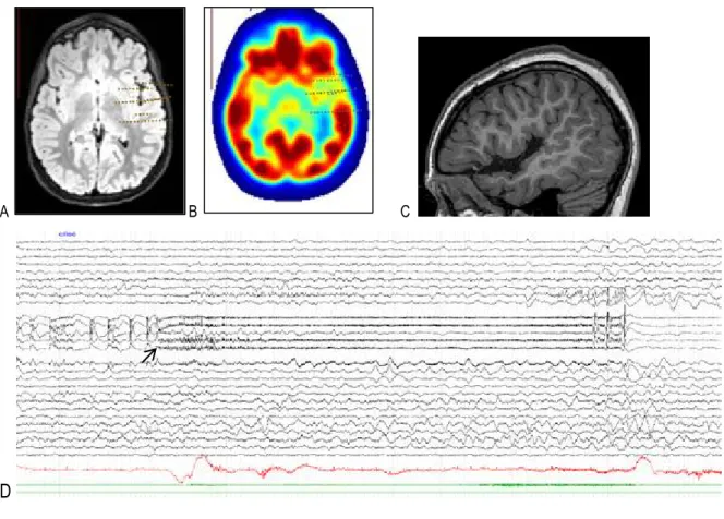
Figure 10 – 8-year-old-girl suffering from a DRE. Seizure semiology (non lateralized auditory hallucinations) suggested a
temporal lobe involvement (STG). Interictal abnormalities and ictal discharges showed a left temporal lobe involvement. 3T-MRI showed a white matter blurring on anterior left STG (A). FDG-PET confirmed the suspicion of a FCD in the same area and allowed a carefully exploration with intracerebral electrodes (B). SEEG recordings (i.e. well localized low voltage fast activity, arrow) confirmed the anterior STG involvement during seizures (D) allowing a tailored resection (C). The patient is seizure free.
The development of functional neurological testing, helping to locate areas of interest as language and memory, can be separated into three different periods. The Wada test was used from 1990 to 2003. F-MRI for language was introducted from 2001, initially (2 years) together with the WADA test (validation of f-MRI in this indication) and since 2003 as the unique tool for language and memory presurgical evaluation. Two populations, before 2001 and after 2003, have been therefore compared. One of the parameters which could have been potentially influenced by functional exploration was surgery lateralisation. Indeed, a significant difference was found regarding surgery lateralisation: 61.8% (214) of patients who had a hemispheric dominance assessment for language using Wada test were operated on the right side, and 38.2% (132) on the left side. Conversely, 46.3% (137) of patients who had hemispheric dominance assessement for language with f-MRI were operated on the right side, and 53.7% (159) on the left side (p<0.001) [Figure 11C].
0% 10% 20% 30% 40% 50% 60% 70% 80% 90% 100%
3T MRI Before 3T MRI
A
Temporal Extra-temporal 0 10 20 30 40 50 60 70 MCD HS LEAT Other P at ie nt n um be rB
FDG-PET No FDG-PETFigure 11 – Recent technical advances’ impact on presurgical assesment. Histogram showing the proportion of patients
operated for temporal or extra-temporal epilepsy according to the subtype of MRI (high field 3T or lower 0.5 or 1.5T) (A). Histogram illustrating the number of diagnosed patients per pathology according to whether or not a FDG-PET had been performed (B). Histogram comparing the number of operated patients classified among surgery’s lateralisation according to the use of WADA test or f-MRI (C). P-value <0.001 for statistical analyses associated with histograms.
4. Discussion
4.1 Seizure outcome
Post-operative seizure outcomes for patients, adults and children, who underwent surgery for DRE in our hospital between 1990 and 2017 account for 70.8% of Engel Class I (free of disabling seizure) and 56.4% of Engel Class IA (completely seizure-free since surgery) at last clinical evaluation with a minimum of two years follow-up. These results are similar to those already published in two randomized clinical trials, with 58% to 73% of seizure free patients at one and two years after surgery respectively.6–
8 Overall, results are heterogeneous, ranging from 34% to 76% of seizure freedom after surgery for
focal epilepsy (temporal, extra-temporal or both) related to variable etiologies in retrospective non controlled, multicenter or large single-center studies, as recently reported in a clinical review. The median of seizure free patients included across all included non-controlled observational studies is 62.4%.7,21 0 50 100 150 200 250
fMRI Wada test
P at ie nt n um be r
C
Right LeftGoing through the analysis of possible prognostic variables in our studied cohort, the age at surgery did not influence the seizure outcome, since seizure free outcomes are similar between children and adults. Indeed, 65.5% of children and 72.4% of adults were in Engel class I (p=0.2), as underlined in other studies.21 In our study, the seizure frequency before surgery could be considered as a favorable
prognostic variable, since the seizure outcome was better in those patients with the lower seizure frequency (rare or monthly seizures) before surgery, this difference being statistically significant (p < 0.001). This data was not reported in other studies.21Shorter epilepsy duration was reported in case of
LEAT, which could be explained by an easily referral context and process until the surgical indication. Concerning the localization of the surgery, extra-temporal lobe surgery was much less rewarding than temporal lobe surgery. Indeed, 58.3% of patients were in Engel class I after surgery of extra-temporal regions, compared to 78.8% of patients in Engel class I after temporal lobe resection. Again, these results are similar to those published in the literature. Indeed it’s widely accepted that patients with temporal lobe epilepsy have the great likelihood of becoming seizure free after surgery. A meta-analysis including 40 studies found 66% of patients with good seizure outcome after temporal lobe surgery22
while another meta-analysis of nearly 1200 patients reported 45.1% of patients free of seizures in case of frontal lobe surgery.23,24 The lower rate of seizure free patients after extra-temporal lobe surgery is
usually explained by functional constraints (i.e. motor strip or functional language area) and thus a possible incomplete resection of the EZ.
Lesional epilepsy was associated with a better outcome than non-lesional epilepsy in our population, with 74.7% versus 55.6% of patients free of disabling seizures in MRI-positive versus MRI-negative patients respectively, accordingly with the previous published data. A meta-analysis including 2860 patients reported a 2.5-times greater likelihood of a seizure free outcome in case of resection of a discrete lesion as compared with when the MRI is negative.25This finding could be easily explained by
the fact that the EZ is more likely co-located with an identifiable lesion on MRI and thus better delineated and possibly completely resected. In addition, patients with MRI lesions may benefit earlier
from surgery and a shorter duration of epilepsy may entail a better outcome. Nevertheless, this prognostic variable was not found in our cohort as well in others and needs to be confirmed. Nevertheless, it is important to underline that in a study conducted during 25 years in 36 European Centers, in 75,9% of patients epilepsy started before 18 years of age and that 72.5% underwent surgery as adults, which is too late to prevent or reverse the disabling consequences of seizures or to affect quality of life.26 A difference with previous publications was found on the proportion of non-lesional
temporal lobe epilepsy in our cohort, that is 18% compared to 30% in the literature.25This difference is
probably related to the criterion used to judge a negative or positive MRI. In fact, in our cohort, MRI images were analyzed by a neuroradiologist and by the epilepsy surgery team including neurologists and neurosurgeons, taking into account the EEG interictal and ictal data and the ictal clinical semiology, thus leading to identify an epileptogenic lesion on an MRI initially considered as normal. Additionally, in our cohort, the percentage of negative MRI is higher in patients with extra-temporal lobe epilepsy (one third of patients), which subsequently could explain the lower rate of seizure freedom in this group. Outcomes for epilepsy surgery vary based on the underlying pathology. Some authors had shown that HS and LEAT resection was associated with better surgical outcome than for other histological subgroups.27We did not found this result in our cohort. We wonder if the significant proportion of lost to
follow-up patients in the first years of inclusion could explain this finding, since LEAT surgery was more frequent at that period. The prognosis of FCD has been evaluated as good as LEAT and HS in several series and similar in any subtype.28Considering the MCD subgroup, we note the high value of average
epilepsy duration for the period 1999-2002, possibly explained by very few patients for this variable in absence of histopathological analysis (disconnections). These data should therefore be interpreted with caution [Figure 8E].
We checked results comparing three subgroups defined as following: positive MRI and positive histopathology, negative MRI and positive histopathology and negative MRI and negative histopathology [Figure 7F]. The worst seizure outcome was in the last group, underlying the epilepsy
complexity in these cases and thus the possibility of either a false localization of the EZ or the presence of a more extended EZ in negative MRI and negative histopathology patients.
Concerning the long-term follow-up on seizure outcomes, our study revealed a gradual reduction in the proportion of patients remaining seizure-free when the available follow-up was 5 to 10 years [Figure 12]. Unfortunately, data was not available regarding the seizure relapse which would have permitted to perform a survival analysis and therefore conclude whether relapses are more frequent after two years or before two years after surgery. This result should also be analyzed in the light of lost to follow-up patients with a significant risk of error. It can be hypothesized that seizure free patients were easily lost to follow-up in the absence of seizure relapse and once AED is discontinued or stabilized. However, previous studies focusing on this subject estimated a 66% of seizure free patients 15 years after surgery, the majority of relapses occurring within the first 24 months.29Another study with an average
follow-up of 26 years showed favorable results with 48% of patients classified Engel I with a significant improvement in the associated quality of life.27
Figure 12 – Post-operative seizure outcome evolution over years. Histogram showing the number of seizure free or not
free patients depending on follow-up duration. There is a trend towards a reduction in time of the proportion of seizure free patients. 0 20 40 60 80 100 120 140 160
2 to 5 years 5 to 10 years > 10 years
P at ie nt n um be r Follow-up duration
A European cohort study published in 2018, which included data from 16 centers and compared seizure outcome between 1990s and 2010s, suggested a modest but significant recent improvement in the proportion of good outcome patients after epilepsy surgery, after adjusting on the fact that surgeries performed after 2010 became gradually more complex. There was also evidence of a decrease in epilepsy duration between these two periods.30Our results did not support these conclusions.
Comparing epilepsy surgery outcomes between studies is challenging because there is little consistency in outcome measurement schemes and the seizure free definition may vary. The Engel seizure outcome scale is the most frequently used (50% of the studies), but another one was the ILAE scale, which classifies patients into six groups according to the percentage of seizure reduction (Appendix 2). The inherent differences in scales may therefore partly explain the differences in results found between studies. For instance, in the ILAE scale authors could consider patients 1, 2 and 3 or 1 and 2 as good prognosis, whether with the Engel scale the choice may differ between those who include ID as a good outcome or not.16 Another potential source of difference between study results was the timing of
outcome measurement. A critic that could also be made is that this method of seizure outcome evaluation does not consider surgery’s impact on quality of life. We used the Engel score at last follow-up and not two years for all patients because results are known to be worst over years: this choice therefore put us in the situation of maximum bias limiting the risk of overestimation of our good results. In a cohort of more than 2500 patients, another presurgical quality management factor was proposed, the use of the operated patient rate at initial assessment: indeed epilepsy surgery centers could select patients with a good postoperative prognosis, rejecting patients whose record were in favor of a bad prognosis.31By doing this, authors wished to take into account the overall activity and not only good
surgical candidates.
4.2 New surgical candidates
Our results showed a global evolution of epilepsy surgery practices in Grenoble University Hospital between 1990 and 2017. The number of surgeries per year increased during the first period, then they
decreased before stabilizing in the last years. We can possibly explain this evolution by two factors. First, in the early 1990s, the neurological team was bigger than the present one and more patients were addressed to our center not only from France but also from Italy, where the epilepsy surgery program developed thereafter. Secondly, in 1990s there were only four centers performing epilepsy surgery in France: Saint-Anne in Paris, Rennes, Bordeaux and Grenoble. This practice thereafter extended to more or less seventeen centers, the recruitment of each center becoming less important.
Patients’ clinical profile evolved over the years and new surgical candidates emerged, partly due to technical advances in the preoperative assessment. Age at surgery and epilepsy duration remained stable during the studied period. The main changes we identified concerned the pathology subtypes and the surgery’s location. Indeed, FCD pathology and thus extra-temporal lobe surgery subtype predominated in more recent years compared to early years when LEAT and HS were the most frequent etiology [Figure 8D]. 3T-MRI and FDG-PET both contributed to this evolution, presumably highlighting previously not visible lesions.
3T-MRI allowed to better identify FCD and HS previously unseen or unclear with a lower magnetic field device (1.5 Tesla). This technique had a better spatial resolution related to a higher signal to noise ratio with high field devices. This detection was facilitated by T1 or T2-FLAIR image analyses.32Contribution
of 3T-MRI in case of previous negative MRI had already been evaluated in several studies suggesting the value of this examination in case of negative 1.5T-MRI for the detection of MCD and mainly FCD.33
FCD were the most common malformations found in children with focal DRE in large series as in our cohort, accounting for about 30% of diagnosed pathologies in this population.34Interestingly, FCD type
II are mainly localized outside the temporal lobe, particularly in the frontal lobe, which may partly explain the evolution concerning the localization of surgery over years.35
FDG-PET, highlighting hypo-metabolic regions, together with clinical, EEG and MRI findings may help to localize the EZ. It is known that approximately 80% of patients suffering from temporal lobe epilepsy showed a temporal lobe hypometabolism on FDG-PET.36This is important not only to corroborate the