Persistently infected cattle stabilise bovine viral diarrhea
virus leading to herd specific strains
C. Hamersa, C. Lecomtea, G. Kulcsarb, M. Lambota, P.-P. Pastoreta
a Dept. of Veterinary Immunology-Vaccinology, Faculty of Veterinary Medicine, University of Liège,
Bld de Colonster, 20 Bat B 43 bis, 4000 Liege, Belgium
b State Control Institute for Veterinary Biologicals, Drugs and Feeds, Budapest, Hungary
KEYWORDS: Cattle-viruses; Bovine viral diarrhea virus; Persistence; Immunotolerance
ABSTRACT
Animals persistently infected with BVDV are important in the epizootiology of the Bovine Viral Diarrhea (BVD) because they are a permanent source of contamination within a herd. These animals produce large quantities of virus and have, therefore, been proposed as responsible for generating antigenic variability. However, limited studies have failed to detect antigenic or genetic changes in viruses isolated at different time from persistently infected animals. One hypothesis to account for this stability is that the immunotolerance is accompanied by a selection against antigenic change. The presence of an immunotolerant persistently infected (IPI) animal in a herd would in turn lead to herd specific strains. To verify this hypothesis, we compared 17 BVDV strains isolated from IPI animals from 3 herds of Eastern Belgium. The comparison was based on the sequence of a 389 bp fragment of E2 - a gene encoding for a highly variable glycoprotein. Sequences were strongly conserved within herds but were quite different between herds, indicating that BVDV herd-specific strains do exist and are associated with the presence of IPI animals.
Introduction
Bovine Viral Diarrhea virus (BVDV) belongs to the genus pestivirus, Flaviviridae family, which also includes two other common viruses of livestock: classical swine fever virus (CSFV) and border disease virus (BDV). The three RNA viruses are genetically closely related yet display considerable diversity between and within each virus species (Paton, 1995).
Sequence variability is unevenly distributed both with respect to the genome and to individual genes. The two most variable genes are E2 and NS2 genes, while NS3 and parts of the 5tuntranslated region are highly conserved. (Paton, 1995). E2 gene encodes for a 53 kd glycoprotein which appears to be a major antigen, inducing virus neutralising antibodies (Donis et al., 1988).
BVDV can cause disease, following either post-natal or fetal infection. Post-natal infections produce a very common, usually mild or unapparent, disease of cattle named Bovine Viral Diarrhea. Infection during fetal development may lead to the development of animals that are immunotolerant to the BVDV and persistent excretors of the virus (Liess et al., 1974; McClurkin et al., 1984). This permanent infection is generally unapparent, although some persistently infected animals will develop a specific lethal illness called Mucosal Disease (Roeder and Drew, 1984; Brownlie et al., 1984).
Persistently infected (IPI) animals have an important role in the perpetuation of BVDV infections (Roeder and Harkness, 1986). These animals multiply BVDV at a high rate of months or years (Corapi et al., 1988) and have therefore been described as BVDV producing factories. Although it might be thought that this considerable virus multiplication would generate diversity, limited studies have, on the contrary, failed to detect antigenic or genetic changes in viruses isolated at different times from IPI (Mignon et al., 1990; Edwards et al., 1991). This is consistent with the hypothesis of immunologically unimpeded multiplication of the resident virus and the immune elimination of emerging variants (Paton, 1995). This contrasts with acute infections which would favour variant BVDV able to escape the immune response (Bolin and Ridpath, 1992). If such selection really occurs, resulting effects should be particularly visible on the 53 kd glycoprotein (E2 gene), known to play a role in protective immunity. The contamination of pregnant females by a permanent excretor may result in the generation of new IPI. According to the hypothesis of strain stability in IPI, strains isolated from these newly generated IPI should be antigenically and genetically close, or even identical to the strain of the contaminator. Paton et al. (1995) suggested the existence of herd-specific strains.
In order to verify the hypothesis of herd specific strains, 17 BVDV strains were isolated from IPI in 3 herds of Eastern Belgium. Partial sequencing of the E2 gene was undertaken after RT-PCR and cloning and intra-herd genetic variation was compared to inter-herd variation.
Material and methods
IDENTIFICATION OF THE IPI
In 24 herds diagnosed positive for a BVDV infection, 49 animals were found viraemic after two consecutive tests at 3-4 weeks interval. These animals, considered as IPI, were all diagnosed in their herd of birth (Onclin et al., 1995). The study presented here involved 17 IPI from 3 herds, harbouring 5 or more IPI with a well documented history. Each farm comprised IPI animals ranging from 4 to 18 months. Each animal was identified by a letter (A, B and L) representing the herd and a digit (1-6) for the birth rank in the herd.
BIOLOGICAL CLONING AND PRODUCTION OF BVDV ISOLATES
Blood samples were centrifuged. Buffy coat were collected and frozen at -80°C. After thawing, 100 µl of these cell suspensions were seeded on subconfluent Calf Testicular (CT) cells, grown in four well multidish (Nunc), and cultured for 7 days in opti-MEM® (Gibco) supplemented with 4% horse serum, 100UI/100 µg penicillin/streptomycin per ml. Isolates underwent later three successive cloning by limiting dilution. Cloned isolates were then multiplied for stock production during 7 days on CT cells grown in tissue culture flasks (Falcon). After one freezing/thawing cycle at -80°C, cells were scrapped. Suspensions were then clarified by centrifugation at 2000 g, 30 min. and stored at -80°C. All cultures and media were screened and found exempt of pestivirus contaminants.
TYPING OF ISOLATES BY REVERSE TRANSCRIPTION (RT)-PCR AND SEQUENCING
OF AN E2 FRAGMENT
Aliquots of the stock production were seeded on CT cells grown in tissue culture flasks (Falcon) and cultured for 6 days. RNA was extracted directly from the cell culture using RNA NOW® (Biogentex). Reverse transcription was performed with the First-Strand cDNA synthesis Kit® (Pharmacia Biotech), primed with random hexanucleotides (pDN6). PCR primers corresponding
to position 2442-2463 (TGATAACAGGGGTACAAGGGC) and position 2875-2852
(ACAGTCCCTGTCCATCCTATGGG) of the NADL strain (Collet et al., 1988) were used to amplify a 389 bp fragment of the E2 gene. The temperature profile used was 35 cycles of 95°C for 1 min, 56°C for 1 min and 72°C for 1 min (with 2 s increment per cycle). PCR products were then cloned in pCR 2.1® plasmids (Invitrogen), according to manufacturer's recommendations. Plasmids were purified by alkaline lysis and two strand sequencing of the inserts was performed with T7 Sequencing Kit@ (Pharmacia Biotech), according to manufacturer's recommendations. Sequences were analysed as such after conversion into amino acid sequences. Corresponding sequences of NADL (Collet et al., 1988) Osloss (De Moerlooze et al., 1993) and SD-1 (Deng and Brock, 1992) were included as references in the analysis.
PARSIMONY METHOD
Dnapars, dnadist and protpars programs of the Phylip, phylogeny inference package (Felsenstein, 1989) were used for analysis and drawing of a tree diagram. Validity of trees was assessed with the boot-strap resampling program of the same package.
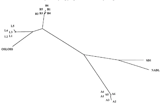
Fig. 1. Unrooted phylogenic tree representing the relationship between pestiviruses from farms A, B and L and reference strains. The tree is calculated by the maximum likelihood method, based on comparison of 389 nucleotide sequence of the E2 gene (NADL position 2463-2852). Branch lengths are proportional to inter-strain differences. Sequences of reference strains are derived from the following authors: Osloss, De Moerlooze et al. (1993); NADL, Collet et al. (1988); SD-1, Deng and Brock (1992).
Results
Sequence homology within farms was obvious: for farm A, isolates A5, A1 and A4 had identical sequences (called type A) while A6, A3 and A2, varied respectively with 1, 2 and 4 bases. For farm L, L1 and L3 sequences were identical (sequence type L), L3 and L5 had each one base difference while L4 had a two bases difference. In farm B, isolates B3 and B2 were identical. Isolates B1, B6, B5 and B4 varied very little from this standard type B sequence, with a maximum variation of 4 bases for isolate B6. Intra farm sequence identity were at least 99.5%. Interestingly, 71% of variations inside farms did result in amino acid variations.
Type A, B and L sequences were clearly different: Type A had only 74.8 and 73.3% identity with respectively type B and type L sequences, while the two later had 89.5% identity. Similar results were found when comparing amino acid sequences.
An unrooted phylogenic tree generated by maximum likelihood method is shown in Fig. 1. Other programs within the PHYLIP package (Neighbour joining, DNA parsimony, protein parsimony) gave essentially the same results. Boot-strap resampling, an assessment of the validity of trees, gave always the same farm-specific clusters, with minor variations inside herds and in the relative position of references strains. L and B appear to be more closely related to the Osloss strain than either A or the SD1 and NADL reference strains.
Discussion
Partial sequencing of E2 gene was proved to be an extremely powerful tool for studying the viruses as the method clearly segregated strains according to their herd of origin. Animals in a farm were evidently contaminated by closely related if not identical (99.5% identity) viruses. The residual variation among strains of the same herd can easily be explained by the mode of contamination of IPI, since it implies the acute infection of the mother, a condition in which virus variation is known to occur. Even then, residual mutation could lie an overestimate as artefactual mutation could have occurred during culturing or PCR.
Paton had already suggested BVDV herd specific strains on the basis of sequence similarities in ovine and bovine virus isolated on the same farm. Our results would suggest that IPI play a central role in establishing herd specificity by stabilizing BVDV strains. Consequently, this finding suggests that BVDV strain variability is the result of horizontal circulation of the virus.
Acknowledgements
We would like to thank M. Loncar technical assistance. This research was supported by grants from the 'Institut pour l'encouragement de la Recherche Scientifique dans l'Industrie et l'Agriculture' (IRSIA).
References
Bolin, S.R., Ridpath, J.F., 1992. Differences in virulence between two non-cytopathic bovine viral diarrhea viruses in calves. Am. J. Vet. Res. 53, 2157-2163.
Brownlie, J., Clarke, M.C., Howard, C.J., 1984. Experimental production of fatal mucosal disease in cattle. Vet. Rec. 114, 535-536.
Collet, M.S., Larson, R., Strick, D., Anderson, D.K., Purchio, A.F., 1988. Molecular cloning and nucleotide sequence of the pestivirus bovine viral diarrhea virus. Virology 165, 191-199.
Corapi, W.V., Donis, R.O., Dubovi, E.J., 1988. Monoclonal antibody analyses of cytopathic and non-cytopathic viruses from fatal Bovine Viral Diarrhea virus infections. J. Virol. 62, 2823-2827.
De Moerlooze, L., Lecomte, C., Brown-Shimmer, S., Schmetz, D., Guiot, C., Vandenbergh, D., Allaer, D., Rossius, M., Chappuis, G., Dina, G., 1993. Nucleotide sequence of the bovine viral diarrhoea virus Osloss strain. J. Gen. Virol. 74, 1433-1438.
Deng, R., Brock, K.V., 1992. Molecular cloning and nucleotide sequence of a Pestivirus genome, non cythopathic bovine viral diarrhea virus strain SD-1. Virology 191, 867-869.
Donis, R.O., Corapi, W.V., Dubovi, E.J., 1988. Neutralizing monoclonal antibodies to the viral diarrhoea virus bind to the 56 K to 58 k glycoprotein. J. Gen. Virol. 69, 77-86.
Edwards, S., Wood, L., Brockman, S., Ibata, G., 1991. Clinical and virological observations of a mucosal disease outbreak with persistently-infected seropositive survivors. Arch. Virol, (Suppl. 3): 125-132. Felsenstein, J., 1989. Phylip: phylogeny intference package (version 3.2.). Cladistics 5, 164-166.
Liess, B., Frey, H.-R., Kittsteiner, H., Baumann, F., Neumann, W., 1974. Observations and investigations on mucosal disease of cattle, a late stage of BVD-MD virus infection with immunobiological explanation and criteria of a slow virus infection? Deutsche Tierarztliche wochenschrift 81, 481-487.
McClurkin, A.W., Littledike, E.T., Cutlip, R.C., Frank, G.H., Coria, M.F., Bolin, S.R., 1984. Production of cattle immunotolerant to BVD virus. Can. J. Comp. Med.; 48, 156-161.
Mignon, B., Schwers, A., Waxweiler, S., Boulanger, D., Dubuisson, J., Brownlie, J., Pastoret, P.P., 1990. Etude de la stabilité antigénique d'une souche non cytopathogène du virus BVD chez des animaux infectés expérimentalement de manière persistante. Ann. Méd. Vét. 134, 325-329.
Onclin, M., Lambot, M., Limbourg, B., Antoine, H., Czaplicki, G., Pastoret, P.-P., 1995. Prévalence des bovins immunotolérants infectés permanents par le pestivirus responsable de la diarrhée virale bovine, dans l'est de la Belgique, au sein de troupeaux infectés. Ann. Méd. Vét. 139, 429-431.
Paton, D.J., 1995. Pestivirus diversity. J. Comp. Path. 112, 215-236.
Paton, D.J., Carlsson, U., Lowings, J.P., Sands, J.J., Vilcek, S., Alenius, S., 1995. Identification of herd-specific bovine viral diarrhoea virus isolates from infected cattle and sheep. Vet. Microb. 43, 283-294.
Roeder, P.L., Drew, T.W., 1984. Mucosal disease of cattle: a late sequel to fetal infection. Vet. Record 114, 309-313.