181 © Springer International Publishing AG 2018
C. Schnakers, S. Laureys (eds.), Coma and Disorders of Consciousness,
Pharmacological Treatments
Olivia Gosseries and John WhyteAbstract We review the current state of knowledge of potentially useful drugs
act-ing on the recovery of consciousness in severely brain-damaged patients. Exploratory and retrospective studies as well as case reports on the sporadic cases of recovery are discussed regarding pharmacological treatments such as amantadine, levodopa, bromocriptine, apomorphine, methylphenidate, zolpidem, baclofen, and lamotrig-ine. Potential underlying mechanisms explaining the effects of these drugs on the awakening and recovery of consciousness in this challenging population are also examined. Finally, we discuss the process of using single-subject methods to assess the off-label use of a specific medication.
Introduction
Disorders of consciousness (DOC) resulting from a severe brain injury include coma [1], the unresponsive wakefulness syndrome (UWS, vegetative state) [2, 3] and the minimally conscious state (MCS) [4]. There are currently only a very few evidence-based guidelines regarding the treatment of patients with DOC. Studies showed that some severely brain-damaged patients benefit from pharmacological treatments, brain stimulation techniques, rehabilitation, and/or sensory stimulation therapies; but, in general, responses to treatment still remain unsatisfactory [5–8]. By targeting various pathways of the central nervous system, several pharmacological agents can contribute to the recovery of consciousness in some patients. Sensory perception is
O. Gosseries, Ph.D. (*)
Coma Science Group, GIGA Research Center and Neurology Department, University and University Hospital of Liège, Liège, Belgium
Department of Psychiatry, University of Wisconsin, Madison, WI, USA e-mail: [email protected]
J. Whyte, M.D., Ph.D.
Moss Rehabilitation Research Institute and MossRehab, Elkins Park, PA, USA e-mail: [email protected]
controlled by a complex neural network, which includes reticulothalamic cholinergic projections and thalamocortical and reticulocortical glutaminergic projections. Lesions in the white matter connections of these networks may affect consciousness and cognition [9], and several drugs including dopaminergic agents can act on this network and support recovery of consciousness.
Using psychoactive medications to enhance cognitive and behavioral perfor-mance is challenging for several reasons. The mechanisms of psychoactive drugs are characterized by the types of receptors they activate or the neurotransmitters or ion transport processes they modulate. In contrast, the goals of treatment are to alter specific cognitive processes such as arousal, memory or behavioral phenomena such as aggression or initiation of functional activities. Unfortunately, there is no simple correspondence between these two levels of analysis. If a drug affects a sys-tem that is relevant to our clinical goals, we can be sure it also affects other syssys-tems that we might not choose to manipulate. Thus, the decision to administer a psycho-active drug typically requires one or more implicit or explicit hypotheses: “This drug binds to receptor class X. Activation of receptor class X is thought to enhance arousal. I hypothesize that increased arousal in this patient will enhance the reli-ability of command following.” This is the motivation for trying the drug. But it may be that activation of receptor class X has a number of other effects that are negative and outweigh its value for enhancing arousal. Moreover, even if we successfully enhance arousal, it might be that the patient’s command following is primarily lim-ited by apraxia rather than lack of arousal, and therefore command following may not result even if arousal is successfully enhanced.
Another challenge is the gap between the words we use to describe psychological and behavioral constructs and our growing knowledge of the complex and interactive nature of the brain. We have one word for “arousal,” but we know that arousal is affected by at least four different neurotransmitters, and we have come to understand that some have greater effects on “readiness to detect” and others on “readiness to act” [10, 11]. So saying that we want to enhance a patient’s arousal, itself, is too crude a statement.
Finally, psychoactive drugs ultimately act on some specific biological target, and to be beneficial, the patient must have that target available. A drug that stimulates the release of a natively produced neurotransmitter or which prolongs its presence in the synapse cannot be effective unless there is sufficient endogenous production of that neurotransmitter. A direct agonist of postsynaptic receptors cannot be effective unless there are sufficient downstream neurons to respond to that agonist. Thus, we hypothesize that there are particular patterns of neural network damage and preser-vation that may predict whether a patient can respond to the drug, though at present we are far from being able to define these patterns and use them in treatment selection.
This chapter will summarize the current state of the art on potentially useful drugs, such as amantadine, levodopa, bromocriptine, apomorphine, methylphenidate, zolpi-dem, baclofen, and lamotrigine, that can act on the recovery of consciousness in DOC patients. Recent neuroimaging studies on the effect of pharmacological treat-ments will be discussed, and we will explore some potential underlying mechanisms
explaining the effects of these drugs on the recovery of consciousness. We will also comment on the process of using individual subject methods to investigate the off-label use of other medications that have not yet been adequately studied.
Potential Pharmacological Treatments
Amantadine
Amantadine is an old dopaminergic agent initially used in the treatment of Parkinson’s disease. It was also employed against influenza due to its antiviral prop-erties, but due to the frequent mutations of the virus and to the advent of new drugs, it is no longer recommended as an antiviral drug. Amantadine increases the avail-ability of dopamine in the striatum both at the pre- and postsynaptic levels. It facili-tates the release of dopamine and delays its reuptake, resulting in an increase of synaptic dopamine concentration [9]. At the postsynaptic level, amantadine increases the number of dopaminergic receptors [12]. It is also a dose-dependent antagonist of the N-methyl-d-aspartate receptors.
The use of amantadine is correlated with a better outcome among severe trau-matic brain-injured patients [13–15]. A retrospective study has shown that in 74 acute traumatic patients diagnosed in UWS, the group treated with amantadine obtained higher scores on the Glasgow Coma Scale (GCS) [16] than the group who did not receive the drug when discharged from the intensive care unit [13]. Mortality was also lower in the treatment group than in the non-treatment group. Another study on 35 patients showed a higher functional improvement, as assessed with the Mini-Mental State Examination [MMSE] [17], the Glasgow Outcome Scale [18], and the Disability Rating Scale [DRS] [19], during a treatment of over 6 weeks in the acute phase of severe traumatic brain injury [20]. Similarly, Whyte et al. showed that patients with traumatic etiology receiving amantadine had better DRS scores 4 months post injury than those who did not receive the treatment [14]. Note that these studies took place when patients were still in the acute or subacute stage and, thus, they do not provide information on patients with a slower recovery process or with chronic DOC and could potentially be biased by early spontaneous recovery.
Zafonte et al. reported a dose-dependent response to amantadine in one MCS patient examined 5 months after a brain trauma. During the treatment, the patient recovered his communication abilities, and the score on the Coma/Near-Coma (CNC) scale [21] increased. This effect was reversible when the treatment was stopped; and during its reintroduction, the patient could communicate again [22]. Another recent case report of a non-traumatic MCS patient also showed a dose- dependent response to amantadine, but when the dosage was increased to 200 mg per day, the patient presented unexplained tachycardia [23].
A well-designed controlled multicenter study has recently been conducted by Giacino and Whyte et al. that has so far the highest level of evidence for the use of
amantadine in promoting recovery of consciousness in patients with DOC [24]. This double-blind, randomized, placebo-controlled trial of a 6-week duration study assessed 184 patients who were either in UWS or MCS 1–4 months after traumatic brain injuries. Patients were randomly assigned to receive amantadine or placebo treatment for 1 month and were followed for 2 weeks after the treatment was dis-continued. In keeping with evidence from the rate of change during inpatient reha-bilitation (i.e., due to spontaneous recovery or stimulation programs), both groups had improved during the 1-month period. Nonetheless, functional recovery (e.g., recovery of consistent response to commands, intelligible verbalization, reliable yes-no communication, functional use of objects) was faster in the amantadine group than in the placebo group, as measured by the improved DRS scores. Although improvements were generally maintained in the amantadine group after the washout period, the rate of functional recovery attenuated after stopping the treatment, and DRS scores were converging between the amantadine and placebo groups at the 6-week follow-up assessment (Fig. 11.1). These results suggest that amantadine accelerated the pace of functional recovery during active treatment in patients with DOC when assessed in the acute and subacute settings. Note that exposure to aman-tadine did not increase the risk of adverse events (e.g., seizures).
Most of the aforementioned studies only involved patients with traumatic brain injury. A recent retrospective study on non-traumatic etiologies explored the effect
23 22 21 20 19 18 17 16 0 DRS score 0 1 2 3 4 5 6 Weeks Placebo Amantadine
Fig. 11.1 Behavioral results of amantadine treatment as compared to placebo during a 6-week
assessment period. DRS scores range from 0 to 29 with higher scores indicating more severe func-tional disability. DRS scores improved faster in the amantadine group than in the placebo group during the 4-week treatment period. During the washout period (the last 2 weeks), the rate of recovery was slower in the amantadine group, and mean DRS scores were similar for the two groups at the 6-week mark. The bars denote the standard error (Taken from [24])
of amantadine and methylphenidate in patients resuscitated after a cardiac arrest [25]. Out of a cohort of 588 acute patients, 16 patients received amantadine, methylphenidate, or a combination of both. Compared to the control group, patients receiving neurostimulants trended toward an increased frequency of goal-directed behaviors at the bedside (i.e., command following) with an improved distribution of the Cerebral Performance Category scale and modified Rankin scale scores. These patients also showed a higher survival rate after hospital discharge. Even if this study suggests a potential therapeutic option for post-cardiac arrest patients in acute setting, it does not account for the spontaneous recovery bias. A controlled prospec-tive trial is still needed to fully determine the effect of amantadine in pathologies other than brain trauma.
Finally, to date, only three studies used electrophysiology or neuroimaging tech-niques to gather objective information about the amantadine efficacy in DOC patients. A first study used electroencephalogram (EEG) to show an increase of alpha activity and a decrease of theta activity in one UWS patient who clinically responded to amantadine [26]. A second case report of a non-traumatic MCS patient showed that during amantadine treatment when the patient was able to communi-cate and use objects (i.e., emergence of MCS), the EEG data also showed an increase in predominant background alpha activity (10–11 Hz), while during base-line and washout periods, the EEG showed moderately abnormal EEG background (7–8 Hz) [27]. Note that this case also presented a dose-dependent effect, but epi-leptic facial myoclonus was observed during treatment, which led to the discontinu-ation of amantadine. The third study conducted by Schnakers et al. used fluorodeoxyglucose positron emission tomography in an ABAB paradigm in a post-anoxic chronic MCS state who responded to amantadine [28]. Behaviorally, the patient improved at the motor level and responded to verbal commands after aman-tadine treatment. The scores at the Coma Recovery Scale-Revised (CRS-R [29]) also increased substantially. Metabolically, amantadine-related increases in brain activity were measured in the fronto-temporoparietal network and in sensorimotor areas. These brain regions were previously hypometabolic when compared to healthy subjects’ scans, and their metabolism increased after 5 weeks of treatment, decreased after withdrawal, and resumed near-normal values after amantadine rein-troduction (Fig. 11.2).
In conclusion, amantadine seems to be a suitable medication to promote recov-ery of consciousness in patients with traumatic DOC, as well as other cognitive functions related to arousal and memory [30], but its effects in non-traumatic DOC are less clear. It can be started days to months post-injury and still produce benefits. Amantadine has a quick onset of action with functional results observed within the first 4 weeks of administration. The administered dosage varies between 100 and 400 mg daily in adults (average of 200 mg a day). A few side effects have been reported so far, mostly in case report studies, ranging from mild to severe. More neurophysiological and neuroimaging studies are also needed to better understand the underlying mechanisms of the positive effect of amantadine in patients with DOC.
30 20 10 –10 –20 –30 0 Effect size RDLPFC B1 B1 B2 B2 A1 A1 A2 A2 C 23 22 20 18 16 14 12 10 8 8 6 5 4 3 7 6 4 2 2 0 1 CRS-R (total score) Time (weeks) 0 2 4 6 8 10 12 14 16 18 20 22 23 24 25 Actimetry (counts/min) * * 21 20 19 18 17 16 15 14 13 12 11 10 9
Fig. 11.2 Behavioral and metabolic effect of amantadine in one anoxic MCS patient. Upper panel:
ABAB design showing treatment-related metabolic changes compared with healthy controls (C) in widespread bilateral fronto-temporo-parietal associative and right-sided sensorimotor areas. Lower
panel: behavioral changes as assessed by the CRS-R total score during 21 weeks (black diamonds). Actimetry monitoring is represented as mean motor activity counted per week (red bars) or per month (white bars). Asterisks represent the significant difference of motor activity between condi-tions (B1 > A2 < B2). RDLPFC, right dorsolateral prefrontal cortex (Taken from [28])
Levodopa
As amantadine, levodopa is a dopaminergic agent initially indicated in the treatment of Parkinson’s disease. Remarkable recovery was observed in the 1990s in a 24-year-old man diagnosed with traumatic UWS for 6 months, who was able to speak a few days after the administration of levodopa [31]. Note that standardized validated diag-nostic behavioral assessment was not used in this case and it was published before the introduction of the criteria of the MCS in 2002, and thus the initial diagnosis of UWS might have been inaccurate. Five other DOC patients with traumatic lesions also became more responsive after taking levodopa, which was initially given to treat extrapyramidal signs [32, 33]. In another uncontrolled unblinded study, eight UWS patients recovered signs of consciousness after the administration of progressive amounts of levodopa. All patients responded to commands within the first 2 weeks of treatment, and seven of them (including two assessed more than 9 months post-injury) were able to interact in a functional way [34]. Finally, in a last prospective case series, some remarkable responses to l-dopa/carbidopa were observed in 9 out of 11 trau-matic and non-trautrau-matic UWS patients. The effects were observed within 10 days of treatment (275 mg/day) and included the recovery of command following and recip-rocal interaction. The authors suggested that the behavioral improvement was due to the treatment itself and not to the spontaneous recovery because the time since injury was between 30 and 180 days, and patients were in a UWS for at least 1 month with-out any improvement before being included in the trial [35]. Nevertheless, none of these studies formally controlled for natural recovery.
Bromocriptine
Bromocriptine is another dopamine agonist used primarily to treat Parkinson’s dis-ease. This agent, less studied, is mainly an agonist of the postsynaptic dopamine D2 receptors. It has been associated with a higher rate of patients recovering from a posttraumatic UWS in a retrospective study [36]. However, in a 6-week double- blind, placebo-controlled, crossover study, bromocriptine (5 mg twice daily) did not improve attentional skills in 12 conscious patients with moderate-to-severe trau-matic brain injury [37]. Moreover, it possibly induced negative side effects (e.g., dizziness) in some patients.
Apomorphine
Apomorphine is a nonselective dopaminergic agonist, which activates D1 and D2 receptors with a preference for the latter [38]. This therapy was initially indicated to treat Parkinson’s disease and erectile dysfunction but it has also shown positive effects in a few severe brain-injured patients. An MCS patient treated with
apomorphine 104 days after a brain trauma suddenly recovered consciousness after 1 day of treatment. He was able to move his legs upon request and to answer yes-no questions, which was not the case before [39]. After stopping the treatment, the patient remained fully conscious, and a considerable functional recovery was still maintained. Diffusion tensor imaging showed a reduction in thalamocortical and corticothalamic projections, as expected in such patients [40]. Another uncontrolled case study of eight UWS and MCS patients with traumatic etiology who were treated continuously with apomorphine showed a recovery of consciousness for all patients except one, with an improvement in CNC and DRS scores [41]. These improvements lasted for at least 1 year, even after stopping the treatment. As above, the design used in these two studies does not distinguish between improvements induced by apomorphine and ones that could have occurred spontaneously.
More studies in DOC patients are needed to confirm the potential benefit of apo-morphine (but also levodopa and bromocriptine) using double-blind placebo- controlled designs and, if possible, complement these with neuroimaging techniques.
Methylphenidate
This neurostimulant was initially used for children presenting attention-deficit hyperactivity disorders, and it was also prescribed for narcoleptic patients. This agent increases the release of dopamine and noradrenalin while blocking their reup-take and inhibiting monoamine oxidase, which increases noradrenergic activity in the striatum and other brain areas such as the caudate nucleus and the medial frontal cortex [9].
Only a few studies using methylphenidate have been conducted in DOC patients to improve the level of consciousness. One study suggested that the early use of methylphenidate in intensive care is associated with shorter hospital stays after severe trauma [42]. In a retrospective study, comatose post-cardiac arrest patients receiving neurostimulants trended toward improved rate of following commands, survival to hospital discharge, and increases at several behavioral scales [25]. Another study including 14 patients with impaired consciousness after acquired brain injury reported improvement in GCS scores after methylphenidate administra-tion. This behavioral amelioration was mainly associated with increased cerebral glucose metabolism in the posteromedial parietal cortex, suggesting that this brain area, which is part of the neural network for consciousness, may be the relevant structure for the pharmacological response to methylphenidate treatment in DOC patients [43]. On the other hand, a meta-analysis of command following and com-munication activity in 22 chronic patients with DOC (17 of traumatic etiology) did not show any clinical improvement on the percentage of responses to command after the administration of methylphenidate [44].
Methylphenidate has mostly been studied for its positive effect on attention and memory in the acute and subacute phases of recovery in patients suffering from moderate to severe brain injury [45–49]. More recently, methylphenidate has been associated with a global reduction of cerebral blood flow and a decreased activity in the left posterior superior parietal cortex and parieto-occipital junction during task performance. This finding suggests a compensatory mechanism by which the drug ameliorates attention impairments in traumatic brain-injured patients [49].
Finally, ten children and teenagers in a UWS and MCS were treated with a com-bination of dopaminergic drugs (amantadine, methylphenidate, bromocriptine, levodopa, pramipexole) and improved their responses to structured stimuli in an uncontrolled, unblinded prospective study [50].
Zolpidem
Zolpidem is an imidazopyridine which acts like an agonist on subtype 1 of the inhibiting receptors of the gamma-aminobutyric acid (GABAA). This agent was
ini-tially recommended in the treatment of insomnia and presents sedative, anticonvul-sive, anxiolytic, and myorelaxant effects.
Many studies have now reported the use of zolpidem as an “awakening” agent among UWS and MCS patients. This drug produces, occasionally, a clear paradoxi-cal temporary effect on the level of consciousness in patients with severe brain dam-age. The effect of zolpidem was described for the first time in 2000 after the fortuitous discovery in an allegedly UWS patient who had had a traumatic brain injury 3 years earlier and who started to communicate 20 min after the administra-tion of the medicaadministra-tion [51]. Clauss and colleagues subsequently reported impressive effects of this drug in four other UWS patients who had suffered a traumatic or anoxic cerebral lesion 3–5 years before [52]. Patients were able to answer questions, speak, and feed themselves shortly after taking a single dose of zolpidem (10 mg). Improvements were also observed at the GCS scale and the Rancho Los Amigos scale [53]. The level of consciousness of these patients returned to its initial state 4 h after the administration of the drug, but an improvement was observed again at the time of readministration. Similar transitory effects have also been reported among patients in MCS resulting from cerebral anoxia or encephalitis [54–57]. Some case studies underlined, however, the absence of improvement among other patients suf-fering from post-anoxic encephalopathy or severe brain trauma [58–60].
The percentage of responders has recently been investigated among patients in UWS and MCS. The first preliminary study showed that among 15 patients, only one indicated a significant clinical response transitioning from UWS to MCS. The remaining 14 patients did not show any improvement [60]. In a subsequent placebo- controlled double-blind crossover study, among 84 DOC patients of at least 4-month duration, only four showed significant recovery such as increased movement, social interaction, command following, and functional object use [61]. The effect typically lasted 1 or 2 h, and mild adverse events occurred in some patients (e.g., shaking or
restless movements). Thus, in these two studies, around 5% of the participants responded to zolpidem, and the responders could not be distinguished in advance from the nonresponders.
An EEG study in a single post-stroke chronic UWS patient showed that zolpi-dem could produce less dramatic changes than the ones previously reported [62]. For instance, after zolpidem, the patient could open her eyes sustainably and start yawning, which was correlated with activation of EEG cortical activity [63]. Thus, although zolpidem may produce rapid and dramatic improvements in a few cases, its effects are subtle or absent in most patients. Along the same lines, a clinical trial in 60 chronic patients with DOC showed that only one MCS patient showed behav-ioral improvements (i.e., functional use of objects) [64]. However, following this performance, the patient was then reassessed in a double-blind placebo-controlled trial but failed to show any clinical improvement. Four other patients showed increased total scores at the CRS-R after zolpidem intake that were never observed before, suggesting that the drug can induce inconsistent effects.
To assess the efficacy of zolpidem treatment according to the patients’ site of injury, 127 subacute patients in UWS were evaluated over a 1-week daily treatment. Patients were divided into two non-brainstem injury (i.e., brain countercoup contu-sion and brain comprescontu-sion injury) and two brainstem injury groups (i.e., primary and secondary brainstem injuries). Under zolpidem, the level of consciousness of the non-brainstem injury groups was better than before treatment, whereas no changes were observed for the brainstem injury groups. SPECT measures also showed increased perfusion in brain-damaged areas in the non-brainstem injury groups, while no changes could be observed in the brainstem groups. These findings suggest positive effects of zolpidem on brain functions only in the absence of brain-stem injuries [65].
Several studies were interested in the mechanisms that could explain the effect of zolpidem. Single-photon emission computed tomography studies showed that zolpi-dem increases the cerebral metabolism of hypoactive areas following traumatic or anoxic lesions [51, 52, 66]. In the same line, using PET scan in one MCS patient, improvement of neuropsychological performances was correlated with an increase in cerebral metabolism in the frontal and postrolandic areas after zolpidem intake. Activations were also observed in anterior cingulate and orbitofrontal cortex, areas known to be involved in motivational processes [54]. Using resting state fMRI in a single post-stroke chronic UWS patient who showed minimal improvement after zolpidem (see above, [62]), increased BOLD signal was transiently measured in a widely distributed cortico-subcortical network (i.e., frontal cortices, anterior cingu-lated areas, thalamus and caudate nucleus). In comparison, a healthy participant showed a deactivation of the frontal, parietal, and temporal cortices after zolpidem administration. Those BOLD signal changes in the UWS patient also correlated with concentrations of extravascular metabolites in the frontal cortex. These findings sug-gest a zolpidem-induced modulation of neurometabolism with an increased metabo-lism related to a dormancy switch-off in a widespread frontoparietal network [67]. Consistently, another PET study showed metabolic level increases after zolpidem intake in a set of hypoactive areas encompassing the limbic loops (i.e., orbitofrontal
cortex) in three chronic post anoxic MCS patients [68] (Fig. 11.3a). All patients recovered functional communication after administration of zolpidem, and none of them presented structural lesions on the brain areas showing increased metabolism after zolpidem. Additionally, zolpidem responders also seemed to present an increase in EEG power at ∼15–30 Hz associated with an attenuation of ∼6–10 Hz power after the zolpidem intake [69] (Fig. 11.3b). Another recent chronic post-anoxic UWS case report showed an increase of amplitude and voltage with a theta-beta rhythm over temporal areas along with an increase of CRS-R score during higher dosage of zolpidem (30 mg instead of 10 mg) without relevant side effects [70].
10 –10 –20 –30 0 Power (dB) 10 –10 –20 –30 0 Power (dB) 10 –10 –20 –30 0 Power (dB) 0 10 20 30 40 Frequency (Hz) 0 10 20 30 40 Frequency (Hz) 0 Frequency (Hz)10 20 30 40
Subject 1 Subject 2 Subject 3
Fz-Cz
Baseline First hour ON b Brain electrical activity in zolpidem responders
a Brain metabolism in zolpidem responders Placebo impaired Zolpidem impaired Zolpidem > Placebo Chatelle et al., 2014 Williams et al., 2013
Fig. 11.3 Neuroimaging and neurophysiology of zolpidem responders. (a) Brain metabolism
assessed with PET scan. Blue areas show decreased brain metabolism after placebo and after zol-pidem intake, and red brain areas show recovery after zolzol-pidem in three MCS patients. (b) Brain electrical activity assessed with EEG. Power spectra measured from midline EEG channel Fz-Cz recordings from three MCS patients. The average spectral power in the hour prior zolpidem dose is shown in red and in blue the average spectral power in the 20–60 min after the zolpidem dose (Taken from [68, 69])
A mechanism of cell dormancy was proposed to explain the effect of zolpidem: certain nonspecific areas of the brain, adjacent or distant to the initially damaged zones (e.g., the ipsilateral and contralateral hemisphere or the cerebellum), might be inhibited by the lesion. These inactive parts of the brain would recover their func-tion after taking zolpidem, generating a recovery of consciousness [51, 54, 66, 71]. In line with this hypothesis, a study using magnetoencephalography showed that zolpidem decreased the number of pathological slow waves associated to dormant cerebral tissue in a patient who had a stroke [72].
From a molecular point of view, changes could take place at the level of glutamate and GABA neurotransmitters close to the cerebral lesions. The release of glutamate produces an excitotoxicity and an excess of inhibitory GABA neurotrans-mitters as well as a long-term oversensitiveness of the GABAA receptors [51, 52].
The inhibitory neurotransmitters, while binding to the receptors of the ionic chan-nels, generate a reduction of metabolism and blood flow in the adjacent cerebral areas, thus causing a state of cell dormancy. While binding to GABAA receptors of
dormant cells, zolpidem provokes the inversion of the abnormal state of the neurons and associated metabolic inhibition. The “GABA impairment hypothesis” was thus proposed to explain the effect of zolpidem on recovery of consciousness, which states that zolpidem may act on the recovery of consciousness by reversing the impairment of GABA and, hence, by restoring normal ratio between synaptic exci-tation and inhibition [73]. According to the mesocircuit model (see below), zolpi-dem could interact with the limbic loops of the brain and modulate subcortical connections, more particularly the globus pallidus, which would bring the thalamo-cortical activity back to normal and would allow a recovery of consciousness [74].
In conclusion, zolpidem responders are rare, i.e., around 5% of UWS and MCS patients from both traumatic and non-traumatic etiology. The dosage varies among studies, but the standard dose is 10 mg with an effect that lasts a few hours. Several hypotheses have been proposed regarding the underlying mechanism of zolpidem paradoxical responses, but future research is still needed to better understand the mechanism of zolpidem in enhancing consciousness and to identify biomarkers that can predict a clinically meaningful treatment response.
Baclofen
Baclofen is an agonist agent of the GABAB receptors, which acts on the posterior
horn of the spinal cord and which is used mainly against spasticity. This symptom is frequently observed after central nervous system lesions and can limit voluntary movements in patients with DOC. The antispasmodic effect of baclofen remains modest when it is administered orally. A direct and continuous perfusion of baclofen in low doses in the cerebrospinal fluid is more effective. Intrathecal baclofen ther-apy can be a useful treatment against severe spasticity among DOC patients, which improves the quality of life by reducing pain-related spasms and contracture
formation [75]. It can also help control persistent autonomic dysfunctions such as tachycardia, tachypnea, fever, and breathing difficulties [76].
In uncontrolled case studies, some impressive cases of recovery were reported in UWS patients who were treated with baclofen in the subacute setting [77–79]. A positive effect of baclofen was also observed in five UWS patients treated for spas-ticity in the chronic stage (at least 19 months post-injury). Two weeks after the start of the treatment, all except one patient presented clinical improvement, which remained stable until the end of the 6-month follow-up interval [80]. Improvements went from an increase in vigilance to a recovery of consciousness, as revealed by changes in CRS-R scores. Similarly, two traumatic MCS patients with spasticity received intrathecal baclofen and emerged from the MCS, but their cognitive defi-cits remained severe [81]. In another recent prospective study, two out of eight DOC patients that had spasticity showed a marked and sustained improvement after intra-thecal baclofen therapy, and they emerged from MCS [82]. Long-term outcome (10-year follow-up) has also been studied in a cohort of 53 severe traumatic or hypoxic patients treated with intrathecal baclofen. A good functional recovery occurred in the traumatic group but not in the hypoxic group, which suggests that hypoxic patients tend to be less responsive to the baclofen treatment than traumatic patients [83]. Among the traumatic group, 21% patients died, 30% patients were severely disabled or in a UWS, and 49% had good recovery of consciousness. Patients who had a good recovery tended to receive baclofen later, and they needed lower doses of baclofen, while poor long-term outcome was associated with early development of severe symptoms of dysautonomia associated with hypertonia [84].
Several assumptions have been made to explain the effects of baclofen on recov-ery of consciousness. Some authors suggest a modulation of motor impulses of the spinal cord on possible cortical reactivation [80]. By improving nervous conduction in the demyelinated axons, Baclofen could possibly accelerate the repair of diffuse axonal injury [85]. It has also been hypothesized that intrathecal baclofen therapy may act by reducing the overload of dysfunctional sensory stimuli reaching the injured brain [75]. A modulation of the sleep-wake cycle has also been considered as a mechanism responsible for the effect of baclofen [80]. However, as in many of the medications discussed above, studies of baclofen which adequately control for natural recovery are lacking.
Lamotrigine
Lamotrigine is an agent used in the treatment of epilepsy and bipolar disorders. By inhibiting the voltage-dependent sodium channels, it stabilizes the neuronal mem-brane and inhibits glutamate release. Its effects on the sodium ion channels contrib-ute to the antiepileptic effects, while the antiglutamatergic agents act more on the psychotropic effects with a possible neuroprotective action [86, 87]. Functional improvement of patients with severe brain injuries has been observed in only one study after administration of lamotrigine, which showed recovery of consciousness
and cognition combined with an earlier discharge from hospital [88]. This uncon-trolled unblinded study suggested a possible effect on functional recovery, particu-larly in patients who had spontaneously emerged from the MCS. This medication might influence other aspects of cognitive performance than the level of conscious-ness per se [89].
Mechanisms Aiming to Explain the Possible Positive Effects
of Pharmacological Treatments
Each drug affects one or more neuronal pathways. Amantadine, levodopa, bro-mocriptine, methylphenidate, and apomorphine act mainly on the dopaminergic system, whereas zolpidem and baclofen affect preferentially the GABAergic system (albeit at different locations in the nervous system). The subjacent neurological mechanisms to the positive effects of these drugs are currently not well understood. As we have seen, amantadine and zolpidem would increase the metabolism of hypo-active cerebral regions [28, 90]. Zolpidem would play a main role in the GABAergic system of the limbic loops in the brain [90], whereas baclofen would act more on the spinal cord and might support the regeneration of motor neurons [80].
More specifically, the favorable effect of dopaminergic agents on arousal and awareness in patients with DOC may reflect enhanced neurotransmission in the dopa-mine-dependent nigrostriatal, mesolimbic, mesocortical, and/or thalamic pathways (Fig. 11.4) [91, 92]. These pathways mainly originate in the brainstem and project forward to interact with different structures of the midbrain and cerebral cortex. The nigrostriatal pathway, which starts in the substantia nigra and ends in the basal gan-glia or striatum, plays a major role in behavior initiation and motor functions. The mesolimbic pathway, which projects from the midbrain ventral tegmental area to the nucleus accumbens in the ventral striatum, is associated with emotional processes, motivation, learning, and memory. The mesocortical circuit, encompassing excitatory projections from the ventral tegmental area to the prefrontal cortex, is believed to be involved in cognition and executive function (via the dorsolateral prefrontal cortex) as well as in emotions and affect (via the ventromedial parts of the prefrontal cortex) [91, 92]. In addition to these three pathways, another system including the thalamus is important for mediating arousal and awareness and hence might play a key role in the functional recovery of severe brain-injured patients. In this thalamic pathway (see the mesocircuit model below), dopamine exerts effects on the thalamus and the basal ganglia, which then connects to the supplementary and primary motor areas, the dor-solateral prefrontal cortex, and the limbic structures [93].
Dopaminergic agents have thus been suggested to increase thalamic tonus firing via the striato-thalamic projections [94]. The mesocircuit model has been proposed to explain the various pharmacological effects on the recovery of consciousness [95] (Fig. 11.5). The central thalamic nuclei (CTN) seem particularly important in
Frontal cortex Striatum Parietal cortex Occipital cortex Thalamus Substantia nigra Ventral tegmental area Globus pallidus Nucleus accumbens Nigrostriatal pathway Mesolimbic pathway Mesocortical pathway
Mesocircuit model (activation) (inhibition)
Fig. 11.4 Schematic illustration of the potential mechanisms of action of pharmacological agents
on the level of consciousness. According to the mesocircuit model [74], dopamine facilitation of the striatum’s output or the direct modulation of the frontal cortex would explain the restoration of anterior forebrain activity within the loop connections of the frontal cortex, striatum, pallidum, and thalamus. Zolpidem would act more on the globus pallidus by directly inhibiting it (Taken from [92])
Frontal cortex Parietal/occipital/temporal cortex
Striatum Dopamine (amantadine, levodopa, bromocriptine,...) Globus
pallidus thalamusCentral
Excitation Inhibition Zolpidem
Glutamate (lamotrigine)
Fig. 11.5 The mesocircuit model that aims to explain the mechanisms of pharmacologically
the emergence of consciousness. They receive ascending projections coming from the brainstem encompassing the arousal systems that control the activity of many cortical and thalamic neurons during the sleep-wake cycle. The CTN are strongly nerved by cholinergic, serotoninergic, noradrenergic afferents of the arousal system in the brainstem. These same neurons of the CTN are also innervated by the down-ward projections coming from the areas of the frontoparietal cortex. Collectively, these ascending and descending pathways seem to modulate the level of conscious-ness [95]. The frontoparietal cortex (and its subcortical modulation via striatum, globus pallidus, and thalamus) is also prevalent for the emergence of consciousness. Thalamocortical projections coming from the CTN activate in normal conditions the neurons of the cortex and striatum. Lesions at this level result in a reduction of cerebral metabolism. Neurons of the striatum inhibit the internal globus pallidus but require a strong basic synaptic activity and elevated levels of dopaminergic innerva-tions in order to maintain their state in activity. Without projecinnerva-tions of the striatum to the globus pallidus (e.g., by a lack of dopaminergic innervations), the globus pal-lidus itself will inhibit the CTN, which in turn will inhibit the cortical structures, and this sequence could, thus, generate consciousness disorders. Disturbances in this mesocircuit influence the total dynamics of the dominating corticothalamic and frontoparietal systems [74]. Dopaminergic drugs could, therefore, facilitate projec-tions of the striatum on the globus pallidus, which would modulate the frontopari-etal cortical neurons and would restore the cortico-subcortical loops. Zolpidem is thought to act directly on the globus pallidus and would make it possible to inhibit it (as it is usually the case due to the action of the striatum), which would also restore the activity of the CTN, whereas the glutamatergic agents (e.g., lamotrigine) would intervene directly on the CTN (Fig. 11.5).
Single-Subject Methods to Assess the Off-Label Use of Specific
Medication
As noted, there are very few drugs for which we have strong evidence of clinical efficacy in treating specific cognitive or behavioral problems after severe TBI. Important questions remain even for the most rigorously studied drugs about the most likely responders and the optimal treatment timing, dosage, and duration. For most of the drugs in this review, we have even less evidence: evidence from other populations coupled with anecdotal evidence or evidence from small or meth-odologically flawed studies. Nevertheless, many of these drugs are in prevalent use in an “off-label” fashion in the hope that they will be effective [96]. If the evidence does not allow us to predict a positive response with confidence before we initiate treatment, then surely we have a responsibility to know after treatment whether the drug resulted in clinical improvement and whether it produced important adverse effects. Thus, a practitioner using psychoactive drugs off-label should have a plan in place for determining the drug’s effects in retrospect and for determining when to consider tapering the drug in the future.
Single-subject experimental designs, also referred to as “N-of-1 studies,” pro-vide useful options for evaluating a drug’s effect in the individual patient [97, 98]. In this approach, the tools of research are used to answer important clinical ques-tions in the individual. The generalizability of this answer is not of concern as long as we know the effects in the patient we are treating. In the facility of one of the authors (JW), this process, within certain well-defined constraints, is not defined as research, does not require individual IRB review, and does not require informed consent to participate in research (though we are always careful to discuss the fact that the treatment itself is off-label). Single-subject designs may include randomiza-tion, specific schedules of drug administrarandomiza-tion, use of specific measurement tools, and sometimes blinded or placebo-controlled administration.
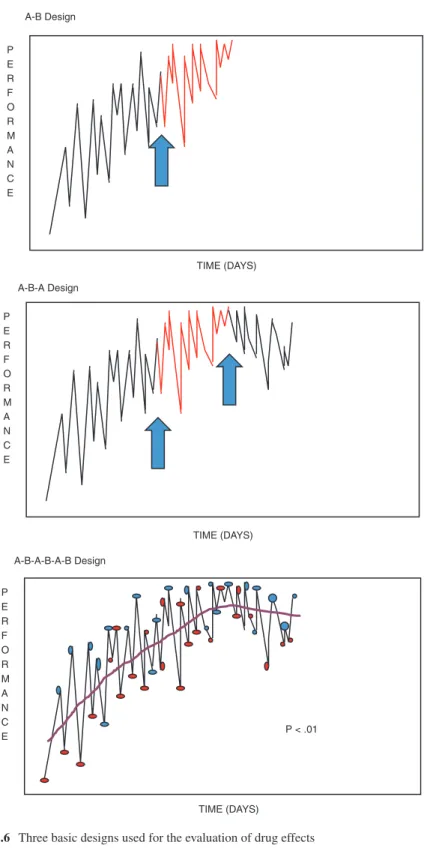
There are three basic designs that are most applicable to the evaluation of drug effects: A-B designs, A-B-A designs, and repeated crossover designs, in increasing order or rigor [99]. For all of these designs, the first task is to select the outcome measures that will be used to assess the drug’s effects. Whenever possible, we select a measure of a very proximal outcome of the drug that may or may not be clinically meaningful, but that will help us ensure that the drug, at the dose given, is physio-logically active. For example, in the hypothetical scenario where we hope to increase arousal to achieve more reliable command following, we might choose a measure of amount of time spent with the eyes open as a measure of the arousal response, though it is not our ultimate clinical goal. We also need, of course, a measure of the clinical goal; in this case, we might choose percent of a standard set of commands that are followed in each session. Finally, when we know that certain adverse effects of the drug are particularly likely, we might have in place a measure of those effects to alert us to their increase.
In planning the drug assessment, we must decide between standardized and psy-chometrically evaluated measures and measures tailored to the individual patient’s problem. We rely on standardized measures where they are clearly applicable to the patient’s problem, but often our treatment goals are too specific to match any exist-ing measure, and then we must create one for the individual. This is typically a team-centered process and may be prompted by such questions as, “How would you know if this drug is having the desired effects? What would you actually observe if the drug does increase ___? How would you measure that change?”
In the A-B design, one begins collecting data with the chosen outcome measures for a period of time (the A phase) before the drug is started, conducting repeated measurements. Then one continues with those same repeated measurements after introducing the drug (the B phase), looking for a change in the measures that cor-responds to the transition from A to B. In some cases visual inspection may reveal a clear change in the level of performance. Statistical evaluation is more controver-sial. In many cases the multiple performance data points are not statistically inde-pendent, violating the assumptions of many traditional statistical tests. One crude statistical approach involves a “celeration line” [100]. In this technique, a regression line is plotted through the A-phase data and continued forward into the B phase. The actual B-phase data points that lie above and below the celeration line are counted and subjected to a binomial test. If there is no drug effect, one would predict that
approximately 50% of B-phase data will likely be above and 50% below the line. Sharp deviation from this 50/50 ratio suggests a drug effect. However, it has been pointed out that, unless there is a very large volume of A-phase data and/or the vari-ability in the A-phase is low, the confidence interval around the celeration line is likely to be wide. This means that having far more than 50% of B-phase data above or below all possible celeration lines could occur easily by chance (Fig. 11.6).
The A-B-A design is identical except that, after an interval of treatment, the drug is withdrawn again and one looks for a drop in performance that corresponds to the transition from B back to A (after which, of course, the drug can be reintroduced, if appropriate). In the repeated crossover design, one transitions back and forth between providing the drug and withholding the drug, ideally at random intervals, collecting the same outcome data throughout. In this design, the repeated crossovers reduce the likelihood that some other intervening events (an illness, another treat-ment, etc.) might actually be responsible for the change, since no other event is likely to follow the same randomly reversing schedule.
The A-B design is the most feasible to implement in the clinical setting, since it corresponds to routine clinical practice aside from the fact that measurement begins prior to treatment. Unfortunately, this design rarely provides a clear conclusion, as noted above. In addition to performance variability, if there is already a non-zero slope, one is challenged to determine that the slope is changed by the treatment. Just as in formal research, more variability requires a larger amount of data to reach conclusions, and this is often not feasible to collect in a time-limited clinical pro-gram. In addition, if the drug being administered requires gradual dose increases, this further undermines the ability to link behavioral changes to the drug.
The A-B-A design overcomes some, but not all, of these problems. Variability in the measured performance still presents a challenge, as does gradual introduction and withdrawal of the drug. However, the confounding between an ongoing recovery slope and the anticipated drug effect is addressed, because in the reversal from B back to A, recovery should still lead to improvement, whereas the drug withdrawal should lead to deterioration (Fig. 11.6). If the data clearly support the notion that the drug was associated with the improvement and deterioration, one can be reasonably confi-dent of that conclusion. However, if improvement and deterioration linked to the drug are not evident, this could be due to excess variability, to the fact that ongoing recov-ery outweighs drug withdrawal effects, or to the fact that recovrecov-ery has proceeded to the point that the drug is no longer needed. Although this is a scientific limitation of the A-B-A design, it is less of a limitation clinically. The main clinical question once a patient is receiving a drug is whether they should stop it or continue it.
The strongest design in terms of the ability to link any performance changes to the drug is the multiple crossover design. If a sufficient number of crossovers are per-formed at random intervals, then drug condition is unlikely to be confounded with time (recovery) or other medical or social events (Fig. 11.6). The main limitation of this design relates to the pharmacokinetics of the drug of interest. This design is well suited to drugs with rapid onsets of action which do not need to be introduced or withdrawn gradually, such as the psychostimulants [44]. Such drugs can be randomized at inter-vals of one to a few days, with little carryover of the drug’s behavioral effects. For drugs with slower onsets of action (e.g., selective serotonin reuptake inhibitors) or those that require gradual drug titration (e.g., bromocriptine), such designs are rarely feasible.
TIME (DAYS) P E R F O R M A N C E A-B-A Design A-B Design TIME (DAYS) P E R F O R M A N C E A-B-A-B-A-B Design TIME (DAYS) P E R F O R M A N C E P < .01
Weighing patient, drug, and measurement factors together suggests an optimal management approach [101]. In the early weeks and months post-injury, off-label prescribing of psychoactive drugs should be minimized. By definition, the effects of such drugs are not known with confidence and have a reasonable likelihood of impairing as well as facilitating recovery. Because of the natural slope and variabil-ity of recovery seen in this phase, it is highly unlikely that the effect of the drug chosen can be determined with confidence. Drugs that have been shown in large group studies to benefit most patients treated can be used in the early period, even though their effects may not be demonstrable in the individual. As recovery slows and variability in performance diminishes, the mandate to intervene to augment natural recovery becomes more pressing, and the ability of the measurement meth-ods to document a treatment response becomes greater.
In practice, it remains useful to define patient-specific goals and measure perfor-mance with quantitative behavioral metrics before committing to pharmacologic intervention. The preliminary data may suggest that it will be impossible to evaluate a treatment response in the time available, that improvement over time is suffi-ciently brisk that there is no urgency to intervene, or that the pattern suggests another treatment approach rather than medication. But where the preliminary data demon-strates modest variability and lack of natural improvement, one can move forward with creative treatment evaluation methods.
Conclusion
Amantadine is the only drug with strong evidence from randomized controlled trials to demonstrate an impact on recovery of consciousness in DOC [24]. Even for aman-tadine, questions remain regarding its benefits for those with non-traumatic injuries, as well as the optimal dose, timing, and duration of treatment. Zolpidem has clearly been shown to lead to abrupt increases in the level of consciousness in a small minority of DOC patients, but, as yet, the factors that predict drug response are not fully known.
No strong evidence currently supports or disproves the use of other pharmacologi-cal agents in order to improve the level of consciousness in DOC patients. As dis-cussed, a number of small and uncontrolled case or cohort studies have reported a positive clinical response. Transitory or permanent improvements have been observed among some UWS or MCS patients of various etiologies. Reported effects were vari-able, ranging from increase in wakefulness, partial recovery of consciousness, and motor, verbal, or communication functions to full recovery of cognitive functioning.
Some of the reviewed treatments (e.g., amantadine, zolpidem, baclofen) seem to benefit patients with severe DOC, whereas others (e.g., methylphenidate, lamotrig-ine) seem to possibly be more beneficial for brain-damaged but conscious patients improving their attention-deficit disorder. Positive drug effects have been observed from a single dose (e.g., zolpidem) or from continuous treatment (e.g., amantadine, baclofen, levodopa).
These studies mainly come from case or cohort reports which cannot disentan-gle the drug effect from natural recovery. Studies are also influenced by the extreme
heterogeneity of DOC, such as the site of neuropathological lesions, time elapsed between injury and the introduction of the treatment, confounding drugs received, and medical comorbidities. Moreover, it is difficult to compare between studies since they lack homogeneity in methodology and differ in the duration of treat-ment, administered doses, and patients’ demographics and clinical status. Measurement tools and behavioral scales are also very different across studies, and a standardization of bedside assessment seems necessary. Additional placebo-con-trolled, double- blind randomized multicenter studies are necessary before drawing conclusions about the role of other medications in these challenging patients.
Importantly, there is little reason to believe that any pharmacologic agent can benefit all patients with DOC, given the heterogeneity of pharmacologic mecha-nisms and the variation in site and severity of neuropathology. Thus, research is needed to understand the pharmacologic targets relevant to restoration of conscious-ness and to identify biomarkers that allow selection of patient subgroups with the ability to respond to specific pharmacologic probes. This will allow conduct of ran-domized trials in patient groups “enriched” with the necessary brain substrates for therapeutic response. Efforts to advance pharmacologic treatments for DOC should focus on the conduct of large parallel group studies. Clinicians practicing in the face of minimal evidence should consider deferring off-label drug intervention to the point in recovery when positive or negative impacts of the drug can be recognized.
References
1. Plum F, Posner JB. The diagnosis of stupor and coma. Philadelphia: F. A. Davis; 1983. 2. The Multi-Society Task Force on PVS. Medical aspects of the persistent vegetative state (1).
N Engl J Med. 1994;330(21):1499–508.
3. Laureys S, Celesia GG, Cohadon F, Lavrijsen J, Leon-Carrion J, Sannita WG, Sazbon L, Schmutzhard E, von Wild KR, Zeman A, Dolce G. Unresponsive wakefulness syndrome: a new name for the vegetative state or apallic syndrome. BMC Med. 2010;8:68.
4. Giacino JT, Ashwal S, Childs N, Cranford R, Jennett B, Katz DI, Kelly JP, Rosenberg JH, Whyte J, Zafonte RD, Zasler ND. The minimally conscious state: definition and diagnostic criteria. Neurology. 2002;58(3):349–53.
5. Ciurleo R, Bramanti P, Calabro RS. Pharmacotherapy for disorders of consciousness: are ‘awakening’ drugs really a possibility? Drugs. 2013;73(17):1849–62.
6. Abbate C, Trimarchi PD, Basile I, Mazzucchi A, Devalle G. Sensory stimulation for patients with disorders of consciousness: from stimulation to rehabilitation. Front Hum Neurosci. 2014;8:616.
7. Klingshirn H, Grill E, Bender A, Strobl R, Mittrach R, Braitmayer K, Muller M. Quality of evidence of rehabilitation interventions in long-term care for people with severe disorders of consciousness after brain injury: a systematic review. J Rehabil Med. 2015;47(7):577–85. 8. Magrassi L, Maggioni G, Pistarini C, Di Perri C, Bastianello S, Zippo AG, Iotti GA, Biella
GE, Imberti R. Results of a prospective study (CATS) on the effects of thalamic stimulation in minimally conscious and vegetative state patients. J Neurosurg. 2016;125(4):972–81. 9. Chew E, Zafonte R. Pharmacological management of neurobehavioral disorders following
traumatic brain injury—a state-of-the-art review. J Rehabil Res Dev. 2009;46(6):851–79. 10. Robbins T. Arousal systems and attentional processes. Biol Psychol. 1997;45(1–3):57–71. 11. Harris CD. Neurophysiology of sleep and wakefulness. Respir Care Clin N Am.
12. Zafonte R, Lexell J, Cullen N. Possible applications for dopaminergic agents following trau-matic brain injury: part 2. J Head Trauma Rehabil. 2001;16(1):112–6.
13. Saniova B, Drobny M, Kneslova L, Minarik M. The outcome of patients with severe head injuries treated with amantadine sulphate. J Neural Transm. 2004;111(4):511–4.
14. Whyte J, Katz D, Long D, DiPasquale MC, Polansky M, Kalmar K, Giacino J, Childs N, Mercer W, Novak P, Maurer P, Eifert B. Predictors of outcome in prolonged posttraumatic disorders of consciousness and assessment of medication effects: a multicenter study. Arch Phys Med Rehabil. 2005;86(3):453–62.
15. Sawyer E, Mauro L, Ohlinger M. Amantadine enhancement of arousal and cognition after traumatic brain injury. Ann Pharmacother. 2008;42(2):247–52.
16. Born JD. The Glasgow-Liège Scale. Prognostic value and evaluation of motor response and brain stem reflexes after severe head injury. Acta Neurochir. 1988;95:49–52.
17. Folstein M, Robins L, Helzer J. The mini-mental state examination. Arch Gen Psychiatry. 1983;40(7):812.
18. Jennett B, Bond M. Assessment of outcome after severe brain damage. Lancet. 1975; 1(7905):480–4.
19. Rappaport M, Hall KM, Hopkins K, Belleza T, Cope DN. Disability rating scale for severe head trauma: coma to community. Arch Phys Med Rehabil. 1982;63(3):118–23.
20. Meythaler JM, Brunner RC, Johnson A, Novack TA. Amantadine to improve neurorecovery in traumatic brain injury-associated diffuse axonal injury: a pilot double-blind randomized trial. J Head Trauma Rehabil. 2002;17(4):300–13.
21. Rappaport M. The Coma/Near Coma Scale. 2000. http://www.tbims.org/combi/cnc
22. Zafonte R, Watanabe T, Mann N. Amantadine: a potential treatment for the minimally con-scious state. Brain Inj. 1998;12(7):617–21.
23. Avecillas-Chasin JM, Barcia JA. Effect of amantadine in minimally conscious state of non- traumatic etiology. Acta Neurochir. 2014;156(7):1375–7.
24. Giacino JT, Whyte J, Bagiella E, Kalmar K, Childs N, Khademi A, Eifert B, Long D, Katz DI, Cho S, Yablon SA, Luther M, Hammond FM, Nordenbo A, Novak P, Mercer W, Maurer- Karattup P, Sherer M. Placebo-controlled trial of amantadine for severe traumatic brain injury. N Engl J Med. 2012;366(9):819–26.
25. Reynolds JC, Rittenberger JC, Callaway CW. Methylphenidate and amantadine to stimulate reawakening in comatose patients resuscitated from cardiac arrest. Resuscitation. 2013;84(6): 818–24.
26. Horiguchi J, Inami Y, Shoda T. Effects of long-term amantadine treatment on clinical symp-toms and EEG of a patient in a vegetative state. Clin Neuropharmacol. 1990;13(1):84–8. 27. Estraneo A, Pascarella A, Moretta P, Loreto V, Trojano L. Clinical and
electroencephalo-graphic on-off effect of amantadine in chronic non-traumatic minimally conscious state. J Neurol. 2015;262(6):1584–6.
28. Schnakers C, Hustinx R, Vandewalle G, Majerus S, Moonen G, Boly M, Vanhaudenhuyse A, Laureys S. Measuring the effect of amantadine in chronic anoxic minimally conscious state. J Neurol Neurosurg Psychiatry. 2008;79(2):225–7.
29. Giacino JT, Kalmar K, Whyte J. The JFK Coma Recovery Scale-Revised: measurement char-acteristics and diagnostic utility. Arch Phys Med Rehabil. 2004;85(12):2020–9.
30. Stelmaschuk S, Will MC, Meyers T. Amantadine to treat cognitive dysfunction in moderate to severe traumatic brain injury. J Trauma Nurs. 2015;22(4): 194–203; quiz E191–2. 31. Haig A, Ruess J. Recovery from vegetative state of six months’ duration associated with
Sinemet (levodopa/carbidopa). Arch Phys Med Rehabil. 1990;71(13):1081–3.
32. Matsuda W, Matsumura A, Komatsu Y, Yanaka K, Nose T. Awakenings from persistent veg-etative state: report of three cases with parkinsonism and brain stem lesions on MRI. J Neurol Neurosurg Psychiatry. 2003;74(11):1571–3.
33. Matsuda W, Komatsu Y, Yanaka K, Matsumura A. Levodopa treatment for patients in persis-tent vegetative or minimally conscious states. Neuropsychol Rehabil. 2005;15(3–4):414–27. 34. Krimchansky B, Keren O, Sazbon L, Groswasser Z. Differential time and related appearance
of signs, indicating improvement in the state of consciousness in vegetative state traumatic brain injury (VS-TBI) patients after initiation of dopamine treatment. Brain Inj. 2004;18(11): 1099–105.
35. Ugoya SO, Akinyemi RO. The place of L-dopa/carbidopa in persistent vegetative state. Clin Neuropharmacol. 2010;33(6):279–84.
36. Passler MA, Riggs RV. Positive outcomes in traumatic brain injury-vegetative state: patients treated with bromocriptine. Arch Phys Med Rehabil. 2001;82(3):311–5.
37. Whyte J, Vaccaro M, Grieb-Neff P, Hart T, Polansky M, Coslett HB. The effects of bro-mocriptine on attention deficits after traumatic brain injury: a placebo-controlled pilot study. Am J Phys Med Rehabil. 2008;87(2):85–99.
38. Millan M, Maiofiss L, Cussac D, Audinot V, Boutin J, Newman-Tancredi A. Differential actions of antiparkinson agents at multiple classes of monoaminergic receptor. I. A multivari-ate analysis of the binding profiles of 14 drugs at 21 native and cloned human receptor sub-types. J Pharmacol Exp Ther. 2002;303(2):791–804.
39. Fridman E, Calvar J, Bonetto M, Gamzu E, Krimchansky B, Meli F, Leiguarda R, Zafonte R. Fast awakening from minimally conscious state with apomorphine. Brain Inj. 2009;23(2): 172–7.
40. Laureys S, Faymonville ME, Luxen A, Lamy M, Franck G, Maquet P. Restoration of thala-mocortical connectivity after recovery from persistent vegetative state. Lancet. 2000; 355(9217):1790–1.
41. Fridman E, Krimchansky B, Bonetto M, Galperin T, Gamzu E, Leiguarda R, Zafonte R. Continuous subcutaneous apomorphine for severe disorders of consciousness after traumatic brain injury. Brain Inj. 2010;24(4):636–41.
42. Moein H, Khalili H, Keramatian K. Effect of methylphenidate on ICU and hospital length of stay in patients with severe and moderate traumatic brain injury. Clin Neurol Neurosurg. 2006;108(6):539–42.
43. Kim YW, Shin JC, An YS. Effects of methylphenidate on cerebral glucose metabolism in patients with impaired consciousness after acquired brain injury. Clin Neuropharmacol. 2009;32(6):335–9.
44. Martin R, Whyte J. The effects of methylphenidate on command following and yes/no com-munication in persons with severe disorders of consciousness: a meta-analysis of n-of-1 stud-ies. Am J Phys Med Rehabil. 2007;86(8):613–20.
45. Kaelin D, Cifu D, Mathies B. Methylphenidate effect on attention deficit in the acutely brain- injured adult. Arch Phys Med Rehabil. 1996;77(1):6–9.
46. Plenger P, Dixon C, Castillo R, Frankowski R, Yablon S, Levin H. Subacute methylphenidate treatment for moderate to moderately severe traumatic brain injury: a preliminary double- blind placebo-controlled study. Arch Phys Med Rehabil. 1996;77(6):536–40.
47. Whyte J, Hart T, Schuster K, Fleming M, Polansky M, Coslett H. Effects of methylphenidate on attentional function after traumatic brain injury. A randomized, placebo-controlled trial. Am J Phys Med Rehabil. 1997;76(6):440–50.
48. Whyte J, Hart T, Vaccaro M, Grieb-Neff P, Risser A, Polansky M, Coslett H. Effects of meth-ylphenidate on attention deficits after traumatic brain injury: a multidimensional, random-ized, controlled trial. Am J Phys Med Rehabil. 2004;83(6):401–20.
49. Kim J, Whyte J, Patel S, Europa E, Wang J, Coslett HB, Detre JA. Methylphenidate modu-lates sustained attention and cortical activation in survivors of traumatic brain injury: a perfu-sion fMRI study. Psychopharmacology (Berl). 2012;222(1):47–57.
50. Patrick P, Buck M, Conaway M, Blackman J. The use of dopamine enhancing medications with children in low response states following brain injury. Brain Inj. 2003;17(6):497–506. 51. Clauss RP, Guldenpfennig WM, Nel HW, Sathekge MM, Venkannagari RR. Extraordinary
arousal from semi-comatose state on zolpidem. A case report. S Afr Med J. 2000;90(1): 68–72.
52. Clauss RP, Nel W. Drug induced arousal from the permanent vegetative state. NeuroRehabilitation. 2006;21(1):23–8.
53. Hagen C, Malkmus D, Durham P. Levels of cognitive functioning. Downey: Rancho Los Amigos Hospital Inc.; 1987.
54. Brefel-Courbon C, Payoux P, Ory F, Sommet A, Slaoui T, Raboyeau G, Lemesle B, Puel M, Montastruc JL, Demonet JF, Cardebat D. Clinical and imaging evidence of zolpidem effect in hypoxic encephalopathy. Ann Neurol. 2007;62(1):102–5.
55. Cohen SI, Duong TT. Increased arousal in a patient with anoxic brain injury after administra-tion of zolpidem. Am J Phys Med Rehabil. 2008;87(3):229–31.
56. Shames JL, Ring H. Transient reversal of anoxic brain injury-related minimally conscious state after zolpidem administration: a case report. Arch Phys Med Rehabil. 2008;89(2):386–8. 57. Appu M, Noetzel M. Clinically significant response to zolpidem in disorders of
conscious-ness secondary to anti-N-methyl-D-aspartate receptor encephalitis in a teenager: a case report. Pediatr Neurol. 2014;50(3):262–4.
58. Lo Y, Tan E, Ratnagopal P, Chan L, Tan T. Zolpidem and its effects on hypoxic encephalopa-thy. Ann Neurol. 2008;64(4):477–8.
59. Singh R, McDonald C, Dawson K, Lewis S, Pringle A, Smith S, Pendland B. Zolpidem in a minimally conscious state. Brain Inj. 2008;22(1):103–6.
60. Whyte J, Myers R. Incidence of clinically significant responses to zolpidem among patients with disorders of consciousness: a preliminary placebo controlled trial. Am J Phys Med Rehabil. 2009;88(5):410–8.
61. Whyte J, Rajan R, Rosenbaum A, Katz D, Kalmar K, Seel R, Greenwald B, Zafonte R, Demarest D, Brunner R, Kaelin D. Zolpidem and restoration of consciousness. Am J Phys Med Rehabil. 2014;93(2):101–13.
62. Machado C, Estévez M, Pérez-Nellar J, Gutiérrez J, Rodríguez R, Carballo M, Chinchilla M, Machado A, Portela L, García-Roca MC, Beltrán C. Autonomic, EEG, and behavioral arousal signs in a PVS case after zolpidem intake. Can J Neurol Sci. 2011;38(2):341–4.
63. Machado C, Estevez M, Rodriguez R, Perez-Nellar J, Chinchilla M, DeFina P, Leisman G, Carrick FR, Melillo R, Schiavi A, Gutierrez J, Carballo M, Machado A, Olivares A, Perez- Cruz N. Zolpidem arousing effect in persistent vegetative state patients: autonomic, EEG and behavioral assessment. Curr Pharm Des. 2014;20(26):4185–202.
64. Thonnard M, Gosseries O, Demertzi A, Lugo Z, Vanhaudenhuyse A, Bruno M, Chatelle C, Thibaut T, Charland-Verville V, Habbal D, Schnakers C, Laureys S. Effect of zolpidem in chronic disorders of consciousness: a prospective open label study. Funct Neurol. 2013;28(4):259–64. 65. Du B, Shan A, Zhang Y, Zhong X, Chen D, Cai K. Zolpidem arouses patients in vegetative
state after brain injury: quantitative evaluation and indications. Am J Med Sci. 2014;347(3):178–82.
66. Cohen L, Chaaban B, Habert MO. Transient improvement of aphasia with zolpidem. N Engl J Med. 2004;350(9):949–50.
67. Rodriguez-Rojas R, Machado C, Alvarez L, Carballo M, Estevez M, Perez-Nellar J, Pavon N, Chinchilla M, Carrick FR, DeFina P. Zolpidem induces paradoxical metabolic and vascular changes in a patient with PVS. Brain Inj. 2013;27(11):1320–9.
68. Chatelle C, Thibaut A, Gosseries O, Bruno MA, Demertzi A, Bernard C, Hustinx R, Tshibanda L, Bahri MA, Laureys S. Changes in cerebral metabolism in patients with a mini-mally conscious state responding to zolpidem. Front Hum Neurosci. 2014;8:917.
69. Williams ST, Conte MM, Goldfine AM, Noirhomme Q, Gosseries O, Thonnard M, Beattie B, Hersh J, Katz DI, Victor JD, Laureys S, Schiff ND. Common resting brain dynamics indicate a possible mechanism underlying zolpidem response in severe brain injury. Elife. 2013;2:e01157.
70. Calabro RS, Arico I, De Salvo S, Conti-Nibali V, Bramanti P. Transient awakening from vegetative state: is high-dose zolpidem more effective? Psychiatry Clin Neurosci. 2015;69(2):122–3.
71. Clauss RP, Nel WH. Effect of zolpidem on brain injury and diaschisis as detected by 99mTc HMPAO brain SPECT in humans. Arzneimittelforschung. 2004;54(10):641–6.
72. Hall S, Yamawaki N, Fisher A, Clauss R, Woodhall G, Stanford I. GABA(A) alpha-1 subunit mediated desynchronization of elevated low frequency oscillations alleviates specific dys-function in stroke—a case report. Clin Neurophysiol. 2010;121(4):549–55.
73. Pistoia F, Sara M, Sacco S, Franceschini M, Carolei A. Silencing the brain may be better than stimulating it. The GABA effect. Curr Pharm Des. 2014;20(26):4154–66.
74. Schiff ND. Recovery of consciousness after brain injury: a mesocircuit hypothesis. Trends Neurosci. 2010;33(1):1–9.
75. Pistoia F, Sacco S, Sara M, Franceschini M, Carolei A. Intrathecal baclofen: effects on spas-ticity, pain, and consciousness in disorders of consciousness and locked-in syndrome. Curr Pain Headache Rep. 2015;19(1):466.
76. Turner M. Early use of intrathecal baclofen in brain injury in pediatric patients. Acta Neurochir. 2003;87:81–3.
77. Kawecki Z, Kwiatkowski S, Grzegorzewski P, Szlachta Jezioro I. Sudden improvement of all neurological functions after general anesthesia and two-day intrathecal infusion of baclofen in a child with primary brain-stem injury. Przegl Lek. 2007;64(2):13–4.
78. Sarà M, Sacco S, Cipolla F, Onorati P, Scoppetta C, Albertini G, Carolei A. An unexpected recovery from permanent vegetative state. Brain Inj. 2007;21(1):101–3.
79. Taira T, Hori T. Intrathecal baclofen in the treatment of post-stroke central pain, dystonia, and persistent vegetative state. Acta Neurochir Suppl. 2007;97(Pt 1):227–9.
80. Sarà M, Pistoia F, Mura E, Onorati P, Govoni S. Intrathecal baclofen in patients with persis-tent vegetative state: 2 hypotheses. Arch Phys Med Rehabil. 2009;90(7):1245–9.
81. Al-Khodairy AT, Wicky G, Nicolo D, Vuadens P. Influence of intrathecal baclofen on the level of consciousness and mental functions after extremely severe traumatic brain injury: brief report. Brain Inj. 2015;29(4):527–32.
82. Margetis K, Korfias SI, Gatzonis S, Boutos N, Stranjalis G, Boviatsis E, Sakas DE. Intrathecal baclofen associated with improvement of consciousness disorders in spasticity patients. Neuromodulation. 2014;17(7):699–704.
83. Hoarau X, Richer E, Dehail P, Cuny E. Comparison of long-term outcomes of patients with severe traumatic or hypoxic brain injuries treated with intrathecal baclofen therapy for dys-autonomia. Brain Inj. 2012;26(12):1451–63.
84. Hoarau X, Richer E, Dehail P, Cuny E. A 10-year follow-up study of patients with severe traumatic brain injury and dysautonomia treated with intrathecal baclofen therapy. Brain Inj. 2012;26(7–8):927–40.
85. Taira T. Intrathecal administration of GABA agonists in the vegetative state. Prog Brain Res. 2009;177:317–28.
86. Coulter D. Antiepileptic drug cellular mechanisms of action: where does lamotrigine fit in? J Child Neurol. 1997;12(1):2–9.
87. Calabresi P, Centonze D, Cupini L, Costa C, Pisani F, Bernardi G. Ionotropic glutamate receptors: still a target for neuroprotection in brain ischemia? Insights from in vitro studies. Neurobiol Dis. 2003;12:82–8.
88. Showalter P, Kimmel D. Stimulating consciousness and cognition following severe brain injury: a new potential clinical use for lamotrigine. Brain Inj. 2000;14:997–1001.
89. Pistoia F, Mura E, Govoni S, Fini M, Sarà M. Awakenings and awareness recovery in disor-ders of consciousness: is there a role for drugs? CNS Drugs. 2010;24(8):625–38.
90. Clauss RP. Neurotransmitters in coma, vegetative and minimally conscious states, pharmaco-logical interventions. Med Hypotheses. 2010;75(3):287–90.
91. Stahl S. Essential psychopharmacology: neuroscientific basis and practical applications. New York: Press CU; 2000.
92. Gosseries O, Charland-Verville V, Thonnard M, Bodart O, Laureys S, Demertzi A. Amantadine, apomorphine and zolpidem in the treatment of disorders of consciousness. Curr Pharm Des. 2014;20(26):4167–84.
93. Oliveira L, Fregni F. Pharmacological and electrical stimulation in chronic disorders of con-sciousness: new insights and future directions. Brain Inj. 2011;25(4):315–27.
94. Schiff ND. Central thalamic deep-brain stimulation in the severely injured brain: rationale and proposed mechanisms of action. Ann N Y Acad Sci. 2009;1157:101–16.
95. Schiff ND. Recovery of consciousness after severe brain injury: the role of arousal regulation mechanisms and some speculation on the heart-brain interface. Cleve Clin J Med. 2010;77(Suppl 3):S27–33.
96. Hammond FM, Barrett RS, Shea T, Seel RT, McAlister TW, Kaelin D, Ryser DK, Corrigan JD, Cullen N, Horn SD. Psychotropic medication use during inpatient rehabilitation for trau-matic brain injury. Arch Phys Med Rehabil. 2015;96(8 Suppl):S256–3. e214


![Fig. 11.5 The mesocircuit model that aims to explain the mechanisms of pharmacologically induced recovery of consciousness (Adapted from [74])](https://thumb-eu.123doks.com/thumbv2/123doknet/6872740.192606/15.659.118.544.542.822/mesocircuit-explain-mechanisms-pharmacologically-induced-recovery-consciousness-adapted.webp)