The Origin and Stimuli Implicated in the
Expression of Nestin
(+)
Cardiac
Myocyte-like Cells in the Ischemic Heart
By
John Assimakopoulos
Department of Physiology
Faculty of Medicine
University of Montreal
Memoir presented to the Faculty of Superior Studies
In light of obtaining the grade of
M.Sc. in Physiology
January 2009
University of Montreal
Faculty of Superior Studies
Memoir Title
:The Origin and Stimuli Implicated in the Expression
of Nestin
(+)Cardiac Myocyte-like Cells in the Ischemic Heart
Presented by: John Assimakopoulos
was evaluated by a jury composed of the following members:
Dr Angelino Calderone
Research Director
Dr Jocelyn Dupuis
President Reporter
Dr Madhu Anand-Srivastava
Jury Member
Abstract
Studies from our lab demonstrated that scar formation and healing was associated with the appearance of nestin(+) cardiac myocyte-like cells predominantly
at the peri-infarct region. The focus of the present study was to identify the underlying mechanism(s) (e.g. hypoxia, neurohormones) implicated in their recruitment and their cellular origin. The presence of these cells was detected as early as 1-week post-myocardial infarction (MI) and persisted 9 months after complete coronary artery ligation. Furthermore, nestin(+) cardiac myocyte-like cells
were also detected in the infarcted human heart. Hypoxia represents a predominant stimulus following MI, however the exposure of normal rats to a hypoxic environment failed to promote the re-appearance of nestin(+) cardiac myocyte-like
cells. By contrast, the infusion of the non-selective β-adrenergic agonist isoproterenol (ISO) in the normal adult Sprague-Dawley rat increased nestin expression in the left ventricle and was associated with the reappearance of nestin(+)
cardiac myocyte-like cells. However, the reappearance of nestin(+) cardiac
myocyte-like cells may not represent a direct effect but was apparently secondary to cardiac myocyte necrosis mediated by isoproterenol. Lastly, we identified a subpopulation of nestin-immunoreactive cells in the normal rat heart that co-expressed cardiac progenitor cell markers Nkx-2.5 and GATA-4. This subpopulation of nestin/Nkx-2.5/GATA-4 cells may represent the progenitor pool that differentiates to a nestin(+) cardiac myocyte-like cell following an ischemic
Résumé
Nos études ont démontrées que la formation de la cicatrice et la guérison sont associées avec l’apparition de cellules de type myocytes cardiaques nestine(+)
dans la région péri-infarcie. Présentement, l’étude examine le mécanisme, tel que l’hypoxie ou les hormones neuronales, possiblement impliqué dans leur recrutement et de dévoiler leur origine cellulaire. La présence de ces cellules a été détectée dans les coeurs infarcies d’une semaine et maintenue après neuf mois suite à une sujétion coronaire complète. Aussi, ces cellules de type myocytes cardiaques nestine(+) ont
été observées dans le coeur infarci humain. L’hypoxie représente un événement prédominant suite à un infarctus de myocarde, mais l’exposition des rats normaux à un environnement hypoxique n’a pas pu promouvoir l’apparition de ces cellules. Autrement, l’infusion de l’agoniste β-adrénergique non-sélectif isoprotérénol (ISO) dans les rats adultes Sprague-Dawley a augmenté la protéine nestine dans le ventricule gauche et a été associé avec la réapparition de cellules de type myocytes cardiaques nestine(+). Cela représente possiblement un effet secondaire suite à la
nécrose des myocytes cardiaques par l’administration d’isoprotérénol. Dernièrement, on a identifié une sous-population de cellules nestine(+) dans le coeur
normal du rat qui co-exprime les marqueurs de cellules cardiaques progénitrices Nkx-2.5 et GATA-4. Cette sous-population de cellules nestine/Nkx-2.5/GATA-4 pourrait représenter des substrats cellulaires qui puissent se différentier en cellules de type myocytes cardiaques nestine(+) suite à une ischémie.
Mots clés: nestine, isoprotérénol, nécrose, cellule souche, cellule progénitrice, myocyte cardiaque
Table of Contents
ABSTRACT ……….………..………. III TABLE OF CONTENTS ………..…...………. VII LIST OF FIGURES AND TABLES ………….…...……….…………...….... VIII LIST OF ABBREVIATIONS ……….………...………... XI ACKNOWLEDGEMENTS ……….………….……….…... XV
INTRODUCTION ……….………...………. 1
1. Prevalence of Myocardial Infarctions ………... 2
1.1 Health Risks ………..…… 2
1.2 Therapeutics ………..…… 2
2. Myocardial Infarction …….………..… 4
2.1 Coronary Occlusion ………..… 4
2.2 Reparative Fibrosis Post-MI ………...……….. 5
2.2.1 Role of Myofibroblasts ………..… 5
2.3 Post-MI Left Ventricular Remodelling …..………... 6
2.3.1 Hypertrophy ………..………...…….. 6
2.3.2 Reactive Fibrosis ……….…..………. 7
2.4 Angiogenesis and Neurogenesis ………..………. 7
2.5 Recruitment of Nestin(+) Neural Stem Cells ………..………….………... 9
2.6 The Appearance of Nestin(+) Cardiac Myocyte-like Cells …..………...……. 11
2.7 Endogenous Cardiac Progenitor Stem Cell Pools …...……..………….…… 12
2.8 Neurohormones and Peptides Implicated in Post-MI Remodelling ……...… 13
3. Sympathetic Nervous System …………...………...…….. 14
3.1 Isoproterenol ………..……….……… 14
3.2 Adrenergic Receptors ……….……… 15
3.2.1 β1 Adrenergic Receptors ………..………... 17
3.2.2 β1 Adrenergic Receptor Signalling ………. 18
3.2.3 β2 Adrenergic Receptors ………. 24
3.2.4 β2 Adrenergic Receptor Signalling ………. 25
OBJECTIVES ………...……….. 27
MATERIALS AND METHODS …………...……….… 29
RESULTS ………...……… 40
DISCUSSION ………. 66
CONCLUSION ………...… 75
LIST OF FIGURES AND TABLES
INTRODUCTION
Figure 1: Myocardial infarction. Figure 2: Nestin(+) neural stem cell.
Figure 3: The structures of epinephrine and isoproterenol. Figure 4: The signalling pathways of the β-adrenergic receptors. Figure 5: Calcium regulation in muscular tissue.
Figure 6: The structure of the sarcomere.
Figure 7: The anti-apoptotic pathway of β2AR stimulation via the PI3K-Akt
pathway.
RESULTS
Figure 1: The absence of nestin-immunoreactive cardiac myocyte-like cells in the normal rat heart.
Figure 2: The presence of nestin(+) cardiac myocyte-like cells in 3-week
post-MI adult rats.
Figure 3: The presence of nestin(+) cardiac myocyte-like cells in 9-month
post-MI adult rats.
Figure 4: Connexin-43 organization in the heart of sham and 1-week post-MI rats.
Figure 5: The presence of nestin(+)/desmin(+) cardiac myocyte-like cells in the
infarcted human heart.
Figure 6: The presence of nestin(+) cardiac myocyte-like cells in the infarcted
human heart.
Figure 7: Nestin expression in the left and right ventricles of normoxic and hypoxic rats.
Figure 8: Nestin(+) cardiac myocyte-like cells undetected in the myocardium
following hypoxia.
Figure 9: Nestin immunoreactivity not detected in Cx43(+) neonatal ventricular
cardiac myocytes following both normoxic and hypoxic exposures Figure 10: Effect of hypoxic exposure on neonatal cardiac myocytes.
Figure 11: Cardiac progenitor cell marker expression in normal adult rat hearts. Figure 12: Distinguished subpopulations of nestin and cardiac progenitor stem
cells are intercalated in between cardiac myocytes in the normal heart.
Figure 13: β1-adrenergic receptor (β1AR) immunoreactivity in nestin(+) stem
cells and nestin(+) cardiac myocyte-like cells.
Figure 14: Numerous nestin(+) cardiac myocyte-like cells expressing β1
-adrenergic receptors (β1ARs) in ISO-treated rats
Figure 15: Smooth muscle α-actin, Bax, Bcl-2, and nestin expression in the left ventricle of sham and ISO-treated rats.
Table 1: Body and heart weights of control and 1-week ISO-treated rats. Table 2: Hemodynamic parameters of control and 1-week ISO-treated rats.
Figure 16: Quantitative analysis of protein expression in the left ventricle of sham and isoproterenol-treated rats.
Figure 17: Sirius Red immunohistological staining of collagen in the left ventricle of sham and ISO-treated rats.
Figure 18: Collagen mRNA expression in the left ventricle of sham and isoproterenol-treated rats.
LIST OF ABBREVIATIONS
α: Alpha
αAR: α-adrenergic receptors AC: Adenylate cyclase
ACE: Angiotensin converting enzyme ADP: Adenosine diphosphate
Akt: Protein kinase B Ang II: Angiotensin II
Apaf-1: Apoptotic protease activating factor 1 ATP: Adenosine-5’-triphosphate
β: Beta
Bad: Bcl-2 associated death promoter protein βAR: β-adrenergic receptor
Bax: Bcl-2 associated X protein Bcl-2: B cell CLL/lymphoma 2
BDNF: Brain-derived neurotrophic factor BSA: Bovine serum albumin
BW: Body weight Ca2+: Calcium
CaMKII: Ca2+/calmodulin-dependent protein kinase II
cAMP: 3’,5’-cyclic adenosine monophosphate cDNA: complementary DNA
Cx43: Connexin-43
DABCO: 1,4-diazabicylo[2.2.2]octane
DMEM: Dulbecco’s Modified Eagle Medium DMSO: Dimethyl sulfoxide
DNA: Deoxyribonucleic acid ECL: Electrochemiluminescence
EDTA: Ethylene diamine tetraacetic acid EGTA: Ethylene glycol tetraacetic acid FBS: Fetal Bovine Serum
γ: Gamma
GAPDH: Glyceraldehyde 3-phosphate dehydrogenase GATA-4: GATA-binding protein 4
GDP: Guanosine diphosphate GPCR: G-protein coupled receptor GTP: Guanosine triphosphate H2O: Water
HBSS: Hank’s Balanced Salt Solution HRE: Hypoxia response element IgG: Immunoglobulin G
IP3: 1,4,5-inositol triphosphate
ISO: Isoproterenol
ITS: Insulin transferase selenium kDa: Kilo Dalton
LV: Left ventricle
LVEDP: Left ventricular end-diastolic pressure LVSP: Left ventricular systolic pressure
MAPK: Mitogen-activated protein kinase MEF-2C: muscle enhancement factor-2C MI: Myocardial infarction
mRNA: Messenger ribonucleic acid Na+: Sodium
NaF: Sodium fluoride NF-M: Neurofilament-M NGF: Nerve growth factor NGS: Normal goat serum
NILV: Non-infarcted left ventricle
Nkx-2.5: NK2 transcription factor related, locus 5 (Drosophila) O2: Oxygen
PBS: Phosphate buffered saline PCR: Polymerase chain reaction PI3K: Phosphatidylinositol-3 kinase
PIP2: Phosphatidylinositol 4,5-bisphosphate
PKA: Protein kinase A
PMSF: Phenylmethylsulphonyl fluoride PVDF: Polyvinylidene fluoride
RV: Right ventricle Sca-1: Stem cell antigen-1 SDS: Sodium dodecyl sulfate SBJ: Super blue juice
SERCA: Sarcoplasmic reticulum calcium-ATPase SMAα: Smooth muscle α-actin
SR: Sarcoplasmic reticulum
TGF-β: Transforming growth factor beta VEGF: Vascular endothelial growth factor
ACKNOWLEDGEMENTS
First and foremost, I would like to thank Dr Angelo Calderone for allowing me the opportunity to work in his laboratory. His vast knowledge and experience is one I aspire to reach coupled to his meticulous scientific analysis. Angelo’s guidance has allowed me to develop as a future researcher and to be able to endure the long hard road that is ahead. He has taught me work efficiently, to look beyond what the results of an experiment show and how to appropriately apply the knowledge I have acquired. Over the past 2 years, his friendship has also provided me with a sense of belonging with our constant anecdotes before I would “get out of his office”. Thank you Angelo for making me a part of your lab, your team, and most importantly, your friend.
Secondly, I would like thank my co-workers Hugues, Robert, Louis, Viviane, and Pauline. Their support and knowledge allowed me to grow as an individual and to realize that mixing up work with a little humor eases the mind. Viviane, your friendship and support has been greatly appreciated over the past 2 years, and our constant conversations have allowed me to fit in and become great friends. Pauline, your hard work ethic is quite admirable, and a trait I hope has rubbed off on me over the last year, because without you pushing me to work I would not have reached my goal today.
Finally, I would like to thank my family for the support, their belief in me, and their confidence that one day I would be achieve great things, and I am one step closer today to reaching that ultimate goal.
1.
Prevalence of Myocardial Infarctions
1.1
Health Risks
Environmental, genetic, and health risk factors can contribute to coronary artery disease ultimately leading to myocardial infarction (MI). The Heart and Stroke Foundation of Canada has reported that since 1994-95 there has been a steady rise in hospitalization due to heart attack with an estimated annual occurrence of 70 000 myocardial infarctions, with approximately 19 000 being fatal (Heart and Stroke Foundation of Canada, 2006). With 60% of Canadians being overweight or obese and nearly half of the population being inactive, the chances of diminishing heart attacks will be a constant uphill battle in the coming years and new tactics must be developed to overcome this rapidly emerging crisis (Heart and Stroke Foundation of Canada, 2004; Statistics Canada, 2007). Preventative measures can be undertaken by at-risk individuals as advised by their physician or by making personal lifestyle choices. In a similar fashion, modern medicine has its limitations to post-MI treatment via the administration of certain drugs as well as preventing damage at the onset of the MI. However, what if the heart possessed its own primitive repair mechanism to decrease the damage caused by this ever-growing pandemic. Stem cell research has constantly been addressing the unlimited potential that stem cells possess and how if cultivated within the proper conditions will be the future of medicine.
1.2
Therapeutics
Following a myocardial infarction, several therapeutic and pharmacological interventions exist that can limit further worsening of cardiac function and maladaptive remodelling. Nitroglycerin administration is a common prescription that has proven beneficial with its vasodilatory effect on the artery that was previously occluded. ACE inhibitors, such as captopril, aid in the treatment of post-MI patients via peripheral vasodilatation, ventricular unloading, and diminishment of ventricular hypertrophy (Sutton et al., 2000). β-blockers, including carvedilol, reduce the onset of post-MI remodelling events (Sutton et al., 2000).
However, what if the potential that lies in stem cells can be harvested, and conform them to the dire need at hand. Many researchers are evaluating whether stem cell cultivation is appropriate during gestation or at time of birth, and to store these cells aside for future use. Similarly, which type of drug administration can promote the differentiation of these cells into the desired cell type is still under intense research and evaluation. Undoubtedly, the prospective properties of these cells cannot be measured and have not fully been determined but still remain a key premise for discovery in times to come despite much ethical debate. This opens up a new realm of possibilities and new strategies for therapeutic treatments in post-MI patients.
2.
Myocardial Infarction
2.1
Coronary Occlusion
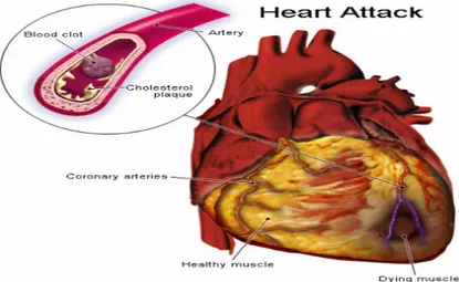
A myocardial infarction occurs when the coronary artery is partially or fully occluded by an atherosclerotic plaque. The ensuing events will lead to left ventricular remodelling which is governed by several hormonal, genetic, and mechanical factors that will overlap and initiate pathophysiological changes in left ventricular morphology and function (Sutton et al., 2000).
Fig. 1 - Myocardial infarction (http://www.straightfromthedoc.com/50226711/flickr_39524 1000.jpg).
The aftermath of the arterial occlusion coincides with a lack of oxygen supply creating a hypoxic environment in the afflicted cardiac area and the resulting death of the local myocyte population. In addition to the increase in diastolic load following an MI, many intracellular cascades will be activated and initiate pathophysiological changes ranging from left ventricular dilatation to the onset of collagen scar formation (Sutton et al., 2000). Similarly, the morphology, thickness,
and location of the scar will determine the amount of time that ventricular remodelling will require to counteract the distending forces with the scar’s tensile strength (Sutton et al., 2000). Subsequently, the drop in pressure will be sensed by baroreceptors and lead to the release of angiotensin II (AII), which in turn will stimulate the production and secretion of catecholamines to increase inotropy and/or chronotropy in the heart via the sympathetic nervous system.
2.2 Reparative Fibrosis Post-MI
2.2.1 Role of Myofibroblasts
The infarct region refers to the hypoxic area where cellular necrosis has occurred and the necrotic cardiac myocyte population is replaced by granular tissue that will eventually undergo a cascade of physiological events leading to scar formation. The initial response consists of lymphocyte invasion to the ischemically damaged region. The subsequent release of inflammatory cytokines such as transforming growth factor-β (TGF-β) will promote the recruitment of cardiac fibroblasts from the non-infarcted left ventricle to the damaged area and initiate the differentiation to a myofibroblasts phenotype (Sutton et al., 2000). Myofibroblasts are characterized by the expression of smooth muscle α-actin and secrete higher levels of extracellular matrix proteins (e.g. fibronectin and collagen) to rapidly heal the ischemically damaged region via the formation of a permanent scar. This response represents an essential physiological mechanism required to heal the damaged heart as is denoted as reparative fibrosis.
2.3
Post-MI Left Ventricular Remodelling
2.3.1 Hypertrophy
Left ventricular remodelling is also separated into 2 stages, early and late remodelling. Early remodelling focuses on the expansion of the collagen scar while late remodelling revolves around the adaptations made by the infarcted portion of the left ventricle to compensate for the increase in wall stress (Sutton et al., 2000).
Cardiac fibroblasts are approximately 2/3 of the total cellular population of the heart, cardiac myocytes are the remaining 1/3 and occupy the majority of the area of the heart (Booz et al., 1995). Early remodelling refers to an early time frame post-MI where myocyte slippage leads to wall thinning and a reduction in the contractile area and volume of the left ventricle (Swynghedauw, 1999). In a study by Olivetti, 2 day post-MI rats had 63% myocytes slippage that led to a 20% increase in ventricular chamber dilatation, 33% decrease in wall thickness, and a 40% reduction in small calibre blood vessels in the scar (Olivetti et al., 1990). This subsequent increase in wall stress will potentiate the onset of cardiac hypertrophy and thus, late-stage remodelling.
In late remodelling, the pathophysiological and structural alterations occurring in the left ventricle due to myocyte death will serve to normalize the increase in systolic and diastolic wall stresses by acting as a potent stimulus for pathological eccentric cardiac hypertrophy (Sutton et al., 2000). In eccentric
cardiac hypertrophy, sarcomere genesis occurs in series leading to an increase in left ventricular chamber volume and is also characterized by a reversion to fetal gene expression affecting proteins such as the conversion of myosin α-heavy chain to the β-isoform and cardiac α-actin to the skeletal isoform (Schwartz et al., 1986). Similarly, there is a small concentric hypertrophic component occurring that will re-establish some slight wall thickness via parallel sarcomere genesis. Also, the onset of reparative fibrosis in the left ventricle will also help diminish the effects of high wall stress.
2.3.2
Reactive Fibrosis
In contrast, to the physiological response of reparative fibrosis, an increased deposition of collagen was also reported in the non-infarcted left ventricle (NILV) and is referred to as reactive fibrosis. The latter process is pathological and ultimately contributes to left ventricular dysfunction. Several studies support the premise that angiotensin II and the sympathetic system may synergistically promote cardiac fibroblast proliferation, thereby initiating the reactive fibrotic response.
2.4
Angiogenesis and Neurogenesis
In the ischemically damaged heart, the compensatory angiogenic response represents an integral event that attempts to prevent cardiac myocyte death in the ischemic region at risk thereby limiting infarct size. The intramyocardial injection of angiogenic peptides and administration of cellular substrates (e.g. endothelial
progenitor cells, bone marrow-derived cells) for de novo blood vessel formation have proven in part therapeutically efficient to limit infarct size. In addition to limiting size, angiogenesis contributes to scar thickening thereby ameliorating the healing process. It has been reported that an increase in scar thickness will limit infarct expansion and concomitantly reduce left ventricular dilatation, thereby improving left ventricular function (Lindsey et al., 2002; Wang et al., 2004).
It has been well established that myocardial infarction promotes sympathetic hyperinnervation, and the study by Vracko et al. (1990) was the first to report sympathetic fibers innervating the infarct region of the ischemically damaged rat heart. Subsequent studies by Drapeau et al. (2005) and El-Helou et al. (2005) confirmed these latter findings and an analogous paradigm was reported in the ischemically damaged human and dog heart as sympathetic fibers projecting from the left stellate ganglion were detected innervating the heart (Zhang et al., 2001; Zhou et al., 2004). Collectively, these data support the premise that sympathetic fiber sprouting and innervation of the damaged region represents a conserved event of myocardial healing across a wide range of species. The local release of nerve growth factor (NGF) by the ischemically damaged heart was identified as a possible candidate to promote sympathetic fiber sprouting (Matsuda et al., 1998).
During cutaneous and corneal wound healing, sympathetic fiber sprouting and innervation were reported and chemical sympathectomy was shown to impair the healing process (Kishimoto et al., 1982; Perez et al., 1987). The underlying
mechanism(s) attributed to the wound healing action of sympathetic fibers includes targeting invading myofibroblasts and angiogenesis. During cutaneous wound healing, nerve fibers were detected innervating myofibroblasts and the release of NGF was reported to promote myofibroblast proliferation and migration (Lui et al., 1999). Moreover, myofibroblasts represent an important source of NGF and acting via a paracrine mechanism promotes sympathetic fiber sprouting (Lui et al., 1999; Matsuda et al., 1998). Furthermore, their role in facilitating angiogenesis was identified because of the fact that sympathetic fiber denervation in skeletal muscle and brown adipose tissue reduced capillary density in response to cold exposure (Borisov et al., 2000; Asano et al., 1997). De novo blood vessel formation in the damaged tissue mediated by sympathetic fiber innervation may occur at least in part via the angiogenic action of brain-derived neurotrophic factor (BDNF) and NGF (Emanueli et al., 2002; Donovan et al., 2000). Thus, sympathetic fiber innervation may play a seminal role in coordinating myofibroblast proliferation and angiogenesis to facilitate proper wound healing. In the ischemically damaged heart, data from Vracko et al. (1990), Drapeau et al. (2005), and El-Helou et al. (2005) suggests that the sympathetic system may play an analogous role.
2.5 Recruitment of Nestin
(+)Neural Stem Cells
El-Helou et al. (2005, 2008) was the first to report that scar formation and healing was associated with the recruitment of a resident population of nestin-expressing neural crest-derived neural stem cells from the normal myocardium to
the ischemic region. Nestin is a type VI intermediate filament protein identified as a putative marker of neural stem cells and plays a role in various cellular functions including cell division and differentiation to axial growth in neurons (Michalczyk et al., 2005). In addition, transient nestin expression during embryonic development may serve as a temporary scaffold until replacement by tissue-specific intermediate filament proteins (Michalczyk et al., 2005). Similarly, the re-expression of nestin fibers following ischemic injury may further add to its speculative role in cell regeneration or remodeling (Michalczyk et al., 2005). Biologically, a subpopulation of neural stem cells isolated from the infarct region have the capacity to differentiate to either a vascular or endothelial cell, thereby representing a substrate for de novo blood vessel formation (El-Helou et al., 2008). Furthermore, during reparative fibrosis, nestin(+) processes emanating from neural stem cells were
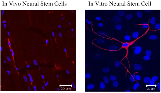
detected innervating the peri-infarct/infarct region and physically associated with newly-formed neurofilament-M (NF-M) immunoreactive sympathetic fibers (El-Helou et al., 2005, 2008). These data suggest that an additional biological role of neural stem cells consists of promoting sympathetic fiber sprouting and subsequently guiding their innervation to the infarct region. These data are moreover clinically relevant as nestin-expressing cells characterized by a refractive cell body and numerous processes were also identified in the viable myocardium and infarct region of human patients and were morphologically identical to that observed in the rat heart (Fig. 2).
Fig. 2 - Nestin(+) neural stem cell.
2.6
The Appearance of Nestin
(+)Cardiac Myocyte-like Cells
Unexpectedly, El-Helou et al. (2005) detected the presence of cardiac myocyte-like cells expressing the intermediate filament protein nestin, localized predominantly in the peri-infarct region. This observation was recently confirmed by the study of Scobioala et al. (2008), as nestin-immunoreactive cardiac myocyte-like cells were detected in the peri-infarct/infarct region of the ischemically damaged mouse heart. The findings are clinically relevant as the presence of numerous nestin-immunoreactive cardiac myocyte-like cells were also identified in the peri-infarct region of an infarcted human heart (El-Helou et al., 2008). Additional studies are required to address whether the appearance of these nestin-immunoreactive cardiac myocyte-like cells represents a transient response to an ischemic insult or is a permanent event associated with scar formation and healing
fibrosis. Furthermore, the stimulus implicated in their recruitment and the source of these cells remains undefined. The focus of the present thesis will address these aforementioned unresolved issues.
2.7
Endogenous Cardiac Progenitor Stem Cell Pools
Previous studies have already shown the multipotency of nestin-expressing stem cells by transdifferentiating into endothelial and neuronal phenotypes, thus resident nestin(+) stem cell population are potential candidates in cardiac
myocyte-like cell formation (Wurmser et al., 2004, Amoh et al., 2004, El-Helou et al., 2005). In addition to the nestin-immunoreactive neural stem cell population identified in the heart (Drapeau et al., 2005; El-Helou et al., 2005), other studies have demonstrated the presence of various cardiac progenitor stem cells, including GATA-4 and Nkx-2.5 cells, adding to the notion that the heart is not a terminally differentiated organ. Tomita et al. (2005) identified multipotent adult cardiac stem cells expressing stem cell antigen-1 (Sca-1) that are capable of differentiating into an endothelial and cardiac myocyte phenotype. Similarly, musashi-1 expressing neural crest-derived stem cells are present in the myocardium and can express the cardiac progenitor marker GATA-binding protein 4 (GATA-4) (Tomita et al., 2005). Muscle enhancement factor 2C (MEF-2C) represents another population of cardiac progenitor cells (Tomita et al., 2005). A small population of bone-derived c-kit(+) (hematopoietic stem cell marker) stem cells have been identified in the
remains to be determined if these cardiac progenitor cells or endogenous nestin(+)
neural stem cells are implicated in the formation of nestin-immunoreactive cardiac myocyte-like cells or other cellular phenotypes.
2.8
Neurohormones and Peptides Implicated in Post-MI
Remodelling
Following an MI, many hemodynamic changes occur that can trigger intracellular cascades ranging from sympathetic nervous stimulation to the renin-angiotensin pathway (Sutton et al., 2000). The predominant physiological role of the sympathetic system following an ischemic insult is to provide inotropic and chronotropic support to the damaged heart. Moreover, the sympathetic system will recruit the renin-angiotensin-aldosterone system to maintain mean arterial blood pressure (Sutton et al., 2000). Furthermore, both the sympathetic system and angiotensin II participate in cardiac hypertrophy (Sutton et al., 2000) and their immediate recruitment following an ischemic insult may directly or indirectly contribute to the appearance of nestin-immunoreactive cardiac myocyte-like cells in the infarcted heart.
3.
Sympathetic Nervous System
3.1
Isoproterenol
Isoproterenol, otherwise known as isoprenaline, is a synthetic sympathomimetic non-specific β1 and β2 receptor agonist with little affinity for
α-adrenergic receptors (Voet et al. 2004). Its chemical structure consists of a benzene ring with multiple hydroxyl groups and an amino group, whose structure closely resembles that of the neurotransmitter and sympathetic agonist epinephrine (Fig. 3).
Epinephrine Isoproterenol
Fig. 3 - The structures of epinephrine and isoproterenol.
Isoproterenol administration represents an established therapeutic approach to treat respiratory disease states such as asthma and bronchitis. In the heart, isoproterenol will act on β1 and β2 adrenergic receptors and elevate heart rate. It
can exert inotropic and chronotropic effects such as elevated systolic blood pressure and vasodilatory effects including decreased diastolic blood pressure. Furthermore,
a high dosage of isoproterenol can create small pockets of necrosis within the myocardium or even induce myocardial infarction and arrhythmia.
3.2
Adrenergic Receptors
Adrenergic receptors interact with the autonomic nervous system and to its composite branches the sympathetic and parasympathetic nervous systems. These systems provide innervation to muscles and organs such as the adrenal glands and initiate biochemical processes that help us initiate the fight-or-flight action via the production of different endocrine hormones (Vander et al., 2001). All sympathetic pre-synaptic neurons will form a synapse with muscular motor end-plates (post-synaptic regions) known as the neuromuscular junction. Upon pre-(post-synaptic membrane depolarization via action potential propagation and Na+ entry, vesicles
containing neurotransmitters will be released within the synapse (Vander et al., 2001). These neurohormones will then bind to specific receptors on the motor end-plates that will initiate Ca2+ entry within the muscle tissue, increase cytosolic Ca2+
levels, and commence the excitation-contraction process.
The cardiovascular system requires input from the sympathetic and parasympathetic nervous system to perform everyday functions. For example, they can influence pacemaker activity or ensure the necessary adaptations of heart rate, cardiac muscle contractility, systolic and diastolic pressures during moments of extreme stress, panic or exercise. Similarly, circulating catecholamines are able to
act on the heart via α- and β-adrenergic receptors (βARs) and exert inotropic and/or lusitropic effects. The heart possesses more βARs than α-adrenergic receptors (αARs) (Vander et al. 2001).
βARs are G-protein coupled receptors that contain conserved 7 hydrophobic transmembrane domains that anchor the receptor to the plasma membrane and have a cytoplasmic C-terminus and an extracellular N-terminus (Zheng et al., 2004). These receptors are coupled to trimeric G proteins involved in signal transduction. There are 2 subtypes of αARs: α1 and α2 and there are 3 subtypes of βARs: β1, β2,
and β3. However, each of these receptor subtypes have functional variations in the
cardiovascular system and differ in the signalling cascades they utilize to perform their physiological roles. The main focus will revolve around the β1 and β2
subtypes and their signalling pathways (Fig. 4).
3.2.1
β
β
β
β
1 Adrenergic ReceptorsThe β1 adrenergic receptor occupies 70-80% of the total βAR subtypes in
the human ventricular myocardium (Brodde, 1991). It maintains 54% and 51% structural homology with the β2AR and β3AR respectively (Zheng et al., 2004). Its
function in the heart serves to increase cardiac output by elevating heart rate via the depolarization of pacemaker cells in the sinoatrial node and heightened myocardial contractility in the left ventricle.
Studies on β1AR knockout mice revealed that when administered
catecholamines, there were no remarkable variations in inotropy or heart rate suggesting the prevalent role of the β1 receptor in cardiac regulation (Rohrer et al.,
1996; Zheng et al., 2004). Clearly, this indicates how each β receptor subtype can constitute a different physiological and pathophysiological function from its cousin receptors.
In terms of functional role, the β1AR pathway can follow two distinct
3.2.2
β
β
β
β
1 Adrenergic Receptor SignallingContraction
The β1AR pathway works via Gsα-coupling. A guanosine triphosphate
(GTP) molecule attaches to the receptor’s α-subunit. In turn, this will cause a conformational change that will separate the α-subunit from the βγ-subunit dimer, leading the α-subunit to activate adenylyl cyclase (AC) that will convert adenosine triphosphate (ATP) to cyclic adenosine monophosphate (cAMP) (Lodish et al., 2001). cAMP, acting as a second messenger, will activate protein kinase A (PKA) by separating the regulatory domain from the catalytic domain, and initiate a cascade of responses that will lead to increased inotropic and chronotropic effects. The intrinsic GTPase activity of the α-subunit, converting GTP to GDP, will then terminate the signal by dissociating from AC and re-binding to the βγ-subunit dimer (Lodish et al. 2001).
Within the ventricular myocytes, the activated PKA will phosphorylate L-type Ca2+ channels on the sarcolemma (Zheng et al., 2004), leading to an influx of
calcium into the myocytes from the T-tubules. The T-tubules contain vast reservoirs of calcium in sacs known as lateral sacs (Vander et al., 2001). Furthermore, phospholamban is inhibiting sarcoplasmic reticulum calcium-ATPase (SERCA) channels on the sarcoplasmic reticulum (SR) from re-absorbing the intracellular calcium. The high calcium levels inside the cell will again activate the ryanodine receptor (a Ca2+-activated Ca2+ channel) on the SR and increase
cytoplasmic calcium levels. This rise in intracellular calcium levels will allow the sarcomere to contract and commence the heart’s pumping action.
Similarly, the rise in intracellular Ca2+ levels can also be regulated by
Ca2+/calmodulin-dependent protein kinase II (CaMKII) independently of PKA
(Zheng et al., 2004). Wang et al. (2004) provides evidence for this notion based on data obtained from inhibiting the PKA pathway and observing that it cannot fully impede the increase in Ca2+ as well as the contractile response. Concomitantly,
blocking CaMKII activity can terminate sustained receptor stimulation (Wang et al., 2004). Furthermore, the data suggests that a time-dependent swap from the PKA pathway to CaMKII occurs based on tracing cAMP production and noting its desensitization following increased CaMKII activity (Hausdorff et al., 1990).
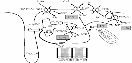
Fig. 5 - Calcium regulation in muscular tissue (http://herkules.oulu.fi/isbn9514267214/ html/graphic11.png). This schematic is a representation of the Ca2+ channels implicated in calcium influx into the cytoplasm to initiate muscular contraction. It also displays the channels involved in the removal of calcium leading to muscular relaxation.
The Sarcomere: Muscular Contractile Unit
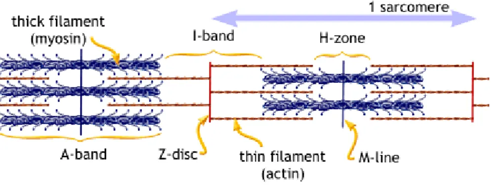
The cardiac muscle fibers, much like skeletal muscle, are basically comprised of several smaller fibers known as myofibrils. Each myofibril has contractile units known as sarcomeres whose organization allows for its organized striated appearance. Each sarcomere is comprised of thick filaments (myosin) and thin filaments (actin). Proteins such as titin and nebulin serve to stabilize and maintain the structure of the thick and thin filaments (Vander et al., 2001).
Each sarcomere is organized into several sections and delineations known as bands and lines (Fig. 5). Firstly there is the Z-line. The Z-line forms the border around each individual sarcomere and distinguishes it from its neighboring sarcomeres. It is where the thin filaments are anchored to the sarcomere at each end (Vander et al., 2001). The I-band refers to area in between the thick filaments of one sarcomere and the adjacent one. This area will decrease in size as the sarcomere contracts. The A-band is the area that myosin thick filaments occupy within the sarcomere. The H-band is the zone where the actin and myosin will interact with each other to reduce the length of the sarcomere and allow for muscular contraction to occur (Vander et al., 2001). Finally, the M-line is in the center of the H-band and delineates the center of the sarcomere where the coupling action between myosin’s crossbridges and actin will occur (Vander et al., 2001).
The sliding-filament mechanism of the sarcomere will commence as soon as there is a calcium influx within the myocyte. The calcium will bind to troponin C
(a subunit of troponin) that will undergo a conformational change that will move tropomyosin with the aid of troponin T (Lodish et al., 2001). Tropomyosin blocks the myosin sites on the actin filaments to prevent maintained contractions. Similarly, molecules such as PKA, mentioned earlier, will also remove the troponin I inhibitory effect on troponin. An ATP molecule will bind to the myosin head and hydrolyze into ADP that will allow it to bind to actin and initiate the sliding movement of thick filament over thin filament. This will cause a shortening of the sarcomere unit and muscular contraction to occur. Once a new ATP molecule binds to the myosin it will detach from actin and begin the process anew (Vander et al., 2001).
Fig. 6 - The structure of the sarcomere (http://www.blobs.org/science/cells/sarcomere2.gif).
Relaxation
Muscular relaxation is simply the removal of the high levels of intracellular calcium within the myocyte by the activation of sarcolemmal and SR channels. Once an inhibitory agonist binds to the receptor or the GTP molecule is hydrolyzed,
it will lead to the eventual re-association of the α-subunit with the βγ-subunit and terminate the activity of adenylyl cyclase. Sodium-calcium exchangers and calmodulin-dependent Ca2+ ATPase on the sarcolemma allow for calcium to exit
the cell. Calcium re-enters the T-tubules and calcium is re-stored in the lateral sacs. Cytosolic calcium can exit also via mitochondrial Ca2+ channels. Similarly,
phosphorylation of phospholamban by PKA removes its inhibition on SERCA allowing for Ca2+ re-entry into the SR.
Also, the second messenger can be adversely affected to terminate the contractile response. Phopshodiesterases serve to degrade cAMP to 5’-adenosine monophosphate (5’-AMP) and halt the activation of PKA’s catalytic domain (Lodish et al., 2001).
The end result is that low levels of Ca2+ will impede troponin’s
conformational change that allows the subsequent binding of the myosin head to actin and thus, allow for sarcomeric length increase and relaxation.
Apoptosis
In addition, to the contraction-relaxation effects of adrenergic signalling, β1
adrenergic stimulation is known to cause apoptosis. Previous studies have discussed the possibility that the cAMP/PKA pathway to be linked to programmed cell death (Communal et al., 1998; Iwai-Kanai et al., 1999). Different types of adenylyl cyclase have been overexpressed to observe their potential effects with
regards to apoptosis. Overexpression of type V and type VI adenylate cyclase do not corroborate with an increase in apoptotic levels but increased PKA activity (Tepe et al., 1999) and increased cardiac contractility (Gao et al., 1999). However, recently the rise in cytoplasmic calcium level that is coupled with an increase in myocardial contractility was in fact due to CaMKII and not linked to the PKA pathway (Zheng et al., 2004). CaMKII blockers significantly reduce the onset of apoptosis following β1 receptor signalling versus using blockers for PKA (Zhu et
al., 2003). Thus, the activity of CaMKII leads to intracellular Ca2+ overload and an
eventual increased wear and tear on the myocytes and subsequent cellular death.
During apoptosis, pro-apoptotic proteins such as Bax are activated that will bind to the mitochondrial membrane of the cardiac myocyte. These pro-apoptotic factors will polymerize together to form pores or associate with the permeability pore complex for subsequent permeabilization of the outer mitochondrial membrane. In contrast, other pro-apoptotic proteins, such as Bad, will bind and inhibit anti-apoptotic proteins, such as Bcl-2, from ensuring caspase activation via Apaf-1 blockade (Lodish et al., 2001). Consequently, the ensuing event will result in apoptotic factor release from the mitochondria, including cytochrome c, that will bind to Apaf-1 leading to the formation of the apoptosome and in turn activate the proteolytic enzymes of the caspase family (Lodish et al., 2001).
Hypertrophy
βAR stimulation is capable of inducing ventricular hypertrophy. Following isoproterenol administration, the chronic activation of the βARs leads to the upregulation of β-adrenergic receptor kinase-1, which phosphorylates and leads to the membrane downregulation of βARs (Iaccarino et al., 1999). The subsequent impairment of β-adrenergic receptor activity may lead to a compensatory ventricular hypertrophic response during pressure overload by which the mechanism to induce hypertrophy remains unknown (Iaccarino et al., 1999).
3.2.3
β
β
β
β
2 Adrenergic ReceptorsThe β2 adrenergic receptor occupies about 20-30% of the total βAR subtypes
in the human ventricular myocardium (Brodde, 1991). It maintains 54% and 46% structural homology with the β1AR and β3AR respectively (Zheng et al., 2004). Its
physiological functions are identical to that of the β1 receptor but to a lesser degree
of intensity most likely due to its lower density presence in the myocardium. Evidence for this is shown in β2AR knockout mice following catecholamine
administration and how a complete cardiac response is elicited (Zheng et al., 2004). Similarly, β1AR knockout mice display a lack of this heightened response
demonstrating how they are not the primary receptors for inotropic and chronotropic reactions (Rohrer, et al. 1996; Zheng et al., 2004).
3.2.4
β
β
β
β
2 Adrenergic Receptor SignallingContraction, Relaxation, and Anti-Apoptosis
Unlike β1ARs that have Gs signalling, β2ARs have dual coupling to Gs and
Gi (Zheng et al., 2004). The associated Gi signalling provides a cardioprotective
effect by reducing the dramatic results of Gs stimulation and promoting cell survival
(Zheng et al., 2004). For instance, β2AR Gi signalling activates the Na+/Ca2+
exchanger on the sarcolemma leading to Ca2+ unloading in the sarcoplasmic
reticulum (Sato et al., 2004). However, it exerts its anti-apoptotic effects by coupling to the PI3K-Akt pathway and not through MAPKs (Chesley et al., 2000; Zheng et al., 2004).
The Gs signalling remains identical to β1AR via the classic adenylyl
cyclase-cAMP-PKA pathway leading to a subsequent rise in intracellular Ca2+ levels,
though not as elevated as through β1AR regulation to induce myocardial
contraction. The relaxation signalling process remains the same as that for β1AR.
The anti-apoptotic effects of β2 stimulation are Gi-dependent following in
vitro β2AR blocking (Communal et al., 1999). The cardioprotective Gi pathway
commences with the activation of phosphotidylinositol-3-kinase (PI3K) (Fig 6). Whether it is Giα- or Giβγ-coupled, it remains unclear. PI3K will in turn
phosphorylate phosphatidylinositol-2-phosphate (PIP2) and convert it to
via phosphorylation, which will phosphorylate and inactivate apoptotic factors such as Bad and ensure cell survival (Chang et al., 2003). Inhibiting the Gi-PI3K-Akt
pathway results in β2AR stimulation switching to promote apoptosis (Zhu et al.,
2001).
Fig. 7 - The anti-apoptotic pathway of β2AR stimulation via the PI3K-Akt pathway (Chang
Scar formation and healing is essential to heal the ischemically damaged heart and involves the interplay of numerous physiological events including inflammation, myofibroblast deposition of collagen, angiogenesis, and sympathetic fiber innervation mediated by cardiac neural stem cells. Work from Calderone’s group revealed the novel observation that following ischemic injury to the rat, the peri-infarct/infarct region contained an abundance of nestin-immunoreactive cardiac myocyte-like cells (El-Helou et al., 2005, 2008). An analogous paradigm was reported in the infarcted human heart. Thus, the primary objective of the present thesis is to identify the phenotype and origin of nestin-immunoreactive cardiac myocyte-like cells and the stimuli implicated in the expression.
Experimental Models
Human MI Tissue
The use of human cardiac tissue was approved by the Clinical Ethics board of the Montreal Heart Institute. Cardiac tissue samples (non-infarcted left ventricle and infarct region) were obtained from patients (3 males, 1 female; range 30-83 years old) who died either 1 week (n = 2) or 3 months (n = 2) following myocardial infarction. These samples were previously used in the study by El-Helou et al. (2008).
MI Rat Model
In this experimental model, we are creating a myocardial infarction in the rat by ligating the left anterior descending coronary artery and then allowing the rat to survive for a specific period of time post-MI ranging from 1 week to 9 months. Male Sprague Dawley rats (Charles River) weighing approximately 200-250 g were under initial anaesthesia using 3% isofluorane, followed by a reduction to 1.5% during the ligating procedure. Following the post-MI survival period, the hearts were excised and stored in either 2-methylbutane to perform immunofluorescence, frozen in liquid nitrogen for use in molecular biology experimentation, or in formalin for immunohistochemistry. The use and care of laboratory animals was according to the Canadian Council for Animal Care and all animal studies were approved by the Animal Care Ethics Committee of the Montreal Heart Institute.
Hypoxia Rat Model
Male Sprague-Dawley rats (200-250 g; Charles River) were placed in a Plexiglas chamber (width 63 cm; height 43 cm; depth 60 cm) into which a continuous flow of air and pure nitrogen mixture was delivered, resulting in an inspired oxygen fraction (FiO2) of 0.12. The flow rates of the gases were controlled
using individual flow meters, and the FiO2 was confirmed using an oxygen analyzer
(Applied Electrochemistry model S-3A/I). Exposure to continuous hypoxia was maintained for 10 days and then the rats immediately sacrificed. The sham rats were maintained in ambient air. The hearts of all animals were subsequently stored in 2-methylbutane.
Isoproterenol Infusion Rat Model
Male Sprague-Dawley rats (200-250 g; Charles River) were implanted (intraperitonially) with osmostic pumps (model 2ML1, 10 µL/hr; Alzet) containing either isoproterenol (infusion rate; 30 mg/kg/day) or saline for a period of 1 week. Rats were subsequently sacrificed, the hearts excised and stored in either 2-methylbutane to perform immunofluorescence or frozen in liquid nitrogen for protein/mRNA experiments.
Hemodynamic Measurements
The rats were anaesthetized using a mixture of ketamine (50 mg/kg) and xylazine (10 mg/kg). The lead electrodes were inserted in the left and right arms, and the left leg to measure and establish Einthoven’s triangle to provide the electrocardiogram to view the electrical activity of the heart. The EMKA 1.8.11.12 program also provided a measure of the heart rate by the addition of the QR or measuring the systolic pressure curves.
Carotid catheter insertion method was used to measure the arterial pressure and the left ventricular pressure. Similarly, a jugular catheter insertion measured the right ventricular pressure. The method also calculated the rate of contraction (dP/dT), based on the steepest point of the ascending pressure curve, and the rate of relaxation based on the steepest point of the descending pressure curve. The diastolic pressure is represented by the smallest pressure reading of the pressure curve. The end-diastolic pressure is the point when the slope begins to rise and the systolic pressure is the highest peak point of the curve.
Cultured Neonatal Rat Cardiac Myocytes and Exposure to Hypoxia
Hearts were removed from 1-3 day old neonatal Sprague-Dawley rats anesthetized with ether and killed by decapitation. Ventricular tissues were placed immediately in chilled Ca2+-free HBSS (GIBCO) and digested with 0.1% trypsin
(GIBCO) in HBSS overnight at 4 oC. Ventricular cells were subsequently
recovered by repeated digestions in 10 mL of 0.1% collagenase type II (GIBCO). The supernatants collected from each digestion were centrifuged and re-suspended in ice cold HBSS, followed by another centrifugation and re-suspension in DMEM containing 7% heat-inactivated FBS (GIBCO). The cells were preplated twice in T75 flasks (VWR) to enrich for myocytes and decrease contamination by nonmuscle cells. The non-adherent myocytes were then plated (100 mm plate; VWR) at a density of 100-200 cells per mm2 for protein and mRNA studies. After
24 hours, the culture medium was changed to serum-free DMEM containing ITS (5 µg/ml insulin, 5 µg/ml transferrin, 5 ng/mL sodium selenite; GIBCO); all experiments were performed 24 hours later.
Neonatal rat ventricular myocytes were placed in a humidified chamber (In Vivo 400 Work Stations) maintained at 37 oC. An oxygen sensor constantly
monitored the atmosphere and ensured hypoxic conditions by maintaining the oxygen level at 1%. Myocytes were exposed to a hypoxic environment for either 8 or 24 hour periods and nestin protein and mRNA levels were assessed. As a positive control, VEGF mRNA expression was determined. The normoxic environment consisted of 5% CO2 and 21% O2 at 37 oC.
Biochemistry Techniques
Protein Extraction
Cardiac tissue or cell cultures were lysed in a buffer containing 10 mM Tris (pH 7.5), 150 mM NaCl, 1 mM EDTA, 1 mM EGTA, 50 mM, NaF, 0.5 mM PMSF (0.174 g/10 mL in ethanol), 1 mM sodium vanadate, 1% Triton X-100, 0.5% Igepal CA-630, 1 µg/mL of aprotinin (1 mg/mL in H2O), and 1 µg/mL of leupeptin (1
mg/mL in DMSO). The tissue sample was placed in a 50 mL tube containing 2-3 mL of the lysis buffer. The tissue was grinded using the Polytron and the samples settled on ice for 1 hour. The homogenate samples were centrifuged at 10 000 RPM for 10 minutes at 4 oC. The supernatant was collected and stored at -20 oC.
The protein concentration was measured using the Bradford assay technique.
Western Mini-Gel Electrophoresis, Transfer, and Blot
The results from the Bradford assay technique were used to calculate and load a protein sample 100 µg in weight. Acetone precipitation concentrated low concentration lysates into pellets followed by dissolution in 10 µL SBJ 1X. Concentrated samples were directly mixed with SBJ 2X for 25 µL loading volume. The SBJ contained 1 M Tris (pH 6.8), 30% glycerol, 6% SDS, 15% β-mercaptoethanol, and bromophenol blue. Prior to loading, the samples were then heated at 70 oC for 1-2 minutes to allow protein denaturation.
The lysates were subjected to SDS-polyacrylamide gel (10-15%) electrophoresis and protein subsequently transferred to a PVDF membrane. Immunoblotting was performed using 5% skim milk or BSA for 1-2 hours followed by successive PBS-Tween washings. The primary antibody was added and incubated at 4 oC overnight. The antibodies employed include mouse monoclonal
anti-nestin (1:500; Millipore), mouse monoclonal anti-GAPDH (1:1000000; Ambion), mouse monoclonal anti-smooth muscle α-actin (1:2000; Sigma), mouse monoclonal anti-Bax and anti-Bcl-2 (1:500; Santa Cruz Biotechnology). Following overnight incubation at 4 oC, goat anti-mouse conjugated to horseradish peroxidase
(1:10000, Santa Cruz Biotechnology) was added and visualization of the bands were detected by autoradiography utilizing the ECL detection kit (PerkinElmer Life Science).
Immunofluorescence
The rat heart was excised, immediately immersed in 2-methylbutane and stored at -80 oC. Primary passage neonatal rat ventricular myocytes were fixed with
4% paraformaldehyde prior to staining. Tissues were sectioned at 14 µm using the cryostat and placed on poly-lysine coated slides and fixed with 4% paraformaldehyde (pH 7.2). Fixed cells or tissue were permeabilized with 2% normal goat serum (NGS) and 0.5% Triton X-100 for 1-2 hours at room temperature, and subsequently incubated overnight at 4oC with the appropriate
antibody diluted in 1% NGS and 0.1% Triton X-100. Antibodies employed include mouse monoclonal anti-nestin (1:500; Millipore), rabbit polyclonal anti-desmin
(1:600; Abcam), rabbit polyclonal anti-NFM (1:500; Millipore) rabbit polyclonal anti-β1-adrenergic receptor, anti-Nkx-2.5, anti-GATA-4, and anti-connexin-43
(Santa Cruz Biotechnology). Following incubation, fixed tissue or cells were incubated for 1-2 hours at room temperature with either goat anti-mouse IgG conjugated to Alexa Fluor 555 (1:600; Invitrogen; emission wavelength at 570 nm) or goat anti-rabbit IgG conjugated to Alexa Fluor 488 (1:800; Invitrogen; emission wavelength at 520 nm) diluted in 0.05-0.1% Triton X-100 in PBS containing 1% NGS. To stain the nucleus, cells or tissue were initially treated with RNase (100 µg/µL; MBI Fermentas) for 20 minutes at 37 oC and then incubated for 20 minutes
at room temperature with the fluorescent label to-pro3 (Invitrogen; 1.5 µM;
emission wavelength 661 nm). Non-specific staining was determined following the addition of either an isotype control antibody (mouse IgG, 5 µg/mL; rabbit IgG, 17 µg/mL) or the secondary antibody alone. The concentration of each isotype control was equivalent to the highest concentration of antibody employed. The slides were mounted on a 1:4 mixture of DABCO and glycerol prior to confocal microscope visualization. Immunofluorescence staining was visualized with either 10X- or 63X-oil 1.4 NA DIC plan apochromat objective mounted on a Zeiss Axiovert 100M confocal microscope.
Molecular Biology Techniques
Real-Time Polymerase Chain Reaction (Real Time-PCR)
Total RNA was isolated from the control and treated samples of cell cultures and/or ISO-treated adult rat hearts by a modification of the guanidine isothiocyanate-phenol-chloroform method. A cDNA library was generated by incubating 5 ng/µL total RNA (each sample), M-MLV reverse transcriptase (800 U, Invitrogen), RNaseOUT (40 U, Invitrogen), random-hexamer primers (0.04 U, Amersham Biosciences), dNTPs (0.5 mmol/L, MBI Fermentas), and supplied optimal buffers. The reaction protocol consisted of 3 successive incubation steps at (1) 25 oC for 10 minutes, (2) 37 oC for 50 minutes, and (3) 70 oC for 15 minutes.
Real-time polymerase chain reaction (PCR) was performed on 2.5 ng of cDNA template containing the appropriate primers (300 nmol/L) and SYBR Green PCR master mix (Applied BioSystems). Primers for each gene were obtained from distinct exons that spanned an intron by using the Ensembl Genome Browser program (http://www.ensembl.org). The sequence specificity of each primer was verified with the Blast program derived from the National Center for Biotechnology Information (http://www.ncbi.nlm.nih.gov). The primers used were as follows: for rat β-actin, forward 5’-AGGCTCTCTTCCAGCCTTCC-3’ and reverse 5’-CATGG-ATGCCACAGGATTCC-3’; for rat nestin, forward 5’-TGCAGGCCACTGATAA-GTTCCA-3’ and reverse 5’-TTCTCCTGCTCCAGGGCTTCCA-3’; for rat collagen type I, forward 5’-CTGACGCATGGCCAAGAAGACA-3’ and reverse
5’-CGTGCCATTGTGGCAGATACAGAT-3’; for rat GATA-4, forward 5’-ATG-GGCACAGCAGCTCCA-3’ and reverse 5’-CATGGCCGGACACAGTACTG-3’; for rat MEF-2C, forward GCCATCTGCCCTCAGTCAGT-3’ and reverse 5’-TGAGATAAATGAGTGCTAGTGCAAGC-3’; for rat Nkx-2.5, forward 5’-CAA-GGACCCTCGGGCG-3’ and reverse 5’-TTTGTCCAGCTCCACCGC-3’; and for rat VEGF-A, forward GAAATCCCGGTTTAAATCCTGG-3’ and reverse 5’-CGCTCTGAACAAGGCTCACAG-3’. Appropriate negative controls were used for each experiment.
Statistics
Data were presented as means ± SE, where n represents the number of rats used in each experiment. Morphological and hemodynamic results were evaluated by a bilateral Student’s unpaired T-test. Protein and mRNA expression were evaluated with a 1-way ANOVA and a significant difference was detected using the Neuman-Keuls post hoc test. A value of P < 0.05 is considered statistically significant.
1.
The phenotype and presence of neural stem cells and
nestin-immunoreactive cardiac myocyte-like cells in the infarcted
rat and human heart.
In the normal rat heart, nestin immunoreactivity was detected exclusively in the neural stem cell population, marked by a refractive cell body and numerous processes, intercalated among terminally-differentiated connexin-43(+) cardiac
myocytes (Fig. 1). Neural stem cells did not stain positive for connexin-43, a gap junctional protein and marker of adult cardiac myocytes, whereas nestin was not expressed in adult cardiac myocytes. Following ischemic damage, neural stem cells were observed in the non-infarcted (Fig. 2A) and peri-infarct region (Fig. 2B) of the 3-week post-MI rats. Moreover, bordering the scar, structurally and morphologically immature nestin-immunoreactive cardiac myocyte-like cells were detected and expressed the cardiac specific marker desmin (Fig. 2B). Similarly, a rarity of nestin-immunoreactive cardiac myocyte-like cells has also been identified in the non-infarcted region (Fig. 2A). The underlying mechanisms attributed to their appearance remain presently undefined. The latter paradigm was not a transient response as nestin-immunoreactive cardiac myocyte-like cells (Fig. 3A, 3B & 3C) were detected in the peri-infarct/infarct region of 9-month post-MI rats. In contrast to the phenotype of cardiac myocytes residing in the viable myocardium (Fig. 4A), immunoreactivity of the gap junctional protein connexin-43 implicated in action potential propagation was either absent, lateralized or in the cytoplasm of nestin-immunoreactive cardiac myocyte-like cells (Fig. 4B). Phenotypically, these data demonstrate that structural and morphological immaturity
nestin-immunoreactive cardiac myocyte-like cells may represent a substrate for arrhthymogenesis (Rucker-Martin et al., 2006).
In the human infarcted heart, nestin-expressing cells were identified in the viable myocardium and scar and exhibited an identical anatomical phenotype to the population of neural stem cells residing in the infarcted rat heart (Fig. 6A & 6B). Concomitantly, structurally and morphologically immature nestin-immunoreactive cardiac myocyte-like cells were also detected in the infarcted human heart of 1-week post-MI patients (Fig. 6B) followed by a robust increase and maintenance of this cellular phenotype following 3 months post-MI (Fig. 6C & 6D). Furthermore, desmin immunoreactivity was detected in nestin-immunoreactive cardiac myocyte-like cells (Fig. 5A, 5B, 5C, & 5D). Lastly, akin to that reported in the rat infarcted heart, connexin-43 immunoreactivity was either absent, lateralized or in the cytoplasm of structurally and morphologically immature nestin-immunoreactive cardiac myocyte-like cells in the infarcted human heart (Fig. 6B, 6C, & 6D).
Fig. 1 - The absence of nestin-immunoreactive cardiac myocyte-like cells in the normal rat heart. Nestin immunoreactivity (red fluorescence) was detected in
nestin(+) neural stem cells (denoted by arrows) intercalated between nestin(-) cardiac myocytes (denoted by *) that stained positive for the gap junctional protein connexin-43 (Cx43) (green fluorescence). The nucleus was identified with the blue fluorescent marker to-pro3.
*
*
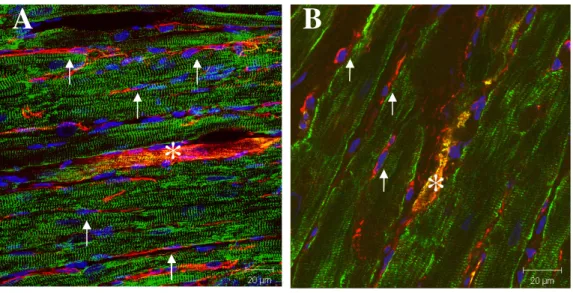
Fig. 2 - The presence of structurally and morphologically immature nestin(+) cardiac myocyte-like cells in 3-week post-MI adult rats. A: Possible terminally
differentiated nestin (red fluorescence) and desmin-immunoreactive (green fluorescence) cardiac myocyte-like cell (denoted by *) in the non-infarcted left ventricle with nestin(+) neural stem cells (denoted by arrows). The underlying mechanisms attributed to its appearance remain undefined. B: Presence of structurally and morphologically immature nestin(+)/desmin(+) cardiac myocyte-like cell in the peri-infarct region surrounded by nestin(+) neural stem cells. The nucleus was identified with the blue fluorescent marker to-pro3.
A
B
*
Fig. 3 - The presence of structurally and morphologically immature nestin(+) cardiac myocyte-like cells in 9-month post-MI adult rats. A: Nestin(+) (red fluorescence) and desmin(+) (green fluorescence) cardiac myocyte-like cell (denoted by *) observed after 9 months in the peri-infarct region reflects a non-transient response. B: Presence of structurally and morphologically immature nestin(+)/desmin(+) (green fluorescence) cardiac myocyte-like cell in the infarct region. C: Nestin(+)/desmin(+) cardiac myocyte-like cells detected in the peri-infarct region. The nucleus was identified with the blue fluorescent marker to-pro3.
A
B
*
*
C
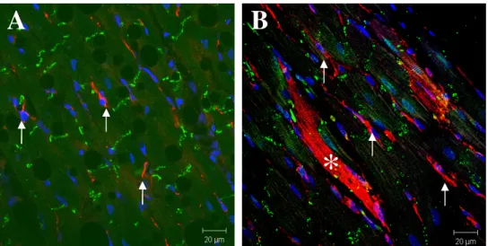
Fig. 4 - Connexin-43 organization in the heart of sham and 1-week post-MI rats. A: Cx43 (green fluorescence) is highly organized in the normal myocardium.
Nestin(+) (red fluorescence) neural stem cells (denoted by arrows) are present in the heart. B: An aberrant pattern of Cx43 staining, either lateralized or cytoplasmic, was detected in nestin(+) cardiac myocyte-like cells (denoted by *). The nucleus was identified with the blue fluorescent marker to-pro3.
A
B
Fig. 5 - The presence of structurally and morphologically immature nestin(+)/desmin(+) cardiac myocyte-like cells in the infarcted human heart. A:
Nestin(+) (red fluorescence) cardiac myocyte-like cell (denoted by *) in the infarct region. The same phenotypical myocyte-like cell was also observed in the infarcted rat heart. B: Desmin(+) (green fluorescence) immunoreactivity of the same myocyte-like cell. C: Nestin and desmin co-localization. D: Structurally and morphologically immature nestin(+)/desmin(+) cardiac myocyte-like cell forming in the scar. The nucleus was identified with the blue fluorescent marker to-pro3.