Warfarin-induced vitamin K deficiency is associated
with cognitive and behavioral perturbations, and
alterations in brain sphingolipids in rats
by
Sahar Tamadon-Nejad
Faculté de Médecine
Département de Nutrition
Mémoire présenté à la Faculté de Médecine en vue de l’obtention du grade de maîtrise
en nutrition
Mai, 2013
Faculté des études supérieures
Ce mémoire intitulé :
Warfarin-induced vitamin K deficiency is associated
with cognitive and behavioral perturbations, and
alterations in brain sphingolipids in rats
Présenté par : Sahar Tamadon-Nejad
a été évalué par un jury composé des personnes suivantes :
Christine DesRosier, président-rapporteur Guylaine Ferland, directeur de recherche
Résumé
La Vitamine K (VK) est largement reconnue pour son rôle dans la coagulation sanguine toutefois, de plus en plus de travaux indiquent son implication dans la fonction cérébrale. La VK est requise pour l'activation de différentes protéines, par exemple la protéine Gas6, et la ménaquinone-4 (MK-ménaquinone-4), le principal vitamère K dans le cerveau, est impliquée dans le métabolisme des sphingolipides. Dans un rapport précédent, nous avons montré qu'un régime alimentaire faible en VK tout au long de la vie était associé à des déficits cognitifs chez des rats âgés. La warfarine sodique est un puissant antagoniste de la VK qui agit en bloquant le cycle de la VK, provoquant un «déficit relatif de VK » au niveau cellulaire. À la lumière du rôle émergent de la VK dans le cerveau, la warfarine pourrait représenter un facteur de risque pour la fonction cérébrale. Ce travail est donc pertinente en raison de la forte proportion d'adultes traîtés à la warfarine sodique. Dans la présente étude, 14 rats mâles Wistar ont été traités avec 14 mg de warfarine/kg /jour (dans l'eau potable) et des injections sous-cutanées de VK (85 mg/kg), 3x/sem, pendant 10 semaines. Quatorze rats témoins ont été traités avec de l'eau normale et injectés avec une solution saline. Les rats ont été soumis à différents tests comportementaux après quoi les niveaux de phylloquinone, MK-4, sphingolipides (cérébroside, sulfatide, sphingomyéline, céramide et gangliosides), et les sous-types de gangliosides (GT1b, GD1a, GM1, GD1b), ont été évalués dans différentes régions du cerveau. Comparativement aux rats du groupe contrôle, les rats traités à la warfarine présentaient des latences plus longues au test de la piscine de Morris (p <0,05) ainsi qu'une hypoactivité et un comportement exploratoire plus faible au test de « l’open field » (p <0,05). Le traitement par warfarine a également entraîné une diminution spectaculaire du niveau de MK-4 dans toutes les régions du cerveau (p <0,001), une altération des concentrations de sphingolipidiques, en particulier dans le cortex frontal et le mésencéphale (p <0,05), et une perte de différences régionales sphingolipidiques, notamment pour les gangliosides. Le traitement par warfarine a été associé à un niveau inférieur de GD1a dans l'hippocampe et un niveau supérieur de GT1b dans le cortex préfrontal et le striatum. En conclusion, la déficience en VK induite par warfarine altère les niveaux de VK et
sphingolipides dans le cerveau, avec de potentiels effets néfastes sur les fonctions cérébrales.
Abstract
Vitamin K (VK) is widely known for its role in blood coagulation, however many studies suggest its involvement in brain function. VK is required for the activation of various cerebral proteins (e.g., Gas6) and menaquinone-4 (MK-4), the main K vitamer in brain, is involved in sphingolipid metabolism. Furthermore, life-long intake of a low VK diet has been associated with cognitive deficits in old rats. Warfarin (W) is a potent VK antagonist that acts by blocking the VK cycle causing a “relative VK deficiency” at the cellular level. In light of this and the emerging role of VK in brain, W could represent a risk factor for cerebral function. The finding of this study is important according to the large proportion of adults with thromboembolic diseases being treated with warfarin drugs. This study was conducted in a rat model where the impact of W was investigated with respect to cognition, behavior, and brain menaquinone-4 (MK-4) and sphingolipid status. Fourteen Wistar male rats were treated with 15 mg W/kg/d (in drinking water) and subcutaneous VK (85 mg/kg), 3X/wk, for 10 wks; 14 control rats were treated with normal water and injected with saline. At the end of the treatment period, rats were subjected to different behavioral tests, afterwhich their brains assessed for VK (phylloquinone and MK-4) and sphingolipids (gangliosides, ceramides, cerebrosides, sphingomyelin and sulfatides) and gangliosides subtypes (GT1b, GD1a, GM1, GD1b). Mean latencies to find the hidden platform were higher in the W compared to the control group (p<0.05) suggesting cognitive deficits as well as hypoactivity and lower exploratory behaviour in the open field test (p<0.05). Warfarin treatment also resulted in a dramatic decrease in MK-4 concentration in all brain regions (p<0.001), altered sphingolipid level, especially in frontal cortex and midbrain (p<0.05), and in a loss of sphingolipid regional differences, notably for gangliosides. W treatment was associated with lower GD1a in the hippocampus and higher GT1b in the striatum and prefrontal cortex. In conclusion, warfarin-induced VK deficiency alters VK and sphingolipid status in brain with potential detrimental effects on brain functions.
Table of contents
Introduction………..….1
Literature review………...3
1. Vitamin K………...3
1.1. Chemical Structure of Vitamin K………...3
1.2. Dietary Sources of Vitamin K………..………..4
1.3. Metabolism……….….6
1.3.1. Absorption………....…6
1.3.2. Transport……….……..7
1.3.3. Tissue Store and Distribution………...7
1.3.4. Turnover………....8
1.3.5. Transformation of Phylloquinone to MK-4………..9
1.4. Vitamin K Dependent Carboxylation………10
1.5. Vitamin K Dependent Proteins………..…12
1.5.1. Blood Coagulation Proteins……….13
1.5.2. Bone Proteins………...14
1.5.3. Proteins Involved in Vascular Calcification ………...15
1.5.4. Other Vitamin K Dependent Protein………..15
2. Vitamin K and Brain Function……….18
2.1. K vitamers in brain………18
2.2. Vitamin K Dependents Proteins in the Brain………18
2.2.1. Gas6……….………...…………18
2.2.2. Protein S ………..…………..20
3. Vitamin K and Sphingolipids………...20
3.1. The Structure and Metabolism of Sphingolipids………...21
3.2. The Physiological Function of Sphingolipids………...25
3.2.1. Ceramide and sphingomyeline………26
3.2.3. Gangliosides..………..27
3.3. Sphingolipids and Alzheimer disease………29
3.4. Vitamin K Involvement in Sphingolipids Metabolism………..30
4. Other Functions of Vitamin K in the Brain………..……...31
5. Vitamin K and Behaviour……….32
5.1. Behavioral Assessments ……….……….……….33
5.1.1. Morris Water Maze ………33
5.1.2. Open Field ………..………35
5.1.3. Elevated Plus Maze ………36
6. Physiological Functions of Brain Regions………38
7. Warfarin- Vitamin K antagonist………41
The objectives and hypothesis ………...43
Article………...44 Abstract……….45 Introduction……….46 Methodology……….47 Results………...53 Discussion……….61 References………65 Discussion………69 Conclusion………...74 References………...75
List of abbreviations
AD Alzheimer's DiseaseAβ Amyloid β
EPM Elevated plus maze GGCX ϒ-glutamyl carboxylase Gla Gamma-carboxyglutamic acid
Glu Glutamic acid
GRP Gla rich protein HD Huntington's Disease HDL High density lipoprotein
HPLC High performance liquid chromatography KH2 Hydroquinone
KO Vitamin K 2,3-epoxide LDL Low density lipoprotein MK-4 Menquinone-4
MPG Matrix Gla protein MWM Morris water maze
OF Open field
PIVKA Protein induced by vitamin K absence or antagonism PRGP Prolin-rich Gla protein
SMS Sphingomyelin synthase TAM Tyro3, Axl, and Mer
TMGs Transmemberance Gla proteins TRGLP Triacylglycerol-rich lipoprotein
UBIAD1 UBiA prenyltransferase domain-containing protein 1
VK Vitamin K
VKDP Vitamin K dependent protein VKOR Vitamin K oxido-reductase
List of tables
Literature reviewTable 1. The vitamin K dependent proteins and their functions ………17
Article
Table 1. MK-4 concentrations in brain regions ………57 Table 2. Sphingolipid concentrations in brain regions ……….…58 Table 3. Ganglioside distribution in brain regions ………..……….60
List of figures
Literature reviewFigure 1. Chemical structures of principal K vitamers ……….4
Figure 2. Conversion of phylloquinone to MK-4 by UBIAD1………....10
Figure 3. Vitamin K cycle………12
Figure 4. General sphingolipds structure……….21
Figure 5. The biosynthesis and metabolic interactions of sphingolipids……….22
Figure 6. Biosynthetic pathways of ganglio-series gangliosides……….24
Figure 7. Morris water maze testing room and apparatus………...34
Figure 8. Open field test……….……….36
Figure 9. Elevated Plus Maze test……….………..37
Figure 10. The flow of information to be encoded in the hippocampus……….39
Figure 11. Cerebellum-cerebro pathways………40
Article Figure 1. Body weight and Food consumption………...……….53
Figure 2. The latency of Morris Water maze test……….54
Figure 3. The results of open field test………55
Figure 4. The results of Elevated plus maze test……….56
Acknowledgement
This thesis would not have been possible without the help, support and patience of my supervisor, Dr. Guylaine Ferland whose advice, encouragement and unsurpassed knowledge contributes to my graduate work and experience. I would have been lost without her.
I also would like to thank the members of research at Hôpital du Sacré-coeur de Montréal-Centre de recherche, Bouchra Ouliass, Elisabeth Bélanger providing valuable comments and their friendly help for my research work.
I owe my loving thanks to the most influential people in my life: my husband, Saeed Bohlooli, and my parents for unconditional support and encouragement to pursue my interest.
Introduction
The history of vitamin K (VK) dates back to 1929, when it was discovered for its role in blood coagulation. Specifically, it acts as a cofactor for the ϒ-glutamyl carboxylase (GGCX) in the posttranslational carboxylation reaction of ϒ -carboxyglutamic acid (Gla) from glutamic acid (Glu), an amino acid common to all VK dependent proteins (VKDP) (ie. Gas6 and protein S), and extends the role of this vitamin beyond coagulation (reviewd in Ferland 2012b).
Gas6 and protein S are ligands for the receptor tyrosine kinases of the TAM family (Tyro3, Axl, and Mer) and are involved in cell signalling actions. Specifically, protein Gas6 has been involved in cell growth and survival, myelination, chemotaxis and mitogenesis. As for protein S, it has been shown to offer neuronal protection during ischemic/hypoxic injury (through its TAM-related actions) in addition to possessing antithrombic properties (Varnum, Young et al. 1995).
Studies have shown the presence of VK in brain tissue, mostly in the form of menaquinone-4 (MK-4) (Thijssen and Drittij-Reijnders 1996). The concentration of MK-4 in the brain of 6 and 21- month old rats was reported at more than 98% of the total VK (Carrie, Portoukalian et al. 2004; Carrie, Belanger et al. 2011). In brain, MK-4 is unevenly distributed among regions; it is present in highest concentrations in midbrain and pons medulla, whereas cerebellum, olfactory bulb, thalamus, hippocampus, and striatum contain the lowest levels (Carrie, Portoukalian et al. 2004). Furthermore, MK-4 concentrations increase as a function of phylloquinone intake, decreases with age and, are higher in female than in male rats (Ronden, Thijssen et al. 1998; Huber, Davidson et al. 1999).
VK is also important to brain as its role in sphingolipid metabolism. Sphingolipids are major components of cell membranes and are highly present in the various cell types of the nervous system (Bartke and Hannun 2009). In addition to their structural roles, sphingolipids play critical roles in cellular events such as proliferation, differentiation, senescence, cell-cell interaction, and transformation (Zeidan and Hannun 2007). The specific role of VK in sphingolipd synthesis was initially discovered in bacteria in the
1970s (Lev and Milford 1972; Lev and Milford 1973), and later confirmed in rats (Sundaram and Lev 1988).
Physiologically, alterations in sphingolipids metabolism have been observed in the aging process and in neurodegenerative disorders such as Alzheimer and Parkinson diseases (Fantini and Yahi 2010; van Echten-Deckert and Walter 2012)
VK status has also been shown to affect behaviour. In a study by Cochetto et al., rats rendered VK deficient through diet or following treatment with the VK antagonist, warfarin, were observed to show hypoactivity and lack of exploratory behaviour (Cocchetto, Miller et al. 1985). In a recent report, lifelong consumption of a low VK diet was linked to cognitive deficit in old rats, an impairment that was associated with an alteration in the sphingolipids profile of the rats’ brain (Carrie, Belanger et al. 2011).
Hence, data published thus far point to detrimental effects of a sub-optimal VK status for brain function. This finding is important in light of the large proportion of adults being treated with warfarin drugs (Božina 2010).
In the Cochetto paper, the VK status of the animals was not assessed, hence the behavioural conclusions cannot be directly linked to VK. The present study aimed to further investigate the role of VK in cognition and behaviour in warfarin-induced VK deficiency with respect to brain’s VK and sphingolipid status.
Literature Review
1. Vitamin KVK was discovered in 1929 by Henrik Dam as part of experiments on sterol metabolism. When chickens were subjected to a fat-free diet, Dam observed that animals developed subcutaneous hemorrhages and anemia. The fat soluble anti-hemorrhagic component was found in liver extracts and several plant tissues and in 1935, it was named VK in reference of the first letter of the German word Koagulationn (Suttie 2009).
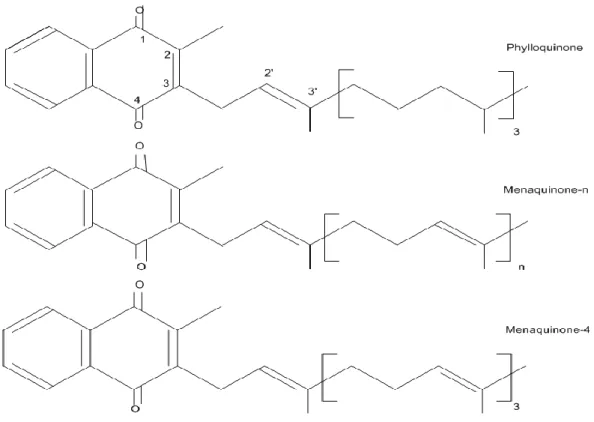
1.1. Chemical Structure of Vitamin K
All K vitamers possess a 2-methyl-1,4-naphtoquinone ring structure called menadione or K3; the K vitamers differ based on the side chain at the 3-position which varies in length and degree of saturation. VK includes two natural forms: 1) phylloquinone, also referred to as K1 (2-methyl-3-phytyl-1, 4-naphtaquinone), is synthesized in plants, and 2) the menaquinones (MK-n) or K2 (2-methyl-1, 4-naphtaquinone), a family of compounds of bacterial origin (Food and Nutrition Board 2001). One of the menaquinone, menaquinone 4 (MK-4), is not a product of bacterial synthesis but is produced from phylloquinone. Animal studies have shown that this K1 MK-4 conversion is tissue specific (Thijssen and Drittij-Reijnders 1994). Menadione is not a natural form of VK, although in avian and mammalian tissues it can be alkylated to MK-4. This synthetic vitamer has been used as the primary source of VK in animal feeds. High performance liquid chromatography (HPLC) is the method of choice for assessing the different K vitamers in biological matrices (e.g. plasma, tissues/organs, foods) (Davidson and Sadowski 1997). The chemical structures of the principal K vitamers are illustrated in figure 1.
Figure 1. Chemical structures of principal K vitamers (Ferland 2012a).
1.2. Dietary Sources of vitamin K
Phylloquinone from plants is the main dietary source of VK in North America and Europe (Shearer and Newman 2008). Green leafy vegetables contain the highest amount of phylloquinone and contribute 40-50% of total daily intake (Booth, Pennington et al. 1996). Phylloquinone content of swiss chard, spinach and kale has been assessed at more than 300 μg/100 g, while broccoli, brussels sprouts, and cabbage contain 100-200 μg/100 g. Vegetable oils such as soybean, olive, and canola contain significant amounts of
phylloquinone varying from 50-200 μg/100 g (Shearer, Bach et al. 1996; Booth and Suttie 1998; Ferland G, Nutrition Foundation. et al. 2011). Mixed dishes prepared with phylloquinone-rich oils contribute approximately 15% of total dietary phylloquinone intake (Booth and Suttie 1998).
Green leafy vegetables contain the highest amount of phylloquinone and contribute 40-50% of total daily intake. Phylloquinone content of swiss chard, spinach and kale has been assessed at more than 300 μg/100 g, while broccoli, brussels sprouts, and cabbage contain 100-200 μg/100 g. Vegetable oils such as soybean, olive, and canola contain significant amounts of phylloquinone varying from 50-200 μg/100 g. Mixed dishes prepared with phylloquinone-rich oils contribute approximately 15% of total dietary phylloquinone intake (Booth and Suttie 1998).
The menaquinones are present in limited amounts in foods, however, low levels of menaquinones have been reported in animal products such as chicken egg yolk and butter (Hirauchi, Sakano et al. 1989). Organ livers (especially pig and bovine) are a good source of long chain menaquinones (MK-6 to MK-13) (Koivu-Tikkanen, Ollilainen et al. 2000) as cheeses with MK-8 and MK-9 content have been estimated at between 5 to 20 μg/100 g (Shearer, Bach et al. 1996). Meat, eggs, and dairy foods are also good sources of MK-4 (Elder, Haytowitz et al. 2006) while MK-7 is found in the highest quantity in fermented soybean products such as Natto, a traditional Japanese dish (Schurgers and Vermeer 2000).
The US Dietary Reference Intakes for VK in 2001 recommended 120 and 90 µg/day for adult males and females, respectively (Food and Nutrition Board 2001).
1.3. Metabolism
1.3.1. Absorption
In the intestine, VK is incorporated into micelles in a pathway which depends on bile salts and pancreatic juices (Yong Ji 2009).
The efficiency of absorption depends on the dietary source of phylloquinone and the presence of fat in the intestine. The absorption efficiency of phylloquinone in healthy adults is estimated at ~ 80% when administered in its free form (Shearer, Bach et al. 1996; Garber, Binkley et al. 1999) but decreases to ~10% when the vitamin is absorbed from green vegetables (Gijsbers, Jie et al. 1996). In one report, the absorption of phylloquinone from spinach was found to be 4-17% (Gijsbers, Jie et al. 1996; Garber, Binkley et al. 1999); similar low absorption efficiency has been reported for kale (Novotny, Kurilich et al. 2010). Reason for the poor bioavailability of phylloquinone in green vegetables is linked to its tight binding to the chloroplasts (Newman and Shearer 1998).
Adding oil to the vegetables has been shown to increase phylloquinone bioavailability (Booth, Lichtenstein et al. 2002). In one study, adding butter to spinach resulted in a 3-fold (8% to 26%) increase in phylloquinone absorption (Gijsbers, Jie et al. 1996).
Recent isotopic studies further indicate that phylloquinone absorption is affected by meal components (Jones, Bluck et al. 2008). In their report Jones et al. (Jones, Bluck et al. 2009) showed that absorption of 13C-phylloquinone was 3 times higher in a fast foods type meal in which 80% of phylloquinone came from oil, than in meals in which 80-90% of the phylloquinone came from vegetables.
1.3.2. Transport
In human, > 50% of phylloquinone is carried in triacylglycerol-rich lipoprotein (TRGLP), the rest being carried equally in low (LDL) and high (HDL) density lipoprotein fractions (~ 15% each) (Lamon-Fava, Sadowski et al. 1998; Schurgers and Vermeer 2002; Erkkila, Lichtenstein et al. 2004). In contrast, the menaquinones are mainly transported in the TRGLP and LDL fractions (Schurgers and Vermeer 2002). Phylloquinone circulates in blood in very small concentrations (0.25-2.7 nmol/L) and has been shown to be strongly linked to circulating triacylglycerols (Sadowski, Hood et al. 1989; Azharuddin, O'Reilly et al. 2007).
13.3. Tissue Store and Distribution
Liver is the main storage site for VK, the long chain menaquinones representing the major VK constituent (Suttie 2009). When assessed in humans, the menaquinones were indeed shown to represent ~ 90% and phylloquinone ~10% of total hepatic VK (Suttie 1995). Phylloquinone stores are generally unstable and can decrease up to 25% within 3 days in conditions of dietary depletion (Usui, Tanimura et al. 1990). It is known from animal studies that the K vitamers (notably phylloquinone and MK-4) are present in non-hepatic tissues (Thijssen and Drittij-Reijnders 1994). Specifically, phylloquinone contents in heart and pancreas are higher than in liver while lower concentrations are observed in brain, kidney, salivary glands and lung. Menaquinone-4 is also widely distributed with low concentration in the liver and high levels in brain, pancreas and salivary glands (Thijssen and Drittij-Reijnders 1994; Thijssen, Drittij-Reijnders et al. 1996). The origin of MK-4 in the brain is discussed in more detail in a later section.
Phylloquinone and menaquinones tissue concentration have been shown to increase as a function of diet in a tissue-dependent manner (Thijssen, Drittij-Reijnders et al. 1996;
Davidson, Foley et al. 1998; Ronden, Thijssen et al. 1998). In a study by Ronden et al., phylloquinone and MK-4 concentrations increased in all tissues following ingestion of a phylloquinone or MK-4 supplemented diet, while the ratio between phylloquinone and MK-4 varied among the tissues; the highest values related to the liver and heart. High levels of MK-4 were observed in the pancreas and testis as a result of exclusive phylloquinone diet (Ronden, Thijssen et al. 1998).
Tissue VK level is also affected by age and gender. In a study by Huber et al. phylloquinone and menaquinones concentration were assessed in different tissues of male and female rats at 3-, 12- and 24-months old. Except in hearts of 3-months of age, phylloquinone concentrations were not affected by gender. In contrast, MK-4 levels in kidney, heart and brain were significantly higher in females than in males. It was also reported that the hepatic phylloquinone and MK-6 concentration elevated with age; while MK-4 level did not change. In the brain and the extrahepatic tissues of males and females rats, MK-4 concentration declined with age (Huber, Davidson et al. 1999). In humans, plasma phylloquinone concentration has been shown to decrease with age (Sadowski, Hood et al. 1989). Another study investigated the gender differences in relation to VK intake and reported the reduced concentration of phylloquinone and MK-4 in the liver of male and female; however supplemented- phylloquinone diet was associated with higher MK-4 in female rats (Huber, Davidson et al. 1999).
1.3.4. Turnover
Phylloquinone and menaquinone are excreted by a degradative pathway in which the polyisoprenoid side chain is cleaved off into two major carboxylic acid metabolites with 7- and 5-carbon side chains. In humans, around 20% of metabolized phylloquinone is excreted in the urine and 40% to 50% in the feces. Urinary metabolites contain
predominantly glucuronide conjugates of derivatives with an oxidized form of phytyl side chain (Shearer, Mallinson et al. 1972).
Compared with other fat-soluble vitamins, the total body pool of VK is very small and the stored VK in liver is depleted rapidly as a result of restricted dietary VK. The turnover of phylloquinone has been estimated at around 1.5 days (Olson, Chao et al. 2002). The consumed phylloquinone in plasma peaks after 6-9 hours and returns to the baseline within 24 hours (Erkkila, Lichtenstein et al. 2004). It was also shown that urinary VK metabolites decrease by 20% within 15 days as a result of restricted diet (11µg/d) and increases rapidly following a rich VK diet (206 µg/d) (Harrington, Booth et al. 2007). However, long-chain menaquinones remain in circulation much longer i.e. ~ 72 hours in the case of MK-7 (Schurgers and Vermeer 2000).
1.3.5. Transformation of Phylloquinone to MK-4
It is now well-established that tissue MK-4 results from phylloquinone conversion with menadione as an intermediate (Thijssen, Vervoort et al. 2006). Two routes have been suggested: 1) the release of menadione from phylloquinone by removal of the phytyl side chain in the intestine followed by prenylation of menadione in tissues, or 2) the side-chain cleavage of phylloquinone and generation of menadione followed prenylation occurring within the target cell (Ansell, Hirsh et al. 2008). Recently, this team identified the human enzyme responsible for MK-4 biosynthesis. This enzyme is UBiA prenyltransferase domain-containing protein 1 (UBIAD1) which is expressed in numerous tissues in mice and is located in the endoplasmic reticulum (Nakagawa, Hirota et al. 2010) (Figure 2).
Figure2. Conversion of phylloquinone to MK-4 by UBIAD1(Nakagawa, Hirota et al. 2010)
1.4. Vitamin K Dependent Carboxylation
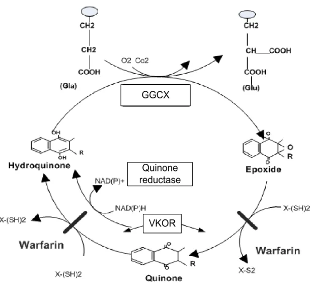
Within the cell, VK acts as a cofactor in the posttranslation of Gla from Glu which is a common residue in all vitamin K dependent proteins (VKDP). The carboxylation is catalyzed by GGCX which is a microsomal enzyme located at the luminal surface of the endoplasmic reticulum. This reaction requires the reduced form of VK, hydroquinone (KH2), and the presence of carbon dioxide and oxygen (Dowd, Ham et al. 1995). The conversion of Gla to Glu increases the affinity of these proteins for calcium, a characteristic that allows them to bind to biological components associated to their specific role. To convert Glu to Gla residues, the carboxylase uses the energy of VK hydroquinone oxygenation, and this carboxylation is facilitated by a carboxylase recognition signal propeptide (Berkner 2008). In the course of the catalytic sequence, hyrdoquinone is oxidized to the epoxide form, an inactive form of VK. The vitamin K 2,3-epoxide (KO) is then recycled to quinone and hydroquinone by the VK oxido-reductase (VKOR) which is dependent on the presence of dithiol cofactors (Suttie 2009). Coumarin derivatives such as
warfarin block the action of VKOR (Wallin and Martin 1985). The formation of KO and its conversion back to KH2 makes the VK cycle (Figure 3).
The liver has a unique reductase enzyme which is not sensitive to coumadin drugs. This enzyme is NAD(P)H dependent and makes possible the reduction of the quinone form of VK to KH2 in the presence of Coumadin drugs, when a high tissue concentration of VK is available (Berkner and Runge 2004) .
In a condition of insufficient dietary VK or the presence of anticoagulant drugs, the carboxylation action is blocked and undercarboxilated forms of proteins are released into the plasma. These inactive forms of proteins are called PIVKAs (protein induced by VK absence or antagonism) and used as a marker of the nutritional status of VK (Lee, Chung et al. 2010) .
GGCX
Quinone
reductase
VKOR
Figure 3. Scheme showing the vitamin K cycle (Ferland G, Nutrition Foundation. et al.
1.5. Vitamin K Dependent Proteins
Although the discovery of VK in 1930 was associated with its specific role in blood coagulation, the discovery of Gla, a new amino acid common to all VKDP, extended the physiological role of VK. These proteins are involved in bone and calcium metabolism and the brain. The VKDP and their function are summarized in Table 1.
1.5.1. Blood Coagulation Proteins
Prothrombine (factor II), factors VII, IX, and X, protein C, S and Z are the VKDPs involved in clotting blood. They are all synthesized in the liver and contain 10-12 Gla residues (Versteeg, Heemskerk et al. 2013). Calcium mediates the binding of the complexes via the Gla residues to the negatively charged phospholipid surfaces expressed by platelets and endothelial cells of the injured cells. The generation of thrombin from prothrombin leads to the formation of fibrin by an activated factor X, which itself is activated by factors VII and IX respectively. Proteins C, S and Z act as the coagulation inhibitors. Protein C inhibits coagulation by deactivating factor Va and VIIIa and elevating the level of fibrinolysis with protein S as a cofactor, while protein Z acts as a cofactor for blocking the activity of factor Xa by protein Z-dependent protease inhibitor (Ferland 1998; Norris 2003). (Davie 2003; Berkner and Runge 2004).
Beside their hemostatic functions the coagulation proteins possess cell signaling activities and are involved in a wide-range of cellular events. For instance, thrombin has been involved in such phenomenon as tumor growth and metastasis, angiogenesis, atherosclerosis, inflammation, survival of neutrophils and monocytes (Sokolova and Reiser 2008; Chen and Dorling 2009). Similarly, protein C has been shown to possess anti-inflammatory and anti-apoptotic properties (Danese, Vetrano et al. 2010)
Three main Gla proteins involved in bone metabolism are osteocalcin, matrix Gla proteins (MGP) and protein S (Ferland G, Nutrition Foundation. et al. 2011). Osteocalcin is a VKDP synthesized by osteoblasts and donotoblasts, and acts as a negative regulator of bone formation (Ducy, Desbois et al. 1996; Boskey, Gadaleta et al. 1998). It contains 3 Glu residues which increase affinity binding to hydroxyapatite in bone (Booth 2009). In vertebrate species, osteocalcin accounts for around 15-20% of the non-collagenous bone protein , and its molecular weight is 5,8 kDa (Price, Otsuka et al. 1976). Approximately 20% of the newly synthesized bone proteins are secreted in blood circulation and are used as biochemical markers for bone formation. Osteocalcin deficient rats by warfarin administration had excessive bone mineralization and also premature closure of the growth plate (Price 1988). Moreover, it was shown that mice with eliminated osteocalcin gene coding had increased bone mass and improved bone functional qualities (Ducy, Desbois et al. 1996). These data suggest that osteocalcin regulates bone formation negatively.
Although MGP only accumulates in calcified tissues, it is expressed in many soft tissues such as vascular smooth muscle cells. Its main physiological role in vascular calcification is explained in the following section (Luo, Ducy et al. 1997).
Typically protein S is known because of its role in blood coagulation; however, it plays a role in maintaining bone homeostasis as well. In early 1990, two cases of osteopenia were reported in protein S deficient children (Pan, Gomperts et al. 1990). Protein S is synthesized and secreted by osteoblasts and increases the bone resorbing activity of mature osteoclasts, an action which is associated to the ability of protein to bind tyrosine kinase receptors (Maillard, Berruyer et al. 1992; Nakamura, Hakeda et al. 1998).
1.5.3. Protein Involved in Vascular Calcification
MGP contains 5 Gla residue and has a molecular weight around 9.6 kDa (Price, Urist et al. 1983). Unlike osteocalcin this protein is widely distributed in body tissues i.e. lung, heart, kidney and spleen (Fraser and Price 1988). It is also expressed in cartilage (Yagami, Suh et al. 1999) and vascular smooth muscle (Shanahan and Weissberg 1998). MGP acts as an inhibitor of tissue calcification (Fraser and Price 1988; Luo, Ducy et al. 1997; Shearer 2000). A study on mice that targeted the deletion of the MPG gene resulted in mortality of the mice within two months as a result of extensive arterial calcification that led to blood-vessel rupture (Luo, Ducy et al. 1997). Similar results with a lower degree of severity was observed in rats treated with high doses of warfarin (Price, Faus et al. 1998). In humans, mutations of the MGP gene results to Keutel syndrome a condition characterized by abnormal cartilage and arterial calcification (Hur, Raymond et al. 2005). Although the mechanism of action of MGP in vascular calcification is not fully elucidated (Proudfoot and Shanahan 2006), it potentially acts by binding to calcium ions and inhibiting crystal growth (Roy and Nishimoto 2002), and modulating the action of bone morphometric protein type 2 and 4 (Zebboudj, Shin et al. 2003; Yao, Zebboudj et al. 2006).
1.5.4. Other Vitamin K Dependent Proteins
Gas6. In 1993, the VKDP Gas6 was discovered as a product of the growth arrest-specific gene 6. It has a molecular weight of 75 kDa and contains 11-12 Gla residues; its 673 amino acids shows 43-44% homology with protein S (Manfioletti, Brancolini et al. 1993). Gas 6 is expressed in numerous tissues and is involved in cellular processes such as cell differentiation, proliferation and activation, phagocytosis and protection against apoptosis (Bellido-Martin and de Frutos 2008). As discussed later, this protein has many actions in the nervous system.
Transmemberance Gla proteins (TMGs). These proteins include proline-rich Gla proteins 1 and 2 (PRGP 1, PRGP2), and TMG3, TMG4. These single-pass integral membrane proteins are widely distributed and although their physiological function are presently unknown, their chemical structures suggest that they could be involved in cell transduction (Kulman, Harris et al. 1997; Kulman, Harris et al. 2001).
Gla rich protein (GRP). This protein was identified from calcified cartilage in 2008. It has a molecular weight of 10.2 kDa and contains 16 Gla residues. GPR is widely distributed in tissues, but its expression is highest in cartilage. The functions of GPR are not clear at present, but it could regulate calcium in the extracellular environment (Viegas, Simes et al. 2008; Viegas, Cavaco et al. 2009).
Periostin. This protein which is associated with the extra-cellular matrix and is involved in cell migration and angiogenesis was recently identified as a VKDP (Coutu, Wu et al. 2008).
Transthyretin. Identified for its association with thyroid hormones and retinol-binding protein; the role of its Gla residues is presently unknown (Ruggeberg, Horn et al. 2008).
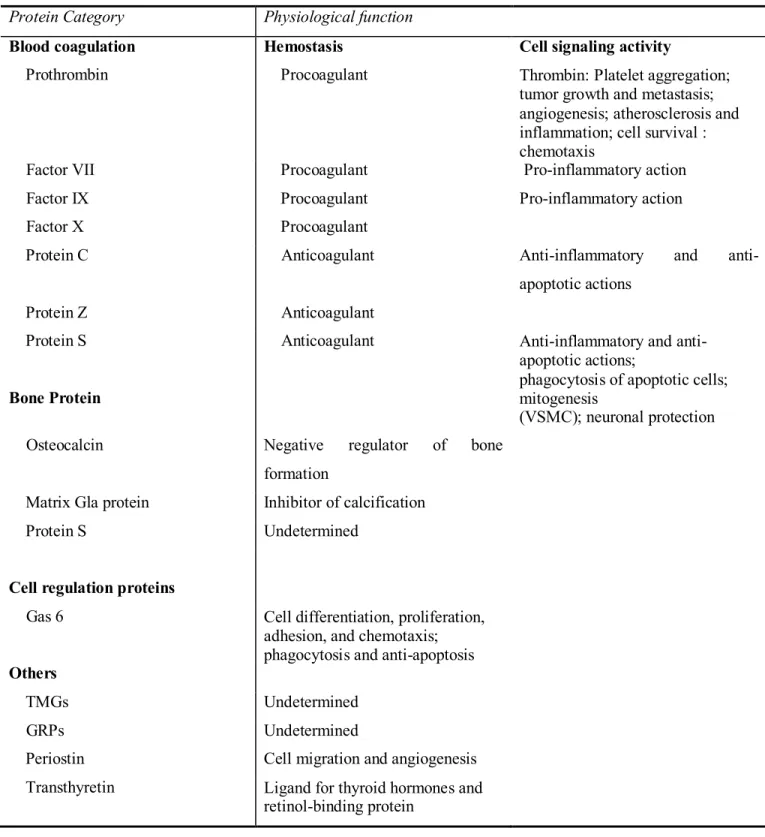
Table 1. The vitamin K dependent proteins and their functions (Ferland G, Nutrition Foundation. et al. 2011).
Protein Category Physiological function
Blood coagulation Hemostasis Cell signaling activity
Prothrombin Procoagulant Thrombin: Platelet aggregation; tumor growth and metastasis; angiogenesis; atherosclerosis and inflammation; cell survival : chemotaxis
Factor VII Procoagulant Pro-inflammatory action
Factor IX Procoagulant Pro-inflammatory action
Factor X Procoagulant
Protein C Anticoagulant Anti-inflammatory and
anti-apoptotic actions
Protein Z Anticoagulant
Protein S
Bone Protein
Anticoagulant Anti-inflammatory and anti-apoptotic actions;
phagocytosis of apoptotic cells; mitogenesis
(VSMC); neuronal protection Osteocalcin Negative regulator of bone
formation
Matrix Gla protein Inhibitor of calcification
Protein S Undetermined
Cell regulation proteins
Gas 6 Cell differentiation, proliferation, adhesion, and chemotaxis; phagocytosis and anti-apoptosis
Others
TMGs Undetermined
GRPs Undetermined
Periostin Cell migration and angiogenesis Transthyretin Ligand for thyroid hormones and
2. Vitamin K and Brain Function 2.1. K Vitamers in Brain
The role of VK in brain function has not been systematically investigated even though VK is widely expressed in brain. It is well established that MK-4 is the predominant form of VK in the brain (Thijssen, Drittij-Reijnders et al. 1996). MK-4 concentration in six months old Sprague-Dawley rats has been shown to account for more than 98% of total cerebral VK and both phylloquinone, and MK-4 increase in brain as a result of dietary VK and are affected by age and sex (section 1.3.3). In rats, MK-4 differs among the different brain regions, the highest concentrations being observed in myelinated regions such as pons medulla and midbrain (Carrie, Portoukalian et al. 2004).
2.2. Vitamin K Dependents Proteins in the Brain
Gas6 and protein S are two VKDPs in the brain. They are not directly involved in cognition, although their signalling actions in neurons (Gas6 and protein S), the glia (Gas6) as well as antithrombic function (protein S), suggest contributions of these proteins to the cognitive process (reviewd in Ferland 2012a).
2.2.1. Gas6
The distribution of protein Gas6 was assessed in the central nervous system of rats using biochemical and histological techniques (Prieto, Weber et al. 1999). In the rat embryo, Gas6 expression is mainly in non-neuronal tissues while in the late embryonic stages and during adulthood it is more widely expressed. In the brain of adult rats, Gas6 is expressed in cerebral and piriform cortex, hippocampus, thalamic and hypothalamic structures, midbrain, and cerebellum. It is also highly expressed in the large neurons of the dorsal root ganglia and the neurons of the spinal cord (Li, Chen et al. 1996; Prieto, Weber
et al. 1999). Compared to other VKDPs such as prothrombin and protein S, plasma Gas6 concentrations are much lower (Garcia de Frutos, Alim et al. 1994); concentrations of 0.25 nM (Balogh, Hafizi et al. 2005) or less (Gibot, Massin et al. 2007) having been published. In rats, Gas6 concentrations have also been shown to decrease with age. When investigated in 6-, 12-, and 24-mo-old Fisher 344 rats, Gas6 expression in 24-mo rats was decreased by > 84% in the frontal cortex and by 55% in the striatum and hippocampus when compared to those aged 6 months (Tsaioun 2000).
Gas6 acts as a ligand for the receptor tyrosine kinases of TAM family (Varnum, Young et al. 1995). In the nervous system, Gas6 has been shown to participate in cell survival, chemotaxis, mitogenesis, cell growth, and myelination. Specifically, Gas6 has been shown to prevent apoptosis of gonadotropin-releasing hormone neurons from undergoing serum deprivation (Allen, Zeng et al. 1999) and of neurons subjected to phospholipase A2-IIA (Yagami, Ueda et al. 2003). Protein Gas6 has also been shown to protect cortical neurons from amyloid β (Aβ) protein-induced apoptosis. Applying Gas6 to rat cortical neuron cultures resulted in cell apoptosis prevention by inhibiting Ca2+ influx and decreasing Aβ -induced apoptotic features (Yagami, Ueda et al. 2002).
In addition to its neuron-related role, protein Gas6 modulates the survival and functions of glial cell, mainly oligodendrocytes, Schwann cells, and microglia. Schwann cells and oligodendrocytes have important roles in neuron myelination in the central and peripheral nervous systems, and in the transmission of the nervous impulses. In the central nervous system, microglia cells are part of the immune effectors involved in tissue homeostasis and repair (Nimmerjahn, Kirchhoff et al. 2005) and possess phagocytic functions, removing pathogens, cellular debris, and apoptotic cells, which amass over time (Binder, Cate et al. 2008). Recent studies have illustrated the role of Gas6-dependent activation of TAM receptors in the survival of glial cells and the modulation of microglial phenotype. In a study, adding Gas6 to the medium of an oligodendrocyte culture of the human fetal cord elevated cell survival (Shankar, O'Guin et al. 2006).
2-2-2. Protein S
Historically known for its role in blood coagulation, protein S is expressed in the central nervous system. Protein S has been identified in the locus coeruleus, choroid plexus, and astrocytes of adult nervous system (Stitt, Conn et al. 1995; Hall, Obin et al. 2002). Protein S mRNA has also been found in pyramidal neurons of the cortex and hippocampus, and granule neurons of the dentate gyrus of rabbit brains (He, Shen et al. 1995).
Although the actions of protein S in the brain have been less investigated, it is a ligand for the TAM receptors like Gas 6 (Varnum, Young et al. 1995). In vivo and in vitro, protein S has been shown to plays a role in neuronal protection during ischemic/hypoxic injury. In one study, brain infarction and edema volumes were significantly reduced and post-ischemic cerebral blood flow was improved in mice treated with protein S. Moreover, treatment with protein S resulted in less fibrin deposition and infiltration with neutrophils, less apoptotic neurons and enhanced motor performance (Liu, Guo et al. 2003). In another study, protein S has been shown to protect neurons from N-methyl-D-aspartate-induced toxicity and apoptosis (Zhong, Wang et al. 2010).
3. Vitamin K and Sphingolipids
Sphingolipids are a class of complex lipids found in all mammalian cell membranes. They are abundantly distributed in the cells of the central nervous system in the forms of ceramide, sphingomyelin, cerebroside, sulfatide, and gangliosides (Bartke and Hannun 2009). In addition to their structural function, they are bioactive molecules involved in cell-cell interactions (Ohanian and Ohanian 2001; Chalfant and Spiegel 2005; Siow and Wattenberg 2011; Tani 2011) and participate in important cellular events such as proliferation, differentiation, senescence and transformation (Zeidan and Hannun 2007). Several studies in vitro and in vivo have demonstrated the contribution of VK in sphingolipid metabolism in the brain (Denisova and Booth 2005).
3.1. The Structure and Metabolism of Sphingolipids
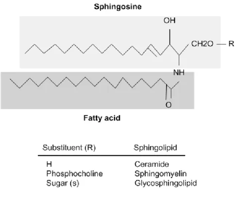
All sphingolipids are composed of a sphingoid long-chain base known as sphingosine that is linked to an acyl chain through an amide bond (Figure 4).
Figure 4. General sphingolipds structure (Malagarie-Cazenave, Andrieu-Abadie et
al. 2002).
The metabolisms of sphingolipids include complex pathways with the sphingoid base subjected to the addition of fatty acids, sugars, or phosphate moieties (Brice and Cowart 2011; Mencarelli and Martinez-Martinez 2012).
Figure 5. Scheme showing the biosynthesis and metabolic interactions of sphingolipids
(Ferland 2012b).
As illustrated in figure 5, the formation of ceramide represents the initial reaction of sphingolipids metabolism and is catalyzed by serine palmitoyl transferase and the combination of serine and palmitoyl-COA. Ceramides can also be generated by the action of sphingomyelinase from sphingomyelin (Mullen, Hannun et al. 2012) .
Sphingomyelin, the major sphingolipid in the plasma membrane, is synthesized in the Golgi apparatus by the action of two isoforms of sphingomyelin synthase (SMS1 and
SMS2). The generation of sphingomyelin occurs by transferring a phosphocholine head group from phosphatidylcholine to ceramide (Milhas, Clarke et al. 2010).
Similarly to sphingomyelin, the glycosphingolipids synthesis occurs in the lumen of the Golgi apparatus. Glycosphingolipids are distributed abundantly in the neuronal cells. They are structurally and biosynthetially derived from lactosylceramide, except GM4, which is a major component of myelin and derived from galactosylceramide. The biosynthetic pathway of the ganglio-series of ganglioside is illustrated in figure 6. Lactosylceramide and the hematosides GM3, GD3 and GT3 are used as precursors for complex gangliosides: the asylio- a-, b-, and c-series respectively. In the successive reactions, carbohydrate- and sialic acid residues are attached to glycosyl acceptors by specific glycosyl transferases.
Initially, GM3 is synthesized by adding a sialic acid to LacCer by the action of CMP-sialic acid: Lactosylceramide α2-3 sialyltransferase. GD3 and GT3 are synthesized by sequential sialic acid addition to GM3, and GD3 by CMP-sialic acid: GM3 α-8 sialyltransferase. The conversion of simple ganglioside to a complex group is catalyzed by GalNAc:LacCer/GM3/GD3/GT3 β1-4 N-acetylgalactosaminyltransferase, UDP-Gal:GA2/GM2/GD2/GT2 β1-3 galactosyltransferase, CMP-sialic acid:GA1/GM1/GD1b/GT1c α2-8 sialyltransferase (Sandhoff and Kolter 2003; Yu, Tsai et al. 2011; Yu, Tsai et al. 2012).
In the early stages of embryonic brain development, simple gangliosides such as GM3 and GD3 are highly expressed, while in later stages the expression of complex gangliosides such as GM1, GD1a, GD1b, and GT1b are up-regulated (Ngamukote, Yanagisawa et al. 2007; Yu, Nakatani et al. 2009).
Figur 6. Biosynthetic pathways of ganglio-series gangliosides (Yu, Tsai et al. 2012).
Galactosylceramide or cerebroside is formed by the condensation of ceramide and UDP-galactose via the action of the enzyme galactosyltransferase. Moreover, galactosylceramide is a precursor for sulfatide synthesis. Sulfotransferase is the enzyme responsible for the synthesis of sulfatide from galactoceramide by activation of a sulfate of phosphodenosine-5-phosphosulfate. Galactosylceramide and its subsequent metabolite, sulfatide, are highly enriched in myelinated regions of the central nervous system. Galactosyltransferase is located in the endoplasmic reticulum transmembrane and its catalytic site is at the lumen of the endoplasmic reticulum (Gault, Obeid et al. 2010).
Most sphingolipids are degraded in lysosomes and to a lesser extent in the plasma membrane. Sphingolipids are catabolized to ceramide, sphingosine, and sphingosine-1-phosphate via the action of the ceramidase. After ceramide is catalyzed into sphingosine, the conversion of sphingosine to sphingosine-1-phosphate occurs by sphingosine kinases. In the final step of sphingolipid degradation, sphingosine-1-phosphate is catalyzed by sphingosine-1-phosphate lyase function in the endoplasmic reticulum to generate hexadecenal and phosphoethanolamine (Gault, Obeid et al. 2010). Byproducts of sphingolipids catabolism, i.e. sphingosine, can then access the cytosol, and through the salvage pathways regenerate ceramide (Zeidan and Hannun 2007).
In human cells, the breakdown of sphingomyelin is a major part of membrane homeostasis. Sphingomyelin hydrolysis results in ceramide and free phosphocholine via the action of the sphingomyelinase enzyme (Gault, Obeid et al. 2010).
Glycosphingolipis catabolism occurs in the acidic compartments of the cells, the endosomes and the lysosomes. Cellular glycolipids are degraded into their building blocks by the digestion of cellular membranes. Parts of the plasma membrane are endocytosed and transferred through the endosomal compartment to the lysosome. Glycosphingolipids are ultimately cleaved into monosaccharides, sialic acid, fatty acids, and sphingosid bases, which can be either degraded or re-used for sphingolipids biosynthesis (Sandhoff and Kolter 2003).
3.2. The Physiological Function of Sphingolipids
Sphingolipids are important components of the cell membranes. Beside their structural role, they have signalling activities and are involved in the regulation of many cellular processes. Here, we review the functions of the main sphingolipids in the nervous system.
3.2.1. Ceramide and Sphingomyeline
Ceramide and sphingomyelin play leading roles in the regulation of cellular events. Ceramide is involved in the regulation of cell growth, differentiation, senescence, necrosis, proliferation, and apoptosis. It has also been shown that the actions of ceramide are through regulation of protein kinase C, raf-1, and the kinase-suppressor of Ras-, notably by changing the phosphorylation level of key substrates (Bartke and Hannun 2009). In a series of studies, ceramide has been shown to inhibit the neuronal survival pathway regulated by phosphatidil-inositol-3-kinase/Akt (Arboleda, Morales et al. 2009), by conveying activity to the caspase-9/caspase-3 pathway (Movsesyan, Yakovlev et al. 2002; Stoica, Movsesyan et al. 2003). Production of ceramide is induced by stimuli such as tumor necrosis factor-α (TNF-α), interleukin (IL)-1, Fas ligand, ionizing radiation, phorbolesters, heat stress, oxidative stress, and chemotherapeutics (Nikolova-Karakashian and Rozenova 2010; Barth, Cabot et al. 2011; Castro, de Almeida et al. 2011; Barth, Gustafson et al. 2012; Li, Gulbins et al. 2012; Martinez, Chen et al. 2012; Pan, Liu et al. 2012; Van Brocklyn and Williams 2012). High levels of ceramide contribute to the inflammation process and generate reactive oxygen species from mitochondria (Ballou, Laulederkind et al. 1996; Jana, Hogan et al. 2009). Furthermore, the transient hydrolysis of sphingomyelin into ceramide has been observed in response to apoptosis-activation of the sphingomyelinases. These enzymes seem to be important for cellular signal transduction (Jana, Hogan et al. 2009; Milhas, Clarke et al. 2010).
3.2.2. Cerbroside and Sulfatide
Myelin is greatly enriched with cerebroside (galactosylceramide) and sulfatide. The critical physiological function of myelin in the brain is that it insulates axons and provides high axon conductivity for nerve impulses. The myelin sheath ensures rapid intercellular communication which is essential for brain functions such as motor control (Sandell and
Peters 2003; Sandvig, Berry et al. 2004). A series of studies showed that mice lacking galactosyltransferase, the enzyme responsible for synthesizing these two myelin lipids, presented thin and unstable myelin with abundant abnormalities (Bosio, Binczek et al. 1996; Coetzee, Fujita et al. 1996; Coetzee, Dupree et al. 1998; Dupree, Girault et al. 1999; Marcus, Dupree et al. 2000). Another study showed that sulfatide is required for myelin membrane maintenance and axon structures, while galactocerbroside is essential for myelin development (Marcus, Honigbaum et al. 2006). Also, a recent study demonstrated an inhibitory role of sulfatide in myelin-associated axon outgrowth (Winzeler, Mandemakers et al. 2011).
Finally, cerebroside and sulfatide play major roles in regulating oligodendrocyte differentiation and survival. During brain development, oligodendrocyte differentiation is critical for reaching the highest level in the elaboration of myelin sheath (Jana, Hogan et al. 2009).
3.2.3. Gangliosides
In the nervous system, gangliosides interact with numerous regulatory proteins (Yu, Nakatani et al. 2009; Furukawa, Ohmi et al. 2011) and are involved in cell survival, proliferation, and differentiation during brain development (Yu, Nakatani et al. 2009). Gangliosides also contribute to the maintenance and repair of nerve tissues (Kittaka, Itoh et al. 2008; Ohmi, Tajima et al. 2009).
GT1b is one of the major ganglioside subtypes in neuronal and synaptic membranes (Viljetic, Labak et al. 2012). It has been shown to influence cellular apoptosis, and to possess neurotoxic functions against dopaminergic neurons. Specifically, cultures of mesencephalic cells deprived of serum were observed to be more susceptible to GT1b-induced neurotoxicity (Chung, Joe et al. 2001). In vivo, GT1b injections into the substantia resulted in the death of nigra dopaminergic neurons, including dopaminergic neurons (Ryu, Shin et al. 2002). Recently, evidence was provided that the Akt/GSK-3/tau signalling pathway is involved in the GT1b-mediated neurotoxic actions (Chung, Bok et al. 2010).
GT1b-induced apoptosis also has been reported in non-neuronal cells like thymocytes and keratinocytes (Zhou, Shao et al. 1998).
GD3, the precursor of GT1b, also possesses an apoptotic function. Induction of apoptosis in cultures of cerebellar granule neurons—by switching the growing medium to a medium containing lower concentrations of potassium—was associated with increased levels of GD3 and the enzyme that synthesizes GD3 from GM3. The exogenous addition of GD3 to the culture accelerated neuronal apoptosis however, no alteration was observed by adding GD1a (Melchiorri, Martini et al. 2002).
GD1a acts as a modulator of cell survival and proliferation. When applied exogenously to the FDC-P1 cell line, GD1a enhanced the proliferation of granulocyte-macrophage colony-stimulating factor (Santos, Maia et al. 2011). In a transgenic model for Huntington’s disease (HD), the poor performances of mice in the footprint test were associated with lower cerebellar gangliosides and decreased numbers of GD1a-enriched granule cells (Denny, Desplats et al. 2010). Also, brains of mice suffering from Rett syndrome, a neurodevelopmental disorder of the grey matter, have been shown to contain 15% lower concentrations of GD1a in the cerebrum/brainstem, a finding associated with poorer motor performances (Seyfried, Heinecke et al. 2009).
GM3 contributes to normal behaviour. Mice lacking GM3 caused by gene disruption exhibited progressive motor, sensory dysfunctions, and deterioration in spatial learning and memory (Tajima, Egashira et al. 2009; Tajima, Egashira et al. 2010). Nimii et al. (Niimi, Nishioka et al. 2011) investigated cognition, motor activity and emotional behaviour of GM-3 knockout mice and found better performances in the Y-maze test, hyperactivity in the motor activity test, and anxiety in the elevated plus maze test. They also exhibited attention-deficit hyperactivity disorder, reduced attention, and increased impulsive behaviour.
Reduced levels of GM1 have been associated with neurodegenerative disorders such as Huntington’s disease (HD). Lowered GM1 synthesis in the brain cells of HD transgenic mice and also fibroblasts from HD patients have been suggested as potentially contributing to the higher susceptibility of HD cells to apoptosis (Maglione, Marchi et al. 2010).
3.3. Sphingolipids and Alzheimer’s Disease
Alzheimer’s disease (AD) is an age-related disorder characterized by the deposition of Aβ peptide components and extensive neuronal apoptosis in the brain, notably in the hippocampus. Recent studies suggest that disturbance in the lipids composition of neurons cell membranes and alteration in sphingolipids metabolism could have a role in the neuropathological process of AD (Haughey, Bandaru et al. 2010).
Decreased levels of sphingomyelin and elevated concentrations of ceramides have been reported in various brain regions in AD (Mielke, Haughey et al. 2011; Filippov, Song et al. 2012; Mielke, Bandaru et al. 2012).
A significantly higher concentration of ceramides was reported in the brains of patients suffering from AD as compared to the age-matched control group (Satoi, Tomimoto et al. 2005; Filippov, Song et al. 2012). Finally, high levels of ceramides and dihydroceramides were associated with the strong progression in AD, although higher plasma concentrations of sphingomyelin, dihydrosphingomyelin were associated with less progression (Satoi, Tomimoto et al. 2005). Furthermore, higher levels of serum ceramides were associated with an increased risk of AD (Mielke, Bandaru et al. 2012).
Sulfatides are also affected in AD. Sulfatide concentrations were shown to decrease in the white and gray matter in the earliest stage of AD (Pettegrew, Panchalingam et al. 2001; Han, D et al. 2002; Cheng, Xu et al. 2003). Han et al. reported that sulfatides content in the gray matter was depleted by up to 93%, and by 58% in the white matter, in all brain regions in subjects with very mild dementia (Han, D et al. 2002). In the nervous system, sulfatide transport and homeostasis is linked to the apolipoprotein E (apo-E) metabolic pathway. Alterations in apoE-mediated sulfatide trafficking has been associated with sulfatide depletion in the early stages of AD (Han 2007; Han 2010)
Lowered levels of total gangliosides and alterations in gangliosides subtypes have been reported in AD (van Echten-Deckert and Walter 2012). Gangliosides form a complex with Aβ that is termed “GAβ” and which accumulates in AD brain (Kakio, Nishimoto et al. 2002; Zou, Kim et al. 2003; Kakio, Yano et al. 2004; Kimura and Yanagisawa 2007;
Okada, Wakabayashi et al. 2007; Yanagisawa 2011). It has been suggested that in AD brains, ganglioside GM3 and GM1 play a role in amyloid neuropathy and the formation of senile plaques. Transgenic mice expressing a mutant amyloid precursor protein have been shown to accumulate GM3 while presenting little GM1 (Oikawa, Yamaguchi et al. 2009; Chan, Oliveira et al. 2012).
3.4. Vitamin K Involvement in Sphingolipids Metabolism
A study conducted in 1958 by Meir Lev’s group was the first to provide evidence for the role of VK in sphingolipids metabolism in bacteria. In this study, VK was shown to act as a growth factor for Bacteroides melaninogenicus (Lev 1958). In 1972, a lowered synthesis of sphingolipids was shown to be linked to the VK-free medium; adding VK to the medium resulted in the stimulation of sphingolipid metabolism (Lev and Milford 1972). Subsequently, it was shown that applying VK to the VK-depleted P.levii raised the activity of serin palmitoyltransferase—the enzyme catalyzes the condensation of palmitoyl-CoA and serine as the first step of sphingolipid synthesis (Lev and Milford 1973).
These findings were confirmed by studies in mice, when warfarin-induced VK deficiency resulted in a 19% decrease in brain serine palmitoyltransferase activity. Also, a general decrease in sphingolipid levels in the brain was observed, with a 42% decrease of sulfatide, a 17% decrease of sphingomyelin, and a 12% decrease of cerebroside. A supplemented VK diet for three days led to a normal 3-ketodihydrosphingosine synthesis, increased levels of sulfatide and ganglioside, and a continuous decrease in levels of cerebroside and sphingomyelin (Sundaram and Lev 1988).
The role of VK in sphingolipids was then confirmed by studies in rodents. Warfarin treatment in mice resulted in a 45% reduction in sulfotransferase activity, the enzyme responsible for sulfatide synthesis, while a supplemented phylloquinone diet led to higher sulfotransferase activity (Sundaram and Lev 1990). Regarding the mechanism whereby sulfotransferase enzyme activity is modulated by VK, it was suggested that phylloquinone
(or menadione) + orthophosphate can provide the required enzyme for ATP and VK involvement in enzyme phosphorylation (Sundaram and Lev 1990; Sundaram and Lev 1992). Similarly, studies showed that the concentrations of sulfatide in the brains of mice with VK- deficient diets were 21% lower than control mice fed VK-replete food (Sundaram, Fan et al. 1996). Studies conducted on mice and rats have shown that VK administration in MK-4 form has an impact on sulfatide levels and also on the activity of galactosyl-ceramide sulphate transferase (Sundaram, Fan et al. 1996).
More recently, brain sphingolipids have been shown to correlate with VK status. A study by Carrie et al. (Carrie, Portoukalian et al. 2004) reported that MK-4 levels in brain are positively correlated with sphingomyelins and sulfatides content, and negatively correlated with gangliosides. Positive correlations between MK-4 concentrations and sulfatide have also been reported in the hippocampus and cortex of 12- and 24-month-old male Fisher 344 rats (Crivello, Casseus et al. 2010).
A study on 6-, 12-, and 20-month-old rats with different dietary amounts of VK has shown that sulfatides, cerebrosides, and sphingomyelin were present in higher concentrations in the pons medulla and midbrain, while ceramides and gangliosides were higher in the striatum and hippocampus. In 20-month-old rats, low dietary VK was associated with higher levels of ceramides in the hippocampus and lower gangliosides in the pons medulla and midbrain compared with rats that received adequate and high VK intake (Carrie, Belanger et al. 2011).
4. Other Functions of Vtamin K and the Brain
In the brain, the K vitamers have actions of their own beside their carboxylation function to the VKDPs. A study showed that phylloquinone and MK-4 could improve neurite outgrowth on PC12D cells; this action is mediated by the protein kinase A and with MAPK signalling pathways. In addition, both K vitamers resulted in elevating nerve growth factor-induced acetylcholinesterase (Tsang and Kamei 2002). These results
confirmed previous studies, which reported that phylloquinone and MK-4 could enhance the survival of cortex, hippocampal, and striatum neuronal cell types at the later stage of embiogenesis (Nakajima, Furukawa et al. 1993). A recent study of primary cultures of oligodendrocyte precursors and immature fetal cortical neurons provided evidence that the K vitamers, particularly MK-4, can inhibit glutathione depletion-mediated oxidative injuries (Li, Lin et al. 2003). This protective effect of MK-4 was insensitive to warfarin treatment suggesting that the action of MK-4 was independent of the VKDPs. Another study confirmed the neuroprotective function of MK-4 in the methylmercury-induced cell death and found a significant association with a reduction in intracellular glutathione (Sakaue, Mori et al. 2011).
5. Vitamin K and Behaviour
Limited animal studies have suggested that VK plays a role in cognition. In 1984, Cocchetto et al. reported that VK deficient rats presented lower locomotor activity when subjected to the open field paradigm; the reduction was reported as 25% compared with the control rats (Cocchetto, Miller et al. 1985). Also, warfarin treatment was linked to an alteration in exploratory behaviour—from more to less exploration, when compared to control rats, and walked less in the center of an open field as a parameter of exploratory behaviour. In the Radial arm maze—a sophisticated test to measure activity and short term memory—depleted VK rats made fewer choices compared with control rats. Rats rendered VK deficient by low VK dietary intake had decreased rates of movement, with no alteration in the accuracy of arm selection.
In a recent study, Carrie et al. showed that lifetime low VK consumption was associated with cognitive perturbations in old rats (Carrie, Belanger et al. 2011). In this study, 6-, 12-, and 20-month-old rats were fed either a low 80µg/kg, adequate 500 µg/kg, or high 2000 µg/kg phylloquinone throughout their lives. In the Morris water maze paradigm – a test to assess the cognition – 20-month-old rats receiving the low VK diet had a greater
latency to find the submerged platform compared to other groups. In contrast, locomotion and anxiety behavior were not affected at 20-month-old rats; also that diet effects were not observed at 6- and 12- month-old rats.
In humans, fetal exposure to warfarin during the first trimester of pregnancy has been shown to result in physical anomalies such as optic atrophy, dilation of the cerebral ventricles, blindness, microencephaly, and mental retardation (Hall, Pauli et al. 1980; Pauli 1988). A study in patients in the early stage of AD reported that the phylloquinone intake was significantly lower in the patient (63 ± 90 µg/d vs. 139 ± 233 µg/d) when compared with age- and sex-matched controls. Green vegetables, the main dietary source of phylloquinone, was consumed in lower amounts in patients with AD as compared to control subjects (33% vs. 49%) (Presse, Shatenstein et al. 2008).
5.1. Behavioral Assessment
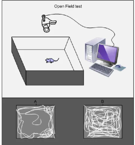
In laboratory rodents, different types of tests are used to determine whether treatments or conditions affect behavioral status. The Morris water maze is widely used to assess learning ability and cognition; while open field and elevated plus maze tests are used to measure locomotion, exploratory and anxiety-related behaviour, respectively (Walsh and Cummins 1976; Walf and Frye 2007; Sharma 2009).
5.1.1. Morris Water Maze Test
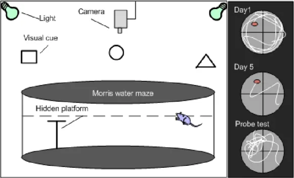
In 1984, Richard Morris developed a device known as the “Morris water maze” (MWM) to assess spatial learning and memory in laboratory rats (Morris 1984). This test has been accepted and extensively used by physiologists and pharmacologists. Although the MWM seems simple at first glance, it is a challenging task for animals and is used in the most sophisticated experiments in neurobiology, neuropharmacology and neurocognition to evaluate the impacts of aging, experimental lesions, and drug effects— especially in rodents. Studies of neurodegenerative and neuropsychiatric disorders where
cognition is damaged (i.e. Alzheimer’s disease, Parkinson’s disease, and schizophrenia) confirm the importance of this test. The MWM involve the acquisition and spatial localization of visual cues which are processed, consolidated, retained, and retrieved to find a relatively small goal (a hidden platform) (Sharma 2009).
The MWM is a large circular pool (diameter: 150 cm) filled approximately halfway with water at room temperature and with a fixed invisible platform, which is submerged below the water surface (~1 cm). The pool is designated with two principal axes by computer software in order to generate a cross imaginary shape. There are then four points at the end of each line: North (N), South (S), East (E), and West (W), and four equal quadrants. The platform is placed in the middle of one of these quadrants. Several objects or images (e.g. circles, squares, and triangles) are located in the testing room or hung on the wall, so rodents are able to use these visual cues for navigating in the maze (Figure 7).
Figure 7. Diagram showing the Morris water maze testing room and apparatus